UNIVERSITÀ POLITECNICA DELLE MARCHE
FACOLTA’ DI MEDICINA E CHIRURGIA
Dottorato di Ricerca XXX CICLO
in
SALUTE DELL’UOMO
MIRNA SIGNATURE TO IDENTIFY
LUNG ASBESTOS-RELATED
MALIGNANCIES.
Relatore: Chiar.mo Dottoranda:
Prof.ssa Lory Santarelli Dr.ssa Simona Gaetani
Correlatore:
Dott. Marco Tomasetti
INDEX
Abstract
………..1 1.INTRODUCTION
………...4 1.1 Asbestos-lung disorders………...9 1.1.1. Asbestosis……….10 1.1.2. Pleural plaques………..101.1.3. Benign asbestos pleural effusion (BAPE)………....12
1.1.4. Diffuse pleural thickening (DPT)……….12
1.1.5. Rounded atelectasis (RA)……….14
1.1.6. Malignant mesothelioma (MM)………...15
1.1.7. Asbestos-related lung cancer………18
1.1.7.1. The attribution of lung cancer to asbestos exposure: a difficult topic………..21
2.1. Characterization of asbestos exposure……….24
2.1.1. Assessment of different exposure routes……….29
3.1. MicroRNA………...32
3.1.1. Discovery and features………33
3.1.2. Biogenesis………....34
3.1.3. MiRNA structure……….38
3.1.4. MiRNAs are gene expression mediators……….39
3.1.5. MiRNAs in tumour development and progression………..42
3.1.6. Epigenetic alterations at miRNA loci………..44
3.1.7. MiRNAs in diagnosis and therapy………...46
3.1.8. MiRNAs as molecular biomarkers………...49
3.1.8.1. MiRNAs as biomarkers in plasma or serum………..52
3.1.9. MiRNA in asbestos-lung malignancies: state of art………53
3.1.10. Effect of environmental carcinogens on the microRNA machinery………...55
4. Purpose of the thesis………..61
5. Materials and Methods………..63
5.1. Study design………....63
5.2. Study population……….66
5.3. Asbestos exposure assessment………70
5.5. Clinical specimen collecetion………...71
5.6. MicroRNA qPCR analysis profiling in FFPE tissue samples………..71
5.7. Circulating miRNA detection………...73
5.8. Reverse transcription………....73
5.9. Quantitative qRT-PCR analysis………...75
5.10. Fibres and chemicals………...76
5.11. Cell cultures and treatments………....77
5.12. Pre-cancerous cells selection………..78
5.13. Cell proliferation assay and mitochondrial reducing activity…………...78
5.14. Assessment of ROS and mithocondrial membrane potential…………...79
5.15. Glucose uptake………...80
5.16. Detection of glucose, lactate and ATP………...81
5.17. Macrophages-induced cytochine production……….81
5.18. Triple co-colture model………...82
5.19. Western blot analysis………...83
5.20. Quantitative RT-qPCR analysis………...84
5.21. Statistical analysis………...84
6. Results………...86
6.1. Discovery phase: identification of miRNA-expression profile……...86
6.2. Verification phase………...91
6.3. Training and validation phases………...93
6.4. Asbestos-related miRNAs with diagnostic value for Lung Cancer……...97
6.5. Asbestos-related miRNAs with diagnostic value for Malignant Mesothelioma………99
6.6. Testing phase: asbestos-related miRNAs in the asbestos-exposed Population………....100
6.7. Asbestos-related miRNAs in carcinogen-induced pre-cancerous cells………..102
6.8. Signal transduction pathway modulates the asbestos-related miRNA expression in the stroma environmental………..107
7. Discussion………....111
8. Conclusions………...119
9. Bibliography………...121
1
ABSTRACT
Introduction. Exposure to asbestos results in serious risk of developing malignant
mesothelioma (MM) and lung cancer (LC). The association between asbestos exposure and lung adenocarcinoma is well established. Nevertheless, precise histopathological data are poorly considered when investigating the asbestos-cancer link in compensatory approach. Occupational lung cancer generates high mortality and is the most common compensated in France. Although, the Helsinki criteria for identifying individuals with high risk of asbestos exposure at work have been accepted, it is insufficiently proved and specific asbestos-related parameters are needed. The presence of pleural plaques is not considered a pre-cancerous condition, while asbestosis, as well smoking is associated with an increased risk of lung cancer. MicroRNAs (miRNAs) have rapidly become an attractive method for profiling cancer. MiRNA expression is early altered by exposure to occupational/environmental carcinogens, thus, useful to identify a novel asbestos-related profile able to distinguish asbestos-induced cancer from cancer with different etiology.
Methods. Consequential study phases have been performed to identify miRNAs associated
to the development of asbestos-induced malignancies. Four groups have been included: patients with asbestos-related malignancies (NSCLCAsb and MM), non-asbestos-related lung cancer (NSCLC), and disease-free subjects (CTRL).Next, the selected miRNAs were evaluated in an asbestos-exposed population, and an in ‘in vitro’ model was performed to identify the mechanism of asbestos-induced miRNA regulation.
Results. Four serum asbestos-related miRNAs consisting of miR-126, miR-205, miR-222
2
and age best depicted the asbestos-related malignancies. The association of miR-222 with miR-222/miR-126 best characterised the non-asbestos related NSCLC group. We found that miR-126 and miR-222 were strongly associated with asbestos exposure and both miRNAs were involved in major pathways linked to cancer. To elucidate the role of mediation effect of these miRNAs between asbestos-exposure and the tumour development, a disease-free population exposed to asbestos, stratified as currently-exposed and ex-exposed with and without asbestos-related diseases (ARDs), was evaluated for the selected miRNAs. Notably, increased expression of 126 and 222 including miR-126/miR-205, miR-222/miR-205, and miR-222/miR-520g ratios were found only in currently exposed subjects. It had been reported that miRNA expression changes in response to environmental carcinogens exposure are transient and revert to normal levels after recovery from exposure. Accordingly, ex-exposed subjects to asbestos did not show any changes in miRNA expression (cf Fig.26). Increased expression of EGFR was found in the asbestos-initiated cells, causing activation of the down-stream effector AKT and p38 MAPK signalling. Asbestos-mediated activation of EGFR-AKT pathway resulted in miR-126 and miR-222 upregulation associated with miR-520g downregulation. Both miR-222 and miR-520g were reversed by inhibiting EGFR (AG1478), suggesting its involvement in asbestos-induced miRNA regulation.Moreover, the upregulation of miR-126 and miR-222 was enhanced when asbestos-induced pre-cancerous cells were cocultured with tumour stromal cells, such as fibroblasts and endothelial cells. Thus, suggesting that cancer stroma cross talk induced the expression of miR-126 and miR-222 to facilitate angiogenesis and invasion growth of lung malignancies.
3
Conclusions. This study uncovers miRNAs that are potentially involved in
asbestos-related malignancies and their expression outline mechanisms whereby miRNAs may be involved in asbestos-induced pathogenesis.
4
1.INTRODUCTION
Asbestos-related diseases are still a major public health problem. The World Health Organization (WHO) has estimated that 107,000 people worldwide die each year from mesothelioma, lung cancer, and asbestosis (WHO, 2010). Although worldwide consumption of asbestos has decreased, consumption is increasing in many developing countries in which exposures may also be high in these areas. All forms of asbestos have been considered carcinogenic by several Health Organizations including the International Agency for Research on Cancer (Int. Agency Res. Cancer (IARC). 1977. Asbestos, Vol. 14. Lyon, Fr.: IARC), and in United States by the Environmental Protection Agency (Int. Programme Chem. Saf. (IPCS). 1988. Chrysotile. Geneva: WHO). At the IARC meeting in March 2009 scientists concluded that all forms of asbestos (chrysotile, crocidolite, amosite, tremolite, actinolite, and anthophyllite) are associated with an increased risk of mesothelioma and lung, laryngeal, and ovarian cancers (IARC, 2012; A Review of Human
Carcinogens: Arsenic, Metals, Fibres, and Dusts; Straif, 2009). The use of asbestos has now been banned in 55 countries worldwide. Nevertheless, asbestos exposure is still widespread in the world: beside active occupational exposure that is estimated to affect about 125 million men and women (IARC. IARC monograph on the evaluation of carcinogenic risk to human, vol. 100 C, Arsenic, Metal, Fibres and Dusts. Lyon: IARC Press, 2012.), a large number of people are exposed to asbestos in areas that may remain in place for decades after the enforcement of asbestos bans. Because of its widespread past use, the epidemic of asbestos-related diseases is known to be nearly worldwide and thus may be described as a pandemic rather than just an epidemic (Fig.1).
5
Figure 1.The Environmental Working Group (EWG) states that mesothelioma, asbestosis and
asbestos-related lung and gastrointestinal cancers claimed as many as 230,000 lives between 1979 and 2001. This chart reflects the EWG's yearly morbidity estimates for three primary asbestos-associated cancers as well as asbestosis, a noncancerous condition that is sometimes diagnosed in asbestos cancer patients.
Asbestos-related diseases (ARDs) resulting from exposure to asbestos include mostly lung cancer (LC) and malignant mesothelioma (MM). Most data on mortality from ARDs came from MM, an aggressive cancer whose association with asbestos exposure is well established. Driscoll et al. (Driscoll et al., 2011) estimated that 43,000 people worldwide die each year by MM although the real cases of MM have been underestimated. Although the proportion of MM attributable to asbestos exposure varies, a fraction of 80% has been reported (Tossavainen, 1997).
In Figure 2 is shown the distribution of age MM incidence rated for males by country between 1998-2002 and these data, which include only cancer registries that had at least 15 years, come from IARC report, Cancer in Five Continent (IARC, 2007).
6
Figure 2. Worldwide age standidardized mesothelioma incidence rates (per 100,000) for males in 1998-
2002. Data from Reference (IARC.2007. Cancer Incidence in Five Continents: Vol. IX, ed.)
Bianchi C. underlined a wide area of the world for which there is not any information on
MM incidence (Bianchi and Bianchi, 2007). The highest incidence of MM in the world was reported in the Italian Genoa Province (5.8 per 100,000). Other areas of the world reporting high rates include the West Cape of Australia (4.7 per 100,000), the Northern Yorkshire (4.2 per 100,000) area of the United Kingdom, Northern Ireland (4.0 per 100,000), and Scotland (3.6 per 100,000). Approximately 95% of the participating cancer registries have reported cases of MM to the IARC program. Delgermaa et colleagues (Delgermaa et al., 2011) established a total of 92,253 MM deaths by 83 countries between 1994 and 2008. The worldwide age-adjusted rate for mesothelioma mortality was 4.9 per million. The United Kingdom was found to have the highest age-adjusted mortality rate (17.8 per million), followed by Australia (16.5 per million), and Italy (10.3 per million). Malignant mesothelioma is the cancer mainly associated with asbestos exposure. However, epidemiologic studies reported several cases of LC in asbestos-exposed workers for all types of asbestos except for crocidolite (Henderson et al., 2004; Stayner et al., 1996).
7
Although the major risk factor for LC is tobacco smoke, an estimated 5-7% of all these cancers are attributable to asbestos exposure (LaDou, 2004). Asbestos-related LC is considered one of the most important occupational cancers (Karjalainen et al., 1994). Hazardous occupational exposures to asbestos fibers have occurred in a variety of industrial operations, including mining and milling, manufacturing, shipbuilding and repair and construction (Current intelligence bulletin 62, 2011).
Current exposures to commercial asbestos occur predominantly during maintenance operations and remediation of older buildings containing asbestos. In addition to occupational asbestos exposure, environmental exposure is highly relevant for the risk to develop asbestos-related malignancies. This has significant health and economic implications that have been well documented. The 20-40-year latency periods of ARDs (Lanphear and Buncher, 1992) and their low incidence rates in the general population make preventive strategies and early treatment extremely challenging. The availability of well-validated diagnostic biomarkers of asbestos exposure would greatly facilitate both prevention and early treatment strategies. Biomarkers are crucial in the screening of patients with a high risk of developing cancer, diagnosing patients with suspicious tumours at the earliest possible stage, establishing an accurate prognosis, predicting and monitoring the response to specific therapies. Biomarkers of response, which reflect a change in biologic function in response to asbestos exposure, have proved to be more useful. MM is the major biological response to asbestos that can be readily monitored and numerous studies have used this disease as confirmation of a previous asbestos exposure. Epigenetic alterations are innovative biomarkers for cancer, due to their stability, frequency, and non-invasive accessibility in body fluids. Recently, circulating microRNAs (miRNAs) were
8
found to be particularly attractive as biomarkers for early diagnosis and prognosis of many cancer types (Song et al., 2017).
Therefore, the identification of asbestos-related molecular-signature has long been a topic of increasing research interest. MiRNA expression is early altered by exposure to occupational/environmental carcinogens, thus, useful to identify a novel asbestos-related profile able to distinguish asbestos-induced cancers from cancers with different etiology.
9
1.1. Asbestos-lung disorders
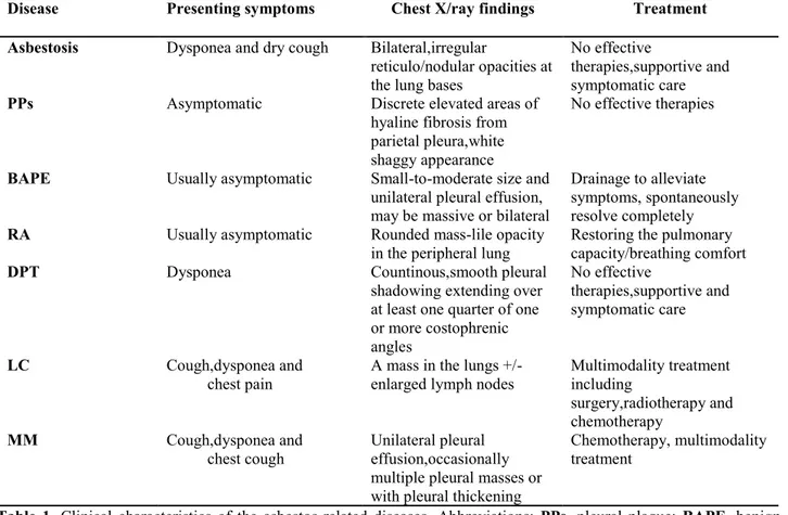
The exposure to asbestos induces the formation of both benign lung disorders such as, asbestosis (diffuse interstitial pulmonary fibrosis due to asbestos inhalation), pleural plaques (PPs), diffuse pleural thickening (DPT), benign asbestos pleural effusion (BAPE), rounded atelectasis (RA), and malignancies, including LC and MM. The clinical characteristics of ARDs are summarized in Tab.1.
Table 1. Clinical characteristics of the asbestos-related diseases. Abbreviations: PPs, pleural plaque; BAPE, benign
asbestos pleural effusion; RA, rounded atelectasis; DPT, diffuse pleural thickening; LC, lung cancer; MM, malignant mesothelioma.
Disease Presenting symptoms Chest X/ray findings Treatment Asbestosis Dysponea and dry cough Bilateral,irregular
reticulo/nodular opacities at the lung bases
No effective
therapies,supportive and symptomatic care
PPs Asymptomatic Discrete elevated areas of hyaline fibrosis from parietal pleura,white shaggy appearance
No effective therapies
BAPE Usually asymptomatic Small-to-moderate size and unilateral pleural effusion, may be massive or bilateral
Drainage to alleviate symptoms, spontaneously resolve completely
RA Usually asymptomatic Rounded mass-lile opacity
in the peripheral lung Restoring the pulmonary capacity/breathing comfort
DPT Dysponea Countinous,smooth pleural shadowing extending over at least one quarter of one or more costophrenic angles No effective therapies,supportive and symptomatic care LC Cough,dysponea and chest pain
A mass in the lungs +/-
enlarged lymph nodes Multimodality treatment including surgery,radiotherapy and chemotherapy
MM Cough,dysponea and
chest cough Unilateral pleural effusion,occasionally multiple pleural masses or with pleural thickening
Chemotherapy, multimodality treatment
10
1.1.1. Asbestosis
Asbestosis is defined as diffuse interstitial pulmonary fibrosis due to inhalation of asbestos fibres. The latency period for disease development is usually 15 years or more, and is influenced by duration and intensity of exposure. Relatively high levels of asbestos inhalation are required to produce asbestosis (cumulative exposure ≥25 fibres /mL-years), although there are some reports that record asbestosis following lower levels of asbestos exposure (Roggli et al., 2010). Asbestosis is interstitial fibrosis that is sub-pleural and initially affects the lung bases. Diagnostic criteria have been published (American Thoracic Society, 2004) and include a compatible exposure history, clinical and radiographic features. Asbestosis usually presents with gradual and progressive dyspnoea and accompanying dry cough. The histological diagnosis of asbestosis requires an appropriate pattern of interstitial fibrosis and demonstration of asbestos bodies in histological section. High-resolution computed tomography (HRCT) is more sensitive and specific than chest radiography (Akira et al., 2003).
1.1.2. Pleural plaques
Another benign ARDs are pleural plaques (PPs) which the commonest manifestation of
asbestos exposure affecting up to 58% of asbestos-exposed workers and up to 8% of general environmentally exposed populations, with a latency period of 20-30 years. They are hypo cellular lesions composed of thick collagen bundles arranged in a ‘basket-weave’ pattern covered by a single layer of normal mesothelial cells (Chapman et al., 2003).
11
PPs are variable in size and number with white or pale yellow appearance, typically distributed on the posterolateral chest wall, the dome of the diaphragm and the mediastinal pleura (Fig.3A,B) (Roach et al., 2002).
Figure 3. (A) Radiological and computed tomography scan of the thorax demonstrating asbestos-related
pleural plaques. (B) Thoracoscopic view of PP on the parietal pleura. Pleural biopsies have done at the level of non-specific inflammation to rule out an early-stage malignant mesothelioma.
They are sharply demarcated from sub-pleural tissue, and calcification is a common late finding (Huggins and Sahn et al., 2004). The diagnosis relies on radiographic findings and a compatible history of exposure. The CXR is a standard diagnostic tool utilizing ILO classification guidelines, although false-positive, false-negative and inter observer variability rates are relatively high. HRCT is more sensitive and specific for making the diagnosis, but because of its high radiation exposure and unavailability, it is inappropriate for screening (American Thoracic Society, 2004). Pleural plaques do not undergo malignant degeneration into MM, and their presence does not increase the risk of developing asbestos-related malignancies (Harber et al., 1987; Weiss, 1993).
12
1.1.3. Benign asbestos pleural effusion (BAPE)
Benign asbestos pleural effusion (BAPE) is an exudative and often haemorrhagic pleural effusion following asbestos exposure and is a diagnosis of exclusion after other possible causes such as malignancies, tuberculosis and other infections (Hillerdal and Ozesmi, 1987). It usually occurs within 10 years after exposure, earlier than other ARDs, although this is not variable (American Thoracic Society, 2004).
BAPE is often unilateral with left-side predominance and usually resolve completely with a mean duration of 3-4 months, but may recur (30%-40%) and usually occur within 3 years of the initial presentation (Epler et al., 1982; Robinson et al., 1981).
A prolonged follow-up is necessary for asbestos-exposed patients with pleural effusions. BAPE does not have specific prognostic implications respect to the subsequent development of MM (Robinson et al., 1981; Gaensler and Kaplan, 1978), but is the witness of exposure to the main risk factor of this malignancy.
1.1.4. Diffuse pleural thickening (DPT)
DPT is characterized by extensive thickening of the visceral pleura, often with adherence to the parietal pleura, and obliteration of the pleural space (American Thoracic Society, 2004; Yates et al., 1996). Many studies showed that DPT is a consequence of BAPE (Epler et al., 1982; MacLoud et al., 1985; Lilis et al., 1985; Fridriksson et al., 1981). However, DPT is not a pathognomonic marker of asbestos exposure: many other causes of exudative pleural effusion can lead to DPT.
13
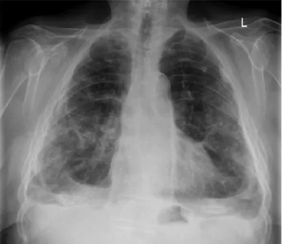
DPT accounts for 22% of all ARDs (Hannaford-Turner et al., 2010). It can develop within a year of exposure but can also take up to 40 years. PPs often coexist. DPT may be associated with dyspnoea and chest pain (Yates et al., 1996; Jeebun and Stenton, 2012). Although symptoms are generally mild, severe restrictive lung disease with hypercapnic respiratory failure and death can rarely occur (Miles et al., 2008). DPT can cause significant restrictive ventilatory impairment. On a chest radiograph, DPT presents as continuous, irregular pleural shadowing that often extends up both chest walls and with blunting of one or more cost phrenic angles (Fig. 4).
Figure 4. Posteroanterior chest radiograph demonstrating asbestos-related diffuse pleural thickening.
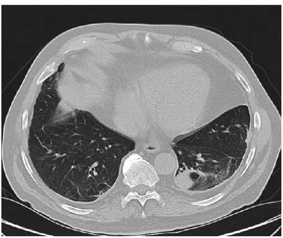
HRCT is more sensitive than CXR for detection of early pleural thickening (i.e. 1–2 mm in thickness) (American Thoracic Society, 2004) (Fig. 5).
14
Figure 5. Computed tomography scan of the thorax demonstrating asbestos-related diffuse pleural
thickening. Note the rounded atelectasis on the left and the sub-pleural interstitial pulmonary fibrosis bilaterally.
Treatment is largely limited to supportive and symptomatic care.
1.1.5. Rounded atelectasis (RA)
Rounded atelectasis (RA) (Blesovsky’s syndrome) may occur with DPT. Pleural adhesions and fibrosis cause deformation of the lung with bending of some small bronchi (Hillerdal, 1989). On CXR, this presents as a rounded opacity in the peripheral lung adjacent to the thickened pleura, with curvilinear opacities (the comet tail sign) extending from the site of atelectasis towards the hilum (Batra et al., 1996). Exposure to asbestos is the principal cause today, but any type of pleural inflammatory reaction can cause RA (Hillerdal, 1989). RA is usually asymptomatic, but may be accompanied by breathlessness.
15
1.1.6. Malignant mesothelioma (MM)
MM is an aggressive and incurable tumour arising from mesothelial cells of the pleura, peritoneum and rarely elsewhere. Although cases due to environmental and para-occu-pational asbestos exposure have been described, most MMs are occupara-occu-pational in origin. MM can develop even after short and low exposure, but a dose-effect relationship has been demonstrated. Median latency is approximately 40 years (range 15-67). MM has a poor prognosis with median survival of 8-14 months (British Thoracic Society, 2007; Musk et al., 2011).
Patients usually present with chest pain (60-70%), dyspnoea (50-70%), cough (20-30%) (Fuhrer and Lazarus, 2011) and restrictive gas exchange abnormality. The radiographic manifestation is usually a unilateral pleural effusion or pleural thickening. CT is the primary modality for the diagnosis, staging and response assessment of MM (Fig. 6A, B).
Figure 6. (A) Posteroanterior chest radiograph of a patient with left-sided malignant mesothelioma.(B)
Computed tomography scan of the thorax of a patient with left-sided malignant mesothelioma showing opacification and very little lung aeration.
16
Fluorodeoxyglucose PET/CT (FDGPET/ CT) has been shown to be a useful diagnostic tool for detection of distant metastasis and to differentiate MM from benign pleural disease (Sharif et al., 2011; Basu et al., 2010).
Staging relies on the International Mesothelioma Interest Group TNM (tumour, nodes, and metastases) staging system (NCCN, 2013). Survival depends on the stage of the disease when diagnosed. Stage I has approximately 12 months of survival, Stage II 4 months, and about 3 months in Stages III–IV (Rusch,1995). The definitive diagnosis of MM requires a tissue sample; however, negative results do not exclude it as sampling problems can occur and the diagnosis is a difficult one histologically.
MM occurs in three main histological subtypes: epithelioid, sarcomatoid and biphasic. These have prognostic significance, with epithelioid being the most common and sarcomatoid subtype predicting the worst outcome (Safe Work Australia, 2011; Musk AW et al., 2011). Several tumour biomarkers measured in either the serum, plasma or the pleural fluid have been evaluated for diagnostic purposes. Biomarkers could also be useful to detect the development of MM at an early, potentially resectable stage, although no randomized data currently exist that confirm prolonged survival with surgical resection. Promising biomarkers include soluble mesothelin-related protein (SMRP) (Wheatley-Price et al., 2010; Park et al., 2008), osteopontin (Pass et al., 2009; Park et al., 2009), and more recently, fibulin 3 (Pass et al., 2012) and integrin-linked kinase (ILK) (Watzka et al., 2013). Although initial results appear promising, most of these biomarkers have yet to be evaluated in prospective studies and primarily apply to epithelial type MM. SMRP has been evaluated in this manner and unfortunately was found to have a high rate of false-positives (Park et al., 2008). High SMRP levels have been shown to correspond with
17
disease volume, suggesting that SMRP may be a useful for detecting the progression of MM and monitoring MM during treatment (Wheatley et al., 2010; Franko et al., 2012). Serum osteopontin levels are elevated in MM but also in patients with non-malignant ARDs (Pass et al., 2005; Park et al., 2009) resulting in rather low specificity and sensitivity (Creaney et al., 2008; Grigoriu et al., 2007), limiting its value as a screening tool. Plasma fibulin-3 levels discriminated between Stage I or II mesothelioma and asbestos exposure without MM, with a specificity of 94% and a sensitivity of 100% (Pass et al., 2012). Integrin-linked kinase (ILK) serum levels have been shown to be significantly higher in patients with MM compared with healthy asbestos-exposed workers (Watzka et al., 2013). There is potential for the use of ILK as a marker of disease progression, as its levels are increased in advanced stages of MM in comparison with early stages (Watzka et al., 2013), but more work in this area is needed. Novel biomarkers such as volatile organic compounds measured in exhaled breath (Chapman et al., 2012), microRNAs (Kirschner et al., 2012) may prove useful in the future and/or a combination of biomarkers.
Many strategies have been tried for treating MM, including surgery, chemotherapy and radiation.
The principal role of radiotherapy is as an adjuvant following surgical resection (Scherpereel et al., 2010). Chemotherapy plays an integral role in multimodality treatment and is also recommended alone as a treatment for inoperable patients (NCCN, 2013), but no highly effective agents are yet available. A first-line regimen of pemetrexed or gemcitabine in combination with a platinum agent (cisplatin or carboplatin) is currently regarded as the best available treatment for MM (NCCN, 2013). Pemetrexed and raltitrexed in combination with cisplatin have been shown to improve survival, global
18
quality of life and pulmonary function, when compared with cisplatin alone, but the survival effect is only approximately 12 weeks (van Meerbeeck et al., 2005; Vogelzang et al., 2003). Currently, chemotherapy has at best only a modest benefit for these patients. Further improvements in drug development and better-designed clinical trials are strongly needed. In recent years, advances in knowledge of molecular and biological mechanisms of MM have led to development of immunologically based and targeted therapies, but these are still under evaluation.
1.1.7. Asbestos-related lung cancer
Lung cancer (LC) has continued to be the most common type of cancer worldwide for several decades, with the highest incidence and mortality rates (Ferlay et al., 2012). Only 13% of patients with lung cancer survive for > 5 years (Balgkouranidou et al., 2013). As the second leading risk factor for lung cancer, asbestos exposure is responsible for an estimated 5-7% of all these cancers (LaDou, 2004.). Asbestos-related lung carcinoma is considered one of the most devastating occupational cancers (Kettunen et al., 2011). In 1997, the Helsinski criteria for identifying individuals with a high risk of asbestos exposure at work were accepted (Tossavainen, 1997). Although the use of asbestos has been banned in many developed countries, asbestos-related lung cancer still poses a great public health threat due to the long latency period from asbestos exposure to the incidence of asbestos-induced cancer (Lin et al., 2007).
Heavy asbestos exposure produces an increased risk of LC, with a latency period of approximately 15-20 years Among asbestos-related lung cancer, non-small cell lung cancer (NSCLC) accounts for at least 80 % of these cases (Balgkouranidou et al., 2004).
19
In 1997 the Helsinki criteria, which have been established that all four major histological types (squamous, adenocarcinoma, large-cell and small cell) can be related to asbestos, have been updated: the current classification (WHO 1999) includes two additional types of lung cancer (sarcomatoid and adenosquamous). These are included as type of lung malignancies that may occur because of asbestos exposure. (Consensus report, Wolff et al., 2015). In the Helsinki criteria, it is reported that there is no threshold of concentration of fibers of airborne asbestos below which the risk of asbestos-related LC is null (Consensus report, Wolff et al., 2015).
Asbestos-related LCs account for about 3-8% of all LCs and is similar in both the type of cancer and in its signs and symptoms in asbestos-exposed and unexposed individuals. Older studies found inconsistent results regarding the lobe of origin and histology of asbestos-related lung cancer (ARLC) (Nielsen et al., 2014).
Some studies showed an upper lobe location similar to tobacco-related lung cancer, whereas other investigators found a lower lobe location (Auerbach et al, 1984; Hiraoka et al., 1990; Johansson et al., 1992; Warnock and Isenberg, 1992; Anttila et al., 1993; Hillerdal et al., 1983; Sluis-Cremer, 1980; Weiss, 1981). Although adenocarcinoma was found to be the most prevalent in some studies, a recent review of the literature by Nielsen
et al (Nielsen et al., 2014) showed that there was no difference in location and cell type
between ARLC and non-ARLC. They concluded that cell type and location of lung cancer were not useful for differentiating ARLC from other lung cancers. Prognosis of ARLC was not different from that of other lung cancers (Nielsen et al., 2014). Because pleural plaques may be associated with low levels of exposure, the attribution of LC to asbestos must be supported by an occupational history of substantial asbestos exposure or measures of
20
asbestos fibre burden. Presence of pleural plaques demonstrated a previous asbestos exposure but was not a precancerous condition. Pleural plaques alone are insufficient for the attribution of LC to asbestos. A possible indicator for attributing LC to asbestos exposure is “bilateral diffuse pleural thickening” which is often associated with moderate or heavy exposure, as seen in cases of asbestosis and should be considered accordingly in terms of attribution. Asbestosis argued for high asbestos exposure and was associated with an increased risk of LC (Nielsen et al., 2014). All asbestos types were associated with LC (Nielsen et al., 2014). The risk of developing LC is linearly related to cumulative asbestos exposure. The issue of whether asbestosis is a necessary precursor for LC is still controversial, but recent consensus statements have concluded that heavy asbestos exposure of ≥25 fibres/mL-years rather than asbestosis is needed (Henderson et al., 2004). A minimum lag time of ten years from the first asbestos exposure is required to attribute the LC to asbestos. Epidemiological evidence indicates that the combined effect of smoking and asbestos exposures on LC incidence appears to be more than additive and is probably multiplicative. Smoking cessation has major health benefits and should be recommended in all patients with past asbestos exposure.
One recent British study showed an inverse relationship between time since smoking cessation and LC mortality risk in asbestos workers (Frost et al., 2011). Former smokers who had stopped smoking for 40 and more years had same risk of LC as asbestos workers who had never smoked (Frost et al., 2011).
The relative lack of symptoms during the early stages of LC frequently results in a delayed diagnosis. However, early diagnosis and treatment can result in long-term survival, improving the 5-year survival to approximately 70%. The treatment of LC and survival is
21
very dependent on disease stage and the presence of co-morbidities and is identical in asbestos-exposed and non-exposed patients. Treatment includes surgical removal of the cancer, chemotherapy, radiotherapy or a combination, and several excellent reviews are available (Rossi et al., 2013; Saintigny et al., 2012). CT screening has been evaluated in a population exposed to asbestos and shown to be feasible, however, with a high incidence of incidental findings (Vierikko et al., 2007). CT screening presents an opportunity for early diagnosis and treatment in asbestos-related LC and possibly also for MM.
1.1.7.1. The attribution of lung cancer to asbestos
exposure: a difficult topic
It is extremely difficult to correlate exposure to asbestos and the onset of lung cancer. According the Helsinki criteria updated in 2014, the “attribution of causation requires a reasonable medical certainty on a probability basis that the agent (asbestos) has caused or contributed materially to the disease” (Consensus report, Wolff et al., 2015). Among the causative factors for asbestos-related lung cancer, it is necessary to consider:
• Occupational exposure
Epidemiological studies indicate that asbestos exposure is associated with an increased risk of lung cancer, (Lash et al., 1997; Hodgson et al., 2000; Berry and Gibbs, 2008) accounting for an estimated 2-5% of new lung cancers (Doll and Peto, 1981; Gustavsson et al., 2003; Marinaccio et al., 2008). An increased proportion of lower-lobe tumour localization (Craighead and Mossman, 1982) of adenocarcinomas (Patz et al., 2000) has been reported among workers exposed to asbestos, but these associations have not been confirmed in other studies (Lee et al.,
22
1998). The risk of developing lung cancer is linearly related to cumulative asbestos exposure, with an estimated increase of 1% for each fiber/mL-year of exposure (Boffetta et al., 1998). However, the strength of the association and the slope of the cumulative dose-response relationships vary considerably across studies and occupations (Mossman and Gee, 1989). The risk appears to be smaller in miners (McDonald et al., 1980), friction product manufacturers, (McDonald et al., 1984), intermediate in asbestos cement (Finkelstein,1983) and product manufacturers (Weill et al., 1979) and highest in textile workers (McDonald et al., 1983; McDonald et al., 1982).
The slope is steeper in asbestos cement manufacturers (Finkelsteinm, 1983) much less in friction product manufacturers (McDonald, 1984) and intermediate in the textile industry (McDonald et al., 1983; McDonald et al., 1982). The Helsinki Report estimates a cumulative exposure of 25 fibers/mL-years to increase the risk of lung cancer by two-fold (Consensus report: Helsinki criteria, 1997). However, a subsequent reevaluation concluded that probably the exposures associated with increased risk of lung cancer are higher (Tossavainen et al., 2000). A recent risk analysis of 14 studies identified a cumulative “no effect” exposure to crysotile for lung cancer in the range of 25-1,000 fibers/mL-years (Pierce et al., 2008). However, these dose estimates have been challenged for accuracy, mainly because of crude surrogates of exposure (Gibbs et al., 2007; Greenberg, 2007; Roggli et al., 2008). Lower-level occupational exposures are not associated with an increased risk of lung cancer (Weiss, 1999; Berry and Gibbs, 2008; Camus, 1998).
23 • Asbestosis and ARLC
There is a controversy on relationship between asbestos-related lung cancer and asbestosis. Several authors hold the notion of an increased risk of LC in asbestos-exposed workers in the absence of evidence of asbestosis (Egilman and Reinert, 2005; Reid et al.,1991), but others conclude that the risk only increases if asbestosis is present (Hughes and Weill, 1991; Jones et al., 2009). However, a high correlation between asbestosis and LC rates was observed in 38 cohorts of workers (Weiss, 1999) and the risks of developing asbestosis and lung cancer are in the same range of cumulative dose (Loomis et al., 2009). The progression of asbestosis was shown to predict the development of lung cancer (Oksa et al., 1998) and this condition is a good indication that exposure of the patient was high enough to put him at risk of lung cancer. In addition to asbestosis, also pleural plaques were put in correlation with greater risk of LC (American Thoracic Society, 2004; Hillerdal and Henderson, 1997), but this conclusion should be challenged because plaques manifest themselves at asbestos exposures much smaller than those associated with increased risk of cancer (Weill and Weill, 2005). When both high asbestos body burden and fibrosis are present, the attribution of LC is clear, but it is also acceptable in the absence of fibrosis (Consensus report: Helsinki criteria, 1997). • Custom tobacco and asbestos interaction
Epidemiological evidence indicates that smoking and asbestos exposures taking place together have more than additive and less than multiplicative effects on LC incidence (Wraith and Mengersen, 2008). Hypotheses consider the mutual influences of asbestos and smoke in the delivery to epithelial cells of smoke
24
carcinogens and fibers, and there is some evidence in humans that smoking increases the penetration of fibers into the bronchial mucosa where the effect is greater for chrysotile than for the amphiboles (Nelson and Kelsey, 2002; Churg and Stevens, 1995). Therefore, extrapolations can hardly be made at levels of asbestos exposure that do not increase the risk of lung cancer.
2.1. Characterization of asbestos exposure
An important point for asbestos-related malignancies that no safe threshold for asbestos exposure has been established. Indeed, Hodgson and Darton suggested that any threshold for mesothelioma is at a very low level and they observed that the proportion of mesothelioma cases in population studies from whom no likely source of asbestos exposure can be identified is often quite high.
These observations suggest that relatively brief exposures may carry a low, but non-zero, risk of causing mesothelioma (Hodgson and Darton, 2000).
Some individuals develop mesothelioma following exposure to small amounts of asbestos, while others exposed to heavy amounts do not (Carbone et al., 2012), suggesting that genetic factors influence risk of this disease. Indeed, in a study Testa et al. reported that the presence of germline BAP1 mutations in membres of families that have a high incidence of mesothelioma, despite very modest exposure to asbestos. These authors showed that BAP1 mutations are associated with a novel hereditary cancer syndrome that predisposes to mesothelioma, uveal melanoma and potentially other cancers and they hypothesized that when individuals with BAP1 mutations are exposed to asbestos, mesothelioma
25
predominates. Alternatively, BAP1 mutation alone may be sufficient to cause mesothelioma (Testa et al., 2012).
In addition, it is important to take a comprehensive occupational and environmental history to identify people at risk because of previous exposure. Factors to establish include the specific occupation, the duration of that occupation, and the intensity of exposure (e.g., was the dust visible or not). A significant exposure can be defined as at least several months’ exposure to visible dust that began more than 10 years earlier. Although much attention is focused on industrial exposure, environmental sources also play a role. These include residence near asbestos and prolonged exposure in buildings with open sources of contamination. Few studies have examined the incidence of asbestos related-disease (ARDs) associated with distinct sources of asbestos exposure (occupational, familial, or environmental).
While the association between occupational exposure to asbestos and asbestos-related disease has been established, the other source of asbestos exposure is less studied. Despite the decrease in asbestos production and use, there is still the possibility for environmental exposure (EE), whose impact on MM occurrence is less know (Carbone et al., 2016; Lemen, 2016). Quantification of the ARDs burden associated with EE to asbestos is further limited by the lack of reliable assessment of type and amount of exposure, smaller sample numbers, and smaller effect sizes compared to those seen in occupational studies. (Lin et al., 2007; Mc Donald, 1985; Goldberg and Luce, 2009; Bourdes et al., 2000). In a recent review, Liu et al. (Liu et al., 2017) classified non-occupational exposure to asbestos as environmental exposure (EE).
26
This exposure was grouped into the following four categories:
1) “non-occupational asbestos exposure” (NOA) to in areas where geological structure has shown the presence of asbestos but asbestos-related industries are absent;
2) “neighbourhood exposure” based on residence near industrial/mining sources of asbestos;
3) household exposure for family members of occupationally exposed subjects, which includes what some authors call familial exposure (Mirabelli et al., 2010; Corfiati et al., 2015);
4) other non-occupational exposures, which includes home-related (Mensi C. et al., 2015) or domestic exposure (Girardi et al., 2014; Goldberg et al., 2010 Gogali et al., 2012). For example, exposure occurring during hobby/leisure activities (Gogali et al., 2016; Marinaccio et al., 2015) do-it-yourself (DIY) projects during home maintenance and renovations (Mensi et al., 20016; Olsen et al., 2011) or residence in urban or polluted areas ( Olsen et al., 2011; Tarrés et al., 2013).
Other authors used the term “familial” to refer studies of genetic susceptibility to MM (Ascoli et al., 2014; de Klerk et al., 2013). For example, Ascoli et al. (Ascoli et al., 2014) used “familial MM” to define a proband or index case in which MM was diagnosed in at least one first-degree relative (parent or sibling), regardless of the type of exposure, in an attempt to disentangle the genetic component of MM from the familial component deriving from a shared environment. Different definitions of EE have been used. For example, some studies referred to any non-occupational exposure (Jung et al., 2012), whereas others referred to non-occupational exposure as any type of exposure other than those occurring in domestic, household, or neighbourhood situations (Mirabelli et al., 2010; Ascoli et al.,
27
2014; Corfiati et al., 2015; Mensi et al., 2015; Mensi et al., 2016). Camiade et al. (Camiade et al., 2013) classified EE as the exposure occurring while living in a town where asbestos-processing plants were located. Bianchi and Bianchi considered “domestic exposure” that occurring in women, who washed clothes of occupationally exposed family members. (Bianchi and Bianchi, 2009).
Another aspect to consider is that different exposure categories tend to overlap. For example, neighbourhood and household exposure often occur simultaneously, because family members of occupationally exposed workers are likely to live in close proximity to asbestos factories or mines. Méndez-Vargas et al. (Méndez -Vargas et al., 2010) used the term “para-occupational exposure” to combine subjects classified as neighbourhood exposure with workers who had intermittent occupational exposure. These authors also classified residents of large polluted cities as environmentally exposed. Mensi et al. (Mensi et al., 2016) divided non-occupational asbestos exposure into three groups: para-occupational (exposure through cohabitants), home related (e.g., exposure through ironing on coated ironing boards, repair works, thermal insulation, and use of asbestos-containing products), and environmental.
Other authors grouped EE into three categories: those with domestic and para-occupational exposure to asbestos-containing material (for the most part, these were people living with asbestos workers or near asbestos mines and manufacturing plants), those with EE from NOA, and those exposed to asbestos-containing material in buildings (Goldberg et al., 2009).
There are also non-identifiable sources of asbestos exposure. In the literature there is a considerable amount of cases which have a non-identifiable source of asbestos exposure
28
(non-IAE i.e. no known, an unknown, or an unspecified/unclassified source). It is possible that these are situations in which not all sources of EE can be properly captured by the traditional exposure questionnaires. Two studies in France attempted to quantify the contribution of different types of sources of asbestos exposure. In the first study, Goldberg
et al. (Goldberg et al., 2010) reported that approximately 27% of MM cases diagnosed
during the decade 1998-2008 were cases with no IAE; they had an M/F ratio of 0.9:1. Further examination of geographic patterns revealed that cases with no IAE were geographically correlated with cases with an identifiable occupational source of asbestos exposure (with the correlation coefficient b= 0.69 [95% CI: 0.14-0.84]). This correlation was likely driven by female cases, as the positive association was restricted to women (for women, b =0.59 [95% CI:-0.05, 0.84]; for men, b= 0.06 [95% CI: -0.40, 0.52]). The results suggest that the cases with no IAE were likely associated with neighbourhood and domestic EEs. In the second study by Camiade et al. (Camiade et al., 2013) the authors used a hybrid clustering analysis of exposure, demographics, and diagnostic variables in 318 French cases in which MM was diagnosed in female patients between 1998 and 2009. They found that more than half of the subjects (59.4%) were clustered together and most of the cases within these clusters had no known asbestos exposure (e.g., no occupational, para-occupational, environmental, or non-occupational exposures to asbestos). These results suggest that there is a large proportion of cases, mostly involving women, for which a clear exposure to asbestos cannot be identified with the commonly used epidemiologic instruments, such as questionnaires. Higher proportions of females among those with no IAE were also reported in other studies. In studying the lifetime exposure of 622 British patients with MM and 1,420 population controls, Rake et al. (Rake et al., 2009) observed
29
that the disease in 14% of male and 62% of female patients could not be attributed to occupational or domestic asbestos exposure.
2.1.1. Assessment of different exposure routes
Most studies reported the overall incidence of ARDs with both occupational and environmental exposure combined, with only a few studies reporting the incidence of these diseases specifically associated with EE. Individuals often had multiple exposure circumstances (occupational and not occupational-related) and the exposure assessment took into account their whole exposure history and computed a single exposure index, reflected the contribution of all sources. As already mentioned, non-occupational exposure included living in proximity to industrial or natural sources of air-bone asbestos (environmental exposures), sharing home with individuals occupationally exposed to asbestos (familial exposure), having asbestos-containing materials installed at home or handling such materials during home repairs or leisure-time activities (domestic exposure). In a study, Ferrante et al. (Ferrante et al., 2016) have defined the asbestos exposure routes (occupational, environmental and domestic) using estimated quantitative exposure and not on a qualitative basis. They observed that a cumulative exposure trend in the risk of asbestos-related diseases (particularly MM) increased with non-occupational exposure as well as with occupational exposure and there is an increased risk of MM for domestic exposure from asbestos exposed family members. The authors have highlighted as any exposure circumstance may entail multiple exposure patterns, which have been separately assessed.
30
Occupational asbestos exposure was primarily identified through questionnaires collecting exposure history, completed by either participants or their proxies. The most appropriated reference value for fibre concentration in each exposure pattern was chosen from collections of fibre measurements organized by job, industry and calendar period available from the literature and the web (Anonimus 2007 and 2015).
The exposure was assessed along the four axes of probability, frequency, intensity and duration. For occupational exposure, the probability was classified as definite, probable, possible and unlikely. The authors used the “definitive probability” when people report that they had work that involves the use of asbestos or materials containing asbestos. Instead, the “probable exposure” refers people who have worked in a firm where asbestos was certainly used, but whose exposure cannot be documented because exposure neither reported nor denied at interview but his prevalence in the specific job, industry and period was estimated to be high. The “possible exposure” refers to people whose have worked in firms in an economic sector where asbestos has been used but the prevalence of exposure was estimated to be low. It was classified as “unlikely” when it had not been reported at interview and the rather had no knowledge of its occurrence under the specific circumstance being evaluated.
The other parameter that the authors used to define the extent of occupational exposure to asbestos is the frequency. Frequency was assessed as the time spent under the exposure pattern under evaluation relative to the duration of a standard 8h work-shift and it is obtained with question on the amount of time that people spent carrying out the different tasks entailed by each job.
31
Then the parameter “intensity” was defined according to an ordinal scale arranged in eight increasing step. The lowest level correspond to fibre concentrations in areas without man-made or natural sources (< 0.3 fibres/L (f/L)) and the highest ones to those typical of unprotected tasks, in presence of very powerful sources of air-bone fibres (30-300 fibres/mL (f/mL)) and eventually, under the influence of other critical factors (≥ 300 f/ mL). Finally, the “duration” of exposure was assessed as the difference between the year of start and the year of end of people’s job reported at interview.
For every occupational exposure pattern, the exposure index was computed by multiplying frequency, intensity and duration. The resulting exposure index had the dimension of fibre per millilitre years (f/mL-years) and intensity correspond to a lifetime cumulative exposure of 0.03 f/ mL-years.
The authors applied the same procedure to define non-occupational exposure circumstances. Assessment of “environmental exposures” was based on the residential distance from identified neighbouring source(s) and from the source characteristics that may determine its emissions. For non-industrial source, such as spaces paved with finely broken AC tailing, assessment was based only on the distance from home and this surface; instead, for industrial sources the exposure assessment was based on areas that involving direct use of asbestos or of asbestos-containing materials and level of production, asbestos consumption. These evidence contributed to define two values: a near field exposure level (30-300 f/L) or a far field (3-30 f/L); instead for the diffuse presence of individually unidentifiable sources the authors used a value of 0.3-3 f /L.
For familial/domestic exposures, the intensity was assessed according to: characteristics of the asbestos-containing material reported at interview to be present at home (asbestos type
32
and content, friability, surface damage, enclosure), type of contact (active, passive) and potential for mechanical damage associated with interventions.
3.1. MicroRNA
It is well established that occupational/environmental carcinogens induce epigenetic alterations (Chappell et al., 2016), thus affecting gene expression, and alteration of microRNAs (miRNAs) expression. Accordingly, altered miRNAs levels can be proposed as biomarkers for early biological effects. MiRNAs are small noncoding RNA, single strand gene products of about 22-24 nt processed from precursors with a characteristic secondary structure (Ambros et al., 2003). They can play important regulatory roles by targeting mRNAs for cleavage or translational repression (Bartel, 2004). Actually, they are the most studied classes of molecules for their involvement in numerous processes, such as growth, differentiation, cell proliferation, maintenance of homeostasis, establishment of disease, regulation of apoptotic mechanisms (Staton and Giraldez, 2008; Kloosterman and Plasterk, 2006).
MiRNAs act as modulators of gene expression programs in different diseases, particularly in cancer, where they act through the repression of genes, which are critical for carcinogenesis. Also these small RNAs have proved to be more efficient in distinguish between tumour histology, classifying undifferentiated tumours and predicting patient outcome compared with traditional gene expression profiling of mRNAs (Wang Q et al., 2009). MiRNAs are fundamental for the life: an organism devoid of any mature form of miRNA cannot neither survive nor reproduce (Barnstein et al., 2003).
33
3.1.1. Discovery and features
The miRNAs discovery is recent and dates only to 1993 when Lee R.C. and her group, investigating the role of LIN-14 protein in Caenorhabditis elegans, found two transcripts of 61 and 22 nt able to regulate LIN-14 translation after interaction with the corresponding mRNA. The 61 nt transcript is the precursor of the mature lin-4 microRNA of 22 nt and the regulation of LIN-14 expression takes place through an antisense RNA-RNA interaction. Lin-4 has a small sequence complementary to a repeated sequence element in the 3’ UTR of lin-14 mRNA and, increasing the lin-4 expression, lin-14 mRNA and protein levels decrease (Lee et al., 1993). For years the discovery made by Lee et al. was ignored, until the work published by Reinhart B.J. et al. They discovered the let-7 microRNA, an ssRNA of 21 nt with sequences complementary to the 3’UTR region of 14, 28, 41, lin-42 and daf-12 mRNAs and involved in the temporal regulation of C. elegans differentiation (Reinhart et al., 2000). Since then, many groups of scientists have discovered thousands of genes coding for microRNAs in eukaryotes such as human, animals, plants, yeasts (Lagos-Quintana et al., 2001; Vaucheret, 2006), and even viruses (Pfeffer et al., 2004; Cullen, 2006). Identified miRNAs may be species-specific or evolutionarily conserved across species: the mechanism depends on the level of conservation of the microRNA gene, on the miRNA expression that varies spatially and temporally among species, and on the conservation of the mRNA target region (Mor and Shomron, 2013). To date, many microRNA families phylogenetically conserved have been identified from nematodes to humans (Altuvia et al., 2005; Arteaga-Vasquez et al., 2006; Lee et al., 2007) and some researchers have proposed their use in phylogenetic studies as an additional line of evidence (Tarver et al., 2013).
34
MiRNAs are also tissue-specific and time-specific: different tissues of the same organism have their own miRNA expression profile (Basso et al., 2009; Lagos-Quintana et al., 2002). It changes in relation to the development phase (Lee et al., 1993; Reinhart et al., 2000) and to the surrounding environment (Kalman et al., 2014; Poy et al., 2004).
3.1.2. Biogenesis
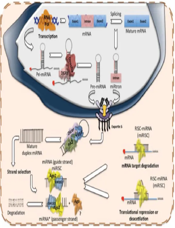
Most of the genes coding for miRNAs are located within intronic regions of known genes (about 80%), suggesting the presence of a convenient mechanism for the coordinated expression of the miRNA and the target protein; the remaining part is positioned in exon or intergenic regions of about 1kb (Rodriguez et al., 2004). Moreover, approximately 50% of the known miRNAs is located in close proximity to other miRNAs: generally, they form clusters of 2-7 genes and present a high similar expression profile (Lagos-Quintana et al., 2001). These groups of miRNAs can be individually transcribed through its own specific promoter and regulated by the same enhancer, or transcribed in the form of a single polycistronic units drive by a single promoter and divided during the maturation process (Lee et al., 2002). The clusters of miRNA genes are the result of segmental duplication events, forming the repeated-derived miRNA families (RdmiRs) (Yuan et al., 2011). Over 500 miRNA genes have been experimentally validated and over 1000 miRNA genes predicted through computer programs in human, representing about the 2% of the whole genome (Li and Zhang, 2015). Recently, scientists have isolated few microRNA genes transcribed by RNA polymerase type III (Borchert et al., 2006) but in the majority of the cases, the RNA polymerase type II transcribes genes coding for miRNAs, as demonstrated by the work of Lee and his group. The primary transcripts, known as “pri-miRNA”, can
35
reach sizes > 1kb: they present the poly-A tail at the 3’-end and the cap of 7-methyl guanosine at the 5’-end, in addition to a complex tertiary structure with different hairpins and loops (Lee et al., 2004) (Fig. 7).
Pri-miRNA undergo to a complex process of maturation in two steps, highly compartmentalized. The process begins in the nucleus through a Microprocessor complex, a protein multi-enzymatic complex of about 650 kDa (Gregory et al., 2004). The complex presents different copies of a protein that binds dsRNA in proximity of the loops: this protein is known as DGCR8 in mammalian and Pasha in Drosophila and C. elegans (Han et al., 2004). Once bound, the pri-miRNA is processed by Drosha, a Microprocessor complex protein characterized by two Rnasi III domains. Drosha cleaves the transcript 11 bp distance from the stem-ssRNA junction, generating a 2 nt overhang on the 3’ end, which is recognized by the successive protein of the maturation process (Han et al., 2006). The product of the cleavage is a hairpin-shaped precursor of dsRNA of about 70 nt, known as “pre-miRNA” (Lee et al., 2003) (Fig. 7).The second step of the maturation process takes place in the cytoplasm; therefore, the pre-miRNA must be transferred from the nucleus to the cytoplasm. The enzyme complex Exportin-5, located in the inner membrane of the nuclear pore, mediates the passage. The protein belongs to a family of receptors able to bind cooperatively the molecule to be exported and a cofactor Ran linked to a molecule of GTP from which derive the energy required for the cytoplasm transfer (Bohnsack et al., 2004). Exportin-5 recognizes the pre-miRNA by the presence of the stem-loop and the overhang of two nucleotides at the 3’ (Lund et al., 2004).
Once in the cytoplasm, Dicer processes the pre-miRNA, an RNase type III very similar to Drosha (Knight et al., 2001). Through PAZ domain, Dicer recognizes the 2 nt overhang at
36
the 3’ end and cleaves the pre-miRNA 22 nt distance from the protrusion. The product is miRNA duplex of about 22 nt without any stem loop (Ma et al., 2004) (Fig. 7). The miRNA duplex immediately becomes part of the ribonucleoprotein complex miRISC (miRNA-containing RNA-induced silencing complex) that contributes to complete the maturation process and supports the miRNA regulation of gene expression.
The pre-RISC complex is composed by Dicer, the core of the complex, TRBP and PACT, two dsRNA binding proteins, Argonaute proteins (Ago 1-4 in mammalian cells) and many other proteins (Redfern et al., 2013).
TRBP and PACT bind the miRNA duplex, recall in site Ago 2 and the other Argonaute proteins and participate to the silence of the gene expression (Thimmaiah et al., 2005; Lee et al., 2006). In particular, Ago1 is responsible for the last step of the miRNA maturation process: it is a RNA endonuclease, which cleaves the passenger strand of the duplex (indicate as miRNA*) (Baumberger et al., 2005), while the helicase Gemin3 separated the two miRNA strands (Mourelatos et al., 2002).
The presence of mismatches, internal loops or G:U pairs could influence the choice of the miRNA guide strand: in most cases, the guide strand is the one whose 5’ end is less tightly paired to its complement. The passenger strand miRNA* can be removed by the helicase or degraded (Schwarz et al., 2003). The guide ssmiRNA of about 22 nt becomes part of the definitive miRISC complex and represents the mature form of the miRNA able to silence the gene expression (Fig. 7).
37
Figure 7: Biogenesis of miRNAs.Production of miRNAs starts in the nucleus with the polymerization of the
primary hairpin miRNA transcript (pri-miRNA) by RNA polymerase II or III, followed by the cleavage and digestion of the pri-miRNA by the microprocessor complex (Drosha–DGCR8). The resulting transcript is the pre-miRNA, which is exported to the cytoplasm by Exportin-5–Ran-GTP. Once in the cytoplasm, Dicer, TRBP and Paz proteins cleave the pre-miRNA hairpin and digest it to produce a mature duplex miRNA. Then, one of the strands is loaded onto the RISC complex and finally this guides the miRNA to its mRNA target to silence it by direct degradation or by translational repression.
38
3.1.3. MiRNA structure
The regulation of gene expression by miRNA is carried out by its pairing with the target mRNA. Experimentally scientists have observed that the miRNA structure regulates the pairing mode (Fig. 8):
• at the 5’ end, miRNAs present the "seed" region, corresponding to the first 2-8 nt of the molecule, which are characterized to be perfectly complementary to the region 3' UTR of the target mRNA, coating a fundamental role in silencing (Lewis BP et al, 2003),
• the downstream region contains "bulges" and "loops" derived by the formation of structural mismatch in the miRNA-mRNA pairing (Lewis et al., 2003),
• the miRNA 3’ end shows a poor complementarity to mRNA but it can provide compensating sites for the binding of the target molecule, including thanks to the formation of G:U pairs (Brennecke et al., 2005).
Figure 8: Structure of microRNAs and pairing with the target m RNA.
These characteristics have been used for the construction of the predictive computer programs of microRNA target, such as miRANDA, Targetscan and Targetminer: these
39
programs have shown that about 30% of human genes is a possible target of the silencing action of microRNAs (Lewis et al., 2005).
3.1.4. MiRNAs are gene expression mediators
MiRNAs are involved in regulating gene expression of the cells via gene silencing of target mRNAs. Generally, miRNAs act at the post-transcriptional level, pairing to specific sequences present on the target mRNA called MRE (miRNA Recognition Element), place most frequently in the 3’UTR region (Lytle et al., 2007). The regulatory mechanism essentially depends on the degree of complementarity between miRNA and mRNA
(Hutvagner and Zamore, 2002).
When the complementarity between the two molecules is imperfect and rather unstable, miRNA carries out a translational repression mechanism. The process can take place in different ways and proteins of the Argonaute family of the miRISC complex, Ago1 and Ago2, coat a key role:
• the silencing can be implemented in the initial phase of translation after the bond between Ago2 and eIF4E, protein involved in the recognition of the 7-methyl-guanosine cap at the 5’ end of the mRNA, a necessary step for the recruitment of messenger in the ribosome, or, following the ATP-dependent deadenilation. Ago1 can recognize the cap through the support of the GW182 protein, going to prevent the assembling of small and large subunits of the ribosome (Iwasaki and Tomari, 2009) (Fig. 9A-B);
40
• the repression can also be performed during the elongation of the polypeptide chain by inducing a premature termination signal or promoting the degradation of nascent polypeptides (Petersen et al., 2006) (Fig. 9C).
The translationally repressed mRNAs are sequestered in P bodies or stress granules in association with several proteins and can be translated later (Liu et al., 2005).
Figure 9. Gene expression silencing by miRNAs associated with miRISC via two different mechanisms: (A)
translational repression in the early stage of the translation with the binding of eIF4E protein. (B) The repression through the association impediment of ribosomal subunits (C) or in the phase of elongation of the polypeptide; the degradation of the messenger thanks to an endonucleotidic cut of the mRNA (D) or following the deadenilation (E).
On the contrary, when the complementarity between miRNA and mRNA is sufficiently high thus leading to the formation of a stable interaction, the mRNA is degraded. The process could happen in three main ways:
• miRNA can promote the mRNA cleavage by the miRISC protein complex: the cut, generally made by Ago 2, takes place between the 10th and the 11th residue and generates two RNA fragments with unprotected ends, easily attacked by exonucleases (Zamore et al., 2000) (Fig. 9D);