Alma Mater Studiorum – Università di Bologna
DOTTORATO DI RICERCA IN
SCIENZE VETERINARIE
Ciclo XXVII
Settore Concorsuale di afferenza: 07/H3
Settore Scientifico disciplinare: VET/05
TITOLO TESI
Development of new molecular methods for the diagnosis
and the study of viral diseases of fish
Presentata da: Ana Cristina de Aguiar Saldanha Pinheiro
Coordinatore Dottorato Relatore
Prof. Carlo Tamanini Dott.ssa Sara Ciulli
CONTENTS
ABSTRACT 1
CHAPTER I - Viral diseases of fish: molecular methods for diagnosis 4
1.1 Introduction 5
1.2 Fish Viruses 7
1.2.1 RNA Viruses of Fish 8
1.2.1.1 Rhabdoviridae 8 1.2.1.2 Paramyxoviridae 11 1.2.1.3 Orthomyxoviridae 12 1.2.1.4 Picornaviridae 13 1.2.1.5 Nodaviridae 14 1.2.1.6 Nidovirales 16 1.2.1.7 Togaviridae 17 1.2.1.8 Caliciviridae 18 1.2.1.9 Retroviridae 19 1.2.1.10 Reoviridae 22 1.2.1.11 Birnaviridae 23
1.2.2 DNA Viruses of Fish 25
1.2.2.1 Iridoviridae 25
1.2.2.2 Herpesviridae 26
1.2.2.3 Adenoviridae 28
1.3 Molecular tools for viral fish diseases diagnosis 29
1.3.1 Conventional Polymerase chain reaction 30
1.3.2 Reverse transcriptase polymerase chain reaction 33
1.3.3 Nested PCR and Semi-Nested PCR 35
1.3.4 Multiplex PCR 36
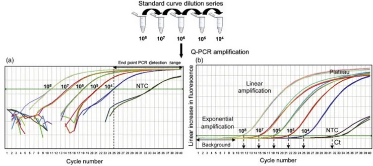
1.3.5 Real-Time PCR 37
1.3.7 Microarray Technology 47
1.3.8 Nucleic acid sequence based amplification 48
1.4 Conclusion 50
1.5 References 51
CHAPTER II 80
Development and application of a Real Time PCR assay for the detection and quantification of lymphocystis disease virus 80
2.1 Abstract 81
2.2 Introduction 82
2.3 Material and Methods 84
2.3.1 Fish samples 84
2.3.2 Virological analysis 85
2.3.3 DNA extraction 86
2.3.4 Conventional PCR, sequencing, cloning and standard preparation of DNA 86
2.3.5. Real-time qPCR assay 87
2.3.6. Quantitation of LCDV load in diseased and asymptomatic fish samples 90
2.4 Results 91
2.4.1. Virological analysis 91
2.4.2. Conventional PCR, sequencing and genotyping 91
2.4.3. Standard curve and reproducibility assays 97
2.4.4. Viral load in diseased and asymptomatic fish 100
2.5 Discussion 101
2.6 Acknowledgments 105
2.7 References 106
CHAPTER III 111
Development of a multiplex RT-PCR assay for simultaneous detection of the major viruses that affect rainbow trout (Oncorhynchus mykiss) 111
3.1 Abstract 112
3.3 Material and Methods 115
3.3.1 Virus titration 115
3.3.2 RNA extraction 115
3.3.3 Primers 116
3.3.4 Optimization of the multiplex RT-PCR 117
3.3.5 Application of the mRT-PCR to clinical samples 117
3.4 Results and Discussion 118
3.5 Acknowledgments 123
3.6 References 123
1
ABSTRACT
The increase in aquaculture operations worldwide has provided new opportunities for the transmission of aquatic viruses; the occurrence of viral diseases remains a significant limiting factor in aquaculture production and for the sustainability of biodiversity in the natural environment. Viruses that infect fish and cause disease are represented in 14 of the families listed for vertebrate viruses by the International Committee on the Taxonomy of Viruses. The viruses are the principal pathogens that are negatively impacting aquaculture both from an economic and sanitary point of view. For this reason, the ability to identify quickly the presence/absence of a pathogenic organism in fish would have significant advantages for the aquaculture systems. At present, the use of cultured eukaryotic cells in a controlled environment for virus propagation and isolation is widely accepted and is still considered the ‘gold standard’ for diagnosis of most viral diseases. The exponential growth of viruses in a cell culture system makes it a very sensitive method for virus detection if the cells used are of high susceptibility and quality, and if the cells are handled by experienced personnel. Furthermore, after the viral growth virus need to be identified and
serological techniques have traditionally played this role. However, for correct
diagnosis, several days of culture are required for cytopathic effects to be observed and subsequent virus identification. Molecular techniques have in recent years alongside the traditional ones, and in some cases, they have replaced traditional methods for the highest advantages in terms of speed and sensitivity of the diagnosis provided. Several molecular methods have found successful application in fish pathology both for confirmatory diagnosis of overt diseases and also for detection of asymptomatic infections. However, a lot of different variants occur among fish host species and virus strains and consequently specific methods need to be developed and optimized for each pathogen and often also for each host species.
For this reason this thesis focus on the molecular diagnosis of some relevant fish viral infections. The PhD dissertation consists of three chapters, the first chapter is a detailed
2
review entitled “Viral diseases of fish: molecular methods for diagnosis”. This work presents a complete description of the viruses that infect fish and cause disease taking
an overview of viruses belonging to 14 different viral families and provides a relevant
information regarding the most common methods and emerging technologies for the molecular diagnosis of viral diseases of fish. Moreover, the molecular methods currently available for diagnosis of viral infections in fish are reported and the advantages and disadvantages that they offer over conventional methods previously available are discussed.
The second and third chapters of the thesis consist of two experimental research for the set up of innovative techniques for the diagnosis of relevant fish diseases.
The second chapter is entitled “Development and application of a real time PCR assay for the detection and quantification of lymphocystis disease virus”. Lymphocystis disease virus (LCDV) is responsible of a chronic self-limiting disease affecting more than 125 teleosts. Viral isolation of LCDV is difficult, time consuming and often ineffective; the development of a rapid and specific tool to detect and quantify LCDV is desirable for both diagnosis and pathogenic study. In this study, a quantitative real-time PCR (qPCR) assay was developed using a Sybr Green based assay targeting a highly conserved region of MCP gene. Primers were designed on a multiple alignment, including all known LCDV genotypes. The viral DNA segment was cloned within a plasmid to generate a standard curve. Limit of detection was as low as 2.6 DNA copies/µl of plasmid and the qPCR was able to detect viral DNA from cell culture lysates and tissues 10 times lower than conventional PCR. Both gilthead seabream and olive flounder LCDV has been amplified and in silico assay showed that LCDV of all genotypes can be amplified. LCDV was detected in target and non-target tissues of both symptomatic and asymptomatic fish. The LCDV qPCR revealed to be highly sensitive, specific, reproducible and versatile to detect and quantify Lymphocystivirus and may be used also for asymptomatic carriers’ detection or pathogenesis study of different LCDV strains. The results of this study were recently published in Journal
3
The third chapter is entitled: “Development of a multiplex RT-PCR assay for simultaneous detection of the major viruses that affect rainbow trout (Oncorhynchus
mykiss)”. In the last 10 years, rainbow trout (Oncorhynchus mykiss) has represented
the second highest annually produced product in European aquaculture. The major viral diseases that affect rainbow trout are viral haemorrhagic septicaemia (VHS), infectious haematopoietic necrosis (IHN), infectious pancreatic necrosis (IPN) and sleeping disease (SD). In the presented study, we developed a multiplex RT-PCR (mRT-PCR) assay for the simultaneous detection of these four rainbow trout viruses in a single assay. The choice of primers was carried out based on the expected size of the fragments, the temperature and time required for the amplification, and the specificity for the target sequence. First, the method was optimized using reference strains of viral haemorrhagic septicaemia virus (VHSV), infectious haematopoietic necrosis virus (IHNV), infectious pancreatic necrosis virus (IPNV) and sleeping disease virus (SDV) cultivated with permissive cell culture lines; subsequently, the method was used for the identification of these viral infections in rainbow trout samples. Twenty-two samples of rainbow trout, clinically suspected of having viruses were analyzed by the developed method to detect the presence of the four viruses, by directly analyzing the animal tissues. The mRT-PCR method was able to efficiently detect the viral RNA in infected cell culture supernatants and in tissue samples, highlighting the presence of single infections as well as co-infections in rainbow trout samples. VHSV/SDV and IHNV/SDV co-infections were demonstrated for the first time in rainbow trout. The mRT-PCR method was revealed to be an accurate and fast method to support traditional diagnostic techniques in the diagnosis of major viral diseases of rainbow trout.
4
CHAPTER I
5
1.1 Introduction
Over the past three decades, aquaculture has developed to become the fastest-growing food-producing sector in the world with 63.6 million tons production and 8.8 % annual growth rate. Driven by population growth, rising demand for seafood and a levelling of production from capture fisheries, the practice of farming aquatic animals has expanded rapidly to become a major global industry (FAO, 2012).
Farming of aquatic animals commonly involves displacement from their natural habitat to an environment that is new and sometimes stressful, the use of feeds that are sometimes live and often unnatural or artificial, and culture in stocking densities that are much higher than occur naturally. This has provided opportunities for exposure to new pathogens and conditions that can compromise defensive responses and facilitate pathogen replication and disease transmission (Walker and Mohan, 2009). Most importantly, the growth in aquaculture and increasing international trade in seafood has resulted in the rapid movement of aquatic animals and their products, with associated risks of the trans-boundary movement of pathogens (Walker and Winton, 2010).
The infectious diseases, represents the most limiting factor to aquaculture production because they increase the production cost due to the losses in dead fish, costs of treatments or decreased growth rate of diseased and convalescent fish. It is difficult to evaluate the real economic losses, due to the different factors related; however, it has been estimated that 10% of all cultured aquatic animals are lost as a result of infectious diseases (Blanco et al., 2000). Among the causative agents of infectious diseases in aquaculture, the viruses are the principal pathogens that are negatively impacting aquaculture. Viral diseases have been difficult to control, due to the high susceptibility of aquatic animals at an early age, the lack of therapeutics, insufficient knowledge of the pathogenesis of viral infections and limited knowledge of natural resistance mechanisms in aquatic animals (Kibenge et al., 2012).
6
Furthermore, aquaculture has been expanded, intensified, and diversified, based heavily on movements of live aquatic animals and animal products (broodstock, seed and feed). The world trade liberalization has accelerated the accidental spread and incursion of diseases into new populations and geographic regions.
Therefore, rapid detection and identification of pathogens is crucial to prevent viral transmission of disease and an effective disease management (Adam and Thompson, 2006).
Detection of aquatic animal viruses historically has been done by growth and isolation of viruses in living cell cultures appropriately chosen for the propagation of target virus. Subsequent virus identification through immunological or nucleotide procedures are then requested. The determination of a testing procedure is a complex decision involving factors of cost, timeliness, sensitivity, specificity, efficiency, and available host tissue and technology (Peters, 2004).
Many viral pathogens of animals are poorly characterized. To date, if a suspected new virus was identified and the virus could be cultured, morphology, physical characteristics, growth characteristics and antigenic nature were determined. However, this method of characterization is very time consuming and is limited to culturable viruses (in established cell lines or readily available primary cells). Usually, because of the time and expense, this characterization is limited to viruses that are associated with an important disease. However, a large portion of viruses is either unculturable, difficult to culture or are not associated with a disease of importance to justify in depth characterization or development of reliable serological reagents. Therefore, the development of broad spectrum diagnostic methods that obviate culture are needed as well as methods to bypass the cumbersome traditional methods of characterizing culturable viruses (Hanson et al., 2006).
Last fifteen years, great advances took place in understanding the molecular biology of fish pathogens and their hosts. Molecular biology has become a routine tool in the search for improved methods of diagnosis and control of fish diseases and for the study of the epidemiology of viral, bacterial, and parasitic diseases. Detection of
7
nucleic acid molecules has demonstrated its usefulness for highlighting hardly culturable, non-culturable, and even dead microorganisms, generating appropriate novel or replacement technologies. The main advantages of molecular techniques are its higher sensitivity and specificity compared with other diagnostic methods such as serological assays and even culture methods in several cases, as well as its possibility to rapidly screen large numbers of samples during disease outbreaks (Cobo, 2012).
From an epidemiological point of view, the ability to screen rapidly numerous samples against pathogens are very important to prevent viral transmission of disease. Moreover, using nucleic acid as targets, molecular techniques can offer a tool for examining the relationships between genotypes of various pathogens, providing essential data for molecular epidemiology studies (Altinok and Kurt, 2003).
1.2 Fish Viruses
Viruses that infect finfish and cause disease are represented in 14 of the families listed for vertebrate viruses by the International Committee on the Taxonomy of Viruses (ICTV). The fish viruses containing DNA genomes are listed in the families
Iridoviridae, Adenoviridae, and Herpesviridae, and those with RNA genomes are listed
in the families Picornaviridae, Birnaviridae, Reoviridae, Rhabdoviridae,
Orthomyxoviridae, Paramyxoviridae, Caliciviridae, Togaviridae, Nodaviridae,
8 1.2.1 RNA Viruses of Fish
1.2.1.1 Rhabdoviridae
Fish rhabdoviruses are considered an important viral pathogen that affecting both wild and cultured fish throughout North America, Asia, and Europe (Kurath and Winton, 2008; Purcell, 2012). The first fish rhabdovirus was described in 1938 by Schaperclaus in European rainbow trout (Oncorhynchus mykiss). Since then, these viruses have been isolated, grown in tissue culture cells, and the genomes have been cloned and sequenced (Leong, 2008).
Members of this group are bullet-shaped, enveloped viruses and share a number of distinct features, including a simple negative-sense, single-stranded RNA (ssRNA) genome. The typical rhabdoviral genome encodes five basic structural proteins including the nucleoprotein (N), phosphoprotein (P), matrix protein (M), glycoprotein (G) and the large polymerase (L) protein (Fig. 1) (Bernard and Brémont, 1995). Three distinct genera of fish rhabdoviruses have been identified: Novirhabdovirus and
Perhabdovirus, representatives of which have all been isolated from fish hosts, and
Vesiculovirus (Whitfield et al., 2011).
Figure 1. The typical rhabdoviral genome. Negative-stranded RNA linear genome, about 11-15 kb
9
Members of the genus Novirhabdovirus are distinguished by the presence a sixth gene located in the genome between the G and the L genes (Fig. 2) encoding a non-structural (‘non-virion’ or NV) protein (Purcell, 2012). It has four important type species: Infectious hematopoietic necrosis virus (IHNV), Viral haemorrhagic
septicaemia virus (VHSV), Hirame rhabdovirus (HIRVV) and Snakehead rhabdovirus
(SHRV) (Gadd, 2013). Additional fish rhabdoviruses that are considered possible members of the Novirhabdovirus genus include eel viruses EEV-B12 and EEV-C26, but these require further genetic characterization to define their taxonomic status. The viruses in the four Novirhabdovirus species differ in several important aspects, including host and geographic range. Both IHNV and VHSV are globally important fish pathogens that are found in wild fish and cause significant disease burdens in fish reared in various aquaculture settings. IHNV has a relatively narrow host range restricted to cold-water salmon and trout fish, collectively referred to as ‘Salmonids’, in the families Oncorhynchus and Salmo. IHNV is endemic to western North America and it has been inadvertently spread to Europe and Asia by aquaculture related activities (Alonso et al., 2003). In contrast, VHSV has extremely broad host range including 80 marine and freshwater fish species from diverse taxonomic families in Europe, North America and Asia. Although the major burden of VHSV in aquaculture is the disease in rainbow trout (Oncorhynchus mykiss) (Kurath and Winton, 2011). IHNV and VHSV are among the seven finfish viral species listed as 'notificable' by the Aquatic Animal Health Code of the World Organization for Animal Health (OIE, 2014a) indicating their recognition as serious pathogen threats to global animal production systems. HIRVV affects hirame, the Japanese flounder (Paralychthys
olivaceus), which is a highly prized food fish in Japan. Its host range includes ayu (Pleuroglossus altivelis) as well as salmonid fish, but it has been isolated outside of Asia (Leong, 2008). Snakehead rhabdovirus (SHRV), a rhabdovirus of warm-water fish, was isolated from a diseased snakehead fish (Ophicephalus striatus) during an epizootic outbreak in Thailand (Kasornchandra et al., 1992).
10
Figure 2. Novirhabdovirus genome. Negative-stranded RNA linear genome, about 11kb in size.
Encodes for six proteins (http://viralzone.expasy.org/viralzone/all_by_species/76.html).
The genus Vesiculovirus comprises an ecologically diverse but genetically similar group of mammalian and fish rhabdoviruses. It currently contains the recognized species Spring viremia of carp virus (SVCV) as the type species and the tentative vesiculoviruses pike fry rhabdovirus (PFRV). However, a new genus has been proposed with the name Sprivivirus and consist of both SVCV and PFRV (ICTV, 2013). Spring viremia of carp virus (SVCV) causes a highly contagious and serious disease of freshwater cyprinid fishes, generating significant economic and ecological impacts throughout the world (Phelps et al., 2012). SVCV was first identified as the etiologic agent of an acute hemorrhagic disease in common carp (Cyprinus carpio) in Europe in 1972. Since then, the disease has been found in Asia, the Middle East, and most recently in South and North America (Leong, 2008). A growing number of fish viruses are related to viruses from the genus Vesiculovirus, such as the Siniperca chuatsi rhabdovirus (SCRV) and the Starry flounder rhabdovirus (SFRV) (Tao et al., 2008; Talbi et al., 2011). The Scopthalmus maximus rhabdovirus (SMRV), originally isolated from farmed turbot (Scophthalmus maximus) affected by lethal haemorrhagic disease in China, also has a phylogenetic relationship with the genus Vesiculovirus, but is genetically distinct from other rhabdoviruses (Zhang et al., 2007; Zhu et al., 2011). A new genus of fish rhabdoviruses recognized since 2013 is Perhabdovirus. It has three species: Perch rhabdovirus (PRV), the type species, Anguillid Rhabdovirus (AngRV) and Sea trout rhabdovirus (STRV). Perhabdoviruses share morphological characteristics, genome organization and sequence similarities with vesiculoviruses and with viruses in the newly proposed genus Sprivivirus (Gadd, 2013).
11 1.2.1.2 Paramyxoviridae
Members of Paramyxoviridae are causative agents of a number of diseases, with a host range that includes mammals, birds, reptiles and fish. The family constitutes a diverse group of enveloped viruses, which possess non-segmented, single stranded, negative sense RNA genomes (Lamb and Kolakofsky, 2001).
The first description of a paramyxovirus-like virus in fish was reported by Winton et al., in 1985 during a routine health assessment of Chinook salmon juveniles in Oregon. This virus, now named Pacific salmon paramyxovirus (PSPV) grow slowly in established fish cell lines and have not been associated with disease in Salmonids (Batts et al., 2008).
Miyazaki et al., in 1989 reported another fish paramyxovirus that caused epidermal necrosis in juvenile black sea bream (Acanthopargrus schlegeli), this virus was identified in Japan by electron microscopy, but it was never cultured in vitro (Miyazaki et al., 1989).
Atlantic salmon paramyxovirus (ASPV), a relatively recent addition to the
family Paramyxoviridae, was first isolated in 1995, from Atlantic salmon (Salmo
salar) suffering from proliferative gill inflammation (PGI) (Kvellestad et al., 2003).
PGI is a respiratory disease of Atlantic salmon, and has been associated with losses in Norwegian aquaculture since the 1980s, with an increase of outbreaks in the past years. The aetiology of the disease appears to be multifactoral. A primary causative agent has not yet been identified, but ASPV is associated with some cases (Kvellestad et al., 2005). Recently, the Atlantic salmon paramyxovirus (ASPV) has been proposed as a species in the new genus Aquaparamyxovirus of the family Paramyxoviridae (Batts et
12 1.2.1.3 Orthomyxoviridae
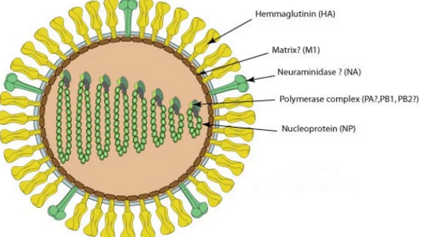
Infectious salmon anemia virus (ISAV) is the only fish orthomyxovirus that has been fully described to date. ISAV virions are pleiomorphic and enveloped with a diameter of 100–130 nm and 10–12 nm surface projections (Fig. 3).
Figure 3. Structure of infectious salmon anemia virus (ISAV). The virions are enveloped and are
90-130 nm in diameter (http://viralzone.expasy.org/viralzone/all_by_species/223.html). .
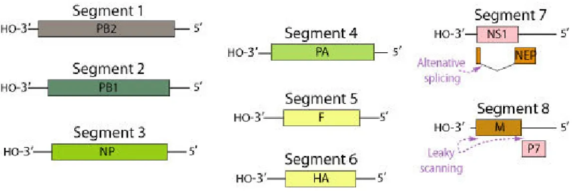
The genome consists of eight single-stranded RNA segments: segment 1 encodes PB2, a component of the virion RNA polymerase; segment 2 encodes PB1; segment 3, the nucleocapsid protein NP; segment 4, the RNA polymerase PA; segment 5, acetylcholinesterase P3 or fusion protein; segment 6, hemagglutinin; segment 7, protein P4 and P5; and segment 8, proteins P6 and P7 (Fig. 4) (Leong, 2008).
Sequence analysis of these segments from different ISAV isolates consistently reveals two genotypes designated according to their geographic origin as European (Genotype I) and North American (GenotypeII) (Godoy et al., 2008). A comparative sequence analysis of the PB1 gene of ISAV and other members of the
Orthomyxoviridae led to its assignment as the type species of a new genus Isavirus (Leong, 2008).
13
Figure 4. ISAV genome. Segmented ssRNA(-) linear genome, encapsidated by nucleoprotein (NP)
Contains 8 segments coding for at least 8 proteins (http://viralzone.expasy.org/viralzone/all_by_species/76.html).
ISAV causes a highly lethal disease, which affected farmed Atlantic salmon displaying severe anemia, leucopenia, ascetic fluids, hemorrhagic liver necrosis, and petecchiae of the viscera. The virus remains an emerging fish pathogen because of the asymptomatic infections in wild and farmed fish and the potential for emergence of new epizootic strains (Godoy et al., 2014). Natural outbreaks of ISA have only been recorded in farmed Atlantic salmon, and in Coho salmon (Oncorhynchus kisutch) in Chile (Kibenge et al, 2001). Subclinically infected feral Atlantic salmon and sea trout (S. trutta) have been identified (Kibenge et al., 2004). Following experimental infection by bath immersion, ISAV has been detected by RT-PCR in rainbow trout (Biacchesi et al., 2007), Atlantic herring (Clupea harengus) and Atlantic salmon (OIE, 2014b).
1.2.1.4 Picornaviridae
The first reported observation of picorna-like viruses in fish was made in 1988 from rainbow smelt (Osmerus mordax) in New Brunswick, Canada (Moore et al., 1988). Since then, picornaviruses have been isolated from barramundi (Lates
14
calcarifer), turbot, sea bass (Dicentrarchus labrax), grass carp (Ctenopharyngodon
idella), bluegill (Lepomis macrochirus), grouper (Epinephelus tauvina), Japanese
parrotfish (Oplegnathus fasciatus), and salmonid fish. In most of these descriptions, the presumptive characterization of the etiologic agent as a picornavirus was based on growth in tissue culture cells and the observation of crystalline arrays in the cytoplasm of small virus particles with a size and morphology consistent with picornaviruses (Leong, 2008).
Members of the Picornaviridae are small (22–30 nm), non-enveloped viruses with icosahedral symmetry. Virions replicate cytoplasmically, form cytoplasmic inclusions and contain a single-strand of positive sense RNA (Iwanowicz et al., 2000). A novel picornavirus was isolated from specimens of a diseased European eel (Anguilla
anguilla) collected from Lake Constance in the Rhine River. This eel presented
symptoms of haemorrhages at the head and the tail. EPV has a typical picornavirus genome layout, but its low similarity to known viral proteins suggests a novel species in the family Picornaviridae (Fichtner et al., 2013). In many cases, sick fish infected with these viruses contain picorna-like virus particles in the brain and medulla and the victims display corkscrew-like swimming and eventually death (Leong, 2008).
1.2.1.5 Nodaviridae
All nodaviruses characterized from fish belong to the genus Betanodavirus which, together with the genus Alphanodavirus whose members infect insects, constitute the family Nodaviridae. Betanodavirus particles are small (20–30 nm), with an icosahedral shaped capsid and a naked single stranded RNA (positive sense) genome consisting of the two segments RNA1 and RNA2. The RNA1 (ca 3.1Kb) encodes the RNA-dependent RNA polymerase (RdRp) and the RNAi antagonist protein B2, whereas the RNA2 (ca 1.4 kb) encodes the capsid protein (Fig. 5) (Munday et al., 1992).
15
Figure 5. Segmented, bipartite linear, ssRNA(+) genome composed of RNA1=3.1 kb and RNA2=1.4
kb. Each genome segment 5’ end is capped. The 3’end has no poly (A) tract (http://viralzone.expasy.org/all_by_species/614.html).
The betanodaviruses are the causative agents of viral nervous necrosis (VNN) or viral encephalopathy and retinopathy (VER) in a variety of farmed marine fish. Yoshikoshi and Inoue (1990) were the first to report viral nervous necrosis (VNN) in hatchery-reared larvae and juveniles of Japanese parrotfish (Oplegnathus fasciatus), the disease was named VNN due to the clinical signs. To date, the disease has been reported in more than 50 fish species, mainly marine with the greatest impact being in striped jack (Pseudocaranx dentex), European sea bass (Dicentrarchus labrax), groupers, and flatfishes (Munday et al., 2002; Sano et al., 2011). The disease mainly affects the larval and juvenile stages. Brain, spinal cord and retina are considered the target organs in which the virus actively replicates causing extensive tissue vacuolization (Munday et al., 2002). Genomic classifications of the betanodaviruses, traditionally, group these into four subtypes designated Barfin flounder nervous
necrosis virus (BFNNV), Striped jack nervous necrosis virus (SJNNV), Red-spotted
grouper nervous necrosis virus (RGNNV), and Tiger puffer nervous necrosis virus
(TPNNV) (Nishizawa et al., 1997). A novel subtype of nodavirus from turbot (Psetta
maxima), was recently described (Johansen et al., 2004) and is pending recognition as
a fifth subtype within the betanodaviruses (Hodneland et al., 2011).
Among the betanodavirus genotypes, the host range of TPNNV is limited to tiger puffer (Takifugu rubripes), whereas SJNNV which was initially isolated almost
16
exclusively in striped jack, have been detected in farm of sea bass (Dicentrarchus
labrax), sea bream (Sparus aurata) and Senegalese sole (Solea senegalensis) (Mori et
al., 1992; Munday et al., 2002). The BFNNV genotype is regarded as the cold-water clade and several species from Northern-Europe, Atlantic coast of North America and Japan, have been shown to be susceptible to this genotype. Virus from this clade has been isolated from cold-water species such as Pacific cod (Gadus microcephalus) (Nishizawa et al., 1997), winter flounder (Pleuronectes americanus) (Barker et al., 2002), Atlantic cod (Gadus morhua) and Dover sole (Solea solea) (Starkey et al., 2001), Atlantic halibut (Hippoglossus Hippoglossus) (Grotmol et al., 1995) and haddock (Melanogrammus aeglefinus) (Johnson et al., 2002). RGNNV genotype was found to have a broad host range causing disease in a wide variety of warm water fish species, particularly groupers and sea bass (Shetty et al., 2012). Furthermore, the salinity seems to have low influence on the occurrence of the disease, some studies, in fact, have shown susceptibility of fish in freshwater (Hegde et al., 2003) or fish reared in freshwater (Athanassopoulou et al., 2003).
1.2.1.6 Nidovirales
The family Coronaviridae comprises two genera, Coronavirus and Torovirus, and is classified together with the families Arteriviridae and Roniviridae in the order
Nidovirales. Members of the Coronaviridae share the common feature of pleomorphic,
enveloped virions with diameters of 126–160 nm and prominent surface projections. The nucleocapsid is helical and contains a single molecule of linear, positive sense ssRNA (Leong, 2008).
Two nidoviruses have been detected in fish, the White bream virus (WBV) and the fathead minnow nidovirus (FHMNV). The first report of White bream virus (WBV) was recovered from a white bream (Blicca bjoerkna L.) collected during a routine examination of wild fish (Granzow et al., 2001). A comprehensive analysis of the complete WBV genome (Schutze et al., 2006) revealed that the virus was sufficiently
17
distinct to represent the type species (White bream virus) of a novel genus Bafinivirus within the subfamily Torovirinae of the family Coronaviridae (De Groot et al., 2012). FHMNV has been isolated from both healthy and diseased fathead minnows (Pimephales promelas) in the midwestern portion of the United States. The isolate was initially characterized as a bacilliform virus, possibly belonging to the family
Rhabdoviridae, although the virus had the uncharacteristic property of inducing
syncytia in infected cell cultures (Iwanowicz & Goodwin, 2002). Based on phylogenetic analysis FHMNV appears to represent a second species in the genus
Bafinivirus (Batts et al., 2012).
1.2.1.7 Togaviridae
The family Togaviridae comprises the genera Alphavirus and Rubivirus among the vertebrate viruses. The genus Alphavirus includes pathogens of salmonids (McLoughlin & Graham, 2007) and marine mammals (La Linn et al., 2001). These viruses have spherical virions, 70 nm in diameter, with a lipid envelope containing glycoprotein peplomers. The positive-sense ssRNA genome containing two ORFs: one occupying the 5’ two-thirds of the genome and encoding four nonstructural proteins (nsP1, nsP2, nsP3, and nsP4 or RdRp) and the other occupying the 3’ one-third of the genome and encoding five structural proteins including the capsid protein and two or three envelope glycoproteins (Fig. 6) (Powers et al., 2001; McLoughlin & Graham, 2007).
Salmonid alphaviruses (SAVs) are recognized as serious pathogens of farmed Atlantic salmon and rainbow trout in Europe (McLoughlin & Graham, 2007). Strains of salmonid alphavirus (SAV), identified to date have been assigned to six subtypes by phylogenetic studies (designated SAV subtypes 1–6) based on the analysis of partial E2 gene sequence data (Fringuelli et al., 2008).
18
Figure 6. Alphavirus genome structure. Monopartite, linear, ssRNA (+) genome of
11-12 kb (http://viralzone.expasy.org/viralzone/all_by_species/625.html)
To date, all freshwater SAV strains collected from rainbow trout have belonged to SAV subtype 2 (Weston et al., 2005; Fringuelli et al., 2008). Prior to 2011, all genotyped strains of SAV reported from both Atlantic salmon and rainbow trout in Norway have belonged to SAV subtype 3 (Hodneland et al., 2005; Karlsen et al., 2006; Jansen et al., 2010). Consequently, it has been assumed that all Norwegian SAV isolates belong to the SAV 3 cluster. However, in 2011, the detection of SAV subtype 2 like viruses in farmed marine Atlantic salmon was reported for the first time in Norway (Graham et al., 2012). In contrast, viruses belonging to multiple subtypes (SAV 1, 2, 4, 5 and 6) have been reported from marine salmonid production in Ireland and Scotland (Fringuelli et al., 2008; Graham et al, 2012).
1.2.1.8 Caliciviridae
Viruses in the family Caliciviridae are known to infect a variety of marine species, including mammals, birds, fish, and invertebrates (Lang et al., 2009). The family Caliciviridae is separated into five genera: Norovirus, Sapovirus, Lagovirus,
19
characterized as marine caliciviruses, which was first isolated from California sea lions (Zalophus californianus) in 1972 (Smith et al., 1973). Later, several species of pinnipeds and cetaceans (including seals, sea lions, walruses, whales, and dolphins), as well as an ocean fish, the opaleye perch (Girella nigricans), have been found to be susceptible to calicivirus infection (Smith et al., 1980).
All the caliciviruses are nonenveloped with icosahedral symmetry. The genome consists of a linear, positive-sense ssRNA (Leong, 2008).
Recently has been identified a novel calicivirus in Atlantic salmon reared in Norway. The virus, named Atlantic salmon calicivirus (ASCV), has a high prevalence in farmed salmon and is found in fish suffering from several diseases and conditions and presumably also in healthy fish. Phylogenetic analysis based on the putative capsid encoding genome region did not cluster the virus with members of the marine
calicivirus subgroup of the Vesivirus genus or any other virus of the known calicivirus
genera or unclassified calicivirus, and may represent a new calicivirus genus (Mikalsen
et al., 2014)
1.2.1.9 Retroviridae
The family Retroviridae consists of two subfamilies, the Orthoretrovirinae, containing six genera, and the Spumaretrovirinae, containing only one genus. The piscine retroviruses constitute the genus Epsilonretrovirus, a genus established within the Orthoretrovirinae precisely to include the piscine retroviruses: Walleye dermal
sarcoma virus (WDSV), Walleye epidermal hyperplasia virus type 1 (WEHV-1),
Walleye epidermal hyperplasia virus type 2 (WEHV-2) (Leong, 2008). Two additional
viruses, perch epidermal hyperplasia virus types 1 and 2 (PEHV-1, PEHV-2), are likely members of this group, but their sequences are incomplete. The exogenous piscine retroviruses, snakehead retrovirus (SnRV) and salmon swim bladder sarcoma-associated virus (SSSV) and the zebrafish endogenous retrovirus (ZFERV) have not
20
yet been assigned to a specific genus (Shen and Steiner, 2004; Rovnak and Quackenbush, 2010).
There are also numerous reports of C-type (retrovirus-like) particles of about 110–150 nm in epidermal papillomas of European smelt (Osmerus eperlanus) and in cells cultured from neurofibromas of damselfish (Pomacentrus partitus) (Leong, 2008). A retrovirus has also been suggested as the etiological agent of plasmacytoid leukemia in chinook salmon and was designated salmon leukemia virus (SLV) (Eaton and Kent, 1992).
Retroviruses from two proliferative skin lesions in walleye (Sander vitreus), walleye dermal sarcoma (WDS) and walleye epidermal hyperplasia (WEV), have been isolated and their sequence determined (Holzschu et al., 1995; Rovnak et al., 2007). These proliferative diseases were first reported by Walker in 1969 in walleye collected from Oneida Lake in New York State and have been reported to occur elsewhere in North America (Walker, 1969; Yamamoto et al., 1985). The most interesting and defining feature of these proliferative diseases is their seasonal cycle (Bowser and Wooster, 1991). The highest incidence of disease occurs throughout the late fall until the spring spawning period at which time the lesions naturally regresses. WEH lesions are broad, flat, translucent plaques of thickened epidermis that range in size from 2 to 3 mm up to 50 mm in diameter. WDS are cutaneous mesenchymal neoplasms that are randomly distributed on the body of the fish, arise from the superficial surface of the scales and range in size from 0.2-1.0 cm in diameter (Rovanak and Quackenbush, 2010). WDSV contained three additional open reading frames, ORF A, ORF B and ORFC. ORF A encodes a D-cyclin homolog (retroviral cyclin) that locates in the nucleus of tumor cells in interchromatic granule clusters. ORF B directly interacts with the receptor for activated C kinase (RACK1) which leads to the activation of the protein kinase C signaling pathway. ORF C encodes a cytoplasmic protein that targets the mitochondria and is associated with apoptosis. It is expressed in regressing tumors when full-length viral RNA is synthesized. (Leong, 2008; Rovnak and Quackenbush 2010).
21
Perch epidermal hyperplasia virus type 1 and type 2 are retroviruses associated with hyperplastic lesions found in yellow perch (Perca flavescens). These lesions are similar to walleye epidermal hyperplasias and occur as thickened plaques on the fish’s body (Lepa and Siwicki, 2011).
The snakehead retrovirus was first reported as a spontaneously productive infection of fish cell line SSN-1 derived from striped snakehead fish (Ophicephalus
striatus). Examination by electron microscopy revealed C-type virus particles. Cell
culture supernatants demonstrated high levels of RT activity and induced a cytopathic effect in the BF-2 cell line derived from bluegill fry (Lepomis machrochirus). All fish from which these cell lines were derived appeared clinically healthy (Frerichs et al., 1991). Experimental infection of juvenile snakehead fish with SnRV showed no lesions in any of the infected fish (Frerichs et al., 1993). The SnRV genome differs from the retroviruses of walleye because it has no ORF between the Unique region in the 5’ LTR (U5) and the gag region (Leong, 2008).
The first outbreak of neoplastic disease involving the swim bladder of Atlantic salmon was observed in 1976, at a commercial fish farm in Scotland (Duncan, 1978). The affected fish were in poor physical condition, had swollen abdomens and presented multinodular masses on the external and internal surfaces of the swim bladder (Lepa and Siwicki, 2011). The swim bladder sarcoma virus (SSSV) provirus is 10.9 kbp in length with a simple gag, pro-pol, env gene arrangement similar to that of murine leukemia virus-like simple retroviruses. Phylogenetic analysis of pol sequences suggests that SSSV is most closely related to the sequenced zebrafish endogenous retrovirus (ZFERV) and that these viruses represent a new group of piscine retroviruses (Leong, 2008).
Zebrafish endogenous retrovirus (ZFERV) was originally isolated from the thymus of zebrafish (Danio rerio). The virus was detected in the sperm of different fish at the same genetic locus, indicating that it is an endogenous virus. The genome of ZFERV is 11.2 kb in length, and, like all retroviruses, contains three principal genetic domains (gag, pro-pol, env), flanked by LTRs. Gag and pro-pol genes are in the same
22
open reading frame. Phylogenetic analysis has shown that ZFERV is closest to murine leukemia virus (MLV)-related retroviruses and to walleye fish retroviruses, although the genome structure is more similar to MLV-related retroviruses (Shen and Steiner, 2004).
1.2.1.10 Reoviridae
Reoviruses that infect aquatic animals are grouped in the genus Aquareovirus in the family Reoviridae and are characterized by a nonenveloped double capsid shell, 11 segments of double-stranded RNA and seven structural proteins (VP1-VP7) which compose the viral particle including the outer capsid and central core (Fang et al., 2005). Aquareoviruses were first isolated in the 1970s from North-American cyprinids and were initially referred to as ‘reoviruslike’ or ‘rotavirus-like’ aquatic viruses (Plumb
et al., 1979). They have subsequently been found in a wide variety of aquatic animals,
including molluscs, finfish and crustaceans. Although these viruses are often isolated from apparently healthy individuals, they can also cause significant clinical signs and even severe disease (Fang et al., 1989). Seven Aquareovirus species have been recognised by the International Committee for the Taxonomy of Viruses (ICTV) (Aquareovirus A to Aquareovirus G), although several other viruses have also been isolated, which may represent additional species (Attoui et al., 2011). Aquareoviruses represent a serious threat to fish breeding and typical clinical signs of infection include haemorrhages, which can be severe (Fang et al., 1989). Grass carp reovirus (GCRV) a strain of Aquareovirus C, causes an important disease in Asia, characterised by severe haemorrhage and up to 80% mortality in fingerling and yearling grass carp (Fang et
al., 1989). Striped bass reovirus (SBRV) included in the species Aquareovirus A was isolated from a moribund striped bass (Morone saxatilis) that was also infected with bacteria (Samal et al., 1990). Golden shiner reovirus (GSRV) is a strain of
Aquareovirus C that was originally isolated in 1977 from a moribund golden shiner
23
with losses of bait fish in the USA, it is nearly identical to a Chinese isolate of GCRV (Attoui et al., 2002). This virus is a significant pathogen of farmed grass carp and fathead minnows (Pimephales promelas) but has also been isolated from wild creek chub fish (Semotilus atromaculatus) in the USA (Goodwin et al., 2006a). American grass carp reovirus (AGCRV) is a new member of the species Aquareovirus G isolated in the USA, from grass carp and golden shiner. This virus was implicated in a winter die-off of grass carp fingerlings on a commercial farm in Arkansas in the USA during 2005. Phylogenetic analyses indicate that golden ide reovirus (GIRV), isolated in Germany represents a second isolate of Aquareovirus G (Neukirch et al., 1999).
1.2.1.11 Birnaviridae
The family Birnaviridae includes four genera: Aquabirnavirus, Avibirnavirus,
Blosnavirus and Entomobirnavirus (ICTV, 2015).
Aquatic birnaviruses are the most abundant and diverse and are grouped in two separate genera: the Aquabirnavirus with the Infectious pancreatic necrosis virus (IPNV) and Tellina virus (TV) type species, and the genus Blosnavirus with the
Blotched snakehead virus (BSNV) type specie (Da costa et al., 2004).
Members of the genus Aquabirnavirus have nonenveloped, icosahedral capsids 60–70nm in diameter, and genomes composed of two segments, A and B, of dsRNA. Segment A encodes a polyprotein which is post-translationally cleaved to form three viral proteins VP2, VP3 and VP4, with VP2 epitopes being responsible for serotype specificity and the target for neutralizing antibodies. Segment B encodes VP1, an RNA-dependent RNA polymerase (Fig. 7) (Dobos et al., 1995).
24
Figure 7. Aquabirnavirus genome structure. Segmented linear dsRNA genome whit 2 segments (A,B)
(http://viralzone.expasy.org/viralzone/all_by_species/571.html).
The type species Infectious pancreatic necrosis virus (IPNV) is the agent of infectious pancreatic necrosis (IPN), a highly contagious viral disease of salmonids, which occurs in all major salmon farming countries (Kibenge et al., 2012). Mortality rates associated with disease outbreaks can be quite variable (5–100%) and it is probable that several host, viral and environmental factors influence the severity of the outbreak (Crane and Hyatt, 2011). Serological classification showed that the IPNV strains are divided into two serogroups (A and B). Within serogroup A, nine distinct serotypes were identified (Hill and Way, 1995; Dixon et al., 2008), whose classification on the basis of deduced amino acid similarities of VP2 demonstrated that the strains clustered into 6 genogroups (Blake et al., 2001) which tend to correlate with geographical and serological characteristics (Crane and Hyatt, 2011). Fish that are exposed to IPNV but either survive disease or do not develop clinical disease may become lifelong carriers of the virus, serving as IPNV reservoirs in populations and transmitting the virus vertically to progeny via the egg (McAllister et al., 1987). IPNV has a wide variety of host species and persistent carriers among recovered hosts, for this reason it is difficult to eradicate, once established (Kibenge et al., 2012).
25 1.2.2 DNA Viruses of Fish
1.2.2.1 Iridoviridae
The family Iridoviridae currently contains five genera: Iridovirus,
Lymphocystivirus, Ranavirus, Megalocystivirus and Chloriridovirus. The genera
Lymphocystivirus, Ranavirus, and Megalocystivirus contain all of the known
iridoviruses that infect fish. Their common features are icosahedral virions, 120–350 nm in diameter that may acquire an envelope, and a viral genome consisting of one molecule of linear dsDNA of 100–303 kbp (Leong, 2008).
Lymphocystis disease virus (LCDV) is the etiological agent of this chronic and self-limited lymphocystis disease (LCD), described in over of 125 species of fish throughout the world (Noga, 2010). The lymphocystis disease occurs both in marine and freshwater fish and is generally associated with stress conditions related to farming, although it is commonly described also in free-living animals (Alonso et al., 2005). Infected fish exhibit macroscopic and nodular lesions on the body surface because of rapid replication and subsequent inhibition of mitosis in the host’s connective tissue cells. The hypertrophied cells, called lymphocysts, are common on skin, fins and occasionally around the mouth (Samalecos, 1986). Although this disease is rarely fatal, the infected fish are more prone to infection by other microorganisms, exhibit anaemia and may have a considerably reduced growth rate (Iwamoto et al., 2002). Therefore, the appearance of this viral infection results in an important economic loss to the aquaculture industry because the diseased fish are not commercially viable. Based on the MCP gene sequence and pathogenicity of lymphocystiviruses, the viruses were divided into nine genotypes. Each genotype includes only one or a very limited number of fish host species: genotype I includes LCDV-1 isolated from the European flounder (Platichtys flesus), genotype II consists of Japanese flounder (Paralichthys olivaceus) isolates, genotype III of rockfish (Sebastes schlegeli) isolates, genotype IV of the sea bass (Lateolabrax sp.) and cobia
26
(Rachycentron canadum) isolates, genotype V of painted glass fish (Pseudambassis
baculis) isolates, genotype VI of gourami (Trichogaster leeri, T. trichopterus) isolates,
genotype VII of gilthead seabream (Sparus aurata) and Senegalese sole (Solea
senegalensis), genotype VIII of largemouth bass (Micropterus salmoides) isolate and
genotype IX of yellow perch strain (Hossain et al., 2008; Cano et al., 2010; Palmer et
al., 2012).
Ranaviruses and megalocytiviruses are recently emerged pathogens. All viruses included in these genera cause severe systemic disease, occur globally and affect several host species. In contrast, lymphocystiviruses cause superficial lesions and rarely cause the death of the fish (Whittington et al., 2010). The ranavirus Epizootic
haematopoietic necrosis virus (EHNV) from Australia was the first iridovirus to cause
epizootic mortality in finfish (Langdon and Humphrey, 1987). The ranavirus European
catfish virus has resulted in periodic high mortality epizootics among cultured
European catfish including sheatfish, brown bullheads (Ameiurus nebulosus), and black bullheads (Ameiurus melas). Epizootics of Frog Virus 3-like viruses have been reported among cultured sleepy gobies (Oxyeleotris marmoratus) in Thailand (Prasankok et al., 2002). There is still uncertainty surrounding the taxonomy of some putative ranaviruses such as Singapore grouper iridovirus (SGIV) and Santee-Cooper
ranavirus (SCRV), both of which cause serious disease in fish. Megalocytivirus is
divided into three major groups; Infectious spleen and kidney necrosis virus (ISKNV) which is reported to cause disease in numerous marine and freshwater fish species, red sea bream iridovirus (RSIVD) that mainly infects red sea bream (Pagrus major) and turbot reddish body iridovirus (TRBIV) that is reported to infect Asian flounder species (Subramaniam et al., 2012).
1.2.2.2 Herpesviridae
Herpesviruses are large double-stranded DNA viruses that infect mammals, birds and fish. They have been classified into three separate families: Herpesviridae,
27
which, predominantly, includes pathogens of mammals, birds and reptiles;
Alloherpesviridae, that consists predominantly pathogens of fish and amphibians;
Malacoherpesviridae, which was identified in mollusk (oyster) (Davison et al., 2009).
Even though alloherpesviruses are distantly related to Herpesviridae, there are many similarities in the way they infect, replicate and persist in the host. The three main characteristics are a high level of host specificity, the apparent ability to intricately interact with the host defenses and the ability to establish long-term latency (Hanson
et al., 2011).
Herpesviruses are associated with disease outbreaks in over 14 species of fish. Alloherpesviruses that infect fish are grouped in three genus: Ictalurivirus,
Cyprinivirus and Salmonivirus (ICTV, 2015). The genus Cyprinivirus includes three
species of ciprinid herpesviruses (CyHV1, CyHV2 and CyHV3) and the Anguillid
Herpesvirus 1 (AngHV1). The genus Ictalurivirus contains Ictalurid herpesvirus 1 and
Ictalurid herpesvirus 2 (IcHV1 and IcHV2) and Acipenserid herpesvirus 2 (AciHV2).
The genus Salmonivirus contains Salmonid herpesvirus 1, 2 and 3 (SalHV1, SalHV2 and SalHV3) (Waltzek et al., 2009; Doszpoly et al., 2011). Ictalurid herpesvirus 1 is also known as channel catfish virus (CCV) and has been isolated from channel catfish (Ictalurus punctatus) (Wolf and Darlington, 1971) and black bullhead (Ameiurus
melas) (Alborali et al., 1996). Clinical signs of CCV disease include erratic swimming,
exophthalmia, distended abdomen and haemorrhage at the fin bases. Epizootics usually involve high mortality and occur sporadically on commercial fish farms in the southern United States during the summer months (Gray et al., 1999). The DNA sequence of the entire CCV genome has been determined and the DNA consists of a 97 kbp unique long component (UL) encoding 65 open reading frames (ORF) bracketed by 18±5 kbp left and right direct repeats (DRL and DRR), each encoding 14 ORF (Davison, 1992).
Acipenserid herpesvirus 2 (AciHV-2), commonly named white sturgeon herpesvirus 2
(WSHV-2), is associated with the disease of farmed and wild white sturgeon, Acipenser
transmontanus, in North America and Italy. (Hedrick et al., 1991; Lepa and Siwicki,
28
The Cyprinid herpes virus 2 (CyHV2) produces a systemic disease with lesions in hematopoietic tissue in common goldfish (Carassius auratus) (Goodwin et al., 2006b).
Cyprinid herpes viruses 1 (CyHV1) is also known as carp herpesvirus, carp pox virus,
and Herpesvirus cyprini. CyHV1 disease is most frequently characterized by mucoid to waxy epidermal growths on the skin of common and koi carp (Cyprinus carpio) (Hedrick et al., 2000). CyHV3 disease occurs as epizootics in common and koi carp and has also been described as koi herpesvirus disease (KHVD). KHVD has been added to the list of notifiable diseases to the World Organisation of Animal Health (OIE) and it is also listed as a non-exotic disease in the recently enacted fish health regulations in the European Union (EU) (Directive 2006/88/EC). Epizootics involving mass mortality occur in spring and autumn, and carps of all ages are susceptible (Hedrick et al., 2000). Anguillid herpesvirus 1(AngHV1) has been isolated from Japanese eel (Anguilla japonica) and European eel (Anguilla Anguilla) (Sano et al., 1990; Van Beurden et al., 2010)
The Salmonid herpesvirus 1 (SalHV-1) was isolated on several occasions from a rainbow trout (Oncorhynchus mykiss) hatchery in the state of Washington in association with excessive mortality in young fish (Wolf et al., 1978). Salmonid
herpesvirus 2 (SalHV-2) was isolated from Oncorhynchus masou, a landlocked
Japanese form of Pacific salmon (Kimura et al., 1981). Salmonid herpesvirus 3 (SalHV-3) was originally found in cultured juvenile lake trout (Salvelinus namaycush) and it causes acute disease with mortality approaching 100%. (McAllister and Herman, 1989; Lepa and Siwicki, 2012).
1.2.2.3 Adenoviridae
Adenoviruses are linear, double-stranded DNA viruses, with a genome ranging from 26 to 45-kbp and an icosahedral capsid (Davison et al., 2003). The family
Adenoviridae comprises five genera: Mastadenovirus, Aviadenovirus, Atadenovirus,
29
A or white sturgeon adenovirus (WSAdV-1) is the single member of the genus
Ichtadenovirus and up to now is the only known fish adenovirus (AdV) (Kovács et al.,
2003). The white sturgeon adenovirus (WSAdV-1) was found associated with infections of the mucosa of the alimentary tract among farmed juveniles of white sturgeon (Acipenser transmontanus) (Hedrick et al. 1991). Adenovirus-like particles have been associated with epidermal hyperplasia in cod (Gadus morhua) (Jensen & Bloch 1980) and in dab (Limanda limanda) (Bloch et al., 1986).
1.3 Molecular tools for diagnosis of viral fish diseases
The rapid detection of pathogens in, both clinical and sub-clinical, infected fish is essential for effective health management in aquaculture. If pathogens can be detected and identified between harvesting and re-stocking or before a disease outbreak, then this can be extremely useful for effective outbreak disease control. Prompt action in the early stages of any disease problem can have an enormous impact on the scale of the outbreaks. Rapid diagnostic methods, therefore, provide powerful tools during emergency management (Adams and Thompson, 2008).
In recent years, great advances have taken place in understanding the molecular biology of fish pathogens and their hosts, and molecular biology has become a routine tool in the search for improved methods of diagnosis and control of fish diseases and for the study of the epidemiology of viral, bacterial, and parasitic diseases (Altinok and Kurt, 2003). The nucleic acid based molecular diagnosis techniques are fast, sensitive and highly accurate. Hence, these techniques efficiently diagnose pathogens from diseased and latent fish as well as aquatic environment that is monitored for ascertaining aquatic animal health status and disease surveillance (Biswas and Sakai, 2014).
30
1.3.1 Conventional Polymerase chain reaction (PCR)
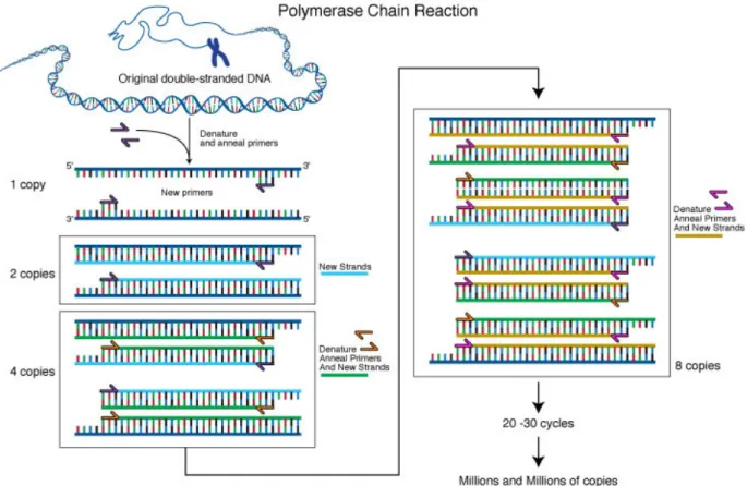
The polymerase chain reaction (PCR) is in vitro technique that consists of an exponential amplification of a DNA fragment, and its principle is based on the simulation of the mechanism of DNA replication in vivo. The PCR technique is based on the use of a thermostable DNA polymerase, which amplifies a specific region of the target. The polymerase activity is initiated by short, 15-30 base pair (bp) long oligonucleotides (primers), interacting with target fragment following the principle of Watson-Crick base pairing. It is a cycling reaction where each cycle contains three steps named denaturation, annealing and extension. The cycle repetition of these steps results in an exponential amplification, producing a vast amount of DNA at the end of the procedure (Fig. 8) (Heid et al., 1996).
Figure 8. Basic principles of the PCR method
31
As the reaction is going on, the used primers and dNTPs are incorporated into the newly synthesized DNA strands, which will compete with the primers on the later stages of the reaction. Finally, the reaction reaches a plateau phase and the amplification ceases or continues with very low efficiency. If the reaction consumes the available chemicals, it also stops the amplification (Kainz, 2000).
To identify the PCR product according to the specific length of the amplicon, gel electrophoresis is used and the DNA molecules can be visualized under UV light after staining with an intercalating dye.
The PCR is a fast and relatively simple technique that can detect a nucleic acid fragment and amplify it. PCR is a highly sensitive procedure for detecting infectious agents in host tissues and vectors, even when only a small number of host cells are infected (OIE, 2012). However, the sensitivity of PCR could be also its major disadvantage since very small amounts of contaminating DNA (from a different sample) can also be amplified producing false-positive results. Moreover, the specificity of the generated PCR products may be altered by nonspecific binding of the primers to others similars sequences on the template DNA (Louie et al., 2000).
The PCR techniques have had a rapid and tremendous progress in recent year. This technique have contributed to the identification and the characterization of several infectious agents that have great impact on human and animal health (Hernández-Rodríguez and Ramirez, 2012). To expand its utility in veterinary diagnostics and pathogen identification, PCR has been extensively modified over the years. This technique often in combination with other techniques has opened up numerous possibilities in epidemiological studies for the identification of individual strains and, in particular, the differentiation of closely related strains (Adams and Thompson, 2008).
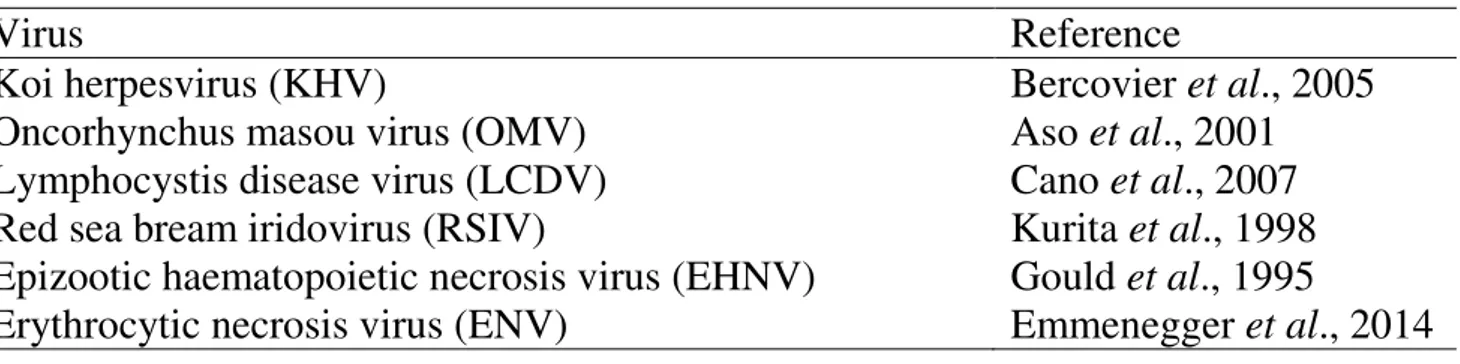
In recent years, several conventional PCR tests have been developed and applied for the detection of the most important fish viruses (Table 1).
32
Specific PCR methods have developed for rapid detection and confirmation of koi herpesvirus in tissues of infected fish (Gilad et al., 2002; Gray et al., 2002). Successively, a robust and sensitive PCR assay based on primers selected from the defined DNA sequence of the TK gene was developed to improve the diagnosis of KHV infection. This assay compared to previously described PCR assays and to viral culture in diseased fish was shown to be the most sensitive method of diagnosis of KHV infection (Bercovier et al., 2005).
The PCR assay described by Gould et al. (1995) provides a rapid method to detect EHNV in infected cell cultures and infected tissues from redfin perch (Perca
fluviatilis L.), rainbow trout and barramundi.
Several conventional PCR assays have been developed to detect LCDV in several species (Hossain et al., 2007; Kvitt et al., 2008; Cano et al., 2009). However, for detection of the virus in apparently healthy carriers, combining PCR with membrane hybridization has been recommended to increase the sensitivity of the assay (Cano et al., 2007).
A fast and sensitive PCR assay has been described for detection of the red sea bream iridovirus (RSV) in infected red sea bream (Kurita et al., 1998). This assay is recommended by the OIE (Office International des Epizooties) for diagnosis and surveillance of red sea bream iridoviral disease (RSIVD).
Table 1. Relevant DNA viruses of fish, targeted by PCR assays.
Virus Reference
Koi herpesvirus (KHV) Bercovier et al., 2005
Oncorhynchus masou virus (OMV) Aso et al., 2001
Lymphocystis disease virus (LCDV) Cano et al., 2007
Red sea bream iridovirus (RSIV) Kurita et al., 1998
Epizootic haematopoietic necrosis virus (EHNV) Gould et al., 1995
33
1.3.2 Reverse transcriptase polymerase chain reaction (RT-PCR)
RT-PCR is one of the variants of the conventional PCR. The RT-PCR is also an
in vitro procedure that combines the action of the reverse transcriptase enzyme with
that of the DNA polymerase. Its main difference whit conventional PCR is that this reaction starts from a RNA template extracted directly from the sample; the RNA is converted to a complementary DNA (cDNA). Subsequently, the newly synthesized cDNA is amplified using traditional PCR (Fig. 9) (Newton and Graham, 1994).
Figure 9. Flowchart of RT-PCR (Wikipedia, 2015)
http://en.wikipedia.org/wiki/Reverse_transcription_polymerase_chain_reaction
The Reverse Transcriptase (RT) reaction can be prepared with random primers, oligo(dT), or a gene-specific primer. The RT-PCR can be carried out either in two-step or one-two-step formats. In two-two-step RT-PCR, each two-step is performed under optimal conditions. cDNA synthesis is performed in a specific RT buffer, then one tenth of the
34
reaction is used for PCR. In one-step RT-PCR, reverse transcription and PCR take place sequentially in a single tube under conditions optimized for both RT and PCR.
RT-PCR is a fast and sensitive method for detection of RNA viruses, but there are some disadvantages, such as the unstable nature of RNA, the risk of contamination. Furthermore, as in the case of PCR, this method cannot distinguish between infection and non-infectious virus (Bootland and leong, 1999).
RT-PCR is widely applied for the detection of many fish RNA viruses (Table 2). Reverse transcriptase-polymerase chain reactions (RT-PCR) assay were developed for the detection of Viral haemorrhagic septicaemia virus (VHSV) and Infectious
hematopoietic necrosis virus (IHNV). With these techniques were possible to detect
viral RNA in acutely and subacutely to chronically diseased fish as well as in asymptomatic VHS or IHN carrier fish (Miller et al., 1998). RT-PCR has been used frequently to identify IHNV and is claimed to be more sensitive than traditional virus isolation (Bruchhof et al., 1995; Miller et al., 1998; Knusel et al., 2007). The RT-PCR method reported by Snow et al. (2004) has proven capable of amplification of a wide range of VHSV genotypes and is recommended for use by the OIE.
The RT-PCR has been shown to be a very sensitive tool for the detection of IPNV in tissues and fish eggs of coho salmon. The use of this method could be an important tool for preventing the horizontal transmission of this virus (Lopez-Lastra et
al., 1994). Other studies have compared RT-PCR assay with traditional virus isolation and have revealed that RT-PCR is the most sensitive method for detection of IPNV. (Blake et al., 1995; Taskdal et al., 2001). A study to optimize and validate a RT-PCR for the detection of Infectious pancreatic necrosis virus (IPNV) was conducted by Kerr and Cunninghan (2006). With this optimized technique was possible to detect all nine serotypes (A1-A9) of IPNV serogroup A.
Several RT-PCR methods were developed for detection of infectious salmon anaemia virus (ISAV). Amplification of parts of segment 8 provides a useful test for presence of the virus (Mjaaland et al., 1997; Rimstad et al., 1999). A RT-PCR was able
35
to detected ISAV in both serum and mucus of fish showing no clinical sign of ISAV or pathological lesions (Griffiths and Melville, 2000).
Table 2. Relevant RNA viruses of fish targeted by RT-PCR assays.
Virus Reference
Infectious hematopoietic necrosis virus (IHNV) Miller et al., 1998 Viral hemorrhagic septicemia virus (VHSV) Miller et al., 1998 Infectious pancreatic necrosis virus (IPNV) Saint-Jean et al., 2001
Sleep diseases virus (SDV or SAV-2) Fringuelli et al., 2008
Infectious salmon anemia virus (ISAV) Devold et al., 2000
Spring viraemia of carp virus (SVCV) Stone et al., 2003
Viral encephalopathy and retinopathy virus (VERV) Nishizawa et al., 1994
1.3.3 Nested PCR and Semi-Nested PCR
Nested PCR is a variant of conventional PCR method that amplifies a target region of DNA with an outer primer pair in an initial reaction, followed by a second amplification step conducted using an internal primer pair (Mothershed and Whitney, 2006). This method increases the specificity and the sensitivity of the reaction since formation of the final product depends upon the bonding of two separate sets of primers and because two sets of amplification (each of the order of 25 cycles) are used (Kawasaki et al., 1990). However, one of the major drawbacks with nested PCR is the risk of introducing contamination when a second PCR reaction is initiated using a portion of the mixture from the first reaction, and consequently it increases the risk of false-positive results. (Rolfs et al., 1992).
Semi-nested PCR is basically the same technique, as the only difference is that one of the primers used in the second amplification is the same used in the first PCR amplification.
Both nested and semi-nested PCR therefore can increase the sensitivity as much as 1000 fold compared with conventional PCR (Kalland, 2009). Besides, the specificity is particularly enhanced because these techniques almost always eliminates any