Evaluation of fluid transport processes in dental enamel.
Methods to assess the relevance of enamel permeability in caries
prevention and etching treatments.
Angelica Bertacci
DOTTORATO DI RICERCA
Biotecnologie Mediche
Ciclo XXI
MED/28 MALATTIE ODONTOSTOMATOLOGICHE
Evaluation of fluid transport processes in dental enamel.
Methods to assess the relevance of enamel permeability in caries
prevention and etching treatments.
Presentata da:
Angelica Bertacci
Coordinatore Dottorato
Relatore
Prof.ssa Marialuisa Zerbini Prof. Carlo Prati
CONTENTS
Enamel Structure ...5
Enamel Rods ...6
Striae of Retius and bands of Hunter-Schreger ...8
Enamel tufts and lamellae ...9
Dentino-enamel junction ...9
Enamel formation...10
References ...11
Fluid transport processes... 13
Enamel permeability...13
Enamel fluid transport processes ...14
Caries and enamel fluid movement ...15
References ...16
Methods to assess enamel permeability and structure ...20
Replica Technique...20
Raman and IR Spectroscopy ...21
SEM-EDX ...23
References ...24
In vivo enamel fluid movement ...26
Introduction...26
Materials and Methods...28
Results ...29
Discussion ...32
References ...34
In vivo fluid release from primary enamel...37
Materials and Methods ...38
Results...39
Discussion...41
References...42
Fluoride: in vivo effects on enamel permeability ...44
Introduction...45
Materials and Methods ...46
Results...47
Discussion...52
References...54
Effects of fluoride release from an orthodontic bondig agent on enamel
demineralization ...58
Introduction...59
Materials and Methods ...60
Results...62
Discussion...68
References...70
Acid treatments modify enamel permeability ...73
Introduction...74
Materials and Methods ...75
Results...75 Discussion...80 References...82
Appendix ...85
Thesis Abstract...85 Acknowledgments...88Scientific papers published as part of this thesis ...89
Other scientific papers published on journals with impact factor ...98
Chapter 1
Enamel Structure
Dental enamel is a composite of mineral, water, protein and lipid by volume and is the most mineralized tissue of the human body consisting of approximately 97 wt% mineral and 3% organic material and water (Simmmer and Hu, 2001; Klocke et al., 2006). Mature enamel can contain less than 1% organic material (Bartlett and Simmer, 1999).
Enamel is composed of mineral rods sized ~30µm in length and 5 µm in diameter that are orientred roughly perpendicular to the dentino-enamel junction (DEJ). In turn, the rods are built up of crystallites sized ~1µm in length and 40 nm in diameter and aligned with their crystallographic c-axis along the rod length (Jongebloed et al., 1975; Klocke et al., 2006).
Crystallites are highly organized, tightly packed and comprise 87% of enamel volume (Simmer and Hu, 2001). In enamel the crystallites are arranged in the enamel prisms and interprismatic substance.
The orientation of the crystallites in enamel is of considerable interest since e.g. the crys-tallite dissolution in the caries process proceeds faster in the radial direction than parallel to crystallite’s c-axis (Arends and Jongebloed, 1978).
Scanning electron microscopic examination showed that demineralization, initiated at core (prism or rod)/wall (prism sheath) interfaces, developed anisotropically along the c- axes (Wang et al., 2006).
The inorganic content of enamel is a crystalline calcium phosphate hydroxyapatite hav-ing an hexagonal symmetry and a general formula [Ca10(PO4)6(OH)2], that contains some
impurities, such as carbonates substituring for phosphate in the crystal lattice (Simmer and Hu, 2001; Klocke et al., 2006). Various ions (eg. strontium, magnesium, and fluo-ride) may be also incorporated into or adsorbed by the hydroxyapatite crystals (Ten Cate, 1998). Dental apatite contains a substantial amount of carbonate groups, which substitute for the OH- groups (A-type CO32-) or for phosphate tetraedal (B-type)
(Klocke et al., 2006). The carbonate content represents 2-4 wt% with a reported 90% of type B and 10% of type A (Penel et al., 1998). The carbonate concentration of human enamel has been found to increase on going from the outside to the inside of enamel layer (Wentrup-Byrne et al., 1997). The structure of HAP can be considered as built up of corner-sharing PO4 and CaO6 polyhedra forming channels along the crystallographic
c-axis, in which the hydroxyl-groups are placed (Klocke et al., 2006).
The bulk of the organic material consists of tyrosine-rich amelogenin polypeptide (TRAP) peptide sequence tightly bound to the hydroxyapatite crystals, as well as nonam-lelogenin proteins (Ten Cate, 1998).
Dental enamel is extremely hard and brittle. The underlying layer of dentin, more resil-ient, is necessary to maintain its integrity. Enamel is translucent, and it also varies in thickness, from a maximum of approximately 2.5 mm over working surface to a feather-edge at the cervical line (Ten Cate, 1998).
Enamel Rods
Enamel is composed primarily of elongated structure called rods. The rod is shaped like a cylinder and is made up of crystals whose long axes run, for the most part, parallel to the longitudinal axis of the rod. Crystals more distant from the central axis, however, flare laterally to an increasing degree as they approach the rod periphery (Ten Cate, 1998).
Enamel crystals are extremely long relative to their thickness and are higly oriented. They generally extend from the underlying dentin toward the surface of the tooth and are organized into bundles, called prisms (Simmer and Hu, 2001).
The interrod region is an area surrounding each rod in which crystals are differently ori-ented. The boundary where crystals of the rod meet those of the interrod region at sharp angles is known as the rod sheath. The basic unit of enamel described as a cylindrical rod has a specific spatial relation to the interrod region directly cervical to it (Ten Cate, 1998).
The forming surface of enamel consists of pits, each surrounded by a wall made up of newly formed interrod enamel. During active secretion, each of these walled pits is oc-cupied by a Tomes’ process. The interrod region is formed slightly earlier than the rod enamel, which thus constitutes the walls of the pits. These walls are formed by secretion from proximal sites that completely encircle each Tomes’ process near its base, where adjacent processes are joined by the distal junctional complexes. Thus each wall (inter-rod region) is formed as a cooperative effort by adjacent secretory ameloblats.
Each ameloblast is responsible for the formation of one rod (by its distal secretory site) and a portion of the surrounding interrod region (by its cooperative proximal sites).
Enamel rods have an average width of 5 µm, but they vary somewhat in size and mor-phology troughout the thickness of enamel. In the first 5 µm, next to the dentine, there is no rod structure. As they traverse the enamel the rods gradually increase slightly in di-ameter. At the enamel surface the rod structure is irregular or absent. Rodless enamel occurs in the outermost 30 µm or so of all primary teeth and in the gingival third of the enamel of permanent teeth. Crystals in these regions are perpendicular to the surface of the enamel.
The rods are aligned in horizontal rows. The rods in each row run in a direction gener-ally perpendicular to the surface of the dentin, with a slight inclination toward the cusp as they pass outward. Near the cusp tip the rows have small radius, and the rods run more vertically. In the cervical enamel the rods run mainly horizontally; only a fey rods are titled apically.
Striae of Retius and bands of Hunter-Schreger
The striae of Retzius are incremental growth lines. They are formed as the results of a temporary constriction of Tomes’ process associated with a corresponding increase in the secretory face forming interrod enamel. As a result enamel structure is altered along the lines. Electron micrographs reveal a possible decrease in the number of crystals in the striae, suggesting that enemel rods bend as they cross an incremental line (Ten Cate, 1998).
The striae of Retius often extend from the dentino-enamel junction to the outer surface of enamel, where they end in shallow furrows known as perikymata. Peikymata run in circumferentially horizontal lines across the face of the crown. In addition, lamellae or craks in the enamel apprear as jagged lines in various regions of the tooth surface. In unerupted teeth the enamel surface consits of a structureless layer some 0.5 to 1.5 µm thick. Immediately below it is a layer of small, losely packed crystallites, some 5 nm thick, with undemineralized material between them. Interspersed among, in and on these fine crystallites are randomly distributed large, platelike crystals. The fine crystallite layer merges into the subsurface enamel where crystals are closely packed and approximately 50 nm in size. In erupted teeth the structureless surface layer and the surface layer of small crystallites are rapidly lost by abrasion, attrition and ersosion (Ten Cate, 1998).
Human enamel is known to form at a rate of approximately 4 µm per day. Situated be-tween the rods at approximately 4 µm intervals are interrod regions. Cross striation probably indicate a daily (or circadian) variation in the secretory activity of the ameloblasts and the striae of Retius represent a weekly rhythm of the same cells (Ten Cate, 1998).
The bands of Hunter and Schreger are an optical phenomenon produced solely by changes in rod direction. They are seen most clearly in longitudinal ground section viewed by reflect light and are found in the inner four fifths of the enamel. They appear as dark and light alternating zone that can be reversed by altering the direction of inci-dent illumination (Ten Cate, 1998).
Enamel tufts and lamellae
Enamel tufts project from the dentino-enamel junction for a short distance into the enamel; they appear to be branched and contain greater concentrations of enamel pro-tein than the rest of the enamel. Lamellae extend for varying dephts from the surface of enamel and consist of linear, longitudinally oriented defects filled with enamel protein or organic debris from the oral cavity. The protein of tufts is a high-molecular-weight vari-ety similar to enamelin. Tufts are believed to occur developmentally baecause of abrupt changes in the direction of groups of rods that arise from different regions of the scal-loped dentino-enamel junction. A different ratio of interrod and rod enamel in these groups creates less mineralized and weakened planes. Faulting of blocks of enamel re-lieves internal strains produced by dimensional changes as the tissue matures. When a fault occurs, it blocks the normal exit for enamel protein, causing the higher organic content of tufts and lamellae (Ten Cate, 1998).
Dentino-enamel junction
Dentin, enamel and cementum formation involves a remarkable mechanism of complex molecular and cellular events that conclude in specific structured tissue joined at distinc-tive interfaces.
The dentino-enamel junction (DEJ) is the natural junction that unites functionally dentin and enamel (Habelitz et al, 2001; Marshall et al., 2001; Schulze et al., 2004).
The junction between enamel and dentin is established as these two hard tissue begin to form and it appear as a series of ridges that increase the surface area and probably en-hance the adhesion between enamel and dentin. The DEJ has a unique structure with at least three levels of microstructure (Schulze et al., 2004).
The DEJ is the junction of coronal dentin and enamel and is formed by the secretion of dentine on one side and of enamel on the other side.
Before enamel forms, some newly forming odontoblast processes push between adjoin-ing ameloblasts and when enamel formation begins, become trapped to form enamel spindles that do not follw the direction of enamel rods (Ten Cate, 1998).
The scallops house microscallops that contain finer nanoscale structures (Marshall et al., 2001; Schulze et al., 2004). The scallops and the presence of a smooth gradient of me-chanical properties at the junction are believed to contribute to reduce stress
concentra-tion. This gradation of properties is initiated by biomineralization starting from the DEJ in both directions (Ten Cate, 1998).
It has been demonstrated that DEJ is resistant to acid attacks as well as to mechanical forces such as cracks propagation (Tramini et al., 2000; Marshall et al., 2001; Schulze et al., 2004).
Enamel formation
Mineralization involves the crystallization of ions from supersaturated solutions (Simmer and Fincham, 1995).
Particularly mineralization process ivolves the net movement of ions out of solutions, where their charges are dissipated by interaction with water molecules, and into a solid structure stabilized by covalent interactions between oppositely charged ions (Simmer and Hu, 2001).
The stages of enamel formation include (Reith, 1970): • secretion of an organic matrix;
• crystal nucleation; • crystal eleongation;
• removal of the organic matrix, and • crystal maturation.
Dentine and enamel formation take place simultaneously starting along the DEJ (den-tino-enamel junction). On enamel side of the DEJ, crystal nuclei elongate into long thin ribbons. These ribbons are evenly spaced, oriented parallel to each other, and extend from the DEJ to the mineralization front just outside the membrane of ameloblasts (the cells lining the extracellular compartment on enamel side) (Simmer and Hu, 2001). The shape and growth of the earliest crystallites appeared at the DEJ can be interpreted as evidence for a precursor phase of octocalcium phosphate (OCP). An OCP crystal displays on its face a surface that may act as a template for hydroxyapatite (OHAp) pre-cipitation. Octocalcium phosphate is less stable than hydroxypapatite and can hydrolyze to OHAp. During this process one unit cell of octocalcium phosphate is converted into two unit cells of hydroxyapaptite (Simmer and Fincham, 1999).
As ameloblasts secrete enamel proteins, the crystallites continue to growth on length but grow very little in width and thickness. The final length of enamel crystals is determined
by how long the ameloblasts continue to add enamel proteins, which also determines the final thickness of the enamel layer as a whole (Simmer and Hu, 2001).
At a certain point, which is decided by the genetic program, ameloblasts undergo a tran-sition that greatly reduced their secretion of enamel proteins. Instead of structural pro-teins, proteinases are secreted and the organic matrix is degraded and suddenly disap-pears from the extracellular compartment. These changes terminate the growth of enamel crystallites in length and vastly accelerate their growth in width and thickness. Crystal elongation is arrested by curbing the secretion of enamel matrix constituents such as amelogenin, ameloblastin, and enamelin. Mineral deposition on the sides of the crystallites accelerates, in part because of the degradation and removal of growth-inhibhiting enamel protein, cleavage products (Simmer and Hu, 2001).
In humans, the maturation stage, during which the crystallites growth in width and thickness, takes about three to four years. This process is necessary to harden the enamel layer and is directed by maturation stage of ameloblasts as they cycle trought smooth and ruffle-ended phases. Fluoride is incorporated into crystal structure during the matu-ration stage. Disturbance during the matumatu-ration stage of amelogenesis results in pathol-ogically soft (hypomaturation) enamel of normal thickness (Simmer and Hu, 2001).
Dental enamel formation is highly specialized and, the proteins most directly involved in enamel biomineralization are specific for it. As a consequence, defect in the gene encod-ing enamel proteins generally cause enamel malformations without affectencod-ing other parts of the body. There are, however, numerous genetic syndromes associated with dental defect of all types (Simmer and Hu 2001).
References
Arends J, Jongebloed WL. Crystallites dimension of enamel. J Biol Buccale 1978;6:161-71.
Bartlett JD, Simmer JP. Proteinase in developing dental enamel. Crtit Rev Oral Biol Med 1999;10:425-441.
Habelitz S, Marshall SJ, Marshall GW, Balooch M. The functional width of the dentino-enamel junction determined by AFM-based nanoscratching. J Struct Biol 2001;135:294-301.
Jongebloed WL, Molenaar I, Arends J. Morphology and size-distribution of sound and acid-treated enamel crystallites. Calcif Tissue Res. 1975 Dec 18;19:109-23.
Klocke A, Mihailova B, Zhang S, Gasharova B, Stosch R, Guttler B, Kahl-Nieke B, Henriot P, Ritschel B, Bismayer U. CO2 laser-induced zonation in dental enamel: a ra-man and IR microspectroscopic study. J Biomed Mater Res Part B: 2007 Appl Biomater 81 B:499-507.
Marshall GW, Balooch M, Gallagher RP, Gansky SA, Marshall SJ. Mechanical properties of the dentino-enamel junction: AFM studies of nanohardness, elastic modulus, and fracture. J Biomed Mater Res 2001;54:87-95.
Reith EJ. The stages of amelogenesis as observed in molar teeth of young rats. J Ultrastruct Res 1970;30:111-151.
Schulze KA, Balooch M, Balooch G, Marshall GW, Marshall SJ. Micro-Raman spectro-scopic investigation of dental calcified tissues. J Biomed Mater Res A. 2004;69:286-93. Simmer JP, Fincham AG. Molecular mechanisms of dental enamel formation. Crtit Rev Oral Biol Med 1995;6:84-108.
Simmer JP, Hu JC-C. Dental enamel formation and its impact on clinical dentistry. J Dent Ed 2001;65:896-905.
Ten Cate AR. Oral histology: development, structure and function, 5th ed. St. Louis,
MO: Mosby; 1998. p218-235.
Tramini P, Pelissier B, Valcarcel J, Bonnet B, Maury L. A Raman spectroscopy investiga-tion of dentin and enamel structures modified by lactic acid. Caries Res 2000;34:233-240. Wang LJ, Tang R, Bonstein T, Bush P, Nancollas GH. Enamel demineralization in pri-mary and permanent teeth. J Dent Res 2006;85:359-363.
Wentrup-Byrne E, Armstrong CA, Armstrong RS, Collins BM. Fourier Transform Ra-man Microscopic Mapping of the molecular components in a huRa-man tooth. J RaRa-man Spectr 1997, 28:151-158.
Chapter 2
Fluid transport processes
Enamel permeability
Enamel does not behave as an inert tissue because water and organic material occur be-tween the prisms (Bartlestone, 1951; Lindén 1968; Pashley 1996; Shellis 1996; Ten Bosch 2000).
Bergman and Lindén showed that small quantities of a fluid passing through the enamel in vivo (Bergman and Lindéen 1965; Lindén 1968).
Enamel fluid flowing is related to permeability but is not well documented in vivo (Berg-man and Lindén 1965; Bakhos et al., 1977).
The transport of water across dental enamel in vitro is not a simple diffusion process in which enamel behaves as an inert porous medium as enamel behaves as an osmotic membrane (Burke and Moreno, 1975). The diffusion of molecules and ions in the dental enamel plays an important role in the development of caries (Dibdin 1993; Kuhar et al., 1999).
Many studies in the literature investigated enamel permeability. In particular most of all related this property with caries trying to explain the tissues changes in the phases of demineralization and remineralization (Fearnhead et al., 1982; Ten Cate 2001) even if the importance of enamel permeability in caries, restorative materials and pulp-dentine-enamel interaction is still not fully understood (Byers et al., 2003).
From the beginning of the last century several authors investigated the penetration of enamel with dyes (Beust 1912, Berggren 1943, Tarbet and Forsick, 1971), the diffusion
of organic components (Poole et al., 1963), the permeability to inorganic ions (Sognnaes and Shaws 1952), radioactive elements (Braden, 1971; Sognnaes and Shows 1952; Joys-tone-Bechal et al., 1971), and water (Poole et al., 1963; Lindén 1968; Shellis and Dibdin, 2000). The main studies in the literature evaluated enamel permeability and fluid flowing in vitro and in vivo through scanning (Whittaker, 1982) and transmission microscopy (Poole et al., 1981), measurement of diffusion coefficients (Lindén 1968; Borggreven et al., 1977) and through physical parameters such as electrical resistance (Hoppenbrou-wers et al, 1986; Wang et al., 2005) conductance (Ie et al., 1995; Ten Bosch et al., 2000), impedance (Scholberg et al., 1984). These results demonstrated the bidirectional perme-ability of enamel.
Enamel fluid transport processes
Zahradnik and Moreno showed that dental enamel has a bimodal pore distribution (Zahradnik and Moreno, 1975) and that the transport processes related to mineralization and demineralization are significantly affected by the amount of water avaible in the tis-sue as well as by its porous structure (Moreno and Zahradnik, 1973).
Diffusion in the aqueous phase which fills enamel pores is the main transport of the ions in the early stages of caries progression, in remineralization, and in fluoride treat-ment (Dibdin, 1993).
The organic matter of the enamel is probably the route of diffusion although the role of organic material needs further investigation to be clearified (Shellis, 2000). Microscopic examinations of enamel section showed that the main channels of diffusion were the in-terprismatic substance (Whittaker, 1982). Microscopical observations show that the prism junction provide the main pathways (Tarbet and Forsick, 1971) although, in inner enamel some transport was observed within the prism (Lindén, 1968).
Because enamel mineral exists as a very small crystals organised in an elaborate structure, the internal pores are small and variable in form, orientation and distribution. The larg-est pores in enamel are associated with the prism junctions, but these constitute only a small fraction of the total porosity, most of which is associated with the prism bodies and tails (Shellis and Dibdin, 2000).
Structure, porosity and enamel solubility are linked. Because the prism-junction material is more soluble than the interprismatic material, it is possible for prism junctions to be opened up under conditions where the enamel fluid is still supersaturated with respect to the intraprismatic mineral (Shellis, 1996).
Enamel permeability is variable depending to age and demineralization (Kotsanos and Darling, 1991) is greatest in teeth with immature enamel, and it appears to require a partnership with dentine (Byers et al., 2003). Epidemiologic studies with animals have suggested that caries susceptibility decreases with age: a process commonly referred as “posteruptive maturation” of enamel may be responsible for this phenomenon reducing the permeability of enamel (Fearnhead et al., 1982; Kotsanos and Darling, 1991).
In extracted young human teeth, cervical enamel has more dye flow than the rest of the crown (Lindén 1968) and appears to be the preferred pathway for fluid flowing (Poole et al., 1981; Byers et al., 2003). Moreover in young teeth the enamel interprismatic region is proportionally greater than in old teeth and the dentin is much thinner and odontoblast processes reach closer to the dentino-enamel junction (Byers and Sugaya, 1995).
Newly-erupted teeth acquire F more readily than older teeth; younger permanent enamel takes up more F, exhibits a higer-water sorption capacity and imbibes more iodide than older permanent enamel (Lindén et al., 1986).
Enamel fluoride concentration of permanent enamel is always higher than that for pri-mary enamel (Issa et al., 2003).
Enamel of deciduous teeth contains more organic matter, more water, less mineral and is more porous in agreement with clinical studies that have shown caries formation and progression to be faster in primary than in permanent teeth (Sønju Clasen et al., 1997; Issa et al., 2003).
Caries and enamel fluid movement
The development of carious lesions in enamel involves transport of acids into and disso-lution of minerals from the tooth surface. Accordingly the rate of diffusion of cariogenic
and cariostatic substances (ions and molecules) plays a crucial role in the dynamic proc-ess of caries (Van Dijk et al., 1983; Featherstone, 1983; Lindèn et al., 1986).
The fluoride content of the mid-coronal buccal surface enamel in increasing age was found to decrease posteruptively with age, therefore not accounting for the decreasing caries susceptibility (Kostanos an Darling, 1991). Recently it has been demonstrated that recently erupted teeth are more sensitive to dental caries than teeth that have remained free from caries lesions for a few years after eruption (Ten Bosch et al., 2000).
This was confirmed with experiments in which artificial caries lesions were produced in extracted teeth of different post-eruptive ages (Kostanos an Darling, 1991; Ten Bosch et al., 2000). It has been hypothesized that these differences could be ascribed to differ-ences in enamel porosity consequent to intra-oral maturation presumably due to incor-poration of calcium-phosphate into the enamel (Ten Bosch et al., 2000).
Studies on enamel physical properties showed that the resistivity of enamel layers in-creased from the DEJ to the outer surface, the permeability increases from the outer surface towards the EDJ (Lindén 1968, Hoppenbrouwers et al., 1986) and that the resis-tivity in erupted teeth was considerable higher than in unerupted teeth confirming the effects of post eruptive mineralization (Hoppenbrouwers et al., 1986).
The formation of caries lesions is strongly influenced by the pathways for diffusion and by electrochemical effects arising from the charge on the pore walls (Shellis and Dibdin, 2000).
Mineral loss during caries progress results in an increase in porosity: the resulting changes in porosity could affect the flow of an electrical current through the enamel (Wang et al., 2005).
References
Bakhos Y, Brudevold F, Aasenden R. In vivo estimation of the permeability of surface human enamel. Arch Oral Biol 1977;22: 599–603.
Bergman G, Lindén L. Techniques for microscopic study of enamel fluid in vivo. J Dent Res 1965; 44: 1409.
Borggreven JMPM, Van Dijk JWE, Driessens FCM. A quantitative radiochemical study of ionic and molecular transport in bovine dental enamel. Arch Oral Biol 1977; 22:467– 472.
Braden M, Duckworth R, Joyston-Bechal S. The uptake of 24Na by human dental
enamel. Arch Oral Biol 1971; 16: 367–374.
Buest T von. A contribution to the study of immunity to dental caries. Dent Cosmos 1912;54:659-663.
Burke EJ, Moreno EC. Diffusion fluxes of tritiated water across human enamel mem-branes. Arch Oral Biol 1975; 20:327-332.
Byers MR, Sugaya A. Odontoblast processes in dentin revealed by fluorescent Di-I. J Histochem Cytochem 1995;43:159-68.
Byers MR, Yoon Lin KJ. Patterns of fluoro-gold entry into rat molar enamel, dentine, and pulp. J Dent Res 2003; 82: 312–317.
Dibdin GH. The water in human dental enamel and its diffusional exchange measured by clearance of tritiated water from enamel slabs of varying thickness. Caries Res 1993; 27: 81–86.
Fearnhead RW, Kawasaki K, Inoue K. Comments on the porosity of human tooth enamel. J Dent Res 1982; 61: 1524–1530.
Featherstone JDB. Diffusion phenomena and enamel caries development. In: Guggen-heim, B, ed. Cariology Today. Basel: Karger, 1983; 259–268.
Hoppenbrouwers PMM, Scholberg HPF, Borgreven JMPM. Measurement of the per-meability of dental enamel and its variation with depth using an electrochemical method. J Dent Res 1986; 65: 154–157.
Ie YL, Verdonschot EH, Schaeken MJ, Vant’Hof MA. Electrical conductance of fissure enamel in recently erupted molar teeth as related to caries status. Caries Res 1995; 29: 94–99.
Issa AI, Preston AJ, Preston AJ, Toumba KJ, Duggal MS. A study investigating the for-mation of artificial sub-surface enamel caries-like lesion in deciduous and permanent teeth in the presence and absence of fluoride. Arch Oral Biol 2003;48:567-571.
Joyston-Bechal S, Duckworth R, Braden M. Diffusion of radioactive ions into human dental enamel. Arch Oral Biol 1971; 16: 375–384.
Kotsanos N, Darling AI. Influence of posteruptive age of enamel on its susceptibility to artificial caries. Caries Res 1991;25: 241–250.
Lindén LA, Björkman S, Hattab F. The diffusion in vitro of fluoride and chlorhexidine in the enamel of human deciduous and permanent teeth. Arch Oral Biol 1986; 31: 33– 37.
Lindèn LA. Microscopic observations of fluid flowing through enamel in vitro. Odontol Rev 1968; 19: 349–365.
Moreno EC, Zahradnik RT. The pore structure of human dental enamel. Arch Oral Biol 1973; 18: 1063–1068.
Pashley DH. Dynamics of the pulpo-dentine complex. Crit Rev Oral Biol Med 1996; 7: 104–133.
Poole DF, Tailby PW, Berry DC. The movement of water and other molecules through human enamel. Arch Oral Biol 1963; 38: 771–772.
Poole DFG, Newman HN, Dibdin GH. Structure and porosity of human cervical enamel studied by polarizing microscopy and transmission electron microscopy. Arch Oral Biol 1981; 26:977–982.
Scholberg HPF, Borggreven JMPM, Driessens FCM. A phenomenological interpretation of the frequency-dependent impedance behaviour of bovine dental enamel. Archs Oral Biol 1984;12:965-970.
Shellis RP, Dibdin GH. Enamel microporosity and its functional implications. In: Tea-ford MF, Ferguson MJ, Smith MM, eds. Teeth: development, evolution and function. Cambridge:Cambridge University Press, 2000; 242–251.
Shellis RP. A scanning electron-microscopic study of solubility variation in human enamel and dentine. Arch Oral Biol 1996; 41: 473–484.
Shellis RP. Transport processes in enamel and dentine. In: Addy M, Embery G, Edgar WM, Orchardson R, eds. Tooth wear and sensivity. London: Martin Duniz, 2000; 19– 24.
Sognnaes RF, Shaw JH. Salivary and pulpal contribuitions to the radiophosphorous up-take in enamel and dentin. J Am Dent Assoc 1952;44:489-505.
Sønju Clasen AB, Øgaard B, Duschner H, Ruben J, Arends J, Sønju T. Caries develop-ment in fluoridated and non fluoridated deciduous and permanent enamel in situ exam-ined by microradiography and confocal laser scanning microscopy. Adv Dent Res 1997; 11: 442-7.
Tarbet WJ, Forsick LS. Permeability of human dental esame to acriflavine and potassium fluoride. Arch Oral Biol 1971; 16:951–961.
Ten Bosch JJ, Fennis IEY, Verdonschot EH. Time-dependent decrease and seasonal variation of the porosity of recently erupted sound dental enamel in vivo. J Dent Res 2000; 79: 1556–1559.
Ten Cate M. Remineralization of caries lesions extending into dentine. J Dent Res 2001; 80: 1407–1411.
Van Dijk JW, Borggreven JM, Driessens FC. Diffusion in mammalian tooth enamel in relation to the caries process. Arch Oral Biol 1983; 28: 591–597.
Wang J, Someya Y, Inaba D, Longbottom C, Miyazaki H. Relationship between electri-cal resistance measurement and microradiographic variables during remineralization of softened enamel lesions. Caries Res 2005; 39: 60–64.
Whittaker DK. Structural variations in the surface zone of human tooth enamel ob-served by scanning electron microscopy. Arch Oral Biol 1982; 27: 383–392.
Zahradnik RT, Moreno EC. Structural features of human dental enamel as revealed by isothermal water vapour sorption. Arch Oral Biol 1975;20:317-25.
Chapter 3
Methods to assess enamel
permeability and structure
Replica Technique
The replica technique performed in these studies has been previously described in the literature (Barnes, 1977; Ittahgarun and Tay, 2000; Chersoni et al., 2005). This technique allows the evaluation of fluid outflow from enamel surface and is able to detect the pres-ence of small quantities of fluid. The specific characteristics of replica technique, that is not invasive and risk-free for the patient, make possible to perform in vivo studies on fluid outflow, that represents enamel permeability, in different clinical conditions.
Hydrophobic polyvinyl siloxane impression material was applied on observational area and after 4 minutes, the polymerised impression material was removed and degassed for at least 48 hours and finally later cast in polyether impression material.
The absence of any chemical reaction between the two impression materials makes this tecniqua effective in showing water exudation.
The in vivo application of this technique yielded qualitative and quantitative findings on outward fluid flow on enamel surfaces by means of scanning electron microscopy in-spection of polyether replicas. This technique uses droplet formation to display the dis-charge of liquid from enamel during the setting time of the polyvinyl siloxane impres-sion material that is able to recall water by osmotic gradient. Moreover water droplets are not incorporated into the polyvinyl siloxane and remain at the observation interface level.
All the replicas obtained, morphological expression of fluid outflow, were gold sputtered and observed by a Scannig Electron Microscope.
Raman and IR Spectroscopy
Raman spectroscopy is an advanced, fast analytic technique to determine the structure and the chemical composition of materials. It provides chemical information based on molecular vibrations of the molecules in the samples.
The Raman spectroscopy technique allows obtaining vibrational spectra of minerals by analysing scattered light caused by monochromatic laser excitation (Tsuda and Arends, 1997). Raman spectra analysis and infrared absorption (IR) spectroscopy are comple-mentary techniques. While IR measures the light absorption by specific molecules using a broadband light source the Raman technique measures the characteristics Raman emis-sion induced from muolecules under monochromatic laser irradiation (Tsuda and Ar-ends, 1997).
Raman signals are emitted in the form of light scattering and can be observed from all directions, unlike the co-linear optical arrangement of IR. The axes of excitation light and detection can be chosen independently, resulting in a considerable instrumental flexibility of Raman. An optical microscope (micro) can be incorporated into a Raman specrtroscopy system (Tsuda and Arends, 1997).
Despite the advantages often Raman technique has the problem of fluorescence exhib-ited by most biological materials when irradiated by laser light. In normal Raman spec-troscopy, fluorescence spectra due to organic materials often dominate the much weakr Raman signals. Therefore Raman spectroscopy studies have been limited to enamel which contains only few organic matter (Tsuda and Arends, 1997).
Raman spectroscopy, as a versatile and non-destructive technique, allows for simultane-ous characterization of the inorganic and organic phases of the tooth. Furthermore, Ra-man spectra exhibit little interference with water, making RaRa-man spectroscopy advanta-geous for the study of many biological specimens (Carden and Morris, 2000).
Raman microspectrometry produces the capability to characterize the spatial distribu-tions of organic and inorganic compounds with spatial resolution of about 1µm. The in-tensity values of the spectra obtained from the inorganic component reveals the
chemi-cal composition; shift analysis of the peaks in the spectra allows the chemichemi-cal contents of the tissues to be differentiate (Schulze et al., 2004).
The micro-Raman technique has considerable potential for studies of crystallite orienta-tion in enamel. Spectral variaorienta-tions were taken as a funcorienta-tion of rotaorienta-tion angle for trans-verse or longitudinal arrangements. Similarly spectra were also obtained with the enamel samples at various orientation angles (Tsuda and Arends 1994).
Applications of Raman spectroscopy in dental research have included studies of enamel powder, artificial apatite, synthetic, carbonated apatite, synthetic fluorapatite (De Mul et al., 1986; Bertoluzza et al., 1996; Penel et al., 1998 Liu and Hsu, 2007). The depeosition of CaF2-like crystals after fluoride treatment and the relative orientation of single crystals
in dental enamel were also investigated through Raman spectroscopy (Tsuda and Ar-ends, 1993; Tsuda and Arends 1994).
The spectra from human dental hard tissue were analyzed in two specific wave number locations, the phosphate stretching band and the C-H stretching mode.
The enamel Raman spectrum is dominated by bands that can be attributing to the min-eral apatite at 591, 961, and 1071 cm-1.
The phosphate/C-H ratio clearly showed that enamel had a different average composi-tion than the adjacent hard tissues. The cementum had the lowest (2.8) and the enamel has the highest ratio (94.2). The phosphate/C-H intensity ratio for dentine was ap-proximatively 10% that of enamel, and varied from 7.1 for dentine to 19.6 for enamel (Schulze et al., 2004).
The apatite crystallites in enamel are preferentially oriented with their crystallographicc-axis perpendicular to enamel-dentine junction, which results in different Raman scatter-ing intensity dependscatter-ing on the experimental geometry (Klocke et al., 2006).
Sound tooth enamel exhibited strong Raman polarization anisotropy, whereas early car-ies consistently showed a lower degree of Raman polarization anisotropy in particular for sound enamel the Raman peak arising from the symmetric ν1 vibration of PO
3-4
FT-IR (Fourier Transform InfraRed) spectroscopy can result in a positive identification (qualitative analysis) of every different kind of material.
An infrared spectrum represents a fingerprint of a sample with absorption peaks which correspond to the frequencies of vibrations between the bonds of the atoms making up the material.
Because each different material is a unique combination of atoms, no two compounds produce the exact same infrared spectrum. In addition, the size of the peaks in the spec-trum is a direct indication of the amount of material present. With modern software al-gorithms, infrared is an excellent tool for quantitative analysis.
Fourier transform infrared (FTIR) spectroscopy and Raman spectroscopy are chemical analytical methods that have been used to collect information about mineral tissues (Wentrup-Byrne et al., 1997; Klocke et la., 2006). The outputs from these methods are FTIR and Raman spectra that contain signals from the organic functional groups in the sample. Since the bands in FTIR spectra are due to polar functional groups while the bands in Raman spectra are due to nonpolar functional groups, FTIR and Raman spec-troscopy are complementary techniques.
SEM-EDX
Energy Dispersive X-ray (EDX) analysis is a valuable tool for qualitative and quantita-tive element analysis. This method allows a fast and non-destrucquantita-tive chemical analysis with a spatial resolution in the micrometer regime. It is based on the spectral analysis of the characteristic X-ray radiation emitted from the sample atoms upon irradiation by the focussed electron beam of a SEM.
The incident beam electrons excite electrons in a lower energy states, prompting their ejection and resulting in the formation of electron holes within the atom’s electronic structure. Electrons from an outer, higher-energy shell then fill the holes, and the excess energy of those electrons is released in the form of X-ray photons. The release of these X-rays creates spectral lines that are highly specific to individual elements. In this way the X-ray emission data can be analyzed to characterize the sample.
The data generated by EDX analysis consist of spectra showing peaks corresponding to the elements making up the true composition of the specimen being analysed.
The technique can be qualitative, semi-quantitative, quantitative and also provide spatial distribution of elements through mapping.
The EDX technique is non-destructive and if required specimens of interest can be ex-amined in situ with little or no sample preparation. The EDX systems also have Image Analysis packages that can be applied to any images generated by the SEM / EDX tech-nique allows for the identification of the critical characteristics of particles. It offers the ability to gather information about finer particles than by optical microscopes and can readily distinguish between clusters and agglomerates of particles in addition to the chemical analysis available by EDX. The strength of this analysis technique is its ability to gather statistically significant data on the size, morphology and composition of the particles in a time efficient manner, beyond the capabilities of conventional optical mi-croscopy.
Severals studies used EDX investigating the effect of different treatment (such as fluo-ride, peroxide etc) on the chemical composition of enamel surface (Takagi et al., 2000; Barbour and Rees, 2004; Schougall Vilchis et al., 2008).
References
Barbour ME, Rees JS. The laboratory assessment of enamel erosion: a review. J Dent 2004;32:591-602.
Barnes IE. The adaptation of composite resins to tooth structure. Part 1. Study 1: intro-duction and the adaptation of composite resins to the unetched enamel cavity wall. Br Dent J 1977; 142: 122–129.
Bertoluzza A, Bottura G, Taddei P, Tinti A. Vibrational spectrs of controlled-structure hydroxyapatite coatings obtained by the polymeric route. J Raman Spectroscopy 1996;27:759-764.
Carden A, Morris MD. Appplication of vibrational spectroscopy to the study of mineral-ized tissue (review). J Biomed Opt 2002;5:259-68.
Chersoni S, Suppa P, Breschi L, Ferrari M, Tay FR, Pashley DH, Prati C. Water move-ment in the hybrid layer after different dentin treatmove-ments. Dent Mater 2004; 20: 796– 803.
De Mul FF, Hottenhuis MH, Bouter P, Greve J, Arends J, ten Bosch JJ. Micro- Raman line broadening in synthetic carbonated hydroxyapaptite. J Dent Res 1986;65:437-40. Itthagarun A, Tay FR. Self-contamination of deep dentin by dentin fluid. Am J Dent 2000; 13: 195–200.
Klocke A, Mihailova B, Zhang S, Gasharova B, Stosch R, Güttler B, Kahl-Nieke B, Henriot P, Ritschel B, Bismayer U. CO2 laser-induced zonation in dental enamel: a
Ra-man and IR microspectrometry study. J Biomed Mat Res Part B: Appl Biomater; 2007: 81B:499-507.
Liu Y, Hsu C-YS. Laser-induced compositional changes on enamel: A Ft-Raman study. J Dent 2007;35:226-30.
Penel G, Leroy G, Rey C, Bres E. Micro-Raman spectral study of the PO4 and CO3
vi-brational modes in synthetic and biological apatites. Calcif Tissue Int 1998;63:475-481. Schulze KA, Balooch M, Balooch G, Marshall GW, Marshall SJ. Micro-Raman spectro-scopic investigation of dental calcified tissues. J Biomed Mater Res A. 2004;69:286-93. Sowa MG, Popescu DP, Werner J, Hewko M, Ko A C-T, Payette J, Dong CCS, Cleg-horn B, Choo-Smith L-P. Precision of Raman depolarization and optical attenuation measurements of sound tooth enamel. Anal Bioanal Chem 2007;387:1613-1619.
Tsuda H, Arends J. Detection and quantification of calcium fluoride using micro-Raman spectroscopy. Caries Res 1993;27:249-57.
Tsuda H, Arends J. Orientational micro-Raman spectroscopy on hydroxyapatite single crystals and human enamel crystallites. J Dent Res 1994;73:1703-1710.
Tsuda H, Arends J. Raman spectroscopy in dental research: a short review of recent studies. Adv Dent Res 1997;11:539-547.
Wentrup-Byrne E., Armstrong CA, Armstrong RS, Bradley MC. Fourier transform Ra-man microscopic mapping of the molecular components in a huRa-man tooth. J RaRa-man Spectroscopy 1997;28:151-158.
Takagi S, Liao H, Chow LC. Effect of tooth-bound fluoride on enamel demineraliza-tion/ remineralization in vitro. Caries Res 2000; 34: 281-8.
Schougall Vilchis RJ, Hotta Y, Yamamoto K. Examination of Six Orthodontic Adhe-sives with Electron Microscopy, Hardness Tester and Energy Dispersive X-ray Micro-analyzer. Angle Orthodontist 2008; 78: 655-61.
Chapter 4
In vivo enamel fluid movement
The aim of this study was to visualize fluid movement through dental enamel in vivo. Fifty permanent upper central incisors, from subjects aged 10–70 yr, and 5 permanent central just-erupted incisors, from subjects aged 6–7 yr, were included in the study. An impression was obtained by vinyl polyxiloxane, and replicas were then obtained by poly-ether impression material. The hydrophobic vinyl polyxiloxane material yielded a mor-phological image in situ of outward fluid flow through tooth enamel. The study con-firmed in vivo that enamel is a permeable substrate, as shown by the presence of droplets on its surface, and demonstrated that age and enamel permeability are closely related. Samples from subjects of different ages showed a decreasing number and size of drop-lets with increasing age: freshly erupted permanent teeth showed many dropdrop-lets covering the entire enamel surface. Droplets in permanent teeth were prominent along enamel perikymata.
Introduction
Enamel is not a completely dense inorganic material as its prismatic structure also con-tains water and organic material (Lindén, 1968; Pashley, 1996; Schellis, 1996; Ten Bosch et al., 2000). Many studies on enamel have focused on caries research to explain the morphology of demineralization and remineralization (Fearnhead et al., 1982; Ten Cate, 2001). Despite what is known about enamel permeability in caries, the efficacy of re-storative materials and pulp–dentine–enamel interactions remain unresolved (Byers and Yoon Lin, 2003).
Throughout the last century, enamel permeability was investigated in different ways, in-cluding dye penetration (Tarbet and Forsick, 1971), diffusion of organic components (Borggrevven et al., 1977), inorganic ions (Byers and Yoon Lin, 2003) or radioactive trac-ers (Braden et al., 1971; Joyston-Bechal et al., 1971), and water (Lindén et al., 1968; Poole et al., 1963; Shellis, 2000). Studies have applied in vitro and/or in vivo monitoring tech-niques, ranging from scanning electron microscopy (SEM) (Whittaker, 1982) and trans-mission microscopy (Poole et al., 1981), to the measurement of diffusion coefficients (Lindén, 1968; Borggrevven et al., 1980), electrical resistance (Hoppenbrouwers et al., 1986; Wang et al., 2005) or conductance (Ten Bosch et al., 2000; Ie et al., 1995).
The diffusion rate of cariogenic and cariostatic substances, ions and molecules through the aqueous phase in the enamel and pores plays a crucial role in the dynamics of the caries process (Van Dijk et al., 1983; Featherstone, 1983; Lindén et al., 1986) and fluoride treatment (Dibdin, 1993). These transport processes are significantly affected by enamel porosity and the amount of water available in the tissue (Moreno and Zaharadnik, 1973).
Fluid flowing through enamel is related to permeability: it is important to correlate enamel permeability to age and the extent of enamel demineralization, as caries suscepti-bility decreases with age (Kostanos and Darling, 1991). In addition, posteruptive (con-tinuing) maturation (Fearnhead et al., 1982; Kostanos and Darling, 1991) could reduce the permeability of enamel, making it clinically important to determine enamel perme-ability in situ, despite the dearth of information currently available (Lindén, 1968, Berg-man and Lindén 1965; Bakhos et al., 1977).
The aim of this study was to visualize fluid flow through tooth enamel in vivo in perma-nent immature and mature teeth using a replica technique and SEM observations to test the effect of enamel posteruptive maturation.
Materials and Methods
Fifty permanent upper central incisors, with no visual signs of caries, cracks, erosion or restorations, in subjects aged 10–70 yr, and 5 permanent central just-erupted incisors, in subjects aged 6–7 yr, were selected for this study.
Four permanent teeth (premolars), extracted for orthodontic reasons from young pa-tients (range age 20–40 yr), were used as controls. The extractions were carried out with great care to prevent any type of alteration to the enamel surface. All subjects enrolled in the study (parents for subjects aged 6–17 yr) gave their informed consent to the proce-dure, which was non-invasive and risk-free.
Enamel surface replica
Each tooth was brushed with a prophylactic paste for 10 s, gently washed and finally air dried for 10 s with a dental chair air syringe (Castellini, Castel Maggiore, Bologna,Italy). The method used to investigate the morphology of enamel, by detecting the presence of droplets, has been described previously (Itthagarun and Tay, 2000; Chersoni et al., 2004). Immediately after enamel preparation, as previously described, an impression of the sur-face was made using polyvinylsiloxane impression material (Affinis ligth body; Coltene, Alstatten, Switzerland). After 4 min, the material was removed from the enamel surface, degassed for at least 48 h and later cast in polyether impression material (Permadyne Ga-rant; 3M ESPE, St Paul, MN, USA). Samples were gold-sputtered and inspected by a scanning electron microscope (Model 5400; JEOL, Tokyo, Japan).
Evaluation and statistical analysis
High, moderate, and low numbers of droplets were evaluated at X200 magnification by two operators, randomly examining, in a double-blind manner, three different points representative of the enamel in the cervical, medium and incisal thirds of each sample. The following visual scale was employed:
• high: more than 75% of the entire enamel surface was covered with droplets;
• moderate: less than 75% but more than 5% of the entire enamel surface was
covered with droplets;
• low: less than 5% of entire enamel surface was covered with droplets. Statistical analysis was performed by the chi-square test.
Results
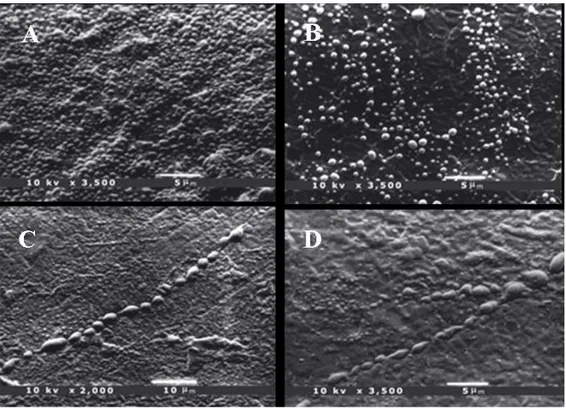
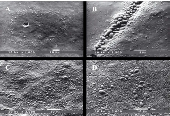
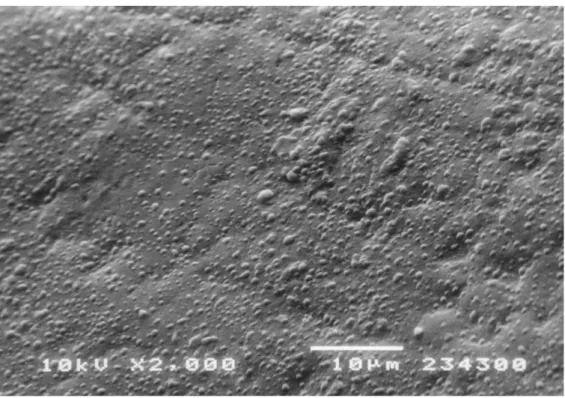
Figure 1 summarizes the statistical analysis and shows the results related to healthy teeth. The percentage distribution revealed a strong relationship (P < 0.01) with age: data showed that all the samples from subjects aged 6–20 yr presented more than 75% of the enamel surface covered with droplets. Samples from older subjects showed a decreasing percentage: samples from the 30–50 yr age group predominantly presented a moderate (5–75%) percentage, whereas in the 50–60 yr age group the number of samples with a low (< 5%) percentage of enamel area covered with droplets increased up to the last group (age > 60 yr), where all the samples showed less than 5% of the enamel surface covered with droplets (Fig. 2A–D and 3A–D). Figure 4A–D shows details of an enamel pore, an enamel crack, and white spot lesions, respectively. The number of droplets dis-closed by SEM observation confirmed that enamel is a permeable substrate. Our results demonstrated that permeability was related to age: freshly erupted permanent teeth showed more droplets covering the entire enamel surface. Samples from subjects of dif-ferent ages showed a decreasing number and size of droplets.
Permanent mature teeth showed many droplets mainly localized along the perikymata, and only a few droplets were detected away from these.
In vitro testing on extracted teeth showed a similar morphology. Droplets were still pre-sent along the perikymata.
Fig. 1 Percentage distribution of enamel area covered with droplets related to age. Barchart for groups by score.
Fig. 2 The arrangement of droplets in samples according to increasing age of the subject. Scanning elec-tron microscopy (SEM) photomicrographs of enamel from 6-yr-old (A) and 17-yr-old (B) pa-tients, showing many more droplets on the enamel surface, covering the whole surface in several areas. Permanent teeth showed many droplets, mainly localized along the perikymata. SEM pho-tomicrograph of 28-yr-old (C) and 30-yr-old (D) patients, showing typical droplet distribution along the perikymata. These droplets measured approximately 1 lm or less in diameter.
Fig. 3 Scanning electron microscopy (SEM) photomicrograph of 33-yr-old (A) and 39-yr-old (B) patients, showing the perikymata covered with droplets that appeared to be much larger than those of the adja-cent enamel. SEM photomicrograph of 67-yr-old (C) and 70- yr-old (D) patients, showing only a few small droplets, probably as a result of the reduced enamel water content.
Discussion
Enamel permeability has been demonstrated in vivo and in vitro (Lindén, 1968; Ten Bosch et al., 2000; Byers and Yoon Lin, 2003). Permeability is more substantial in teeth with immature enamel and appears to require a partnership with dentine (Byers and Yoon Lin, 2003, Shellis and Dibidin, 2000). Permeability is also correlated with enamel pores, which may cause water uptake and release (Shellis, 1996). Most permeability studies re-corded electrical variables, such as electrical resistance (Hoppenbrouwers et al., 1986; Wang et al., 2005) or conductance (Ten Bosh et al., 2000; Ie et al., 1995), providing an in-direct evaluation of enamel thickness, mineral loss and uptake (Ten Bosch et al., 2000), and enamel porosity (Flaitz et al., 1986; Rock and Kidd, 1988; Huysmans et al., 1995; Ricketts et al., 1996). The present study yielded qualitative and quantitative findings on outward fluid flow on tooth enamel surfaces in vivo by means of scanning electron mi-croscopy inspection of polyether replicas. This technique uses droplet formation to dis-play the discharge of liquid from enamel during the setting time of the impression mate-rial, as demonstrated in vitro by Barnes (Barnes, 1977). The fluid forming these droplets may come from free, unbound water in blind outer enamel porosities and partly in deeper structures, as suggested by the droplet distribution on enamel surface related to age. Presumably, the mechanism of droplet formation is simply diffusion. When a water-free impression material is applied to hydrated enamel, water diffuses out of the enamel down its concentration gradient and accumulates over the pores, without wetting or spreading, on the light-bodied hydrophobic material.
Droplet formation appeared to be typical in its location on the enamel surface of per-manent mature teeth, with a strong preference for the perikymata. The enamel surface of recently erupted teeth presents these and other open structures containing proteins produced during tooth development. Indeed, the enamel of freshly erupted permanent teeth showed more discharge of droplets than mature enamel. It is likely that these structures and interprismatic spaces form diffusion pathways, which alter with time in the oral cavity as a result of intermittent pH shift, traumas, and mineral deposition (Shellis and Dibdin, 2000). The results of this study appear to predict that the water con-tent of outer enamel decreases with age.
Moreover, increasing enamel maturation and age involve a progressive localization of outward fluid flow on the enamel surface along perikymata those are anatomically corre-lated to deep enamel structures.
The results of this in vivo study, obtained with a new, non-invasive technique, could be correlated to epidemiological data on caries.
Recently erupted teeth are more prone to dental caries than teeth that have remained free from caries lesions for a few years after eruption (Ten Bosch et al., 2000), as con-firmed by experiments in which artificial caries lesions were produced in extracted teeth of different posteruptive ages (Ten Bosh et al., 2000, Kostanos and Darling, 1991).
This may be ascribed to differences in enamel porosity dependent on intra-oral matura-tion, presumably caused by congestion of the pathways by deposition of calcium-phosphates in the outer layer of the tooth surface (Ten Bosh et al., 2000).
Therefore, enamel surface alterations, interpreted as posteruptive maturation and, con-sequently, enamel permeability, are of paramount importance for caries pathogenesis. Enamel fluid could also interfere with adhesive procedures. On the other hand, clinical procedures, such as acid etching and reshaping of teeth by grinding off some of the enamel outer surface, will increase the permeability of dental enamel. Partial recovery from such damage takes several months in vivo, and in the meantime the tooth is more susceptible to carious decay (Kuhar et al., 1999).
The replica procedure described identified the location of the pathway openings in the outer surface of tooth enamel in vivo by demonstrating fluid outflow, namely along the perikymata. Furthermore, the null hypothesis was rejected; the enamel of freshly erupted teeth presented higher outflow than mature enamel. We speculate that this outflow re-flects both enamel permeability and, possibly, caries susceptibility. Specific obstruction of these pathways may increase caries resistance.
References
Bakhos Y, Brudevold F, Aasenden R. In vivo estimation of the permeability of surface human enamel. Arch Oral Biol 1977;22: 599–603.
Barnes IE. The adaptation of composite resins to tooth structure. Part 1. Study 1: intro-duction and the adaptation of composite resins to the unetched enamel cavity wall. Br Dent J 1977; 142: 122–129.
Bergman G, Lindén L. Techniques for microscopic study of enamel fluid in vivo. J Dent Res 1965; 44: 1409.
Borggreven JMPM, Driessens FCM, Van Dijk JWE. Diffusion through bovine tooth enamel as related to the water structure in its pores. Arch Oral Biol 1980; 25: 345–348. Borggreven JMPM, Van Dijk JWE, Driessens FCM. A quantitative radiochemical study of ionic and molecular transport in bovine dental enamel. Arch Oral Biol 1977; 22:467– 472.
Braden M, Duckworth R, Joyston-Bechal S. The uptake of 24Na by human dental enamel. Arch Oral Biol 1971; 16: 367–374.
Byers MR, Yoon Lin KJ. Patterns of fluoro-gold entry into rat molar enamel, dentine, and pulp. J Dent Res 2003; 82: 312–317.
Chersoni S, Suppa P, Breschi L, Ferrari M, Tay FR, Pashley DH, Prati C. Water move-ment in the hybrid layer after different dentin treatmove-ments. Dent Mater 2004; 20: 796– 803.
Dibdin GH. The water in human dental enamel and its diffusional exchange measured by clearance of tritiated water from enamel slabs of varying thickness. Caries Res 1993; 27: 81–86.
Fearnhead RW, Kawasaki K, Inoue K. Comments on the porosity of human tooth enamel. J Dent Res 1982; 61: 1524–1530.
Featherstone JDB. Diffusion phenomena and enamel caries development. In: Guggen-heim, B, ed. Cariology Today. Basel: Karger, 1983; 259–268.
Flaitz CM, Hicks MJ, Siverstone LM. Radiographic, histologic, and electronic compari-son of occlusal caries: an in vitro study. Pediatr Dent 1986; 8: 24–28.
Hoppenbrouwers PMM, Scholberg HPF, Borgreven JMPM. Measurement of the per-meability of dental enamel and its variation with depth using an electrochemical method. J Dent Res 1986; 65: 154–157.
Huysmans MC, Verdonschot EH, Rondel P. Electrical conductance and electrode area on sound smooth enamel in extracted teeth. Caries Res 1995; 29: 88–93.
Ie YL, Verdonschot EH, Schaeken MJ, Vant’Hof MA. Electrical conductance of fissure enamel in recently erupted molar teeth as related to caries status. Caries Res 1995; 29: 94–99.
Itthagarun A, Tay FR. Self-contamination of deep dentin by dentin fluid. Am J Dent 2000; 13: 195–200.
Joyston-Bechal S, Duckworth R, Braden M. Diffusion of radioactive ions into human dental enamel. Arch Oral Biol 1971; 16: 375–384.
Kotsanos N, Darling AI. Influence of posteruptive age of enamel on its susceptibility to artificial caries. Caries Res 1991;25: 241–250.
Kuhar M, Cevc P, Schara M, Funduk N. In vitro permeability and scanning electron mi-croscopy study of acid-etched and ground enamel surfaces protected with dental adhe-sive coating. J Oral Rehabil 1999; 26: 722–730.
Lindén LA, Bjo¨rkman S, Hattab F. The diffusion in vitro of fluoride and chlorhexidine in the enamel of human deciduous and permanent teeth. Arch Oral Biol 1986; 31: 33– 37.
Lindén LA. Microscopic observations of fluid flowing through enamel in vitro. Odon-tol Rev 1968; 19: 349–365.
Moreno EC, Zahradnik RT. The pore structure of human dental enamel. Arch Oral Biol 1973; 18: 1063–1068.
Pashley DH. Dynamics of the pulpo-dentine complex. Crit Rev Oral Biol Med 1996; 7: 104–133.
Poole DF, Tailby PW, Berry DC. The movement of water and other molecules through human enamel. Arch Oral Biol 1963; 38: 771–772.
Poole DFG, Newman HN, Dibdin GH. Structure and porosity of human cervical enamel studied by polarizing microscopy and transmission electron microscopy. Arch Oral Biol 1981; 26:977–982.
Ricketts DN, Kidd EA, Liepins PJ, Wilson RF. Histological validation of electrical resis-tance measurement in the diagnosis of occlusal caries. Caries Res 1996; 30: 148–155. Rock WP, Kidd EA. The electronic detection of demineralization in occlusal fissures. Br Dent J 1988; 164: 243–247.
Shellis RP, Dibdin GH. Enamel microporosity and its functional implications. In: Tea-ford MF, Ferguson MJ, Smith MM, eds. Teeth: development, evolution and function. Cambridge:Cambridge University Press, 2000; 242–251.
Shellis RP. A scanning electron-microscopic study of solubility variation in human enamel and dentine. Arch Oral Biol 1996; 41: 473–484.
Shellis RP. Transport processes in enamel and dentine. In: Addy M, Embery G, Edgar WM, Orchardson R, eds. Tooth wear and sensivity. London: Martin Duniz, 2000; 19– 24.
Tarbet WJ, Forsick LS. Permeability of human dental esame to acriflavine and potassium fluoride. Arch Oral Biol 1971; 16:951–961.
Ten Bosch JJ, Fennis IEY, Verdonschot EH. Time-dependent decrease and seasonal variation of the porosity of recently erupted sound dental enamel in vivo. J Dent Res 2000; 79: 1556–1559.
Ten Cate M. Remineralization of caries lesions extending into dentine. J Dent Res 2001; 80: 1407–1411.
Van Dijk JW, Borggreven JM, Driessens FC. Diffusion in mammalian tooth enamel in relation to the caries process. Arch Oral Biol 1983; 28: 591–597.
Wang J, Someya Y, Inaba D, Longbottom C, Miyazaki H. Relationship between electri-cal resistance measurement and microradiographic variables during remineralization of softened enamel lesions. Caries Res 2005; 39: 60–64.
Whittaker DK. Structural variations in the surface zone of human tooth enamel ob-served by scanning electron microscopy. Arch Oral Biol 1982; 27: 383–392.
Chapter 5
In vivo fluid release from
primary enamel
A relationship between caries susceptibility and enamel permeability has been proposed for permanent teeth by detecting in vivo outward fluid flow on tooth enamel surface. The aim of this study was to reveal in vivo the occurrence of fluid release from primary tooth enamel.
Four primary upper canines with no visual signs of caries, cracks, erosion or restorations from 6 to 10 years old subjects and two retained primary upper canines from 33 and 40 years old subjects were included in the study. The enamel surface was gently polished and air dried for 10 s. An impression was immediately obtained by vinyl polyxiloxane. Replicas were then obtained by polyether impression material, gold coated and inspected under SEM. The hydrophobic vinyl polyxiloxane material enabled to obtain in situ a morphological image of the presence of droplets, most likely resulting from outward flu-ids flow through outer enamel.
Primary enamel showed a substantive permeability as confirmed by droplets presence on its surface. Droplets distribution covered the entire enamel surface in all the samples, without any specific localization. No signs of post-eruptive maturation with changes in droplets distribution were observed in samples from adult subjects.
SEM evaluation of droplets distribution on enamel surface indicated a substantive per-meability in primary teeth, accordingly with histological features, without changes during aging and suggested a strong relationship between enamel permeability and caries sus-ceptibility.
Introduction
Primary enamel is less-mineralized (81.3-94.2 wt%), more porous, contains more organic matter, more water, and shows a greater diffusion coefficient than enamel of permanent teeth (Lindén et al., 1986; Cuy et al., 2002; Wang et al., 2006; Lussi et al., 2000). Moreover, overall mineral density is lower in the outermost layers but shows no significant differ-ences closed to enamel-dentine junction (Wang et al., 2006; Wilson and Beyond, 1989). The primary enamel is not more susceptible to erosion even though it is reported to be statistically significantly softer and less elastic and was reported that dissolves considera-bly faster than permanent enamel (Lussi et al., 200; Lippert et al., 2004).
The structured nature of enamel allows transport of ions, molecules and water (Lindén, 1968; Lindén et al., 1986; Shellis, 1996; Ten Bosh et al., 2000). Fluid flowing through enamel is related to its water content and its permeability. As clinical studies showed car-ies formation and progression to be faster in primary than in permanent teeth (Kostanos and Darling, 1991; Sønju Clasen et al., 1997; Issa et al., 2003), an eventual correlation be-tween caries susceptibility and fluid flow might be existing.Moreover enamel fluid flow has been correlated with post-eruptive maturation as permanent teeth showed a decreas-ing permeability with age (Bertacci et al., 2007).
The aim of this study was to visualize in vivo fluid flow through tooth enamel in primary teeth with a replica technique and SEM observations to investigate the effects of intra-oral staying on primary enamel.
Materials and Methods
Patient and tooth selection
Four primary upper canines with no visual signs of caries, cracks, erosion or restorations from six to 10 years old subjects and 2 retained upper primary canines from 2 adult sub-jects (aged 33 and 40 years) were included in this study. The study has been conducted in full accordance with ethical principles of the World Medical Association Declaration of Helsinki. Parents for all subjects aged 6 to 10 years old included in the study, gave written consensus to the procedure that was non-invasive, and did not create any risk for the patients.
Enamel treatment procedures
Each tooth was brushed with a prophylactic paste for 10 s, gently washed and finally air dried for 10 s with a dental chair air syringe (Castellini, Italy). The method used to inves-tigate the morphology of enamel detecting the presence of droplets was previously de-scribed (Bertacci et al., 2007).
Immediately after enamel preparation as previously described, an impression of the sur-face was made using a polyvinilsiloxane impression material (Affinis® light body
COLTENE, Alstatten, Switzerland). After 4 min, the material was removed from the enamel surface and was degassed for 48 h and poured out in polyether impression mate-rial (Permadyne Garant®, 3M ESPE, St. Paul, MN, USA).
Samples were gold-sputtered and inspected by scanning electron microscope (SEM, JEOL, Model 5400, Tokyo, Japan).
Evaluation and statistical analysis
The evaluations on presence of droplets were performed at x 2000 and x 3500 magnifi-cation by two double blind operators examining randomly for each sample three differ-ent points represdiffer-entative of the enamel in the cervical, medium and incisal third.
Results
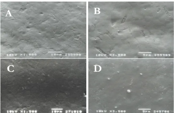
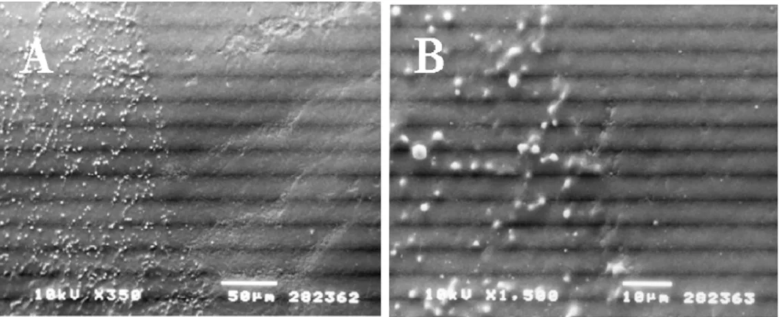
SEM replicas observation showed that all enamel surfaces release equal droplet forma-tion. All the samples from patients aged 6-10 yr showed many droplets that covered the entire enamel surface (Fig 1). Enamel of primary teeth from adult subjects showed no difference in droplets distribution: droplets covered the entire surface without any spe-cific localization (Fig.2).
Fig. 1 SEM photomicrograph of 10-yr-old patient upper canine shows the typical droplets arrangement. Many droplets cover the whole enamel surface.
Fig. 2 SEM photomicrograph of 33-yr-old patient enamel surface. According to increasing subject’s age