R E S E A R C H
Open Access
Conservative treatment in Scheuermann
’s
kyphosis: comparison between lateral curve
and variation of the vertebral geometry
Angelo G. Aulisa
1*, Francesco Falciglia
1, Marco Giordano
1, Giuseppe Mastantuoni
1, Andrea Poscia
2and Vincenzo Guzzanti
1,3From 12th International Conference on Conservative Management of Spinal Deformities - SOSORT 2015 Annual Meeting Katowice, Poland. 7-9 May 2015
Abstract
Background: Conservative treatment in the Scheuermann’s kyphosis obtain, during skeletal growth, remodelling of the deformed vertebras. In a previous paper on Scheuermann’s kyphosis, we have studied the geometry variations of all vertebrae included in the curve, before and after the treatment.
The purpose of this study was to confirm the effectiveness of conservative treatment in Scheuermann’s kyphosis and was to evaluate and compare the variation of the vertebral geometry with the curve trend in Cobb degrees, before and after conservative treatment.
Methods: From a consecutive series of patients, we selected 90 patients with thoracic Scheuermann’s kyphosis, treated using anti-gravity brace: 59 male, 31 female. The mean age at the beginning of the treatment was 14 years. Radiographical measurements were performed on radiographs from a lateral projection, at the beginning (t1) and at the end of the treatment (t5). Vertebral geometry modifications at t1 and t5 were analysed according to the following parameters and evaluated by three independent observers: Anterior wedging angle (ALFA) of the apex vertebra and Posterior wall inclination (APOS) of the limiting lower vertebra. The curve was measured in Cobb degrees. Results: The results from our study showed that of the 90 patients with a thoracic curve mean value of Cobb degrees was 57.8 ± 6.0 SD at t1 and 41.3 ± 5.6 SD at t5. The differences between t1(angle at baseline) and t5 (end of treatment) were calculated for Cobb, ALFA and APOS angle and were respectively−16.4 ± 4.5, −6.4 ± 1.4 and −2.7 ± 1.2; tested with paired t-test were significative (p < 0.01). The results of the regression analysis to test the relationship between the three measures for the kyphosis (Cobb degree, ALFA and APOS) showed that the best association was between Cobb t5 and ALFA t5 (p < 0.01) and Cobb t1 and APOS t1 (p < 0.01). No significative association was found between the difference between ALFA and APOS.
Conclusion: We sustain that using new parameters to study vertebral remodelling allows us to reach a better comprehension of Scheuermann spine response to anti-gravity brace treatment. Furthermore, the evaluation of the ALFA angle of the apex vertebra confirms to be more reliable than Cobb’s angle because it cannot be affected by the radiological position.
Abbreviations: ALFA, Anterior wedging angle; APOS, Posterior wall inclination
* Correspondence:[email protected]
1U.O.C. of Orthopedics and Traumatology, Children’s Hospital Bambino Gesù,
Institute of Scientific Research, Rome 00165, Italy
Full list of author information is available at the end of the article
© 2016 The Author(s). Open Access This article is distributed under the terms of the Creative Commons Attribution 4.0 International License (http://creativecommons.org/licenses/by/4.0/), which permits unrestricted use, distribution, and reproduction in any medium, provided you give appropriate credit to the original author(s) and the source, provide a link to the Creative Commons license, and indicate if changes were made. The Creative Commons Public Domain Dedication waiver (http://creativecommons.org/publicdomain/zero/1.0/) applies to the data made available in this article, unless otherwise stated.
Background
In 1920 Scheuermann [1] first described the associ-ation of developmental Kyphosis and wedging of thoracic vertebrae; he used the term “osteochondritis juvenilis dorsi” [2], but the condition is universally known today as Sheuermann’s kyphosis. Sorenson [3], proposed a diagnosis based on the presence of three or more adjacent vertebrae wedged 5° or more and no evidence of congenital, infectious or traumatic disorders of the spine. These criteria are widely ac-cepted and used today. The prevalence in the general population ranged from 4 to 10 % [4, 5]. The patho-genesis is still not clear some authors write that “The weakness of the vertebral endplate probably re-sults from a predisposing genetic background that in-fluences the quality of matrix components (collagen types II and IX) and chondrocytes” [5, 6] other said that mechanical stress influences the severity of spinal impairment [7].
Vertebral geometry alterations in Scheuermann’s kyphosis and results of the orthopedic treatment have been measured by radiographic measure of both curve entity and vertebral wedging on longitudinal section [8–11]. Clinical evolution of the deformity is not always correlated to presently used radiographic parameters. On the other hand, it is possible that vertebal morphology alteration in kyphotic curve could be explained by a more complex theory model than the currently accepted one [12]. For this rea-son, in a previous paper on Scheuermann’s kyphosis, we have studied the geometry variations of all ver-tebrae included in the curve, before and after the treatment [13].
The purpose of this study was to confirm the effective-ness of conservative treatment in Scheuermann’s kyphosis and was to evaluate and compare the variation of the vertebral geometry with the curve
trend in Cobb degrees, before and after conservative treatment.
Methods
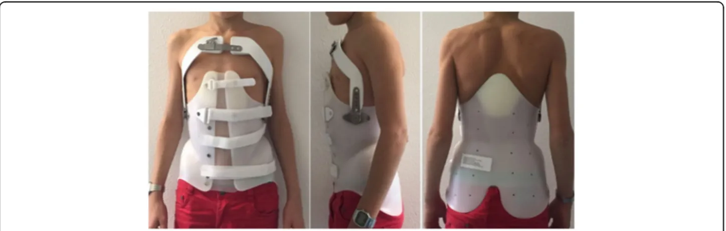
We selected, from a consecutive series of patients, included in a prospective database, 90 patients with thoracic Scheuermann’s kyphosis, treated using anti-gravity brace between 2004 and 2010 (Fig. 1). Other type of kyphosis were excluded. 59 patients were male, 31 were female. The mean age at the beginning of the treatment was 14.2 ± 1.8 years. The mean curve entity before treatment, measured by Cobb’s method, was 57.8 °, a value that, according to the lit-erature data, requires orthopaedic treatment [14].
All cases were treated with anti-gravity brace. Time bracing prescribed was max 20 h daily, min 16 h daily. In order to maximize the adherence to treatment, patients were always followed by the same doctor. Furthermore, controls were performed every 3 months. Frequent checks allowed to verify and implement compliance establishing an open and friendly relationship with the patients. Close checks were also performed to maximize bracing effec-tiveness over the time. Weaning was started when a full recovery of vertebral geometry was seen on a latero-lateral radiograph view. Not exercises were performed.
Radiographical measurements were performed on radiographs from a lateral projection, at the begin-ning (t1) and at the end of the treatment (t5).
To avoid the great variance in the range of curve angles in thoracic kyphosis that rely on the radio-logical position, x-rays were performed all at our Radiology Department observing the following pos-ition: standing with head straight, arms bent at 45° and hands placed on a support.
Vertebral geometry modifications at t1 and t5 were analysed according to the following parameters and evaluated by three independent observers:
Cobb degrees for curve magnitude
Anterior wedging angle (ALFA) of the apex vertebra Posterior wall inclination (APOS) of the limiting lower vertebra.
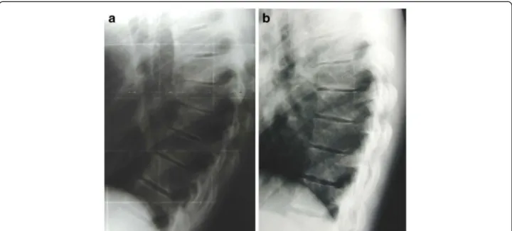
In particular, the measurement of ALFA angle was the calculation of the convex angle formed by two lines perpendicular to the lines passing through pos-terior and anpos-terior limit, respectively of superior and inferior disk plates of the vertebral body (Fig. 2). In-stead the measurement of the posterior wall inclin-ation APOS, was conducted using disk plate limits of
each vertebra. More specifically, we have calculated the angles between the line perpendicular to the in-ferior plate and the line passing through superior and inferior limit of posterior wall.
These parameters were chosen because they had shown to be the most significant in a previous study (13).
Statistical analysis
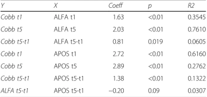
Mean and Standard Deviation of kyphosis degree as Cobb, ALFA and APOS angle at baseline (t1) and at the end of treatment (t5) were calculated and differences be-tween t1 and t5 were tested with paired t-test. Further-more, a linear regression model was used to test the relationship between Cobb, ALFA and APOS angle at t1, t5 and at t5-t1 difference.
Results
The results from our study showed that of the 90 patients with a thoracic curve mean value of Cobb degrees was 57.8 ± 6.0 SD at t1 and 41.3 ± 5.6 SD at t5 (Fig. 3). The mean duration of treatment was 32.9 ± 18.44 months and the mean follow-up was 30.02 ± 18.85 months. The differences between t1(angle at baseline) and t5 (end of treatment) were cal-culated for Cobb, ALFA and APOS angles and were respectively −16.4 ± 4.5, −6.4 ± 1.4 and −2.7 ± 1.2; tested with paired t-test were significative (p < 0.01) (Table 1). No difference statistically significative be-tween male and female was reported. The results of the regression analysis to test the relationship be-tween the three measures for the kyphosis (Cobb Fig. 2 Measure of posterior wall inclination APOS, conducted using
disk plate limits of every vertebra. More specifically, we have calculate the angles between the line perpendicular to inferior plate and the line passing trough superior and inferior limit of posterior wall
degree, ALFA and APOS) are shown in Table 2. The best association was found between Cobb t5 and ALFA t5 (p < 0.01) and between Cobb t1 and APOS t1 (p < 0.01). No significative association was found be-tween the difference bebe-tween ALFA and APOS.
Discussion
The results confirm that conservative treatment in Scheuermann’s Kyphosis, during skeletal growing, is effectiveness and we can obtain a remodelling of the deformed vertebrae.
In particular the antigravity brace, based on bio-mechanical action of the three points principle: one force is applied behind the curve apex and the other two forces are applied at the end of the vertebrae at the curve ends. Moreover, in according to the vector calculation principles, a force applied to a curve structure is divided in two components with direction and course determined by the application point and by the space orientation of the resultant. Therefore it is logical that forces applied by an antigravity brace could produce different effects on the vertebral re-modelling, depending on the vertebral position. On this basis, we sustain that using ALFA angle of the apex vertebra and APOS angle of the limiting lower vertebra to study vertebral remodelling allows us to
reach a better comprehension of Scheuermann spine response to anti-gravity brace treatment.
Conclusion
The correlation between Cobb and ALFA at follow-up and Cobb and Posterior Wall Inclination at base-line confirm the complexity of vertebral remodelling and allows us to reach a better comprehension of Scheuermann spine response to anti-gravity brace treatment.
Moreover the evaluation of the ALFA angle of the apex vertebra confirms to be more reliable than Cobb’s angle because it cannot be affected by the radiological position.
Declarations
This article has been published as part of Scoliosis and Spinal Disorders Volume 11 Supplement 2, 2016. Research into Conservative Management of Spinal Deformities: Short Articles from the SOSORT 2015 Meeting. The full contents of the supplement are available online http://scoliosisjournal.biomedcentral.com/ articles/supplements/volume-11-supplement-2.
Availability of data and materials
The data will be shared only with a direct request to the author by email.
Authors’ contributions
AGA participated in the conception, design and coordination, and to acquisition of data, analysis and interpretation of data and drafted the manuscript. MG, FF and GM helped to draft the manuscript. AP participated in the design of the study and performed the statistical analysis. VG participated in the conception. All authors read and approved the final manuscript.
Competing interests
The authors declare that they have no competing interests.
Consent for publication
Informed consent was obtained for the Fig. 1.
Ethics approval and consent to participate
All procedures performed in studies involving human participants were in accordance with the ethical standards of the institutional and/or national research committee and with the 1964 Helsinki declaration and its later amendments or comparable ethical standards.
Ethics approval was not required as stated by Ethics Committee Ospedale Pediatrico Bambino Gesù IRCCS*.
Informed consent was obtained from all individual participants included in the study.
Table 1 Mean, Standard Deviation (SD) of kyphosis in terms of Cobb degree, ALFA and APOS angle at baseline (t1) and at the end of treatment (t5)
Condition Time Mean sd Differ ence T5-T1 mean (SD) P value (Paired T test)
Cobb t1 57.8 6.0 −16.4 (4.5) <0.01 t5 41.3 5.6 ALFA ant t1 14.0 2.2 −6.4 (1.4) <0.01 t5 7.6 2.4 APOS t1 4.6 1.7 −2.7 (1.2) <0.01 t5 1.9 1.0
Table 2 Regression analysis to test the relationship between Cobb, ALFA and APOS angle
Y X Coeff p R2 Cobb t1 ALFA t1 1.63 <0.01 0.3545 Cobb t5 ALFA t5 2.03 <0.01 0.7610 Cobb t5-t1 ALFA t5-t1 0.81 0.019 0.0605 Cobb t1 APOS t1 2.72 <0.01 0.6160 Cobb t5 APOS t5 2.89 <0.01 0.2762 Cobb t5-t1 APOS t5-t1 1.38 <0.01 0.1322 ALFA t5-t1 APOS t5-t1 −0.20 0.09 0.0307
Author details
1U.O.C. of Orthopedics and Traumatology, Children’s Hospital Bambino Gesù,
Institute of Scientific Research, Rome 00165, Italy.2School of Hygiene and
Preventive Medicine, University Hospital“Agostino Gemelli”, Catholic University of the Sacred Heart School of Medicine, Rome 00168, Italy.
3University of Cassino, Cassino (FR) 03043, Italy.
Published: 14 October 2016
References
1. Scheuermann HW. Kyphosis dorsalis juvenilis. Ugeskr Laeger. 1920;82:385–93. 2. Scheuermann HW. Kyphosis juvenilis (Scheuermann Krankheit). Fortschr Geb
Rontgenstr. 1936;53:1–16.
3. Sorenson KH. Scheuermann’s Juvenile Kyphosis. Cophenagen: Munksgaard; 1964.
4. Palazzo C, Sailhan F, Revel M. Scheuermann’s disease: an update. Joint Bone Spine. 2014;81(3):209–14.
5. Damborg F, Engell V, Nielsen J, Kyvik KO, Andersen MO, et al. Genetic epidemiology of Scheuermann’s disease. Acta Orthop. 2011;82:602–5. 6. Esapa CT, Hough TA, Testori S, et al. A mouse model for
spondyloepiphysealdysplasia congenita with secondary osteoarthritis due to a Col2a1 mutation. J Bone Miner Res. 2012;27:413–28.
7. Fotiadis E, Kenanidis E, Samoladas E, et al. Scheuermann’s disease: focus onweight and height role. Eur Spine J. 2008;17:673–8.
8. Ascani E, Montanaro A. Scheuermann’s disease. In: Bradford D, Hensinger R, editors. The pediatric spine. New York: Thieme Verlag; 1985.
9. Aufdermaur M. Juvenile kyphosis (Scheuermann’s disease): radiography, histology and pathogenesis. Clin Orthop. 1981;154:166–74.
10. Bradford DS, Moe JH. Scheuermann’s juvenile kyphosis. A histologic study. Clin Orthop. 1975;110:45–53.
11. Zaina F, Atanasio S, Ferraro C, et al. Review of rehabilitation and orthopedic conservative approach to sagittal plane diseases during growth: hyperkypho-sis, junctional kyphosis, and Scheuermann disease. Eur J Phys Rehabil Med. 2009;45:595–603.
12. Tsirikos AI, Jain AK. Scheuermann’s kyphosis; current controversies. J Bone Joint Surg Br. 2011;93(7):857–64.
13. Pola E, Lupparelli S, Aulisa AG, Mastantuoni G, Mazza O, De Santis V. Study of vertebral morphology in Scheuermann’s kyphosis before and after treatment. Stud Health Technol Inform. 2002;91:405–11.
14. De Mauroy J, Weiss H, Aulisa A, Aulisa L, Brox J, Durmala J, Fusco C, Grivas T, Hermus J, Kotwicki T, Le Blay G, Lebel A, Marcotte L, Negrini S, Neuhaus L, Neuhaus T, Pizzetti P, Revzina L, Torres B, Van Loon P, Vasiliadis E, Villagrasa M, Werkman M, Wernicka M, Wong M, Zaina F. Conservative treatment of idiopathic & Scheuermann’s kyphosis. 7th SOSORT consensus paper: Scoliosis. 2010; 5:9
• We accept pre-submission inquiries
• Our selector tool helps you to find the most relevant journal
• We provide round the clock customer support
• Convenient online submission
• Thorough peer review
• Inclusion in PubMed and all major indexing services
• Maximum visibility for your research Submit your manuscript at
www.biomedcentral.com/submit