Università degli Studi di Cagliari
DOTTORATO DI RICERCA
Scuola di dottorato in scienze e tecnologie chimiche e farmaceutiche
Indirizzo/ corso in scienze e tecnologie chimiche
Ciclo XXIII
TITOLO TESI
DNA based biosensors for environmental and medical applications
Settore scientifico disciplinari di afferenza
CHIM/02 CHIMICA FISICA
Presentata da:
Francesca Cugia
Coordinatore Dottorato
Prof. Mariano Casu
Tutor
Prof.ssa Maura Monduzzi
Dott. Andrea Salis
Abstract
iii
ABSTRACT
In the present thesis two electrochemical DNA based biosensors were developed using screen printed electrode as transducers.
Biosensors are defined as a self-containing integrated devices, capable of providing specific quantitative or semi-quantitative analytical information using a biological recognition element which is in contact with a transduction element.
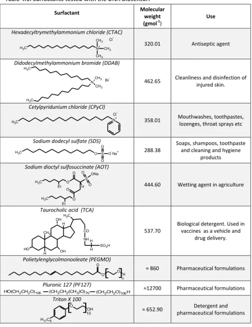
The first DNA biosensor realized was applied to the rapid screening of toxic substances. The biosensor was constructed immobilizing a double helix DNA (Calf Thymus DNA) onto screen-printed electrodes. Subsequently, the biosensor was used for the determination of the toxicity of different kinds of common surfactants.
Surfactant interactions with double stranded DNA were evaluated measuring the height of the guanine oxidation peak. Indeed, the interactions with toxic substances raises structural and conformational modifications of DNA causing decrease of guanine peak. The intensity of the guanine oxidation peak, was measured through Square Wave Voltammetry (SWV).
Moreover, the toxicity of some selected surfactants was investigated both in sea water and tap water, and data were compared to those obtained in acetate buffer. The interaction between surfactants and Calf Thymus DNA in solution and adsorbed on the sensor surface was also investigated through FTIR and FTIR-ATR spectroscopy respectively.
The second kind of biosensor studied was a Genosensor, that is an analytical device where the biological recognition element is a single strand oligonucleotide sequence. These sequences referred as capture probe are capable to recognize selectively a complementary sequence (RNA or DNA), named target, by a hybridization reaction. Among the sequence probes, modified locked nucleic acid (LNA) and the peptide nucleic acid (PNA) were used. In this case the screen printed electrode
Abstract
iv
was used only as transducer while the hybridization assay was conducted onto paramagnetic micro beads.
The genosensor was used for the analytical detection of DNA and RNA sequences. In particular, the analytical properties of PNA and LNA capture probes with classical DNA sequences were compared. Hybridization with RNA target as well as with the corresponding DNA sequence was also performed. Differential pulse voltammetry (DPV) was used to perform the electrochemical measurements.
Acknowledgements
v
ACKNOWLEDGEMENTS
First of all I would like to express my sincere gratitude to my supervisor Prof. Maura Monduzzi for her encouragement and precious suggestions during this work and Dr. Andrea Salis for his invaluable help and guidance. Thanks are due to Projects MIUR DM28142 of the Sardinian Biomedicine District, MIUR Prin 2008, grant number 2006030935, for financial support. Sardegna Ricerche Polaris is thanked for free access to the instruments belonging to the Nanobiotechnology laboratories. In addition thanks are due to CSGI and CNBS for general expertise support.
Thanks are due Sardinia Region, Project Master & Back. A particular acknowledgement is dedicated to Prof. Marco Mascini and to his group for the expert and friendly environment at the Department of Chemistry of Florence University. A special mention goes to Dr. Serena Laschi for her dedication in helping me and answering all of my numerous questions. Thanks to all my colleagues (Amita, Brajesh, Daniela, Elisabetta, Luca, Marcella, Marco and Viviana) at the Biocatalysis laboratory with whom I spent many great moments and who have always encouraged and helped me.
I would also like to thank: Davide Espa, Maria Varotto, Elisa Sessini and Flavia Artizzu for their support, enthusiasm and friendship.
Lastly, I offer my regards to all of those who supported me during the completion of the project.
Contents vii
CONTENTS
ABSTRACT iii Acknowledgements v CONTENTS viiLIST OF ABBREVIATIONS AND SYMBOLS xi
CHAPTER I. GENERAL INTRODUCTION 1
1.1 BIOSENSOR: DEFINITION, FUNCTION AND APPLICATIONS 3
1.2 BRIEF HISTORY OF BIOSENSORS 4
1.3 BIOSENSOR CLASSIFICATION 6
1.3.1 Receptor: biological recognition element 6
1.3.2 Detection or measurement mode: electrochemical
transduction 8
1.3.2.1 Screen-printed electrodes (SPE) as electrochemical
transducers 9
1.3.2.2 Preparation of screen printed electrodes 10
1.4 NUCLEIC ACID BASED BIOSENSOR: DEFINITION AND
APPLICATIONS 12
1.4.1 Structure of Nucleic Acids 12
1.4.1.1 Deoxyribonucleic acid (DNA) 13
1.4.1.2 Ribonucleic acid (RNA) 17
1.4.1.3 Analogous of Nucleic Acid: PNA and LNA 18
1.4.2 DNA-based biosensor for environmental application 20
1.4.2.1 DNA biosensor principle 21
1.4.3 Genosensors 22
REFERENCES 25
CHAPTER II. ANALYTES 27
2.1 SURFACTANTS 29
Contents
viii
2.1.2 Surfactants Toxicity 33
2.2 MicroRNA 35
2.2.1 Implications of microRNAs in cancer 35
REFERENCES 38
CHAPTER III. INSTRUMENTAL TECHIQUES 41
3.1 INTRODUCTION 43
3.2 VOLTAMMETRY 43
3.2.1 Excitation signals 45
3.2.2 Voltammetric Instruments 47
3.2.3 Voltammograms 48
3.2.4 Differential Pulse Voltammetry (DPV) 49
3.2.5 Square-Wave Voltammetry (SWV) 50
3.3 ZETA POTENTIAL 52
3.3.1 Zeta potential measurements 53
3.4 INFRARED SPECTROSCOPY 56
3.4.1 Molecular Vibrations 57
3.4.2 Fourier Transform Spectrometers 59
3.4.2.1 ATR-FTIR spectroscopy principles. 60
REFERENCES 62
CHAPTER IV. SURFACTANTS TOXICITY TOWARDS AN
ELECTROCHEMICAL DNA BIOSENSOR 63
4.1 INTRODUCTION 65
4.2. MATERIALS AND METHODS 67
4.2.1 Chemicals 67
4.2.2 Toxalert®100 procedure 67
4.2.3 Electrochemical oxidation of guanine and adenine 68
4.2.4. DNA-Biosensor functioning principle 69
4.2.5 Analysis of surfactant toxicity through DNA-biosensor 71
Contents
ix
4.2.7 DNA-Screen Printed Electrodes storage 72
4.3. RESULTS 72
4.3.1 Comparison between Toxalert®100 of AOT and Triton X
100 toxicity toward and DNA-biosensor. 72
4.3.2 Toxicity of surfactants 76
4.3.3 Effect of the aqueous matrix on the toxicity 79
4.3.4 Stability towards storage of the immobilized DNA on
screen-printed electrode 82
4.4. DISCUSSION 82
4.5. CONCLUSIONS 84
REFERENCES 85
CHAPTER V. FTIR STUDIES ON INTERACTIONS BETWEEN SURFACTANTS AND CALF THYMUS DNA IN SOLUTION AND ADSORBED ON SCREEN PRINTED ELECTRODES
89
5.1 INTRODUCTION 91
5.2. MATERIALS AND METHODS 92
5.2.1 Chemicals 92
5.2.2 Interactions between DNA and surfactants in buffer
solution through zeta potential and FTIR 93
5.2.3 Interactions between DNA adsorbed on SPE and surfactants
trough ATR- FTIR spectroscopy 93
5.3. RESULTS AND DISCUSSION 94
5.3.1 Zeta potential measurements 94
5.3.2 FT IR measurements: study of interactions between DNA
and surfactants in aqueous solution 96
5.3.3 Characterization of DNASPE -surfactants interactions 103
5.3.4 FTIR-ATR spectra of oxidized DNASPE 108
5.4. CONCLUSIONS 111
Contents
x
CHAPTER VI. HYBRIDIZATION ASSAY COUPLED TO MAGNETIC
BEADS FOR NUCLEIC ACID DETECTION 115
6.1 INTRODUCTION 117
6.2 MATERIALS AND METHODS 118
6.2.1 Chemicals 118
6.2.2 Steptavidin- Biotin binding 120
6.2.3 Biomodification of streptavidin-coated magnetic beads 121
6.2.4 Hybridization assay 123
6.2.5 Labelling with alkaline phosphatase and electrochemical
detection 123
6.3 RESULTS AND DISCUSSION 125
6.3.1 Assay for detection of DNA target using DNA, LNA and
PNA probes 125
6.3.2 Assay for detection of DNA target using different probes 126
6.4 CONCLUSIONS 127
REFERENCES 128
CONCLUDING REMARKS 131
xi
LIST OF ABBREVIATIONS AND SYMBOLS
Abbreviation Meaning
A Adenine
AOT Sodium dioctyl sulfosuccinate
ATR Total reflection accessory
DDAB Didodecylmethylammonium bromide
DNA Deoxyribonucleic acid
DPV Differential Pulse Voltammetry
dsDNA Double strand DNA
C Cytosine
CCP Critical packing parameter
CMC Critical micelle concentration
CPyCl Cetylpyridunium chloride
CTAC Hexadecyltrymethylammonium chloride
EC Effective concentration
G Guanine
HLB Hydrophilic lipophilic balance
LC Lethal concentration
LNA Locked nucleic acid
miRNA MicroRNA
NA Nucleic Acid
PEGMO Polietylenglycolmonooleate
PF 127 Pluronic 127
PNA Peptide nucleic acid
RNA Ribonucleic acid
RSD % Relative standard deviation
SDS Sodium dodecyl sulfate
ssDNA Single strand DNA
SPE Screen printed electrode
SWV Square Wave Voltammetry
T Thymine
TCA Taurocholic acid
Chapter I
General Introduction
Chapter I
General Introduction
3
1.1 BIOSENSOR: DEFINITION, FUNCTION AND APPLICATIONS
According to IUPAC, a biosensor is defined as a self-containing integrated device, capable of providing specific quantitative or semi-quantitative analytical information using a biological recognition element which is in contact with a transduction element (Figure1.1).1
Figure 1.1: Biosensor detection principle.
Both the biological and the transduction elements are essential. The first works as a bioreceptors (biorecognition element), and has a powerful molecular recognition capability. The biological element can be an enzyme, a single or double DNA strand, an antibody or a cellular component of a living system. The transducer element translates the interaction of the biorecognition element into a detectable signal. If the signal intensity is proportional to the concentration of the analyte quantitative analysis can be carried out.2 The biorecognition element enables the sensor to respond selectively to a particular analyte or group of analytes, thus avoiding interferences from other substances. This property, together with compact size, one-step reagentless analysis, and sensitivity make biosensors very attractive in comparison with conventional analysis techniques. Some interesting commercial biosensors are already available for the detection of glucose, lactate,
Chapter I
4
penicillin and urea, but their number is still limited in comparison with the research efforts for their development.
1.2 BRIEF HISTORY OF BIOSENSORS
The modern concept of biosensor is due to Leland C. Clark Jr. He invented the oxygen electrode, and its subsequent modification with enzymes. In 1962 at a New York Academy Sciences symposium he described “how to make electrochemical sensors (pH, polarographic, potentiometric or conductometric) more intelligent” by adding “enzyme transducers as membrane enclosed sandwiches”. The first example was illustrated by entrapping the enzyme Glucose Oxidase in a dialysis membrane over an oxygen probe. The decrease of oxygen concentration was proportional to glucose concentration. The term enzyme electrode was coined by Clark and Lyons.3 Clark's ideas became a commercial product in 1973 with the successful launch of the glucose analyser commercialized by Yellow Springs Instrument Company (Ohio). This was based on the amperometric detection of hydrogen peroxide and was the first biosensor-based laboratory analyser .
Guilbault and Montalvo were the first to develop a potentiometric enzyme electrodes. They realized a glass electrode coupled with urease to measure urea concentration in the blood.4 Starting from 1970, several other authors started to couple an enzyme with an electrochemical sensor to develop a biosensor. In 1975 Divis suggested that bacteria could also be used as the biological element in microbial electrodes for the measurement of ethanol.5 In 1975 Lubbers and Opitz6 proposed the term “optode” to describe a fibre-optic sensor to measure carbon dioxide or oxygen. They developed an optical biosensor that was used for ethanol detection by immobilizing alcohol oxidase at the end of a fiber-optic oxygen sensor.7
General Introduction
5 In 1976, Clemens et al.5 incorporated an electrochemical glucose biosensor in an artificial pancreas and this was later marketed with the commercial name the Biostator. In the same year, the pharmaceutical group La Roche (Switzerland) introduced the Lactate Analyser LA 640 for which the soluble mediator, hexacyanoferrate, was used to carry electrons from lactate dehydrogenase to the amperometric electrode. This was not a commercial success at that time, but subsequently it became an important forerunner of a new generation of mediated-biosensors for lactate analysis in sport and clinical fields.
In 1982, Shichiri et8 al. described the first needle-type enzyme electrode for subcutaneous implantation. This result was a major advance in the in vivo application of glucose biosensors. Companies are still pursuing this possibility, but no device for general use is available yet.
The biosensors based on the use of enzymes involving catalytic action are referred as catalytic biosensor. Lately bioaffinity biosensors that make use of antibodies and receptor molecules having affinity towards analytes have been developed. In 1980s, the first bioaffinity biosensor was developed using radio-labelled receptors immobilized onto a transducer surface. Biosensor based on ELISA have also been developed using labeled antibody or labeled antigen coupled with a suitable transducer. The “cell biosensors” were also developed during 1980s, making use of whole microbiological cells or organelles to measure the level of various drugs or environmental toxicants. Biosensor research is currently investigating a wide variety of devices using biological element, such as enzymes, nucleic acids, cell receptors, antibodies and intact cells, in combination with various transduction mechanisms.9 These biosensors can be applied to a wide range of analytical systems in health care, food and drink, process industries, environmental monitoring defence and security.9
Chapter I
6
1.3 BIOSENSOR CLASSIFICATION
Biosensors may be classified according to the biological element and the mode or signal transduction.
1.3.1 Receptor: biological recognition element
Biocatalytic recognition element: these biosensors are based on a
reaction catalysed by bio-macromolecules. Continuous consumption of substrate (S) is obtained due to the action of biocatalyst incorporated into the sensor. The responses are monitored by the integrated detector. Three types of biocatalysts are commonly used.
Enzymes (mono or multi-enzyme): these are the most common and well-developed recognition systems.
Whole cells (micro-organisms, such as bacteria, fungi, eukaryotic cells or yeast) or cell organelles or particles (mitochondria, cell walls).
Tissues (plant or animal tissue slice).
The biocatalyst based biosensors are the most studied and the most frequently applied to analysis of biological matrices since the pioneering work of Clark & Lyons.3 One or more analytes, usually named substrates (S and S’) react in the presence of enzyme (s), whole cells or tissue culture and yield one or several products (P and P’) according to the general reaction scheme:
Biocomplexing or bioaffinity recognition element: the biosensor principle
is based on the interaction of the analyte with macromolecules or organized molecular assemblies that have either been isolated from their
General Introduction
7 original biological environment or engineered. The equilibrium is usually reached and there is no further net consumption of the analyte by the immobilized biocomplexing agent. The equilibrium responses are monitored by an integrated dectector. In some cases, this biocomplexing reaction is itself monitored using a complementary biocatalytic reaction. Transient signals are then monitored by the integrated detector.
a. Antibody-antigen interaction.
The most developed biosensors that use biocomplexing receptors are based on immunochemical reactions, i.e. binding of the antigen (Ag) to a specific antibody (Ab). Formation of such Ab-Ag complexes has to be detected under conditions where non-specific interactions are minimized. Each Ag determination requires the production of a particular Ab, its isolation and, usually, its purification. In order to increase the sensitivity of immuno-sensors, enzyme labels are frequently coupled to Ab or Ag, thus requiring additional chemical synthesis steps.
b. Receptor/antagonist/agonist.
Protein receptor-based biosensors have recently been developed. The result of the binding of the analyte, here named agonist, to immobilized channel receptor proteins is monitored by changes in ion fluxes through the channels.1
A developing field in electrochemical biosensors is the use of chips and electrochemical methods to detect binding of oligonucleotides (gene probes). There are two approaches currently developed. The first one intercalates into the oligonucleotide duplex, during the formation of a double stranded DNA on the probe surface, a molecule that is electroactive. The second approach directly detects guanine oxidation.1
Chapter I
8
1.3.2 Detection or measurement mode: electrochemical transduction
An electrochemical biosensor is a self-contained integrated device, where the biological recognition element is in contact with an electrochemical transduction element.
Electrochemical biosensors are mainly used for the detection of hybridized DNA, DNA-binding drugs, glucose concentration, etc. The basic principle for this class of biosensors is that many chemical reactions produce, or consume, ions or electrons which, in turn, cause some change in the electrical properties of the solution which can be sensed out and used as measuring parameter. Electrochemical biosensors can be classified on the basis of the electrical parameters measured as: (1) conductimetric, (2) amperometric and (3) potentiometric:
1) Conductimetric biosensors
Many enzymatic reactions, such as that of urease, and many biological membrane receptors may be monitored by conductometric or impedimetric devices, using microelectrodes.1 Since the sensitivity of the measurement is hindered by the parallel conductance of the sample solution, usually a differential measurement is performed in the presence and in the absence of an enzyme.
2) Amperometric biosensors
Amperometry is based on the measurement of the current resulting from the electrochemical oxidation or reduction of an electroactive specie. It is usually performed by maintaining a constant potential at a Pt, Au- or C based working electrode, or an array of electrodes with respect to a reference electrode, which may also serve as the auxiliary electrode, if currents are low (10-9 to 10-6 A).
General Introduction
9
3) Potentiometric biosensors
In this type of sensor the measured parameter is the potential difference between a working and a reference electrode or two reference electrodes separated by a perm selective membrane, when there is no significant current flowing between them. The transducer may be an ion-selective electrode (ISE), which is an electrochemical sensor based on thin films or selective membranes as recognition elements.1
1.3.2.1 Screen-printed electrodes (SPE) as electrochemical transducers
In recent years, with the aim of developing rapid, inexpensive and disposable biosensors, the use of screen-printing technology for the production of electrodes has obtained significant importance.10 The most common disposable electrodes are produced by thick-film technology. A thick-film biosensor configuration is based on different sequentially deposited layers of inks or pastes onto an insulating support or substrate. The process allows the realization of a film with determined thickness and shape, by the use of different inks. This technique is more advantageous since it allows for the fast mass production of highly reproducible electrodes, at low cost for disposable use and high reproducibility and definition. In addition, these sensors avoid the contamination between samples, and show a reproducible sensitivity.
The possibility to use different inks to print electrodes permits to obtain sensors having different features. With regard to the supports, the inks can be printed on glass, ceramic and plastic sheets. The choice of material depends on the final use of the cell, and on the kind of ink used in the printing process. All supports have common characteristic such as chemical inertia and high properties of electric insulation. The most used inks are based on noble metals such as gold, platinum and silver. The most interesting materials for printed electrochemical sensors are the
Chapter I
10
graphite-based inks, because of their low polymerization temperature (from room temperature to 120°C) and the possibility to be printed on plastic sheets. Besides, this material permits to obtain easily modified sensors and biosensors since graphite can also be mixed with different compounds, for example metals.
1.3.2.2 Preparation of screen printed electrodes
The screen printing process consists in forcing inks of different characteristics through a screen into a surface of a polyester sheet with a squeegee. Typical thickness of the film is around 20 µm. The inks consist of finely divided particles of different materials in a mixture with thermoplastic resins. In order to obtain the silver pseudo-reference electrode the first layer printed is the silver based ink. The auxiliary and working electrodes are obtained through the second layer, and are made of graphite ink. After each step, the sheets are heated at 120° C for 10 min to achieve the polymerization of the printed films. In the last step an insulating ink is used to delineate the working electrode surface (∅=3mm) and then heated at 70°C for the curing. Each electrode printed on the polyester flexible sheet can easily be cut by scissors and fits a standard electrical connector. In some cases to facilitate handling, the screen-printed electrochemical cells are stuck on a rigid polycarbonate-based support. Each electrode can be used only once. The scheme of these three printing steps of a screen-printed electrode sheet is reported in Figure 1.2 a. Figure 1.2 b shows the final appearance of SPE.
In chapter five screen-printed electrodes were used for transduction and as a support for the immobilization of DNA, and in the chapter six only for transduction.
General Introduction
11
a
b
Figure 1.2. a) Scheme of printing steps of screen-printed electrodes produced using thick film technology; b) Screen-printed electrodes produced using thick film technology.
Chapter I
12
1.4 NUCLEIC ACID BASED BIOSENSOR: DEFINITION AND APPLICATIONS
A nucleic acid (NA) biosensor is an analytical device incorporating an oligonucleotide (original or modified) or a more complex structure of NA (like double stranded DNA) integrated with a signal transducer. NA biosensors can be used to detect DNA and RNA fragments or other biological and chemical species. Most NA biosensors are based on the highly specific hybridization of complementary strands of DNA or RNA molecules. Hybridization is the process of establishing sequence-specific interactions between two or more complementary strands of nucleic acids into a single hybrid. DNA or RNA will bind to their complement under normal conditions. The biosensors based on this principle are referred as genosensor.11 In other applications selected NAs play the role of highly specific receptor of biologic and/or chemical species, such as target proteins, pollutants or drugs. In addition, the interaction of chemical compounds with DNA molecules has been exploited for toxicity screening assays.
Nucleic acid biosensors can be of different types:
those containing single strands of DNA or RNA which can hybridize with specific complementary sequences. Such biosensors can be used to detect nucleic acids, distinguish between DNA or RNA, and search for specific sequences;
those containing double stranded DNA or RNA which can bind to specific compounds such as drugs or proteins;
1.4.1 Structure of Nucleic Acids
A nucleic acid is a polyelectrolyte that carry genetic information used in the development and functioning of all known living organisms, with the exception of some viruses.
General Introduction
13 The most common nucleic acids are deoxyribonucleic acid (DNA) and ribonucleic acid (RNA). Artificial nucleic acids include peptide nucleic acid (PNA) and locked nucleic acid (LNA), as well as glycol nucleic acid (GNA) and threose nucleic acid (TNA). Each of these differs from naturally-occurring DNA and RNA by changes in the backbone of the molecule.
1.4.1.1 Deoxyribonucleic acid (DNA)
DNA is a polyelectrolyte whose monomeric unit is called nucleotide. Nucleotides are composed by different subunits: a nitrogenous base, a sugar (pentose), and a phosphate group (Figure 1.3 ).
P O -O -O O O H H O Base Pentose Phosfate
Figure. 1.3: Structure of a nucleotide
Nitrogenous bases are derivatives of two compounds, pyrimidine and purine. DNA contains two purine bases, adenine (A) and guanine (G),and two pyrimidine bases, cytosine (C) and thymine (T) (Figure 1.4).
Purines Pyrimidines N N NH N NH2 N NH NH N NH2 O N NH NH2 O NH NH O O NH NH O O
Adenine (A) Guanine (G) Cytosine (C) Thymine (T) Uracil (U)
Chapter I
14
In the case of RNA, the uracil replace the thymine ( paragraph 1.4.1.2). The nucleotides are covalently bonded through phosphate groups bridges by a phosphodiester linkage. Therefore the covalent bonds of nucleic acid consist of alternating phosphate and pentose residues, while the nitrogenous bases are side groups connected to the backbone at regular intervals. The backbone is hydrophilic, the phosphate groups have a very low pKa (≈1), and are completely ionized and hence negatively charged (one charge for nucleotide) at pH 7.12 Figure 1.5 shows the structure for a strand backbone, that constitutes the primary structure of DNA.
O O O H O O -O P O O O O -O P O O O O -O P O O OH O -O P N N NH N NH2 O N N N N N N NH2 N NH2 O NH O O C H3
Figure 1.5: Covalent backbone structure of DNA
Free pyrimidines and purines are weakly basic compounds. Pyrimidines are planar molecules and purines are quasi planar. This geometry, and the
General Introduction
15 fact that bases are hydrophobic and low soluble in water (at quasi neutral pH), makes them packed in a base-stacking configuration, in which two o more bases are positioned with the plane of the rings parallel. The hydrophobic stacking interaction involves van der Waals and dipole-dipole interaction between the bases. The stacking minimizes the contact with the water and is one of the two important modes of interaction between bases in nucleic acids. The other is the hydrogen bond formed between the bases that allows for the complementary association of the strands of DNA.
Two strands of DNA form a "double helix" structure, which was firstly discovered by James D. Watson and Francis Crick in 1953. Watson and Crick proposed the base pairing rule: A pairs only with T, and G pairs only with C. These two types of base pairs are responsible of the formation of double-stranded DNA.
Different configurations of ds-DNA do exist (A,B and Z-form).
Figure 1.6. The structures of DNA: A, B and Z
In native form (B) DNA strands are organized through a double helical conformation with a diameter of about 20 Å and two periodicities along their long axis. The primary periodicity is 3.4 Å, and corresponds to the
Chapter I
16
separation of the adjacent bases. The bases undergo a rotation of 36°. Hence the helical structure is repeated after 10.5 base pairs on each chain, that is, for an interval of 36 Å, the secondary periodicity.
The spaces between the turns of the phosphate groups in the external part of the helix structure are termed grooves. Due to of the asymmetry in the base pairs in the B-form, the grooves have unequal width, the narrower referred as minor groove and the wider referred as the major groove. The major groove is easily accessible to proteins.13
The B-form described above is the most stable structure under physiological conditions. However, two other structures have been well characterized in crystallographic studies. They are believed to occur in nature, named the A-form and the Z-form.
DNA adopts the A-form upon dehydration. It has also been suggested that it forms when DNA is complexed with oppositely charged species, that is, when the electrostatic repulsions between the phosphate groups decrease.
Table 1.: Structural characteristics of the A,B and Z Forms of DNA14
Helical sense A-Form
Right-Handed B-Form Right-Handed Z-Form Left-Handed Diameter ≈ 26 Å ≈ 20 Å ≈ 18 Å
Bp per helical turn 11.6 10.5 11.6
Helix rise per bp 2.6 Å 3.4 Å 3.7 Å
Charge density 0.77 e-/Å 0.59 e-/Å 0.54 e-/Å The Z form of DNA structure differs from the other two forms since it has a left-handed helical sense. This form has one more base turn and rise of 0.38 nm per base pair. Whereas all the nucleotides along the B-DNA have the same conformation, the nucleotides along the left-handed DNA alternate the syn and the anti conformations of the bases.
General Introduction
17 The different configurations of ds-DNA due to the asymmetry in shape and linkage of nucleotides, and each backbone has an observable direction ability. The two strands in a DNA are oriented in different directions, that is an antiparallel orientation. This means that one of the extremities of the DNA chain terminates at the hydroxyl(-OH) group of the third carbon in the sugar ring (3’end), and the complementary chain at the chemical group attached to the fifth carbon of the sugar molecule (5’end). The direction ability has consequences on the biological function of DNA ( for example in the replication).
1.4.1.2 Ribonucleic acid (RNA)
The primary structure of ribonucleic acids (RNA) is very similar to that of DNA, but differs in two important structural details: RNA nucleotides contain ribose instead of deoxyribose and the base uracil instead of thymine.
RNA has a single-stranded structure in most of its biological roles and has a much shorter chain of nucleotides than DNA. However, RNA molecules can form double helix structure in the presence of complementary sequences.
It is possible to find several classes of RNA in the cell, each one with a distinct biological function. There are three major types of RNA that are mainly involved in protein synthesis. Messenger RNA (mRNA) carries the genetic information from one, or more genes to the ribosomes where the corresponding protein is synthesized. Ribosomal RNA (rRNA) is a component of the ribosomes where proteins are synthesized. Transfer RNA (tRNA) are small nucleotides molecules (74-93 nucleotides) that translate the information of mRNA into a specific sequence of amino acids. In addition, there are many other types of RNAs playing other roles in the cell.
Chapter I
18
1.4.1.3 Analogous of Nucleic Acid: PNA and LNA
Peptide Nucleics Acids (PNA) are the most known of the neutral
analogues of nucleic acids. These are synthetic molecules where the sugar phosphate backbone of natural nucleic acids has been replaced by a synthetic peptide usually formed by N-(2-amino-ethyl)-glycine units (Figure 1.7), resulting in an achiral and uncharged mimic.
a O N NH O . Base . b O N NH O Base . N O NH O N Base O NH O . Base
Figure 1.7: (a) Structure of PNA nucleotide; (b) structure of PNA oligonucleotide
PNAs show considerable hybridization properties and have many interesting applications. They are chemically stable and resistant to hydrolytic (enzymatic) cleavage, and thus not expected to be degraded inside a living cell. PNAs are able to recognize sequence specific of DNA and RNA according to Watson-Crick rules. PNA oligomers also show great specificity in binding to complementary DNA strands. Since the backbone
General Introduction
19 of PNA does not contain charged phosphate groups, the binding between PNA and DNA strands is stronger than between DNA and DNA strands, due to the lack of electrostatic repulsion. In addition the hybrid complexes exhibit high thermal stability .
Since their discovery, PNAs have attracted the attention of chemists and biologists because of their interesting chemical, physical, and biological properties, and their potential to act as active components for diagnostic as well as pharmaceutical applications.
However, PNAs applications are limited since they have low water solubility; they are not recognized as substrates for DNA enzymes; they cannot easily go across cellular membrane.15
Synthetic peptide nucleic acid oligomers have recently been used in molecular biology and diagnostic assays. Due to their high binding strength there is not need to design long PNA oligomers. Usually oligonucleotide probes constituted by 20–25 bases are required.
LNA oligonucleotides are defined as DNA or RNA nucleotides containing one or more Locked Nucleic Acid (LNA) nucleotides. LNA nucleotides are a class of nucleic acid analogues where the ribose ring is “locked” by a methylene bridge connecting the 2’-O atom and the 4’-C atom (Figure 1.8).
LNA nucleotides contain the same bases that form DNA and RNA and are able to form base pairs according to standard Watson-Crick rules. The locked ribose conformation enhances base stacking and backbone organization. This decreases the flexibility of the ring and blocks the ribofuranose structure in a rigid frame bicycle. This structure is very stable and has a high hybridization capability.16 LNA oligonucleotides have a high affinity and specificity towards complementary nucleotide strands of DNA and RNA.17 Indeed, double helices containing LNA oligonucleotides have greater thermodynamic stability than a double helix of DNA and RNA. The “bridge” blocks the ribose in 3’- endo conformation, as in the case of DNA and RNA in the A-form. The change of helical conformation and the
Chapter I
20
higher stability open new perspectives concerning the studies of affinity with DNA. LNA nucleotides are used to enhance the sensitivity and the specificity in Microarray based DNA, real-time PCR and in other molecular biology techniques that need highly specific oligonucleotide probes.
a O Base O O O P O O -. . b O Base O O O P O O -. O Base O O O P O O -O Base O O O P O O -.
Figure 1.8: (a) Structure of LNA nucleoside; (b) structure of LNA oligonucleotide
1.4.2 DNA-based biosensors for environmental application
More recently there has been a great interest for the use of nucleic acid based biosensors for environmental applications. These sensors have rapidly found applications in areas such as screening of impurities in pharmaceutical products, the search for the release of genetically
General Introduction
21 engineered microbes in the environment, or to investigate mutations in gene sequences.
In this thesis, two different kinds of biosensors-based on screen printed electrode transducers-were used. Firstly, an electrochemical DNA-based biosensor was used for the determination of surfactants toxicity. Then, in order to develop a new kind of NA biosensor, we investigated the properties of PNA and LNA as capture probes. For the realization of an electrochemical hybridization assay a screen printed electrode was used as the transducer.
1.4.2.1 DNA Biosensor Principle
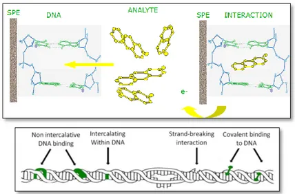
The guanine oxidation peak obtained through square wave voltammetry was used as the transduction signal to detect DNA toxic agents. The result of the interaction between double stranded calf thymus DNA and a toxic substance is the decrease of guanine oxidation peak (Figure1.9).
a 0 1 10-5 2 10-5 3 10-5 4 10-5 5 10-5 6 10-5 0.2 0.4 0.6 0.8 1 1.2 1.4 DNA DNA + Analyte i ( u A ) E (V) b -1 10-6 0 1 10-6 2 10-6 3 10-6 4 10-6 5 10-6 6 10-6 7 10-6 0.2 0.4 0.6 0.8 1 1.2 1.4 DNA DNA + Analyte i ( u A ) E (V)
Figure 1.9. Redox behavior of guanine (+1.0V) and adenine (+1.25V) bases after a square wave voltammetric scan carried out with graphite screen printed working electrode. a) the oxidation peaks before and after the interaction with a toxic agent. b) the same peaks after baseline correction.
This decrease is due to the interaction of genotoxic substances with the DNA helix that causes structural and conformational modifications (Figure 1.10).
Chapter I
22
Figure 1.10 : possible kind of interactions between chemical compounds and DNA
As reported in chapter five, DNA modifications are estimated as the percentage of guanine oxidation peaks decrease (G%). Also the adenine oxidation peak could be used but the guanine peak is preferred since it gives more reproducible results.
1.4.3 Genosensors
In the last two decades, the field of molecular diagnostics has grown rapidly due to the discovery of new genes involved in different diseases. The development of novel therapeutics based on the regulation of gene expression provides revolutionary opportunities in the area of pharmaceutical new science. To improve patient care, the analysis of gene sequences and the study of gene regulation play fundamental roles in the rapid development of molecular diagnostics and in drug discovery. Some methods more commonly used in the diagnostic laboratory are based on the analysis of specific gene sequences. In particular, the analysis of specific gene sequences exploits DNA hybridization reaction
General Introduction
23 for the its simplicity. In DNA hybridization, the target gene sequence is identified by a DNA probe that forms a double-stranded hybrid with its complementary nucleic acid (Figure 1.11). The reaction is highly efficient and extremely specificity also in the presence of a mixture of many different, non-complementary, nucleic acids. DNA probes are single-stranded oligonucleotides, labelled with either radioactive or non-radioactive material, to provide detectable signals for DNA hybridization. In order to make DNA testing more convenient, more economically feasible, and ultimately more widely used, DNA biosensors (genosensor) have been developed. A genosensor is a biosensor that employs an immobilized oligonucleotide as the biorecognition element. Typically, the design of an electrochemical genosensor involves immobilization of the DNA probe, the hybridization with the target sequence, the labelling and the electrochemical investigation. However, deviations from this general scheme have to be considered when using modified magnetic beads for electrochemical genosensing.
Chapter I
24
We have used this kind of DNA biosensor for the analytical detection of DNA as well as RNA sequences (chapter six). In particular, we have studied RNA sequences related to microRNA (miRNAs). MicroRNAs regulate target gene expression through translation repression or mRNA degradation. These non-coding RNAs are emerging as important modulators in cellular pathway, and they appear to play a key role in tumors genesis.
To this aim is important to identify miRNAs and their targets that are essential to promote cancer development and metastasis. Hence, these microRNAs may provide new therapeutic opportunities.18
General Introduction
25
REFERENCES
1. Thévenot, D. R.; Toth, K.; Durst, R. A.; Wilson, G. S., Electrochemical biosensors: recommended definitions and classification.
Biosensors and Bioelectronics 2001, 16, (1-2), 121-131.
2. Mairal, T.; Cengiz Özalp, V.; Lozano Sánchez, P.; Mir, M.; Katakis, I.; O’Sullivan, C., Aptamers: molecular tools for analytical applications.
Analytical and Bioanalytical Chemistry 2008, 390, (4), 989-1007.
3. Clark, L. C.; Lyons, C., Electrode systems for continuous monitoring cardiovascular surgery
Ann. N. Y. Acad. Sci. 1962, 102, (1), 29-45.
4. Guilbault, G. G.; Montalvo, J. A., Urea Specific Enzyme Electrode.
J. Am. Chem . Soc 1969, 91, 2164–2169.
5. Joshi, R., Biosensors Gyan Books ed.; Delhi, 2006.
6. Arnau Vives, A., Piezoelectric transducers and applications Springer ed.; 2004.
7. Völkl, K. P.; Opitz, N.; Lübbers, D. W., Continuous measurement of concentrations of alcohol using a fluorescence-photometric enzymatic method. Fresenius' Journal of Analytical Chemistry 1980, 301, (2), 162-163.
8. Shichiri, M.; Yamasaki, Y.; Kawamori, R.; Hakui, N.; Abe, H., Wearable artificial endocrine pancreas with needle-type glucose sensor.
The Lancet 1982, 320, (8308), 1129-1131.
9. Collings, A. F.; Caruso, F., Biosensors: recent advances. Rep. Prog.
Phys 1997, 60, (11), 1397–1445.
10. Bergveld, P.; Turner, A. P. F., Fabrication and mass production. In:
Advances in Biosensors Suppl. 1: Chemical Sensors for In Vivo Monitoring. London, 1993.
11. Palchetti, I.; Mascini, M., Biosensor Technology: A Brief History. In
Sensors and Microsystems, Springer, Ed. 2010; pp 15-23.
12. Cantor, C. R.; Schimmel, P. R., Biophysical Chemistry: Part I: The
Conformation of Biological Macromolecules San Francisco, 1980.
13. Nekludova, L.; Pabo, C. O., Distinctive DNA conformation with enlarged major groove is found in Zn-finger-DNA and other protein-DNA complexes. Proc Natl Acad Sci U S A 1994, 91 (15), 6948-6952.
Chapter I
26
14. Dias, R.; Lindman, B., DNA interaction with Polymers and
Surfactants. 2008.
15. Bonham, M. A.; Brown, S.; Boyd, A. L.; Brown, P. H.; Bruckenstein, D. A.; Hanvey, J. C.; Thomson, S. A.; Pipe, A.; Hassman, F.; Bisi, J. E.; Froehler, B. C.; Matteucci, M. D.; Wagner, R. W.; Noble, S. A.; Babiss, L. E., An assessment of the antisense properties of RNase H-competent and steric-blocking oligomers Nucl. Acids Res. 1995, 23, (7).
16. Dominick, P. K.; Keppler, B. R.; Legassie, J. D.; Moon, I. K.; Jarstfer, M. B., Nucleic acid-binding ligands identify new mechanisms to inhibit telomerase. Bioorganic & Medicinal Chemistry Letters 2004, 14, (13), 3467-3471.
17. Vester, B.; Wengel, J., LNA (Locked Nucleic Acid): High-Affinity Targeting of Complementary RNA and DNA. Biochemistry 2004, 43, (42), 13233-13241.
18. Cho, W. C. S., MicroRNAs in cancer- from research to therapy.
Biochimica et Biophysica Acta (BBA) - Reviews on Cancer 1805, (2),
Chapter II
Analytes
Chapter II
Analytes
29
2.1 SURFACTANTS
Surfactants are amphiphilic molecules which consist of two parts, a water soluble hydrophilic head and a water insoluble hydrophobic tail. The word surfactant derives by the contraction of surface-active-agent and indicates a substance which exhibits surface or interfacial activity.
Hydrophobic groups tend to minimize the contacts with water and the chains in water self-assembly to reduce the free energy of the system. In addition, these substances tend to be adsorbed at interfaces. When surfactant molecules are at the liquid-gas or liquid-liquid interface, the hydrophobic tails extend out of the bulk water phase, while the water soluble heads remain in the water phase. This alignment of surfactant molecules alter the surface properties of water at the water/air or water/oil interface. The driving force of the phenomena is the lowering of the interfacial free energy. When the boundary between water and air is covered by surfactant molecules the surface tension is reduced.
In water surfactants tend to form aggregates referred as micelles. Micelle formation, or micellization, can be viewed as an alternative mechanism to adsorption at interfaces. Micelles are formed at very low surfactant concentrations in water. The concentration at which micellas start to form is definite as the critical micelle concentration (CMC). This important feature depends mostly on the chemical structure of the surfactant but also on co-solutes, for instance, salts in the case of ionic surfactants or temperature, particularly for nonionic surfactants.
CMC can be determined by measuring the variation of different physicochemical parameters (surface tension, equivalent conductivity, self-diffusion, osmotic pressure, turbidity and solubilization) of an aqueous solution, as a function of the surfactant concentration.1
Surfactant self-assembly leads to different structures, some of which are shown in Figure 2.1.
Chapter II
30
Fig. 2.1: Some example of molecular structure resulting from surfactant self-assembly. Critical packing parameters (CPPs) of surfactant molecules and preferred aggregate structures for geometrical packing.
The aggregate structure forms as a result of the balance between the polar and non polar parts of surfactants molecule, generally described as the hydrophilic lipophilic balance (HLB). However, different approaches are based on the concepts of surfactant packing and the spontaneous curvature of the surfactant film. The critical packing parameter (CCP) is estimated by the following equation:
Analytes
31 Where is the headgroup area, is take as 80% of the extended length, and is the volume of the hydrophobic part of a surfactant molecule. As Figure 2.1 shows, simple geometrical consideration can give an indication of the structure formed by a given amphiphile, depending on the relative value of CCP, from normal structure to reversed structures.
2.1.1 Classification of surfactants
Surfactants are classified according to the nature of their hydrophilic head in four classes: cationic, anionic, non ionic and zwitterionic surfactants.
Cationic surfactants are dissociated in water into an amphiphilic cation
and an anion. Usually the polar head of cationic surfactants is an ammonium groups bound to different alkyl chains, according to the general formula: R N+ c a b + X
-where R is a long hydrocarbon chain (C10-C18), X is a halide, sulfate, or
methosulphate ion; and a, b and c may be H, small alkyl groups.
Also double-chained ammonium surfactants, where two R groups are present, are commonly used. The primary use of cationic surfactants is related to their tendency to adsorb at negatively charged surfaces. Hence they can be used as anticorrosive agents for steel, dispersants for inorganic pigments, for fertilizers and bactericides.
Anionic surfactants are historically the earliest and the most common
surfactants. They are dissociated in water as an amphiphilic anion, and a cation. The most commonly used hydrophilic groups are carboxylates,
Chapter II
32
sulphates, sulphonates and phosphates. General formulas of anionic surfactants are as follows:
Carboxylates: CnH2n+1COO-X
Sulphates: CnH2n+1OSO3- X
Sulphonates: CnH2n+1SO3- X
Phosphates: CnH2n+1OPO(OH)O- X
where n≥8 atoms and the counter ion X is usually Na+, K+ or NH4+.
Non ionic surfactants contain polar groups unable to dissociate, but
possessing a significant affinity to water and other polar substances. Usually these groups incorporate atoms of oxygen, nitrogen, phophorpous or sulphur (alcohols, amines, ethers, etc.). Among the non ionic surfactants, the most common are oxyethylated alkyl phenols, fatty alcohols, fatty acids, amines and block-copolymer surfactants (oxyethylene non ionic surfactants), where the polar parts of the molecules consist of repeated oxyethylene group – CH2-O-CH2- and
closing –OH, -COOH or –NH2 group.
They are compatible with charged molecules and easily used in mixtures with other ionic surfactants, which often result in beneficial associations.
Zwitterionic or amphoteric surfactants present both acid and basic
functional groups. This is the case of synthetic products like betaines or sulfobetaines, and also natural substances such as aminoacids and phospholipids. They are usually used in association with other surfactants (anionic or nonionic) for particular applications. Since the optimal surface activity of amphoteric surfactants takes place around neutral pH, they are particularly appreciated in personal care products (shower gels, foam baths, shampoos, etc.) for their mildness and skin compatibility.
Analytes
33
2.1.2 Surfactants Toxicity
Surfactants harmful effects on the environment are well known. The acute toxicity of surfactants to organisms is highly variable, depending on the chemical structure of the surfactant and the organism. In general, aquatic organisms are more susceptible to surfactants than terrestrial organisms. Surfactants can be adsorbed at the biological membranes and disrupt biological functions. Moreover, surfactants can be remove inhibit the enzyme activities.2
Several types of toxicity tests indicate that chronic toxicity of anionic and non ionic surfactants occur at concentrations < 1ppm.3 Generally, non ionic and anionic surfactants tented to be more toxic at lower concentration than cationic surfactants.4
Toxicity caused by surfactants is influenced by several factors including the molecular structure of the surfactants, water hardness, temperature and dissolved oxygen. The most important factors are the molecular structure of the surfactants. Their toxicity is probably due to damage that surfactants cause to cellular protein and cell membrane.4
Acquatic toxicity of surfactants is usually measured on fish, daphnia and algae. The toxic index is expressed as LC50 (for fish) or EC50 (for daphnia
and algae), where LC and EC stand for lethal and effective concentration, respectively. Values below 1 mgL-1 after 96 h testing on fish and algae, and 48 h on Daphia are considered toxic. Environmentally benign surfactants should, preferably, be above 10 mgL-1.5
Verge and Moreno6 studied the effects of anionic surfactants on Daphnia
Magna. In this study, the acute toxicity of various linear alkyl benzene
sulphonates (LAS), alkyl sulphates and alkyl ethoxy sulphates was determined. The study was carried out to obtain a valid set of data of the above surfactants for environmental classification and labeling according to European legislation. The results indicate that commercial LAS should
Chapter II
34
be classified as dangerous for the environment with respect to their effects on Daphnia Magna.
R.J Rosen at al.7, 8 studied the relationship between the interfacial properties of surfactants and their toxicity to aquatic organism. They found that the toxicity of surfactants depends on their tendency to be adsorbed by the organisms and on their ability to penetrate the cell membranes of the organisms. The interfacial activity is expressed by the physic-chemical parameter :
Equation 2.2
where ΔG0ad is the standard free energy of adsorption of the surfactant at
the air-solution interface and Amin is the minimum cross-sectional area of
e surfactant at the liquid/air interfaces.
The analogous parameter at the liquid/solid interface:
Equation 2.3
where: is a standard free energy of adsorption at the liquid solid
interface between the surfactant solution and an immobilized artificial membrane designed to mimic a cell membrane; is the minimum
cross sectional area of the surfactant at the interface.
The solid is an immobilized artificial membrane that mimics a biological cell membrane. The results show that the toxicity increases with: an increase in the length of the hydrophobic chain and, in linear polyoxyethylene (POE) alcohols, with the decrease of the number of oxyethylene units in the molecules. Besides, for isomeric materials the toxicity decreases with branching or movement of the phenyl group to a more central position in the linear alkyl chain. This is due to the expected changes in the value of both ΔG0ad and Amin.9
Analytes
35 The general nature of the relationships between interfacial activity of the surfactants and their biological effects in aqueous systems indicate that adsorption to a biological membrane is a critical parameter for predicting and understanding environmental effects.
Differences in toxicity potential between classes of surfactants exist. However, such classification does not allow an exact determination of such capacity of each surfactant. Even within the same class, each surfactants exhibits its own specific effects distinguishable from the others.10
2.2 MicroRNA
MicroRNAs (miRNAs) are a family of endogenous ≈22 nucleotides non-coding RNAs that regulate gene expression with a strong sequence specificity.1112
To date, over 8600 miRNAs have been identified and deposited in the online miRBase sequence database, including currently more than 690 miRNAs sequences for the human genome.11
Every cellular process is likely to be regulated by microRNAs, and an aberrant microRNAs expression signature is a hallmark of several diseases, including cancer. MicroRNAs expression profiling has indeed provided evidence of the association of these tiny molecules with tumor development and progression. An increasing number of studies have then demonstrated that microRNAs can function as potential oncogenes or oncosuppressor genes, depending on the cellular context and on the target genes they regulate.13
2.3.1 Implications of microRNAs in cancer
Defects in normal cell processes such as differentiation, proliferation, and apoptosis are all well-known to be involved in cancer pathogenesis. The
Chapter II
36
connection between miRNA and cancer was initially made since miRNAs were found to be involved in many cancer diseases. This connection promoted studies which further reinforced the correlation between miRNAs and cancer development. Researchers discovered that there is an aberrant miRNA expression when comparing various types of cancer with normal tissues. Although the association between miRNA and cancer has initially been suggested, the question still remain “ whether the altered miRNA expression was a cause or a consequence of cancer”.14 In addition very few informations about the specific targets and functions of miRNAs are still available.
Some miRNAs are thought to have oncogenic activity while others have tumor suppressor activity (Table 2.1). It is important to note that these distinctions may not be so strict and that some miRNAs may express different activities depending on the situation and tissue type. Nevertheless, the majority of recent research provides results that point toward one category or the other. It is also possible to group miRNAs based on their various functions. Some play a single role while others contribute to cancer through multiple cellular functions.
Analytes
37
Table2.1: Various Oncogenic and Tumor Suppressor miRNAs 15
miRNA Tumor suppressor
Activity Oncogenic Activity Espression in cancer Let-7 family X - miR-9 X + miR-10a X + miR-15a/16-1 X - miR-17-5p X - miR-17-92 cluster X + miR-21 X + miR-29b X - miR-34 a X - miR-106 a X + miR-124 a X - miR-127 X - miR-141 X + miR-142 X + miR-143 X - miR-145 X - miR-146 b X + miR-155/bic X + miR-181 b X - miR-197 X + miR-200b X + miR-221 X + miR-222 X + miR-346 X +
Chapter II
38
REFERENCES
1. Dias, R.; Lindman, B., DNA interaction with Polymers and
Surfactants. 2008.
2. Hrenovic, J.; Ivankovic, T., Toxicity of anionic and cationic surfactant to Acinetobacter junii in pure culture. Central European Journal
of Biology 2007, 2, (3), 405-414.
3. Board, N., The Complete Technology Book on Detergents National Institute of Industrial Research: 2003.
4. Gerardi, M. H., Toxicity. John Wiley & Sons, Inc.: 2006; p 173-209. 5. Tadros, T. F., Applied Surfactants -Principles and Applications wiley: 2005.
6. Verge, C.; Moreno, A., Effects of anionic surfactant on Daphnia magna. Tenside Surfact. Det 2000, 37.
7. Rosen, M.; Fei, L.; Zhu, Y.-P.; Morrall, S., The relationship of the environmental effect of surfactants to their interfacial properties. Journal
of Surfactants and Detergents 1999, 2, (3), 343-347.
8. Rosen, M. J.; Li, F.; Morrall, S. W.; Versteeg, D. J., The Relationship between the Interfacial Properties of Surfactants and Their Toxicity to Aquatic Organisms. Environmental Science & Technology 2001, 35, (5), 954-959.
9. Milton, D.; Rosen, J., Chapter I: Characteristic features of surfactants. In Surfactant and interfacial phenomena, Wiley-Interscience, Ed.
10. Effendy, I.; Maibach, H. I., Surfactants and experimental irritant contact dermatitis. Contact Dermatitis 1995, 33, (4), 217-225.
11. Beier, V.; Hoheisel, J., MicroRNAs: small Molecules with big impact in cancer. BIOforum europe 2008.
12. Pang, Y.; Young, C. Y. F.; Yuan, H., MicroRNAs and prostate cancer.
Acta Biochim Biophys Sin 2010, 42, 363–369.
13. Lorio, M. V.; Croce, C. M., MicroRNAs in Cancer: Small Molecules With a Huge Impact. Journal of Clinical Oncology 2009, 27, (34), 5848-5856
14. Sassen, S.; Miska, E. A.; Caldas, C., MicroRNA—implications for cancer. Virchows Arch 2008, 452, 1-10.
Analytes
39 15. VandenBoom II, T. G.; Li, Y.; Philip, P. A.; Sarkar, F. H., MicroRNA and Cancer: Tiny Molecules with Major Implications. Current Genomics
Chapter II
Chapter III
Instrumental techniques
Chapter III
Instrumental techniques
43
3.1 INTRODUCTION
The following paragraphs describe the general principles of the analytical techniques used for this study. Square Wave Voltammetry (SWV) was used to study surfactants toxicity (chapter 4) and Differential Pulse Voltammetry (DPV) was used for the detection of specific DNA and microRNAs sequences (chapter 6). FTIR spectroscopy and zeta potential measurements were used to study the interactions between DNA and surfactants (chapters 4 and 5 respectively).
3.2 VOLTAMMETRY
Voltammetry is an analytical technique based on the measure of the current flowing through an electrode dipped in a solution containing electro-active compounds, while a potential scanning is imposed upon it. The resulting current-potential and current-time curves are analyzed to obtain information about solution composition. Voltammetric techniques can be used for the determination at trace level at trace level of many organic compounds and inorganic substances (mainly metal ions).The effects of the applied potential and the behavior of the redox current are reported by the laws described below.
A reversible electrochemical reaction involving the reduction of an analyte Ox to give a product Red can be writer as:
Equation 3.1
the application of a potential E modifies the respective concentration of Ox and Red species at the surface of the electrode in agreement with the Nernst equation: Equation 3.2
Chapter III
44
where is the potential difference between the working electrode
and the reference electrode, is the standard electrode potential for the reaction, R is the ideal gas constant (8.3144 Jmol-1K-1), T is the absolute temperature (K), n is the number of exchanged electrons, F is the Faraday constant (96,485C/mol), and are the molar concentrations of Red and Ox species in a thin layer of solution close to the electrode surface.
For some voltammetric techniques it is useful to use the Butler-Volmer equation that gives the current as a function of concentration:
where: i: intensity current, A K0: rate constant
Eapp: electrode potential, V
A: electrode active surface area, m2
T: absolute temperature, K
n: number of electrons involved in redox reaction
F: Faraday constant, C/mol
R: ideal gas constant, J/mol∙K
α: transfer coefficient, dimensionless
In most cases the current also depends directly on the flow of material to the electrode surface. When new species are produced at the surface, the increased concentration provides the driving force for its diffusion towards the bulk of the solution to the electrode surface. On the contrary, when species are destroyed, the decreased concentration promotes the diffusion of new material from the bulk solution. The
Instrumental techniques
45 resulting concentration gradient and mass transport is described by Fick’s law:
Equation 3.4
where (mol/m3) is the flux of matter, DO (m2/s )is the diffusion
coefficient of oxidized specie, A is the electrode active surface area (m2) and (m) is the distance from the electrode surface. An analogous equation can be written for the reduced species.
3.2.1 Excitation signals
In voltammetry the voltage of the working electrode is varied systematically while the current response is measured. Several different voltage-time functions, called excitation signals, can be applied to the working electrode. The waveforms of four of the most common excitation signals used in voltammetry are shown in Scheme 3.1. The classical voltammetric excitation signal is a linear scan (Scheme 3.1a) in which the potential of the working electrode is changed linearly with time. The current flowing in the cell is then measured as a function of the applied voltage. Two pulse-type excitation signals are shown in Scheme 3.1 b and c: differential pulse voltammetry and square wave voltammetry. Currents are measured at various times during the lifetimes of these pulses, as discussed in paragraph 3.2.3 and 3.2.4. In the (Scheme 3.1 d) the potential is varied linearly between two values at a fixed rate (cyclic voltammetry). When the voltage reaches V2 the scan is reversed and the
voltage is swept back to V1. This process may be repeated several times
Chapter III
46
Type of pulse Waveform Type of
voltammetry
a Linear Scan Linear
voltammetry b Differential Pulse Differential pulse voltammetry
c Square wave Square wave
voltammetry
d Triangular Cyclic
voltammetry