1
SAPIENZA
Università di Roma
Facoltà di Scienze Matematiche Fisiche e Naturali
PhD Course in Cell and Developmental Biology
XXXII Cycle
(A.A. 2018/2019)
The host-pathogen interactions in the Caenorhabditis elegans animal model: the role of
glycosylation and a tool to screen for probiotic bacteria
PhD student
Emily Schifano
Supervisor
Prof. Claudio Palleschi
Coordinator
INDEX
Aim of the study pag. 3
English summary 4 Italian summary 4 Introduction 5 Results 8 Discussion 28 Methods 31 References 35
3
Glycans, one of the main macromolecular components of cells, exhibit a certain diversity in line with the biological processes in which they are involved. Recent studies have shown their involvement in the innate and acquired immunity of vertebrates (Marth and Grewal, 2008). They are an excellent target to study molecular mechanisms involved in the host-pathogen interaction. The adhesion to the surface of the host cell is a common mechanism used by pathogens to infect organisms. Therefore, subtle differences in the glycan’s structure could affect resistance and susceptibility.Pmr-1 gene encodes a P-type ATPase localized in the Golgi apparatus, regulating Ca2+ and Mn2+ homeostasis. Studies on this protein in S. cerevisiae and K. lactis yeasts discovered its involvement in alterations of the glycosylation mechanism. PMR-1 function is evolutionary conserved from yeast to human, where mutations in the orthologous gene ATP2C1 cause Hailey-Hailey disease. A correct concentration of the Ca2+ ion within the Golgi apparatus and the Endoplasmic Reticulum seems to be necessary for a correct activity of the enzymes involved in N- and O-glycosylation, for a regular secretion of glycoproteins and a normal protein degradation after incorrect folding.
In recent years, the C. elegans glycome has been described (Paschinger et al., 2008). However, it has been poorly characterized in the context of glycosylation’s role in the infective process during infection by several bacterial and fungal pathogens. C. elegans is widely used to study microbes and host interactions of various human pathogens, such as Pseudomonas aeruginosa, Salmonella enterica,
Staphylococcus aureus, and Enterococcus faecalis (Sem and Rhen, 2012; Kirienko et al., 2013).
Growing evidences indicate that lipids and proteins glycosylation seem to be required for infection in this nematode. For instance, the Bre genes encode proteins required for glycolipid synthesis and they are resistant to Cry toxins produced by Bacillus thuringiensis. Similarly, bah, srf, and bus mutants are resistant to a number of bacterial infections. Bah mutants have an altered cuticle surface and are resistant to Yersinia pestis and Y. peudotuberculosis biofilm adhesion (Darby et al., 2002). In addition,
C. elegans has become increasingly used for screening antimicrobials, including preselecting probiotic
bacteria against different pathogens (Zanni et al., 2017; Kwon et al., 2016; Zanni et al.,2015; Uccelletti et al., 2010). However, the molecular mechanisms at the basis of the pathogen resistance are not known.
This research will explore interactions between pathogens and the host cell surface; special focus will be placed on the host innate immune response. It should be noted that C. elegans does not have a humoral immune response, so innate immunity study is simplified. Questions to be addressed include:
if silencing pmr-1 could impact on pathogen resistance and oxidative stress responses in C.
elegans;
what are possible changes in glycoconjugates expression on cell surfaces in pmr-1 mutant worms;
if pmr-1 mutant strain show alteration in expression of genes involved in innate immune system and/or in glycosylation pathways;
what are the effects of probiotics administration in C. elegans mutants during infections. This project can provide an opportunity to investigate glycosylation role in pathogen recognition as well as innate immune response. The main signaling pathways involved in infection responses can be explored using studies of interaction between C. elegans and different pathogens. Finally C. elegans can be used as in vivo screening model to identify potential probiotic bacteria and to study microbe-host interactions when the glycosylation process is affected.
English summary
In this project, the role of the pmr-1 gene in the glycosylation process and in the host-microrganism interaction was analyzed in C. elegans model system. Glycosylation produces a large number of glycans, polysaccharides whose different morphology reflects the multiplicity of biological processes in which they are involved. The emerging role of glycans in the innate and acquired immunity of vertebrates makes them a target for the study of the molecular mechanisms underlying the host-pathogen interaction, in order to develop new therapeutic approaches. The Ca2+-ATPase, encoded by the pmr-1 gene, appears to be involved in alterations of glycosylation mechanisms, as demonstrated by studies on the homologous PMR-1 protein of Saccharomyces cerevisiae and Kluveromyces lactis. In this work, pmr-1 mutant nematodes, obtained through RNA interference, were exposed to different pathogenic microorganisms, showing an increased survival capacity compared to wild-type individuals, following infection by the Gram-positive Staphylococcus aureus and Enterococcus
faecalis pathogens. This increased resistance seems to be related to defects in the oligosaccharide
structure of glycoproteins of the cell surface caused by their altered glycosylation. The lack of regular recognition of C. elegans surface glycoproteins by the two pathogens could, in fact, determine the difficult adhesion of bacteria to the nematode and the reduction of their pathogenicity. Furthermore,
pmr-1 worms showed a different innate immune and oxidative stress responses, as well as an alteration
of the glycocalyx in their gut. Since glycosylation is also involved in beneficial role of host microbiota against pathogens, nematodes were used to preselect probiotic bacteria, whose efficacy will be evaluate after administration on pmr-1 mutants, to measure the response of worms to pathogen infection. Therefore, this study represents a first step in understanding the role of PMR-1 protein in the host-microrganism interaction in C. elegans.
Italian summary
In questo progetto è stato analizzato il ruolo del gene pmr-1 nel processo di glicosilazione e nell'interazione ospite-microrganismo nel sistema modello C. elegans. La glicosilazione produce un gran numero glicani, la cui diversa morfologia riflette la molteplicità dei processi biologici in cui sono coinvolti. Il ruolo emergente dei glicani nell’immunità innata e acquisita dei vertebrati li rende un bersaglio di studio per l’approfondimento dei meccanismi molecolari alla base dell’interazione ospite-patogeno, al fine di sviluppare nuovi approcci terapeutici. Studi condotti sulla proteina omologa PMR-1 dei lieviti Saccharomyces cerevisiae e Kluveromyces lactis hanno dimostrato che la Ca2+-ATPasi, codificata dal gene pmr-1, sembra essere coinvolta in alterazioni del meccanismo di glicosilazione. In questo lavoro i nematodi mutanti per il gene pmr-1, ottenuti mediante RNA interference, sono stati esposti a diversi microrganismi patogeni, mostrando un’aumentata capacità di sopravvivenza rispetto agli individui wild-type, in seguito a infezione dei patogeni Gram-positivi Staphylococcus aureus e
Enterococcus faecalis. Tale incremento di resistenza sembrerebbe correlato a difetti nella struttura
oligosaccaridica di glicoproteine della superficie cellulare che potrebbero essere causati dalla loro alterata glicosilazione. La mancanza di un regolare riconoscimento delle glicoproteine di superficie di
C. elegans da parte dei due patogeni potrebbe, infatti, determinare la difficile adesione dei batteri al
nematode e la riduzione della loro patogenicità. I nematodi mutanti, inoltre, hanno mostrato una diversa risposta immunitaria innata e allo stress ossidativo, nonché un'alterazione del glicocalice a livello intestinale. Poichè il processo di glicosilazione è anche coinvolto nel ruolo di difesa che il microbiota intestinale esercita nei confronti di patogeni, il nematode è stato usato nello screening di batteri probiotici, la cui efficacia è stata testata in mutanti pmr-1, per analizzare come varia la risposta degli animali al patogeno. Questo studio, dunque, rappresenta un primo passo per la comprensione del ruolo di pmr-1 nell’interazione ospite-microrganismo in C. elegans.
5
IntroductionGlycosylation is a key modification of proteins and lipids and is involved in various important intermolecular interactions, such as cell signalling, differentiation and adhesion, leading to the formation of glycan linkages that are essential for cell viability and biochemical communication (Helenius and Aebi, 2001).
The gastrointestinal mucus gel is continuous and can be divided into an attached inner layer and an outer layer, which is less dense and unattached. In addition, the membrane-bound glycosylated proteins and lipids create a glycocalyx, which is dynamic but remains adherent to each cell. The secreted glycans form a mucus gel layer that serves as a physicochemical sensor and is composed of highly glycosylated mucins and associated peptides. Mucin are large and continuously renewed proteins, which are important to protect the gastrointestinal lumen (Hansson, 2012).
During pathogen colonisation and infection, microbes commonly interact with glycan structures of the host glycocalyx (Hooper and Gordon, 2001). The glycosylation state of both host and pathogen change in response to the presence of the other, modulating pathogenesis and immune response. Adhesion to host cells is considered to be mediated by hydrophobic interactions, among which the binding of bacterial lectins to the corresponding glycosylated receptors associated with host cells.
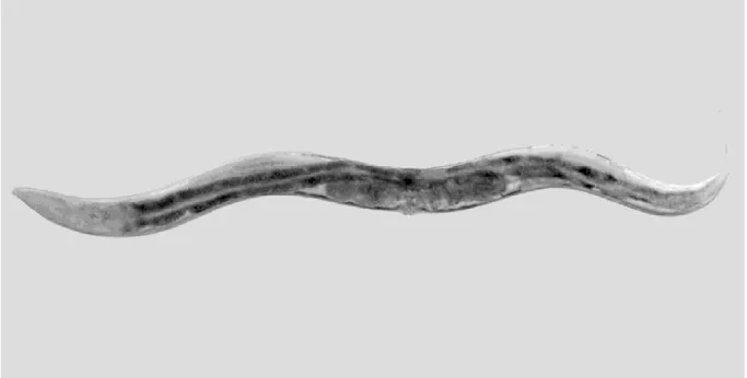
The in vivo model host Caenorhabditis elegans provides a valuable tool to study host-microbe interactions mediated by glycans (Fig. 1). In particular, its many advantages include: the presence of only 20 non-renewable intestinal cells and a well-characterized immune system; the ability to access genome sequences, the possibility to visualize infection in situ and to monitor the distribution of glycoconjugates in tissues using lectins and antibodies thanks to the transparency of the body. Moreover, many mutant strains are available and it is susceptible to infection from more than 40 pathogens (Jiang and Wang, 2018; Palaima et al., 2009).
C. elegans has no cell-mediated immunity mechanisms; however, it is characterized by systemic
immune responses depending on the synthesis and action of antimicrobial molecules, including lectins, lysozymes and other antibacterial factors (Alper et al., 2017; O'Rourke et al., 2006).
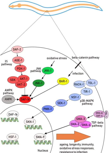
In their natural environment, nematodes coexist with a variety of microbial pathogens, including bacteria, fungi, protozoa, and viruses (Félix and Duveau, 2012; Engelmann and Pujol, 2010; Félix and Braendle, 2010). Infection can occur in C. elegans through different pathways: the cuticle and epidermis, rectum or the subsequent colonization of the intestine. These different tissues are made up of dedicated immune cells responsible for ensuring the host's defenses. These defense responses are regulated by different signaling pathways, among which the p38 Mitogen-activated-protein(MAP) kinase, the Insulin/insulin-like growth factor (IIS) pathway and the transforming growth factor β (TGF-β) signaling (Marsh and May, 2012). p38 MAP kinase, IIS and TGF-β pathway regulate the activation of SKN-1, DAF-16 and SMA-9 transcriptional factor, respectively (Fig. 2). They activate genes involved in longevity, oxidative stress responses and development. In particular, the p38 MAP kinase pathway, which includes the NSY-1, SEK-1 and PMK-1 kinases, is an evolutionarily conserved signal transduction system and is involved in the defense against pathogens both in intestinal and hypodermis cells (Kim et al, 2002). It regulates about 86 genes, responsible for the secretion of defense molecules such as lectins, lysozymes, and antimicrobial peptides (Troemel et al., 2006).
7
differences in structure, density and distribution of glycoconjugates could lead to resistance or susceptibility to pathogens. Therefore, it is important to study the glycosylation process for dissecting the molecular basis of the host-pathogen interaction. Indeed, it plays a decisive role in the exchanges of C. elegans with the external environment because it represents the first mechanism the nematode effects towards microorganisms (Parsons et al., 2014).The intestinal barrier function may be enhanced by mucus secretion promoted by probiotics (Zareie et al., 2006). Probiotics, defined as living organisms improving the host’s health when ingested in adequate amounts, facilitated cross talk with intestinal epithelium, influencing the O-glycosylation pattern of mucins (Silva et al., 2014; Mattar et al., 2002).
The Ca2+-ATPase PMR-1, located in the Golgi apparatus and encoded by the pmr-1 gene, appears to be involved in alterations of the glycosylation mechanism, as demonstrated by studies on the homologous protein of S. cerevisiae and K. lactis yeasts (Uccelletti et al., 2005; Antebi and Fink, 1992). Indeed, a normal concentration of the Ca2+ ion inside the Golgi apparatus and the Endoplasmic Reticulum seems to be necessary for the functional activity of the enzymes involved in N- and O-glycosylation, as well as for a regular secretion of glycoproteins and a normal degradation associated with the RE of misfolded proteins.
C. elegans can synthesize N-glycans, various O-glycans, glycolipids and chitin (Schachter, 2004).
However, the complex and hybrid N-glycans that are highly abundant in vertebrates are either absent in C. elegans or present at very low levels. Although many C. elegans N-glycans are fucosylated, covering roles in development, host–microbe interactions, signal transduction, the most abundant and the most conserved among eukaryotes class of glycans in C. elegans, are the high mannose type glycans (Moran et al., 2011).
In addition, C. elegans is a useful model host not only for a wide variety of human pathogens, but also for probiotic bacteria, which are important for promoting human health and longevity. Nematode has become increasingly used for preselecting probiotic bacteria against different pathogens (Zanni et al., 2017; Kwon et al., 2016; Zanni et al., 2015); its advantages allow to use nematodes for probiotic screening, thanks to easiness of monitor anti-aging markers, as well as body fat storage, which represent aging indexes (Park, 2014; Lee, 2015). Indeed, pharyngeal pumping rate, locomotion ability, body size and lipofuscin accumulation are three of the most examined markers (Son et al., 2019). In particular, lipofuscin consists of intestinal autofluorescent granules accumulating during aging (Pincus et al., 2016).
Many studies on worms have indeed reported the beneficial effects of probiotics, including antimicrobial effects, microbial interference, and protection from infections (Nakagawa et al., 2016; Wang et al., 2011). Among them, different probiotic strains isolated from fermented food were found to increase nematode lifespan and showed anti-aging effects (Guantario et al., 2018; Gerbaba et al., 2017). In this work, the role of the pmr-1 gene in the glycosylation process and in the response to pathogens was evaluated, using the nematode C. elegans as an in vivo model system. Moreover, studies on nematodes were performed to identify potential probiotic bacteria and to understand how they influenced host glycosylation and response to pathogens.
Results
pmr-1 suppression in C. elegans induced resistance to Staphylococcus aureus infection
In order to inactivate the Ca2+-ATPase PMR-1 in C. elegans, pmr-1 gene was silenced through RNA interference by feeding. The transcript levels of this gene were reduced in wild-type nematodes with respect to control worms fed E. coli with the empty vector. RT-qPCR analysis demonstrated that, after 48 hours of treatment, the gene silencing occurred: indeed, the levels of pmr-1 transcript of individuals subjected to RNA interference, normalized for the housekeeping gene act-1, were considerably lower than the transcript derived fromnematodes used as controls (Fig.3).
Figure 3. RT-qPCR analysis of pmr-1 gene. Expression of pmr-1 mRNA level in N2 worms after 48 h of RNAi, as compared to control interfered with empty vector. Bars represent the mean of three independent experiments. Asterisks indicate significant differences (***p <0.001).
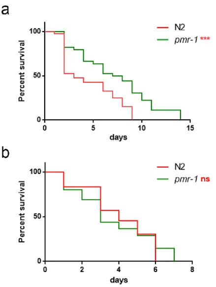
After RNAi silencing, the amount of pmr-1 transcript was reduced of about 70% in pmr-1 worms compared to the levels showed by control worms. After 48 hours from RNAi, nematodes were exposed to the Gram-positive Staphylococcus aureus pathogen and effects on worm physiology were evaluated. In lifespan analysis, pmr-1 worms showed a resistance to infection as compared to control. In particular, the median survival of worms was recorded at days 1 and 7 for N2 and pmr-1 worms, respectively (Fig. 4a). Moreover, intestinal colonization capability was explored as an indicator of infection by N2 or pmr-1 nematodes, after 48 h from C. elegans exposure to pathogen. In pmr-1 worms, a relevant reduction of about 60% in the intestinal CFUs was observed, as compared to control (Fig. 4b). This result probably correlated with the increase of longevity observed above.
9
Figure 4. Effect of pmr-1 silencing on C. elegans viability and gut colonization. a) Kaplan-Meier survival plot of pmr-1 mutant worms with respect to control. n = 60 for each data point of single experiments. (b) Bacterial colony forming units (CFU) recovered from nematodes after 48h to infection with S. aureus. Bars represent the mean of three independent experiments. Asterisks indicate significant differences (**p <0.01, ***p <0.001).pmr-1 gene was specifically involved in C. elegans responses to Gram-positive pathogens
Pmr-1 nematodes showed resistance to pathogen when infected with S. aureus. In subsequent
experiments, worms were exposed to another Gram-positive pathogen, Enterococcus faecalis: Fig. 5a shows the viability analysis of nematodes exposed to these bacteria: an increase in the survival capacity of pmr-1 worms, compared to control, was observed. In particular, the median survival of worms was recorded at days 3 and 8 for N2 and pmr-1 worms, respectively.
In order to analyze lifespan of nematodes also infected with Gram-negative bacteria, experiments with
Pseudomonas aeruginosa were performed. Lifespan analysis did not show, in this case, significant
Figure 5. Effect of pmr-1 silencing on worms infected with different pathogens. Kaplan-Meier survival plot of N2 and pmr-1 mutant worms infected with a) the Gram-positive E. faecalis, and b) the Gram-negative P. aeruginosa. n = 60 for each data point of single experiments. Asterisks indicate significant differences (***p <0.001, ns: not significant).
Moreover, nematodes were exploited to compare the in vivo virulence of two uropathogenic
Escherichia coli (UPEC) isolates, ECP45 and ECP110. Different studies have tried to associate
virulence and antibiotic resistance traits to environmental E. coli clones (Amos et al., 2014; Anastasi et al., 2010), highlighting the need of simple models to perform in vivo studies. C. elegans could serve as an in vivo model to distinguish, among uropathogenic E. coli, different virulence behavior. When worms were infected, after 24 hours almost 50% of ECP110-fed worms were dead (Fig. 6a). ECP110 strain was significantly more virulent than both ECP45 and the MG1655 strain, used as control together with OP50. Notably, the lifespan shortening was dependent on the viability of the urinary strains used as food source. Indeed, dietary administration of heat killed UPEC did not produce any effect on nematode lifetime (Fig. 6b), indicating that viability of the strain was required for pathogenicity.
11
Figure 6. Effect of UPEC E. coli strains on C. elegans viability. a) Kaplan–Meier survival plots of worms infected with the indicated E. coli strains are shown. Infections were performed at 25°C and worm mortality was monitored every day. E. coli CFT073-fed worms were taken as control. b) Survival of C. elegans fed heat killed E. coli strains. n=60. Statistical analysis was evaluated by Log-rank (Mantel-Cox) test; asterisks indicate significant differences (**p< 0.01; ***p < 0.001; ns: not significant).After 2 days of infection, gut colonization was analyzed through colony forming units (CFU) method. ECP110 strain showed a colonization capability almost 2- and about 3-fold higher than CFT073 and OP50 controls, respectively (Fig. 7a). To visualize the bacterial colonization in the nematode intestinal tract, the GFP-expressing E. coli strains were then used to feed C. elegans. After a 48h-exposure to infection, a high fluorescence was observed in animals fed with ECP110–GFP strain, where bacterial cells had spread along the entire nematode gut (Fig. 7b), highlighting a well evident difference when compared to ECP45-GFP or CFT073-GFP infections.
Figure 7. Colonization of UPEC strains in C. elegans gut. a) Colonization of UPEC strains within the nematode gut. b) Fluorescence photomicrographs of 10 representative nematodes infected with the GFP-expressing CFT073, ECP45 and ECP110 strains for 2 days are reported (scale bar, 100 µm). Based on these findings, C. elegans model was used to test E. coli ECP110 strain in animals with silenced pmr-1, to analyze possible involvement of glycosylation pathway in worm infection responses.
Lifespan experiments highlighted that pmr-1 worms infected with the Gram-negative E. coli ECP110 did not show resistance to S. aureus, when compared to control (Fig. 8).
13
d a y s P e r c e n t s u r v iv a l 0 5 1 0 1 5 0 5 0 1 0 0 N 2 p m r - 1 n sFigure 8. Effect of pmr-1 silencing on worms infected with E. coli ECP110. Kaplan-Meier survival plot of N2 worms, after 48h of RNAi, infected with E. coli ECP110. n = 60 for each data point of single experiments. Statistical analysis was evaluated by Log-rank (Mantel-Cox) test (ns: not significant).
Role of pmr-1 was also evaluated in C. elegans response to Candida albicans: worms infected with the yeast did not show an increase of lifespan when pmr-1 gene was silenced, when compared to control (Fig. 9). Hence, pmr-1 seemed to be involved in C. elegans specific-response to Gram-positive pathogens. d a y s P e r c e n t s u r v iv a l 0 5 1 0 1 5 0 5 0 1 0 0 N 2 p m r - 1 n s
Figure 9. Effect of pmr-1 silencing on worms infected with C. albicans. Kaplan-Meier survival plot of N2 worms, after 48h of RNAi, infected with C. albicans. n = 60 for each data point of single experiments. Statistical analysis was evaluated by Log-rank (Mantel-Cox) test (ns: not significant).
Pmk-1 gene was involved in immune responses mediated by pmr-1
To investigate the mechanism of action of PMR-1 protein in the stress response and in the defense against pathogens, transcriptional levels of pmk-1, sek-1 and hsf-1 genes, which are involved in the main pathogen-defense pathways in C. elegans (Singh et al., 2006; Kim et al., 2002), were evaluated. After 48h from RNAi, an increase expression of hsf-1 and pmk-1 transcripts was observed in pmr-1 nematodes, as compared to control (Fig. 13a). In particular, pmk-1 gene showed a more than 2-fold higher expression in pmr-1 nematodes compared to control, while transcript levels of hsf-1 gene increased of about 50% in pmr-1 worms. The subsequent lifespan analysis of pmk-1 mutants confirmed the involvement of the pmk-1 gene in cellular responses mediated by PMR-1, since any significant difference was observed (Fig. 13b). This result suggests that the increase in stress resistance, previously found in wild type nematodes, could occur only in the presence of MAP kinase PMK-1. Moreover, the involvement of the transcription factor HSF-1, which inactivation it has been shown to render worms infection sensitive, in the protection to S. aureus infection was evaluated. As shown in Fig. 8a, 1 animals were characterized by increased hsf-1 expression. Consistently,
pmr-1/hsf-1 mutants were more susceptible to S. aureus than control animals (fig. 13c). On the contrary, sek-1 transcripts resulted reduced in nematodes having pmr-1 silenced with respect to control.
Lifespan analysis of C. elegans sek-1 mutant strain showed that pmr-1 silenced worms were resistant to S. aureus infection, demonstrated that SEK-1 protein is not involved in stress resistance response observed in wild type nematodes (Fig. 13d). These results indicate that pmr-1 inactivation leads to protection by eliciting multiple antimicrobial defense pathways.
15
Figure 13. Impact of pmr-1 silencing on C. elegans immunity. a) Expression of hsf-1, sek-1 andpmk-1 mRNA level in pmr-1 worms, as compared to control. Bars represent the mean of three
independent experiments. Kaplan-Meier survival plot of b) pmk-1 c) hsf-1 and d) sek-1 mutant worms with silenced pmr-1, after 48 h to infection with S. aureus, as compared to control. Asterisks indicate significant differences (*p <0.05, **p <0.01, ***p <0.001, ns: not significant). n = 60 for each data point of single experiments.
Pmr-1 influenced oxidative stress response before and during infection
It has been reported that C. elegans is able to mount a protective oxidative response upon infection against many pathogens (Chávez et al., 2007). Since the response of pmr-1 nematodes against S.
aureus could relate to the ability to trigger a different oxidative host defence, oxidative stress and
production of reactive oxygen species (ROS) were evaluated in those animals.
In C. elegans, SKN-1, the orthologue of the mammals NRF2, mainly control the transcription of several enzymes that sustain the detoxification reactions in response to ROS. Consistently, SKN-1,which is downstream in sek-1/pmk-1 pathway, is required for the survival of the worms and provides protection against pathogenic bacteria. (An and Blackwell, 2003). Therefore, we investigated whether the extended lifespan of the pmr-1 worms fed S. aureus was associated with the expression of skn-1 gene. We observed that skn-1::GFP transgenic pmr-1 nematodes constitutively express SKN-1 protein with a higher fluorescence detected at the level of intestinal cell nuclei, as compared to control (Fig. 12a and b). Additionally, SNK-1 expression resulted in significantly increased in the pmr-1 worms compared to the control worms after S. aureus infection (Fig. 12c and d). The involvement of SKN-1 was also demonstrated in lifespan experiment, where skn-1/pmr-1 mutant strain exhibited increased susceptibility to S. aureus similarly to control animals (Fig. 12e).
Therefore, collectively these observations indicate that the life-extending effect of pmr-1 inhibition might be mediated through activation of an antioxidant response.
17
1::GFP worm strain after 48h of RNAi and b) related median fluorescence intensity. c) Fluorescence
microscopy of skn-1::GFP worm strain after RNAi and S. aureus infection and d) related median fluorescence intensity. Statistical analysis was evaluated by one-way ANOVA with the Bonferroni post-test; asterisks indicate significant differences (**p<0.01; ***p<0.01). Bars represent the mean of three independent experiments. Scale bar = 100μm. e) Kaplan-Meier survival plot of skn-1 mutant worms with silenced pmr-1, after 48h to infection with S. aureus, as compared to control. (ns: not significant). n = 60 for each data point of single experiments.
Among several genes regulated by SKN-1, the sod-3 gene in C. elegans encodes the mitochondrial MnSOD isoform 3 of superoxide dismutase. In nematodes, as in mammals, the enzymes SOD family represents the first line of defence against oxidative stress, contributing to neutralize reactive oxygen species (ROS) through the reaction that transforms superoxide ion into hydrogen peroxide and molecular oxygen. Then, hydrogen peroxide can be neutralized by catalase or glutathione peroxidase, but SOD proteins are the only ones able to neutralize the superoxide ion (Moreno-Arriola et al., 2014). After 48h from RNAi, RT-qPCR analysis in N2 worms highlighted that the sod-3 transcript levels were about 5-fold higher in pmr-1 worms, as compared to control (Fig. 11a). Moreover, the use of C.
elegans sod-3::GFP transgenic strain allowed to evaluate the protein expression in pmr-1 nematodes,
as compared to control. Silencing pmr-1, a strong induction of the sod-3::GFP reporter in pmr-1 worms was observed (Fig. 11b). Consistently, endogenous ROS level was also decreased upon pmr-1 treatment (Fig. 11c). As shown in Fig. 11d, fluorescence signal of SOD-3 protein was also increased in response to pathogen exposure both in pmr-1 and in control worms. Interestingly, when fed with S.
aureus, ROS levels were robustly reduced in pmr-1 nematodes compared to control (Fig. 11e). The
reduced oxidative stress levels were also consistent with the higher fluorescence signal of SOD-3 protein (Fig. 11e).
Figure 11. Analysis of oxidative stress in sod-3::GFP strain and ROS evaluation. a) Expression of
sod-3 mRNA in N2 and pmr-1 worms. b) Fluorescence microscopy of sod-3::GFP worm strain. Scale
bar = 100μm. c) Measurement of ROS levels in N2 worms and pmr-1 mutants. d) Fluorescence microscopy of sod-3::GFP worm strain after 48h of infection with S. aureus. Scale bar = 100μm. e) Measurement of ROS levels in N2 and pmr-1 worms after 48h of infection. Statistical analysis was evaluated by one-way ANOVA with the Bonferroni post-test; asterisks indicate significant differences (**p<0.01; ***p<0.01). Bars represent the mean of three independent experiments.
Pmr-1 silencing altered glycoconjugates expression in C. elegans
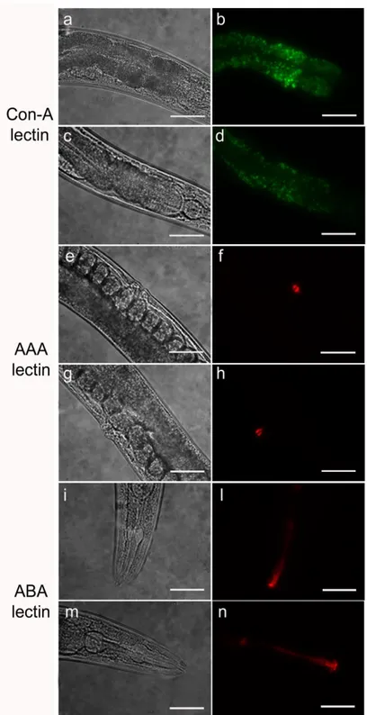
Different lectins were used to characterize cellular surfaces and surface coat of wild type nematodes after RNAi. Glycoconjugates on the cuticle, intestine, including the anus and rectum, allow the interaction with the external environment, acting as ligands for receptors exposed on microorganisms cell surface. Therefore, any changes could contribute to the observed bacterial resistance (Parsons et al., 2014). The use of lectins allowed to determine and localize the presence of alterations of glycoproteins on C. elegans surfaces, which could contributing to worm resistance to S. aureus. A reduced fluorescent signal was observed in pmr-1 nematodes, when Con-A lectin staining was
19
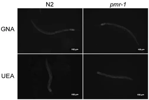
more intense in control nematodes than in the pmr-1 mutants (Fig. 14). Moreover, an altered fluorescent signal was also observed in vulva of pmr-1 nematodes, when AAA staining was carried out (Fig. 14). The ABA lectin labeling was more evident in the region of the pharynx and gut of pmr-1 nematodes, compared to control nematodes. On the contrary, no changes were observed when nematodes were treated with UEA or GNA lectin staining (Fig. 15). These data indicate that silencingpmr-1 gene expression dramatically alters C. elegans glycoproteins abundance and distribution. In
order to identify possible alterations in gene expression involved in glycosylation processes, RT-qPCR analysis was performed for N-acetylgalactosaminyltransferase gly-11, the GDP-mannose 4,6-dehydratase gmd-2 and mucin-like let-653 and osm-8 transcripts. The results showed that pmr-1 silencing induced an increased transcription of gly-11 and let-653, and a reduction in the mRNA levels for gmd-2 and osm-8, further supporting the involvement of pmr-1 gene in glycoconjugates formation (Fig.16).
Figure 14. Lectin staining of N2 e pmr-1 mutant worms. Nematodes were stained with FITC conjugated Con-A (a, b, c and d), Texas red conjugated AAA (e, f, g and h) or ABA (i, l, m and n) lectins after RNAi. Panels a, b, e, f, i and l indicate worms interfered with the empty vector. Panels c, d, g, h, m and n indicate pmr-1 mutant worms. n = 10 for each sample. Scale bar = 100μm.
21
Figure 15. Lectin staining. N2 and pmr-1 nematodes were stained with FITC conjugated GNA and UEA lectins before infection. n= 10 for each sample. Scale bar = 100μm.Figure 16. RT-qPCR analysis of glycosylation genes. Expression of (a) gly-11, (b) gmd-2, (c) let-653 and (d) osm-8 genes in N2 worms and pmr-1 mutants. Histograms show the expression of glycosylation related genes detected by RT-PCR. Bars represent the mean of three independent experiments. Asterisks indicate significant differences (**p <0.01, ***p <0.001).
Pmr-1 mutant worms showed microvilli surrounded by a dense glycocalyx
Transmission electron microscopy confirmed the general organization of the classic intestinal morphology of C. elegans, in both the control and the pmr-1 worms. The microvilli are anchored into a strong cytoskeletal network of intermediate filaments at their base, called the terminal web. The core of each microvillus has a bundle of actin filaments that connects to this web. Over the microvilli, there is an extracellular electron-lucent coating of highly modified glycoproteins (a glycocalyx), which may function to localize digestive enzymes, protect microvilli from physical or toxic injury or serve as a filter (Lehane, 1997). The intestinal cells are each very large and contain large nuclei with a prominent nucleolus, many mitochondria, extensive rough endoplasmic reticulum (RER), many ribosomes, and an extensive collection of membrane-bound vesicles and vacuoles. The nature of these organelles changes gradually as the animal ages. The digestive and metabolic activities of the intestine are central to the growth and development of the animal, and correspondingly, these organelles include yolk granules, recycling endosomes, autophagic vacuoles, and autofluorescent (gut) granules. Using light microscopy, some of these gut granules become visible as birefringent objects in older adults and are inferred to be secondary lysosomes involved in catabolism (Clokey and Jacobson, 1986).
Through TEM analysis, cross or longitudinal sections revealed intestinal cells linked by adherens junctions at their apical borders, forming a narrow lumen bordered by dense microvilli and coated along the entire luminal surface by a dense glycocalyx (Fig. 10a and d). After infection with S. aureus, the intestinal lumena of all pmr-1 mutants analyzed appeared dilated and full of bacteria, displaying shorter microvilli relative to the infected worms (Fig. 10b and e). Mainly in controls, bacteria strictly adhered to the microvilli, binding their bases, or were even observed embedded into them (Fig. 10b and c). Indeed, in C. elegans the pathogens caused local damage via degradation of the microvilli, a process described during aging. Differently, in mutant worms, the microvilli were mostly surrounded by a dense glycocalyx and the intestinal lumen appeared to be filled mostly with dense soluble material (Fig. 10f), suggesting a protective role of the glycoproteins composing the glycocalyx. Many microvilli extend into the lumen from the apical face, forming a brush border (IntFig. 10).
23
Figure 10. Ultrastructural analysis of control worms and pmr-1 mutants, before and after infection with S. aureus. Electron micrographs show the intestinal surface of both control and pmr-1 nematodes with a typical apical domain, including brush border with microvilli, terminal web, and apical junctions (a, d; arrowheads). Panels a and d indicate N2 and pmr-1 worms before S. aureus infection, respectively. Panels b and c indicate control worms after S. aureus infection, while pmr-1 infected worms were indicated in panels e and f. After infection (b, e), the intestinal lumena of both control and defective worms are dilated. However, in mutant worms, a denser and thicker glycocalyx (asterisc) separates the microvilli from the intralumenal bacteria (e-f). Differently, in control worms, bacteria adhere to the microvilli (b, c). Bars 1 µm; Fig. c, f. Bars 0,5 µm.Pmr-1 silencing enhanced resistance in C. elegans bus-4 mutants
Since it has been reported that C. elegans glycosyltransferase BUS-4 is involved in S. aureus resistance (Parsons et al., 2014), bus-4 mutants were subjected to RNA interference for pmr-1 gene, to better understand the mechanism of action of this protein. When worms were exposed to S. aureus, an increase in the lifespan of bus-4 nematodes defective for pmr-1, compared to bus-4 or N2 control worms, was observed (Fig. 17). In particular, the median survival of bus-4 worms was recorded at days 5 and 3 for pmr-1 and control nematodes, respectively. Hence, the double mutant showed an additional effect with respect to the two single mutants, suggesting that pmr-1 might act on S. aureus resistance independently on the glycosylation impairment.
Figure 17. Lifespan analysis. Kaplan-Meier survival plot of N2 and bus-4 worms, after 48 h of RNAi, infected with S. aureus. n = 60 for each data point of single experiments. Asterisks indicate significant differences (*p <0.05; **p <0.01, ***p <0.001).
Probiotics and glycosilation: how foodborne Lactobacillus fermentum MBC2 strain is involved in host responses?
Probiotic bacterial strains are able to modulate host-pathogens interactions in the gut. Different studies reported that glycosylation were modulated to adapt the host defense toward pathogens (Freitas et al., 2003; Silva et al., 2014). The intestinal microflora may participate to the defense against intestinal pathogens known to use host glycan patterns as targets to invade intestinal cells, underlining an important role of probiotics in host-pathogen cross-talk.
In this project, C. elegans was employed to explore potential health promoting features of L.
fermentum MBC2, previously isolated from Mozzarella di Bufala Campana microbiota (Zanni et al.,
2015). To investigate whether this strain induced beneficial effects on C. elegans physiology, the survival rate was first examined. Feeding wild type nematodes with L. fermentum MBC2, the median lifespan resulted significantly extended as compared with the probiotic L. rhamnosus GG (LGG) and OP50 controls (Fig. 18a). In particular 50% of worm viability in L. fermentum MBC2-fed nematodes was recorded at days 18, in comparison with day 11 and 14 in E. coli- and LGG-fed animals, respectively. By contrast, the prolongevity effect was not observed when the dietary administration was based on heat-killed bacteria (Fig. 18b). Therefore, the positive effect on C. elegans lifespan was dependent on the viability of L. fermentum MBC2 strain. Afterwards, to determine whether bacteria strains were consumed by the worms, intestinal colonization capability was explored plating worm lysates at different time points and evaluating the bacterial colony forming units (CFU). Results demonstrated that the intestinal CFUs of L. fermentum MBC2 diet increased along the lifespan. Notably, at 5 days of adulthood the CFU number relative to L. fermentum MBC2 resulted to be about 2-fold and about 4-fold more than that relative to LGG and OP50, respectively (Fig. 18c).
25
Figure 18. Effect of L. fermentum MBC2 on nematode lifespan and colonization capability. (a) Kaplan-Meier survival plot of N2 worms fed L. fermentum MBC2. Lifespans of E. coli OP50- and LGG-fed animals are reported as controls; n = 60 for each data point of single experiments. (b) Effect of heat-killed strains on C. elegans viabilty. (c) Bacterial colony forming units (CFU) recovered from nematodes were obtained by plating whole lysates of L4 and 5-day-old adults fed L. fermentum MBC2, LGG or OP50. Bars represent the mean of three independent experiments. Asterisks indicate significant differences (*p <0.05, **p <0.01, ***p <0.001, ns: not significant).To investigate whether feeding with L. fermentum MBC2 increased the quality of life, changes in age-related biomarkers, as body movement, pumping, and lipofuscin accumulation were measured. The locomotory rate of C. elegans was evaluated in the range between 3 and 11 days from hatching. L.
fermentum MBC2 fed nematodes displayed, from 6 to 9 days, a higher motility than control worms
(Fig. 19a).
The pharyngeal pumping rate measures muscle function, and the pumping activity is associated with food intake ability. Although the frequency of pharyngeal pumping generally declined with age, statistic analysis results showed that in the case of L. fermentum MBC2 fed nematodes it was two times higher with respect to OP50 fed worms, at 13 days of adulthood. Noteworthy, pumping rate measured in Lactobacillus strains fed-worms was similar to that of young adult OP50-fed worms (Fig. 19b). Since intracellular lipofuscin is a marker of cellular damage during aging, the autofluorescence of worms was also analysed (Fig. 19c). Nematodes fed on L. fermentum MBC2 showed at 13 days of adulthood a reduced fluorescence signal compared with LGG and OP50 fed-young adult, while OP50-fed animals at 13 days adult displayed instead a marked accumulation of fluorescent granules along the intestine, typical of aged animals. Based on these findings, experiments feeding C. elegans on L.
fermentum MBC2 after RNAi will be performed to analyze the role of potential probiotics on
27
Figure 19. Analysis of aging markers in C. elegans fed L. fermentum MBC2. (a) Body bending ofC. elegans fed Lactobacillus strains with respect to OP50, measured for 30 seconds. Bars represent the
mean of three independent experiments. Different letters indicate significant differences (p < 0.05) (b) Pumping rate of 13-days-old worms, measured for 30 seconds and determined from the mean of 10 worms for each bacterial strain. Worms fed OP50 or LGG were used as controls. Statistical analysis was evaluated by one-way ANOVA with the Bonferroni post-test; asterisks indicate significant differences (**p <0.01, ***p <0.001). Bars represent the mean of three independent experiments. (c) Autofluorescence of lipofuscin granules in C. elegans fed L. fermentum MBC2, LGG and OP50 on day 13. Ten worms were used for each measurement. Scale bar = 100μm.
Discussion
The aim of the study was to evaluate of the role of the pmr-1 gene in the host-microorganism interaction in the C. elegans model system. Nematodes subjected to RNAi for pmr-1 gene showed an increased longevity compared to control individuals, after infection of S. aureus pathogen. The increased resistance to infection was related to the low colonization capability of the Gram-positive bacteria observed in pmr-1 mutants, as compared to control. The protein encoded by pmr-1, expressed in all eukaryotes, is an ATPase for the transport of the Ca2+ ion, localized on the membranes of the Golgi apparatus (Antebi and Fink, 1992). It plays a fundamental role in the homeostasis of the ion in the Golgi and in the endoplasmic reticulum (RE). Indeed, as already reported, it is involved in different cellular mechanisms. Studies on S. cerevisiae and K. lactis demonstrated that normal levels of Ca2+ in these cellular compartments are necessary for the functional activity of enzymes involved in N- and O-glycosylation of glycoproteins (Antebi and Fink, 1992). The inactivation of PMR-1 homologous proteins produces phenotypes with defects in glycosylation, in cell wall morphogenesis and in mitochondrial metabolism, and it is involved in the response to oxidative stress (Durr et al., 1998; Farina et al., 2004; Uccelletti et al., 1999). Glycoconjugates are involved in the interaction of C.
elegans with the external environment since they are targets for the adhesion of microorganisms to the
cuticle and to the cell surface. Several studies showed the importance of the structure, density and distribution of glycoconjugates in the host-pathogen interaction and in the determination of the susceptibility or resistance of the nematode to bacterial attack (Gravato-Nobre et al., 2011; Palaima et al., 2010). Intestinal pathogens exploit host immune responses to compete with the indigenous microbiota and therefore to colonize gut. High colonization capability of in C. elegans gut, followed by death of the worms, could permit bacterial survival, protection by stress so that promoting environmental spread (Zhang and Hou, 2013). Some of these effects were observed when C. elegans was infected with UPEC E. coli ECP110 strain.
The availability C. elegans mutants for genes involved in the glycosylation of extracellular matrix proteins allowed to observe an increased resistance of the nematode to the infection of the Gram-positive Microbacterium nematophilum and to the formation of biofilm by the Gram-negative Yersinia
pestis (Parsons et al. 2014; Palaima et al., 2009). In particular, it was observed that C. elegans mutants
for the glycosyltransferase bus-2 gene, resistant to the M. nematophilum, showed an increase in fucosylated N-glycans and alterations in the central core Gal (β1,3) GalNAc of the O-glycans (Parsons et al., 2014). Therefore, composition of the cuticle glycoproteins and of C. elegans cell surface is fundamental for the correct recognition by the receptors of Gram-positive bacteria and for the subsequent phases of adhesion and pathogenesis. S. aureus show adhesion molecules to the host cells that include the MSCRAMM family (surface microbial components that recognize matrix adhesive molecules) and capsular polysaccharides. MSCRAMM are specific for a series of ligands, different from species to species, and include extracellular matrix proteins, such as glycoproteins and collagen (Clarke and Foster, 2006; Speziale et al., 2009). Alterations of the glycosylation process, due to the absence of the functional activity of PMR-1 and to changes in the levels of Ca2+ concentration in the RE and in the Golgi, may cause changes in the structure of glycoproteins exposed on the cell surface. These glycoproteins could be implicated in binding to S. aureus receptors; hence, their alteration would compromise the adhesion of the bacterium to the nematode, reducing its ability to infect the host.
To verify the presence of changes in the glycan composition of the cell surface glycoproteins, staining of the nematodes after RNAi with fluorescent lectins were carried out. The binding specificity to mono and disaccharides of these proteins allowed to collect information on the structure of the N- and O-glycans of C. elegans surface and to detect structural modifications. In pmr-1 defective worms, the fluorescent signal related to the binding of the AAA lectin to the surface glycans is altered with respect
29
different presence of fucosilated surface glycans in pmr-1 mutants compared to nematodes with normal transcript levels. Fucose residues in the glycan structures are typical of invertebrate organisms. There are divergent opinions on the location of fucose residues in C. elegans: fucose could be linked to terminal residues or one or two fucose residues could replace the N-acetylglucosamine core linked to asparagine (Schachter, 2004). Therefore, defects in glycosylation process could cause alterations of the central nucleus linked to the Asn residue or the terminal stage of the process in which an abnormal addition of fucose residues occurs. The ABA lectin is specific to the disaccharide Gal (β1,3) GalNAc, which represents the central nucleus that binds to the Ser/Thr residue in the O-glycans. The signal related to ABA lectin staining was more intense in nematodes defective for pmr-1 and it was located at the level of the pharynx of worms. This result probably was due to alterations of the central core of O-glycans, caused by defects in the glycosylation process. The observed alterations in lectin staining correlated to the different expression of genes involved in glycosylation processes, confirming the presence of modifications in glycoconjugates. In particular, gmd-2 catalyzes the conversion of GDP-Man to GDP-Fuc, involved in the synthesis of fucosyl glycoconjugates. The reduction of transcript levels of gmd-2 in pmr-1 mutants, were consistent with the weaker fluorescence signal observed inpmr-1 worms stained with AAA lectin. These changes could result in increased resistance of the pmr-1
mutant nematodes to S. aureus. Indeed, the exposure, on C. elegans cells, of altered ligands of the receptors of S. aureus could prevent or make the interaction between the bacterium and the nematode difficult, reducing the ability of the pathogen to infect the worm. This result could be related to the low colonization capacity of S. aureus observed in pmr-1 mutants. Moreover, worms subjected to RNAi for the pmr-1 gene showed an increased expression of SOD-3 already before infection. In C. elegans the SOD enzymes are constitutively expressed in the pharynx, in the tail and in the vulva; under conditions of oxidative stress their expression increases considerably in all regions of the animal's body (Van Raamsdonk and Hekimi, 2012). These levels increase significantly when hsf-1 gene expression was analysed. hsf-1 gene encodes a transcription factor of Heat Shock Protein (HSP). In the
K. lactis Hsp60 protein is able to compensate for the absence of the homologous protein of PMR-1 and
to eradicate the conditions of oxidative stress in which the cells are found in the absence of it (Uccelletti et al., 2005). Hence, the PMR-1/HSP60 system was hypothesized as a mechanism of resistance to oxidative stress in this organism (Uccelletti et al., 2005). The increase in expression of the SOD-3 enzyme and the transcriptional factor HSF-1, observed in this work, could be related to the increase of oxidative stress in pmr-1 mutant nematodes, due to the absence of a functional activity of the protein PMR-1. After infection, levels of the Oxygen Reactive Species are further reduced. This result could be explained by an induction of the expression of genes coding for enzymes involved in the oxidative stress response, in addition to the sod-3 gene and the transcriptional factor SKN-1, following exposure to the pathogen. All together, these results allowed evaluating the involvement of the PMR-1 protein in the resistance to infection of the S. aureus and in the oxidative stress response. Consistently, we observed an increased resistance of the pmr-1 mutant nematodes to S. aureus. However, the additive phenotype of the double mutant indicates that the two mutated genes function independently from the other by differentially influencing the expression of surface-exposed glycoconjugates involved in bacterial adhesion colonization or alternatively pmr-1 plays a role in controlling immune response of C. elegans to infections. In line with a model in which pmr-1 is involved in response to the infection, the central signaling of the antimicrobial response of C. elegans seemed to be dyregulated by loss of pmr-1. We found that upon pmr-1 knockdown both PMK1/SKN-1 and PMK1/HSF-1 signaling, which mediate stress and immune responses, were activated (Van Raamsdonk and Hekimi, 2012; Uccelletti et al., 2005). The extended lifespan in S. aureus-fed pmr-1 worms thus might largely depend on the activation of these pathways that contribute to enhance pathogen resistance and longevity. Moreover, the additional effect observed in bus-4/pmr-1 mutants
with respect to the two single mutants, suggested that pmr-1 might act on S. aureus resistance independently on the glycosylation impairment.
Futhermore, C. elegans was also used to evaluate host-foodborne bacteria interactions. Lactic acid bacteria (LAB) are known to be components of the microbiota of humans and animals. The many properties of LAB, including the ability to provide health benefits modulating host glycosylation state, are strain-specific. L. fermentum is one of the most common cultivable and predominant microbes in fermented dairy products. Positive impact of L. fermentum MBC2 on wild-type nematodes was dependent on the viability of bacteria, in agreement with the fact that microflora in fermented dairy products must be alive and is able to colonise human gut. Recently, different L. fermentum strains were tested on C. elegans to investigate the relationship between feeding foodborne bacteria and aging, reporting that L. fermentum LA12 isolated from food and infant feces could contribute to enhance immune responses and prolong the life span of animals (Lee, 2011). The effects exerted by L.
fermentum MBC2 on C. elegans physiology could be due to the high survival rate of the bacterial
strain in the nematode gut. In fact, L. fermentum MBC2-fed animals showed a higher colonization capability than that of nematodes fed E. coli OP50 or LGG. The prolongevity effects observed in lifespan experiments correlated to a delay in aging processes.
Therefore, C. elegans resulted to be a valuable model system for screening probiotic bacteria. By analysis of lifespan with co-culture in pmr-1 worms, L. fermentum MBC2 was not able to contrast effects of pathogenic bacteria. As previously described, mucin glycoproteins are major macromolecular constituents of epithelial mucus; probiotics may promote mucus secretion as one mechanism to improve barrier function and the exclusion of pathogens (Bermudez-Brito et al., 2012). Several Lactobacillus species increase mucin expression in human intestinal cell lines. However, this protective effect is dependent on Lactobacillus adhesion to the cell monolayer (Mack et al., 2003; Mattar et al., 2002). For instance, probiotic formulation VSL3, which contains some Lactobacillus species, increases the expression of MUC2, MUC3 and MUC5AC in HT29 cells (Otte and Podolsky, 2004). The sugar residues of mucins can act as ligands for bacterial membrane receptors, and in fact, changes in the glycosylation pattern have been associated to dysbiosis during intestinal inflammation (Larsson et al. 2011; Sommer et al. 2014). Species of Lactobacillus and Bifidobacterium are the most commonly used probiotic bacteria. Both genera are characterized as Gram-positive lactic acid bacteria and share common surface molecules such as lipoteichoic acid (LTA), surface layer associated proteins (SLAPs) and mucin biding proteins (Mubs) that play an important role in the interaction with mucus components (Lebeer et al. 2010). The microbial adhesion process of LAB includes also passive forces, electrostatic interactions, hydrophobic interactions, steric forces.
Cell surface associated LTA often serves as major virulence factors in Gram-positive bacteria. These complex glycolipid structures mediate interaction with host receptors, triggering signalling pathways, resulting in probiotic/pathogenic effects (Lebeer et al. 2010). It has been reported that pathogens such as S. aureus, Listeria monocytogenes, or E. faecalis produce wall techoic acids (WTA) with diverse, usually strain-specific glycosylated structure and they are responsible of virulence or resistance to infection (Winstel et al., 2014).
Based on this, the data could explain why L. fermentum MBC2 was able to prolong lifespan in wild type worms, while in pmr-1 mutants, survival was similar to that based on S. aureus diet. Indeed, the Gram-positive probiotic probably present recognition molecules similar to those of Gram-positive pathogens, and the lack of pmr-1 could cause the inhibition of binding of these bacteria to intestinal cells.
31
Bacterial and fungal strains and growth conditionsGram-negative bacteria strains used in experiments were Escherichia coli OP50, E. coli ETEC K88 and Pseudomonas aeruginosa ATCC 15692; Gram-positive bacteria were Enterococcus faecalis JH 2.2 and Staphylococcus aureus ATCC 25923. All the strains were grown in Luria Bertani (LB) broth at 37°C overnight, in agitation. Candida albicans ATCC 10231 was tested in experiments of fungal infection and was grown in Yeast Extract-Peptone-Dextrose (YPD) broth at 28°C overnight, under shaking. After overnight growth, 30 μl of each culture (about 1*108 cells/mL) was spread onto respective agar-plates. For S. aureus ATCC 25923 and C. albicans ATCC 10231, Trypticase soy agar (TSA) and Brain Heart Infusion (BHI) agar were used, respectively. E. coli strains, E. faecalis JH 2.2 and P. aeruginosa ATCC 15692 were spread on 3.5 cm diameter NGM plates.
E. coli strains were isolated in a tertiary teaching hospital in Rome from urine; ECP45 strain from a
patient in a medical ward, with uncomplicated UTI, and ECP110 from a catheterized patient in the neurological intensive care unit. The E. coli K12 MG1655 (Guyer et al., 1981) and the uropathogenic E.coli CFT073 strain, isolated from blood of a patient suffering from acute pyelonephritis, were used for as controls (Mobley et al., 1990). UPEC E. coli cultures were grown exponentially in LB at 37°C. Bacterial lawns were prepared by spreading 30 μl of each culture corresponding to 1*108 cells on the NGM agar plates (35mm). E. coli cultures were grown exponentially in LB at 37°C. Bacterial lawns used for C. elegans infection assays were prepared by spreading 30 μl of each culture corresponding to 1*108 cells on the NGM agar plates (35 mm). Lactobacillus fermentum MBC2 and Lactobacillus
rhamnosus GG (LGG, ATCC53103) were routinely grown in De Man Rogosa Sharpe (MRS) medium
for 24-48h at 37°C under anaerobic conditions. Afterward, 25 μL of each overnight culture, suspended in M9 buffer, corresponding to 10 mg of bacterial cells, was spread on 35 mm diameter mNGM plates. Heat-killed UPEC and lactobacilli cells were prepared as follows: after overnight growth, cells were then incubated at 65°C for 90 min and deposited onto NGM and mNGM agar plates, respectively. Heat-killed cells were also plated on LB or MRS agar in parallel to ensure that no viable cells remained. OP50 cells were treated in the same manner.
C. elegans strains and infection assay
Wild type C. elegans strain,
skn-1(QV225),
pmk-1(KU25), sek-1(AU1), hsf-1(PS3551) and bus-4(e2693)CB5443 mutant strains and transgenic sod-3::GFP(cf1553) strain were grown onnematode-growth media (NGM) plates seeded with E. coli OP50 at 16°C according to standard procedures (Stiernagle, 2006).
RNAi of pmr-1 was performed by feeding as described previously (George et al., 2014). In RNAi experiments, the empty vector L4440 E. coli HT115 strain was used as the negative control. Then, synchronized young adult worms, grown on E. coli OP50 as described before, were transferred onto NGM plates seeded with E. coli strain HT115 carrying the RNAi clone of pmr-1 gene.
After 48 hours of RNA interference, animals were transferred onto infection plates, as described above, and grown at 25°C. Animals were monitored daily and scored as dead when they no longer responded to gentle prodding with a platinum wire.
For E. coli uropathogenic infections, the plates, prepared as described above, were incubated at 37°C for 24h before being seeded with young adult nematodes, grown at 16°C, from a synchronized culture (Brenner, 1974). The infections were performed at 25°C for several days, as indicated. E. coli cultures were grown exponentially in LB at 37°C. Bacterial lawns used for C. elegans infection assays were prepared by spreading 30 μl of each culture corresponding to 1*108 cells on the NGM agar plates (35mm).
Estimation of bacterial CFU within the nematode gut
For infection experiments, 10 animals for each sample, after 48h from infection, were washed and lysed according to (Uccelletti et al., 2010). Whole worm lysates were plated onto LB-agar plates. The number of CFU was counted after 24 h of incubation at 37°C, aerobically.
For experiments with foodborne bacteria, 10 animals at L4 stage and at 5 days of adulthood were washed and lysed. Lysates were plated onto MRS-agar plates. The number of CFU was counted after 48h of incubation at 37°C, anaerobically. Instead, OP50-fed worm lysates were plated onto LB-agar plated and incubated at 37°C.
Brood size and body size measurements
Synchronized worms obtained as above were grown on mNGM plates seeded with bacteria and then allowed to lay embryos at 16°C. Next, animals were transferred onto a fresh bacteria plate every day, and the number of progeny was counted with a Zeiss Axiovert 25 microscope. The procedure was repeated until the mother worms stopped laying eggs. Each day the progeny production was recorded and was compared with the OP50- or LGG-fed nematodes.
Pumping rate and body bending measurements
Pharyngeal pumping was analyzed under Zeiss Axiovert 25 microscope by counting the number of grinder contractions of 10 animals for each treatment, during a period of 30 s. The analysis was performed in 13-days-adult worms, grown on different bacteria starting from embryo stage.
Lipid droplets visualization
Approximately 100 L4 nematodes, grown on LAB, LGG, or OP50 containing mNGM plates, were suspended in 1 mL of M9 buffer and washed three times. Subsequently, worms were incubated with a solution of 6.7 μg/mL BODIPY 493/503 (Life technologies) for 20 min. Afterwards, worms were mounted onto 3% agarose pads containing 20 mM sodium azide and observed with a Zeiss Axiovert 25 microscope. BODIPY images were acquired using identical settings and exposure times to allow direct comparisons.
Lipofuscin analysis
The autofluorescence of intestinal lipofuscin was measured as an index of senescence at day 13 of adulthood. Randomly selected worms from the plate lawned with bacteria were washed three timeswith M9 buffer. Worms were then placed onto a 3% agar pad containing 20 mM sodium azide. Lipofuscin autofluorescence was detected by fluorescence microscopy (Zeiss Axiovert 25).
RT-qPCR analysis.
After 48 h from infection, total RNA from 200 worms for each sample was isolated with RNeasy midi kit (Qiagen) according to manufacturer’s instructions and then digested with 2U/μL DNAse I (Ambion). 1 μg of each sample was reverse-transcribed using oligo-dT and enhanced Avian reverse transcriptase (SIGMA, Cat. Number A4464). For realtime qPCR assay, each well contained 2 μL of cDNA used as template, SensiMix SYBR & Fluorescein Kit purchased from Bioline, and the selective primers (200 nM) designed with Primer3 software and reported in Table 1. All samples were run in triplicate. Rotor-Gene Q Real-Time (QIAGEN) was used for the analysis. The RT-qPCR conditions are described by Gorietti et al. (2014). Quantification was performed using a comparative CT method (CT = threshold cycle value). Briefly, the differences between the mean CT value of each sample and the CT value of the housekeeping gene (act-1) were calculated: ΔCTsample = CTsample − CTact-1. Final
33
hsf-1 FOR 5’-ATGACTCCACTGTCCCAAGG
REV 5’-TCTTGCCGATTGCTTTCTCT
pmk-1 FOR 5’-AAATGACTCGCCGTGATTTC
REV 5’-CATCGTGATAAGCAGCCAGA
sek-1 FOR 5’-CAGAGCCGTTTATTGGGAAA
REV 5’-TGCATCCGGCTTGTACAGT
sod-3 FOR 5’-AGAACCTTCAAAGGAGCTGATG
REV 5’-CCGCAATAGTGATGTCAGAAAG
act-1 FOR 5’-GAGCGTGGTTACTCTTTCA
REV 5’-CAGAGCTTCTCCTTGATGTC
gly-11 FOR 5’-GGACCTGCGGTGGAGAACT
REV 5’-GCGGAAAATGTGGCCAACT
gmd-2 FOR 5’-AAAGCGAGCTGACCCCATT
REV 5’-ATACATCTTGGCGACCGCATA
let-653 FOR 5’-CTGTCTCGTGAGAATATGTCC
REV 5’-TTCCACGTCGTCGCATGT
osm-8 FOR 5’-AGAAGCCCCACCACTGATTG
REV 5’-TTGTTTTTGCCACGGTTCAA
act-1 FOR 5’-GAGCGTGGTTACTCTTTCA
REV 5’-CAGAGCTTCTCCTTGATGTC
Analysis of C. elegans strain sod-3::GFP and skn-1::GFP fluorescence
Synchronized transgenic worms were transferred on RNAi NGM plates and then infected, as described above. After 48h from RNAi and 24 h or 48h from infection, worms were anesthetized with sodium azide (20 mM) on 3% agarose pad on a glass slide and the fluorescence was viewed under a Zeiss Axiovert 25 microscope. The experiments were repeated three times and 15 worms per group were used in each experiment.
Measurement of reactive oxygen species (ROS)
ROS formation in C. elegans was measured using the fluorescent probe H2DCFDA according to (Kampkötter et al., 2007) with some modifications. Before and after infection, adult worms were washed in M9 buffer and then transferred individually into wells of a 96-well microtiter plate, containing 50 μL of M9 buffer. Subsequent to the complete transfer of the worms 50 μL of 100 μM 2,7dichlorodihydrofluorescein diacetate (H2DCF-DA, SIGMA-ALDRICH) in methanol was added to the wells. H2DCF-DA is a membrane-permeable non-fluorescent probe, which can enter the cells of the worm and it is intracellular converted to H2DCFs. This probe can be oxidized by ROS to yield the fluorescent dye DCF. The changes of fluorescence in worms indicate the accumulation of ROS at different time points (30, 60 and 120 minutes). The measurement was performed using a multiplate reader (Promega, GloMax multidetection system) at excitation/emission wavelengths of 485 and 520 nm.
Lectin staining
Lectin staining of live nematodes after 48 h from infection, was performed as described previously (Palaima et al., 2010). After fixation, 10 μL of stained worms, were mounted on glass slides and