The Department of Health Sciences Università degli Studi del Piemonte Orientale
Characterizing TG2 autoantibody response in
Celiac Disease
Dottorato In Biotecnologie Per L’Uomo
PhD Program In Biotechnologies For Human Health Cycle XXVIII
PhD student: Szilvia Bakó
Tutor: Prof. Daniele Sblattero
Coordinator: Prof. Claudio Santoro
Characterizing TG2 autoantibody response in
Celiac Disease
A thesis submitted to
The Department of Health Sciences
Università degli Studi del Piemonte
By Szilvia Bakó
In partial fulfillment of the requirements for the degree of
Doctor of Philosophy
Acknowledgements
The PhD. thesis includes results undertaken at the Department of Health Sciences in Universitá degli Studi del Piemonte Orientale. The research project was realized within the framework of TRANSPATH-Transglutaminases in disease- a novel therapeutic target?, supported by the European Commission Seventh Framework Programme (FP7).
I would like to express my gratitude to Prof. Daniele Sblattero and Prof. Claudio Santoro for the opportunity to participate the PhD program within the Università degli Studi del Piemonte Orientale and be a member of laboratory Biologia Applicata. Beyond this I am thankful for the couching by providing advices and endless patience in the course of my project. Im also thankful for teaching me how I can improve not only as a researcher but also as an individual by asking myself the correct questions.
I would like to thank to Dr. Diego Cotella for his support, patience and for being always able to provide help in the time of need by his extraordinary knowledge in science.
Thanks to all my colleagues with whom I had the privilege to work together: Cecilia Deantonio, Frank Antony, Silvia Saragozza, Eleonora Rizzato, Maria Felicia Soluri, Laura Patrucco, Olga Tarasiuk, Andrea Chiesa, Gabriela Forestieri for the scientific and non scientific dialogues, their comments and advices and last but not least their friendship through all these years.
I would like to thank Prof. Ludvig M. Sollid for supporting me in the course of my secondment in Centre for Immune Regulation, University of Oslo.
I want to express my heartfelt thanks to Erika Tóth, for being my friend for more than ten years. Thank you for being always there for me and make me laugh in the times when it was most needed.
ABSTRACT
Characterizing TG2 autoantibody response in Celiac Disease
PhD Student: Szilvia Bakó1
PhD Program In Biotechnology For Human Health, Cycle: XXVIII
Year: 2012-2015
Tutor: Prof. Daniele Sblattero1 Coordinator: Prof. Claudio Santoro1
1 Department of Health Science and Interdisciplinary Research Center on Autoimmune Diseases, University of Eastern Piedmont, Novara, Italy
Background: Celiac Disease (CD) is a gluten sensitive enteropathy with autoimmune features, characterized by the destruction of intestinal epithelium and the production of IgA and IgG antibodies against gluten and self antigen tissue transglutaminase (TG2). Because of the fact that TG2 acts not only as key player in the disease but also as a self antigen studying the autoimmune response against this enzyme is crucial for understanding CD. Numerous studies aimed to investigate the antibody response due to the possible role anti TG2 antibodies play in the onset of the disease, however there are still many questions to be answered. Phage display antibody technology have been proved to be an efficient tool in the study of autoimmune diseases, and because of this we selected this method for dissecting the autoimmune response in CD.
Results: We were able to generate and classify antibodies in phage expressed scFv format first due to their specificity and second by competing them with reference antibodies. Antibodies were clustered into four Epitope groups, with great majority belonging to Epitope 1., the VH5 cluster. Antibodies representing each epitope cluster were analyzed in scFv-Fc format. It was shown that Epitope 1. antibodies recognize a region in TG2 molecule overlapping with fibronectin binding site while other epitope groups recognized a different region. Three out of four epitope clusters were able to stain cell surface TG2 (csTG2). Of the four epitope groups only Epitope 2. had inhibitory
effect on TG2 enzymatic activity in vitro. By reconstructing a VH5 antibody from IgG1 format into scFv format we generated two constructs
with different VH/VL orders that retained specificity. The VH chain was substituted with Vk chains originating from three cDNA libraries generated from CD patients. The Vk usage was restricted IGKV1-5*03 F, IGKV1-39*01 F, IGKV1-12*01 F chains, IGKV1-39*01 being most abundant chain. Two construct with original VH/VL and four selected clones with Vk chains coming from cDNA libraries were characterized in scFv-Fc format. All clones were shown to recognize the region on TG2 overlapping with fibronectin binding site. Most but not all of scFv-Fcs recognized csTG2. None of the antibodies showed inhibitory effect on TG2 enzymatic activity corresponding to what we saw in the case of VH5 antibodies .
Conclusion: In this work we demonstrated that phage antibody display is an extraordinary tool to study the autoantibody response in CD. The generated antibodies in phage expressed scFv format were able to mimic the natural antibody response, and a straightforward reconstruction in scFv-Fc format allowed the precise analysis of the disease. The study not only provided an insight into the pathology of CD but also revealed the possibilities of a novel technology to analyze autoimmune diseases.
TABLE OF CONTENTS
Introduction
Chapter 1. Tissue transglutaminase……….9
1. 1. Transglutaminases………..……….9
1. 2. Tissue transglutaminase a multifunctional enzyme.………..….11
1. 3.Enzymatic functions of TG2……….………..13
1. 3. 1. Transamidating activity………..………..13
1. 3. 2. GTPas activity of TG2………..………14
1. 3. 3. Pro and antiapoptotic effect of TG2………..………..14
1. 3. 4. PDI activity of TG2………..15
1. 3. 5. Protein kinase activity of TG2………16
1. 4. Functions of Extracellular TG2………16
1. 5. Interacting partners of TG2……….18
1. 5. 1. Fibronectin and TG2 interaction in the ECM………..19
1. 5. 2. Other extracellular interacting partners of TG2………..20
1. 6. Pathological implications of TG2………23
1. 6. 1. TG2 in neurodegenerative diseases………..23
1. 6. 2. TG2 in fibrozis and scarring ………25
1. 6. 3. TG2 in cancer……….25
1. 7. Inhibition of TG2………26
1. 7. 1. Reversible TG2 inhibitors……….26
1. 7. 2. Irreversible TG2 inhibitors………..28
Chapter 2. Celiac disease………30
2. 1. Antigen presentation in Celiac disease……….31
2. 2. T cell response in Celiac disease………36
2. 3. Antibody response in Celiac disease………..39
2. 4. TG2 specific autoantibody response. ………40
2. 4. 3. Targeted epitopes of anti TG2 antibodies………42
2. 4. 4. Collaboration of TG2 specific B-cells and gluten specific T-cells in CD………44
2. 4. 5. Pathogenic role of anti TG2 antibodies………46
Chapter 3. Expression libraries ………..48
3. 1. Display systems……….48
3. 1. 1. In vitro display systems………..49
3. 1. 2. Cell based display systems……….49
3. 2. Selection methods……….49
3. 2. 1. Physical selection methods……….49
3. 2. 2. Genetic selection methods………..50
3. 3. Phage display………51
3. 3. 1. Phage antibody libraries……….53
3. 3. 2. Phage display for the study of Celiac disease………56
Chapter 4. Recombinant antibody technology………58
4. 1. Early systems for therapeutic antibody generation………..58
4. 2. Recombinant antibody formats………..59
4. 2. 1. Single domain antibodies………59
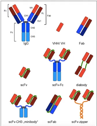
4. 2. 2. Multi domain antibodies……….60
4. 3. Recombinant antibody expression………..62
4. 3. 1. Prokaryotic expression systems………..62
4. 3. 2. Eukaryotic expression systems………..63
RESULTS Chapter 1. Developing TG2 inhibitory antibody………64
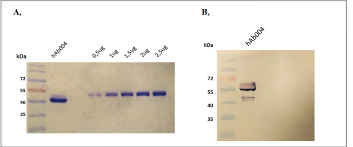
1. 1. Construction of antibody in scFv-Fc format in pMA-T vector………65
1. 2. Cloning and expression in pMB-SV5 vector………..65
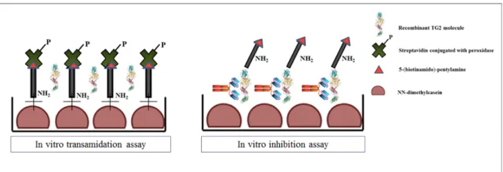
1. 3. Developing TG2 inhibition assay on solid surface and in solution………..68
2. 2. Construction of phage antibody libraries.………..72
2. 3. Characterizin antigen specificity in scFv format.………..74
2. 4. Clustering antibodies into epitope groups in scFv format..………74
2. 5. Cloning and expression in scFv-Fc format.………77
2. 6. Dissecting TG2-autoantibody response in ECM………80
2. 6. 1. Analyzing the effect of TG2-FN interaction on antibody binding………..80
2. 6. 2. Investigate the staining properties of antibodies on csTG2………81
2. 7. Investigating inhibitory effect of antibodies in scFv-Fc format………..82
Chapter 3. Defining the role of VH/VL pairing in antigen recognition.………84
3. 1. Introduction to the results.………..…85
3. 2. Reformatting Epitope 1. IgG1 into scFv format….………86
3. 3. Constructing clones in scFv format from minilibraries of CD patients………..87
3. 4. Cloning and expression in scFv-Fc format……….91
3. 5. Dissecting TG2-autoantibody interaction in the extracellular environment...………93
3. 5. 1. Analyzing the effect of TG2-FN interaction on antibody binding.………..93
3. 5. 2. Investigate the staining properties of scFv-Fcs on csTG2………94
3. 6. Investigating inhibitory effect of antibodies in scFv-Fc format.………..95
DISCUSSION Chapter 1. Background……….97
Chapter 2. Developing TG2 inhibitory antibody………98
Chapter 3. Dissecting anti TG2 antibody response………..100
3. 1. Characterizin and clustering Abs in scFv-Fc format………..100
3. 2. Cloning and expression in scFv-Fc format………..101
3. 3. Dissecting TG2-autoantibody interaction in ECM………..101
3. 3. 1. Analyzing the effect of TG2-FN interaction on antibody binding……….101
Chapter 4. Defining the role of VH/VL pairing in antigen recognition………..103
4. 1. Reformatting Epitope 1. [1] from IgG1 into scFv format………103
4. 2. Construction ad analysis of clones in scFv format from Vk libraries of CD patients……..104
4. 3. Cloning and expression in scFv-Fc format..……….104
4. 4. Dissecting TG2-autoantibody interaction in the extracellular environment………105
4. 4. 1. Analyzing the effect of TG2-FN interaction on antibody binding……… 105
4. 4. 2. Investigate the staining properties of antibodies on csTG2………..105
4. 5. Investigating the inhibitory effect of antibodies in scFv-Fc format……….106
CONCLUSION………107
MATERIALS AND METHODS Chapter 1. Abbreviations………108
Chapter 2. Materials………109
Chapter 3. Standard protocols………110
Chapter 4. Methods……….113
BIBLIOGRAPHY……….117
INTRODUCTION
Chapter 1. Tissue transglutaminase
1. 1. Transglutaminases
Transglutaminases (TGases) are widely distributed group of enzymes catalyzing the post- translational modification of proteins through acyl transfer reactions involving γ-carboxamide group of peptide-bound glutamine and the ε-amino group of peptide-bound lysine, resulting in a ε-(γ- glutamyl) lysine isopeptide bond [1]. The cross linked products often of high molecular mass are highly resistant to proteolysis and their accumulation can be found in various tissues and processes where they play an important role. The term transglutaminase was first introduced by Clarke et al in 1957 [2] where they were describing the transamidating activity observed in guinea pig liver. Since these findings enzymes having TGase activity have been identified in micro organisms [3], plants [4], amphibians [5], fish [6], and birds [7]. In mammals nine TGase isoenzyme have been identified at the genomic level, however only eight have been characterized at protein level. All mammalian forms of TGases having structural homology are members of the papain -like superfamily of cysteine proteases [8], and the tissue content of them is tightly regulated at transcriptional level (Table1.1). Transglutaminase 2 (TG2) is the most abundant and studied member of the TGase enzyme family including TG1, TG3 and TG5 isoforms expressed in epithelial tissue, TG4 expressed in prostate gland, factor XIII expressed in blood, TG6 and TG7 most prominently distributed in the testis and lungs [9]. A further member of TGase family is band 4.2 an enzymatically inactive protein component of erythrocyte membrane [10]. The characteristics of each member of the family include:
• Keratinocyte transglutaminase (TG1): localized in squamous epithelia, keratinocytes and cytosolic membrane, activated by proteolysis, has a barrier function in stratified squamous epithelia and plays a central role in keratinocyte differentiation.
• Tissue transglutaminase (TG2): exists in extracellular and intracellular form, ubiquitously distributed in various tissue types. Plays role in many biological processes including apoptosis, cell survival, signaling, cell differentiation, matrix stabilization, endocytosis and so forth.
• Epidermal transglutaminase (TG3): cytosolic protein, requires proteolysis to become active and get involved in the terminal differentiation of keratinocytes, hair follicles.
• Prostate transglutaminase (TG4): expressed in prostate gland, prostatic fluids, extracellular. Plays key role in the fertility of rodents where it is involved in semen coagulation.
• TG5: Ubiquitously expressed cytosolic protein, predominant in female reproductive tissues and skeletal muscle, playing role in epidermal differentiation.
• TG6: Showing high homology with TG2, distributed in testis, lungs, brain involved in the development of central nervous system and late stage cell envelope formation in the epidermis and hair follicle.
• TG7: Ubiquitously expressed , prominent in testis and lungs.
• Factor XIII: Converted into the active TGase Factor XIIIa (plasma TGase) by a thrombin-dependent proteolysis, distributed in chondrocytes, platelets, astrocytes, macrophages and dendritic cells in the dermis. Involved in wound healing, blood clotting and bone growth.
• erythrocyte protein band 4.2 (Band 4.2): distributed in the surface of erythrocyte membranes, bone marrow, fetal liver and spleen. Key component of erythrocyte skeletal network, maintaining erythrocyte shape and mechanical properties.
Table 1.1. Transglutaminases characterized at protein level [11]
1. 2. Tissue transglutaminase a multifunctional enzyme
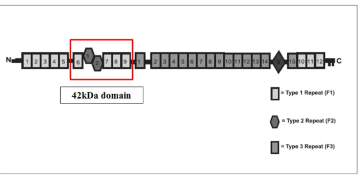
Tissue transglutaminase (TG2) is the most ubiquitous and diverse member of the transglutaminase family with a great variety of biological functions. The human transglutaminase gene localized to the chromosome 20q11-12 is composed of 13 exons and spams 37kb.[12] The monomeric TG2 protein is composed of 687 amino acids, (MW ~ 78kDa) including four distinct domains. N terminal β-sandwich with the fibronectin and integral binding site, catalytic core containing the catalytic tridad Cys277-His335-Asp358 for the acyl- transfer reaction, a conserved Trp essential
for catalytic activity [13] a Ca2+binding region and two C terminal β-barrels, with β-barrel 1
containing the GTP binding site, while β-barrel 2 containing a phospholipase C binding sequence [14] (Figure 1.1.)
Figure 1.2. TG2 conformations. The N-terminal β-sandwich is shown in blue (N), the catalytic domain
(Core) in green, and the C-terminal β-barrels (β1 and β2) in yellow and red, respectively. (A) GDP-bound TG2 [16]. (B) TG2 inhibited with the active-site inhibitor [17]. (C) The N-terminal β-sandwich and catalytic domains of the two structures are superimposed, highlighting the conformational change. The GDP-bound structure is shown in blue and the inhibitor-bound structure in gold.
In spite of the fact that TG2 was found with high expression in various tissues such as in endothelial cells, red blood cells and smooth muscle cells, it is still considered as a stress related protein, with its expression up regulated by physiological and pathological stimuli. Retionic acid (RA) is a well known inducer of TG2 expression both at mRNA and protein level [18]. A number of inflammatory cytokines and growth factors can induce TG2 expression such as transforming growth factor β1 (TGFβ1) [19], interleukins [20] and tumor necrosis factor α (TNFα) [21], thought to up regulate TG2 by activating nuclear factor κB (NF-κB). The activity of TG2 is tightly controlled within the intracellular environment, due to the presence of low Ca2+ and the inhibitory effects of GTP and
GDP on its cross-linking ability [22]. In normal physiological conditions TG2 exists in a closed conformation where the active site of TG2 is hidden in a cleft preventing substrate binding and the C-terminal β-barrel domains tightly fold back for the catalytic core domain [23] (Figure 1.2.). Under stress, disturbance of the intracellular Ca2+ balance may cause transient loss of Ca2+
homeostasis activate TG2 into its catalytically active, open conformation. Ca2+ binding activates
TG2 by inducing a conformational change increasing the inter domain distance between the catalytic core and the two C-terminal β-barrel domains up to 15nm. This results in the relaxation, widening of the whole molecule making the active site accessible for TG2 substrates [16, 17, 24]. In contrast, GTP binding likely stabilizes the closed conformation. Although GTP is considered to be a negative regulator of TGase activity, it has been indicated that the GTP binding is required to display transamidation activity of TG2 [25].
1. 3. Enzymatic functions of TG2
1. 3. 1. Transamidating activity
Two main biological functions of TG2 are transamidation and GTP-binding, with Ca2+levels acting
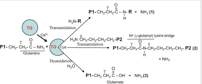
as a switch between them. When the transamidating activity is turned on the catalysis of post-translational modifications of proteins is hallmark of TG2 activity [26]. The two step process involves as a first step the formation of a thioester bond with the enzyme’s active cysteine site via the transamination of γ-carboxamide group of a specific protein-bound glutamine substrate, accompanied by the release of ammonia. The second step includes the transfer of acyl intermediate to a nucleophilic substrate either the ε-amino group of a distinct protein-bound lysine residue [27] or primary amines such as polyamines and histamine. These bonds are resistant to chemical and physical degradation and are essential for stabilizing the ECM [28, 29]. Isopeptide bonds formed by TG2 also play a key role in apoptosis where they prevent inflammation by ensuring that intracellular contents of dying cells are not released to the extracellular environment [30]. Water can also act as a nucleophile causing deamidation where the acyl-donor glutamine residue is converted to a glutamate residue [31] (Figure 1.3.).
Figure 1.3. Enzymatically active TG2 catalyzes Ca2+-dependent acyl-transfer reaction between
γ-carboxamide group of a protein-bound glutamine and either the ε-amino group of a distinct protein-bound lysine residue (covalent protein cross-linking) or primary amines such as polyamines and histamine. Water can replace amine donor substrates, leading to deamidation of the recognized glutamines. [32].
1. 3. 2. GTPase activity of TG2
Under normal conditions in the intracellular environment the transamidating activity of TG2 is turned off due to the low level of calcium and the high level of GTP, inhibiting this function of the enzyme. TG2 is involved in transmembrane signaling by its GTPase activity combined with its ability to interact and signal through receptors [33-35]. TG2 was shown to interact with several receptors, facilitating hormone-receptor mediated transmembrane signaling pathways, involving the classical G-protein-coupled receptors.TG2 has been shown to directly interact with PLC δ1, a key player in the signal transduction process for many receptors [36]. A region of 12 amino acids
between Leu661 and Lys672 in TG2 was identified as the PLC interaction site while the TG2 interaction site in PLC δ1 was shown to be located within the C2 domain of the enzyme [14, 37]. PLC δ1 not only serves as a guanine nucleotide exchange factor (GEF) but also as GTP hydrolysis inhibiting factor (GHIF) for TG2. These functions of PLC δ1 for TG2 facilitate TG2-mediated signaling. Other receptors, which interact with TG2 and mediate transmembrane signaling, are the thromboxane receptor [38] and the oxytocin receptor [39]. Both of the interactions are regulated by activation of PLC.
1. 3. 3. Pro and Anti-apoptotic effect of TG2
TG2 was reported to be involved in apoptosis [40] and it has been proposed that the cell type, the kind of stressor, intracellular localization, and transamidation activity of TG2 determine whether it promotes pro-apoptotic or anti- apoptotic responses [41]. The pro-apoptotic effect of TG2 is due to its cross linking activity, which requires millimolar concentration of calcium. Stressful conditions can generate reactive oxygen species (ROS) and also trigger the release of Ca2+ from the
endoplasmic reticulum (ER), resulting in the activation of TG2 and cross linking of intracellular proteins initiating the apoptotic process [42, 43]. TG2 initiates apoptosis by mediating the crosstalk between dying and phagocytic cells and also ensures that once apoptosis initiated it is completed without inflammation or tissue injury [44]. TG2 can indirectly promote apoptosis by prompting the activation of TGF -ß release by macrophages, which can lead to the death of various cells [45, 46] . Additionally, TG2 can promote chemoattractant formation and the release of phosphatidylserin, to aid macrophage migration to the site of apoptosis and eventually the recognition of apoptotic cell [47].
TG2 may also have anti-apoptotic effect, which is independent from its transamidation and cross-linking activities. Nuclear TG2 protects cells by interacting with retinoblastoma protein pRb, polymerizing the alpha-inhibitory subunit of the transcription factor NF-kappaß, regulating the transcription of several anti-apoptotic genes [48]. Similarly TG2 can translocate to the plasma membrane where it serves as a co-receptor for integrin, promoting its interaction with fibronectin. A plausible scenario is that pro-apoptotic and anti-apoptotic effects of TG2 are dependent on the activation pathways and localization of the protein, with nuclear and extracellular TG2 as anti-apoptotic and cytosolic TG2 is pro-anti-apoptotic [49].
1. 3. 4. PDI activity of TG2
TG2 was proposed to have protein disulphide isomerase (PDI) activity. PDI is a typical resident protein of the lumen of the endoplasmic reticulum (ER) on the surface of eukaryotic cells, catalyzing the formation, breakup, and exchange of disulphide bonds via cysteine residues in various proteins [50-52]. The PDI activity of TG2 was found to be independent from Ca2+ and GTP
[53] but required free sulfhydryl groups of the protein for catalysis. Oxidants/antioxidants influence PDI activity, which is strongly amplified by oxidized glutathione but inhibited by its reduced form. These indicate that TG2 might be able to function as PDI in cytosol, where the majority of the enzyme is found in cells and where the concentrations of Ca2+ are very low and of nucleotides fairly
high. The distribution of PDI is generally believed to be specific in the lumen of the ER, but there have been recent reports of its distribution in non-ER fractions including cytosol, nucleus and cell-surface fractions [54]. PDI function of TG2 was also supported by analysis of TG2-/- mice which had abnormalities in the mitochondrial respiratory chain and ATP production [55]. The underlying molecular mechanism may depend on defective disulphide bond formation in the ATP synthase complex and other components of the respiratory chain [56], including ADP/ATP transporter adenine nucleotide translocator (ANTI) which was incorrectly assembled and dysfunctional in the absence of PDI activity of mitochondrial TG2 [57].
1. 3. 5. Protein kinase activity of TG2
Evidence shows that cytosolic TG2 under specific physiological conditions can be translocated into the nucleus where it either crosslinks proteins or interact with them non-covalently [58]. The increase in Ca2+ concentration can result in the translocation of TG2 into the nucleus and
subsequent increase in its transamidation activity [59]. Sequence analysis of TG2 showed the presence of a putative bipartite nuclear localization sequence (NLS) which indicates the active transport of TG2 to the nucleus [60]. The p53 oncoprotein was implied to be a substrate for the protein kinase activity of TG2 in the nucleus. TG2 induced phosphorylation was shown to interfere with Mdm2 binding suggesting that this mechanism can facilitate apoptosis [61]. Additional substrates of nuclear TG2 protein kinase activity include histones H1 and H3, indicating that TG2 night be involved in modulating the conformation of nucleosomes [62]. It is proposed that the interaction is involved in the condensation of chromatin in apoptotic nuclei [63]. TG2 was also reported to catalyze the cross-linking of polyamine binding proteins [64] and polyamines, involved in the modulation of chromatin structure and function [65]. Because putrescine, a polyamine, is incorporated by histones via a transglutaminase reaction, TG2 could contribute to polyamine-mediated chromatin condensation by direct interaction and covalent modification of histones.
1. 4. Functions of extracellular TG2
TG2 is predominantly localized within cells in the cytosol but can also be found in other cell compartments such as plasma membrane and the nucleus. TG2 is externalized from various cell types including fibroblasts, osteoblasts, endothelial cells, smooth muscle cells and monocytes/ macrophages [66, 67]. However it is not fully understood how TG2 is secreted as there are no secretory signal sequences and hydrophobic or transmembrane domains in the protein. Available data suggests that TG2 is externalized by an unconventional mechanism. A plausible scenario is that the unconventional secretion pathway of TG2 involves phospholipid dependent delivery into recycling endosomes [68] (Figure 1.4).
Figure 1.4 Unconventional secretion pathway of TG2 involving phospholipid-dependent delivery into recycling endosomes. [29]
The process occurs most likely in two steps where TG2 initially tethers to the endosomal phosphoinositides followed by binding the endosomal membrane through unidentified membrane proteins. Once secreted outside the cell TG2 promotes cell adhesion, migration and stabilization of the ECM by cross-linking several ECM proteins. A number of proteins are potential substates of TG2 cross-linking including fibronectin (FN), vitronectin, osteonectin, osteopontin, laminin, fibrillin, collagen I, collagen II, collagen V, collagen VI and XI. [69-71]. Collagen I can also be cross-linked by TG2 leading to high resistance to protease degradation and matrix turnover. TG2 cross-liked collagen shows low toxicity to cells providing an ideal biomaterial in wound healing, enhancing cell adhesion, proliferation and differentiation compared to native collagen [72] ECM and cell–matrix interactions are regulated by cross-linking of ECM proteins and also by proteolysis of them, where matrix metalloproteinases (MMPs) are playing a key role [73]. These proteinases, such as MT1-MMP, participate not only in ECM degradation, but also degradation of TG2 on tumor cell surfaces [74]. MT1-MMP is also activator of MMP2 [75, 76] which interact with the core domain of TG2 and direct its cleavage leading to the elimination of the catalytic and adhesion activity [77]. The degradation of TG2 by MMP2 plays a significant role in adhesion/migration related physiopathological conditions.
1. 5. Interacting partners of TG2
TG2 is a multifunctional enzyme with several substrates and interacting partners.Until now, 159 TG2 substrates, and 46 interaction partners, have been identified, according to TRANSDAB online database (http://genomics.dote.hu/wiki/). The two-third of the intracellular TG2 substrates in TRANSDAB are reported to be primarily in the cytoplasm, supporting the fact that TG2 is predominantly a cytoplasmic protein. STRING, the database of known and predicted protein interactions (http://string-db.org/), predicts over 100 possible protein interaction for TG2 with 50% confidence. Identified interacting partners of TG2 include proteins involved in interaction with ECM such as fibronectin and cell signaling as integrin α subunit and PLC-δ1 (Figure 1.5)
1. 5. 1. Fibronectin and TG2 interaction in the ECM
Fibronectin (FN) is a high molecular weight glycoprotein present in plasma and extracellular matrix (ECM) produced by hepatocytes and many other cell types [78, 79]. FN is a dimer of 230-250 kDa subunits joined by disulfide bonds in physiological conditions, consists of highly structural domains separated by flexible polypeptide segments. Each subunit is composed of three type of modules of which there are 12 type 1 (F1), two type 2 (F2) and 15-17 type 3 (F3) modules per subunits depending on splice variation (Figure 1.6) [78, 80]. These modules compose various functional domains, from the N-terminal there are heparin and fibrin-binding domain (30 kDa), collagen-binding domain (40 kDa), fibrin-collagen-binding domain (20 kDa), cell-collagen-binding domain (57 kDa), heparin-binding site (35 kDa) and fibrin-heparin-binding site (30 kDa). The domains are relatively resistant to proteases and contain the binding sites for macromolecules such as collagen, fibrinogen, fibrin, and proteoglycans, as well as cells [81].
Figure 1.6. Structural domains of fibronectin molecule. The 42kDA domain responsible for TG2 binding
is indicated in red. (modified from [82] )
FN has been reported to be a substrate for TG2 enzymatic activity and high molecular weight covalently cross-linked FN complexes may be formed by TG2 [83, 84]. However the formation of matrix fibrils does not require the enzyme’s cross-linking function but depends on the binding to FN. TG2 binds FN with high affinity (Kd ~8nM) and 2:1 stoichiometry [85]. The binding involves
The N-terminal of TG2 is involved in the interaction with FN and the functional sequences required for the binding are located within the amino acids 89-140 of the protein [87]. The major functional sequence has been demonstrated that the β5/β6 hairpin of the first domain represents the major recognition site on TG2 molecule for the interaction with FN. This hairpin forms a prominent “finger” which is extended well beyond the globular first domain with its tip located aside from any other parts of TG2 molecule [16] making the β5/β6 hairpin well positioned for interaction with the large FN molecule. Surface TG2 on isolated hepatocytes and endothelial cells was shown to bind FN and mediate it’s cross-linking into high molecular weight complexes [88]. Reduced expression of TG2 in endothelial cells led to an inhibition of the cross-linking of Fn [89] and, conversely, up-regulation of TG2 in Swiss 3T3 fibroblasts increased the amount of the cross-linked FN.
1. 5. 2. Other extracellular interacting partners of TG2
TG2 is involved in cell adhesion and migration by interacting with integrins mediating their association with FN which potentiates signaling. TG2 interacts with integrins through non covalent interaction involving the β1, β3 and β5 intern subunits of integrins [90, 91]. The exact mapping of integrin binding site on TG2 molecule has not been reported yet, however its most likely involves the first and fourth domains of TG2 whereas the TG2 binding site in integrins includes several membrane-proximal epidermal growth factor (EGF) like repeats of β subunit away from the FN binding site. Integrins are relatively low affinity receptors for ECM proteins including FN. In contrast TG2 binds high with affinity the 42 kDa fragment of FN, lacking the integrin binding site and this way creates additional binding sites between the two molecules.[90]. This potentially doubles the number of sites in the FN matrix that cells can access in adhesion and spreading. There are different models proposing the role of TG2 in cell adhesion (Figure 1.7). In one scenario TG2 serves merely as a bridge between integrin and FN. This strengthen adhesion because of the higher affinity and by allowing a second integrin molecule to access the FN chain. However there is also the possibility for even more stable ternary complexes where each protein interact with two other. Either scenario provides an explanation for increased cell adhesion and spreading on FN.
Figure 1.7. Model proposing the role of TG2 in cell adhesion and spreading by forming ternary complexes with integrin and FN. (A) Integrin-mediated adhesion to FN in the absence of TG2. (B) TG2
enhancing adhesion acting as a bridge between integrins and FN. (C) TG2 enhancing adhesion by mediating the formation of ternary complexes where all three proteins interact with each other. [90]
Increased expression of TG2 has been described in epithelial malignancies, specifically in ovarian, breast and pancreatic cancers [92-94]. TG2 has been linked to various functions in cancer but most importantly it acts as a promoter of chemotherapy resistance [95, 96] and a facilitator of metastasis [94, 97, 98]. It has been demonstrated that TG2 increases peritoneal metastasis [94, 98] and linked this process to β integrin mediated ovarian cancer cell adhesion to the peritoneal matrix. TG2 induces epithelial-to-mesenchymal transition (EMT) [98] which is a critical step in the initiation of metastasis and that the FN-binding domain of TG2 is sufficient to initiate this process [99, 100]. In addition, the TG2-mediated interaction between β integrin and FN activates cell survival pathways [90] and contributes to doxorubicin resistance in breast cancer cells [101], as well as cisplatin and dacarbazine resistance in melanoma cells [102]. Because of these findings TG2-FN interaction has become a potential target for cancer therapies. TG2 and FN interaction has been analyzed by AlphaLISA assay [103] and the application was used to study a ChemDiv library of chemical compounds leading to the discovery of potent TG2 inhibitors [104]. TG2-FN interactions characterized by a TG2 hairpin inserting a deep pocket of FN generating an attracting target for small molecular inhibitors, the most potent a diamino-pyrimidine derivate TG53 [104].
Another important binding partner of TG2 is the heparin sulphate proteoglycan, syndecan-4 (Figure 1.8). Syndecan-4 transmembrane component present together with integrins in focal adhesions where it interacts with the Hep-2 region of fibronectin. [105]. The high affinity interaction of TG2 and syndecan-4 is thought to maintain the activation of protein kinase Cα, important for controlling integrin levels and clustering throughout cell surface [106, 107]. Furthermore the fibronectin-TG2 heterocomplexes interact with syndecan-4 what may serve as a parallel adhesive/signaling platform cells may utilize in the case of integrin-deficiency [108, 109]. TG2 also reported to act as a scaffolding protein between platelet-derived growth factor receptor (PDGFR) and integrin, amplifying signaling from the membrane to the cell’s interior this way stimulating cell adhesion and migration [110] (Figure 1.8).
Figure 1.8 Types of TG2-containing adhesive/signaling complexes present on the cell surface involving
1. 6. Pathological implications of TG2
TG2 has been implicated in a wide variety of pathological states:neurodegenerative disorders (Huntington‟s, Alzheimer‟s), fibrosis and scarring, cancer and autoimmune disease such as Celiac disease. One of the most well known and studied pathological role of the enzyme is involved in Celiac disease, which will be discussed more detailed in the next chapter.
1. 6. 1. TG2 in neurodegenerative diseases
TG2 has been implied to be involved in several neurological diseases such as Huntington’s disease [111, 112], Alzheimer’s disease [113, 114] and Parkinson’s disease [115].
Huntington’s disease
Huntington's disease (HD) is a dominant, monogenic, neurodegenerative disorder affecting 1 in 10,000 individuals. The disease is characterized by progressive loss of gross motor skills, development of chorea (involuntary movements), subcortical dementia and emotional disturbance. The disease is caused by a mutation in the huntingtin gene, an expanded CAG repeat, which encodes an abnormally long polyglutamine (polyQ) repeat in the N-terminus of huntingtin protein. When the length of the polyQ domain exceeds 35-40 glutamines, the disease occurs. Despite the widespread expression of huntingtin, the brains of HD patients show selective neuronal loss in the striatum and the deep layers of the cerebral cortex. Aggregation of mutated huntingtin, transcriptional dysregulation, altered energy metabolism, impaired axonal transport and altered synaptic transmission culminate in neuronal dysfunction and cause death. Huntingtin was found to be primarily associated with microtubules as well as other huntingtin-associated proteins [116] [117]. TG2 was shown to co-localized with huntingtin and β-tubulin and it was proposed that microtubules could serve as binding site, bringing together TG2, huntingtin, and other TG2 substrates for TG2 dependent cross-linking [118]. Another protein reported to interact with TG2 and contribute to HD pathology was a calcium binding protein, calmodulin. Calmodulin was found to co-localize with TG2 and mutant huntingtin in the intranuclear inclusions in the HD brain. It has been shown that calmodulin is might increase TG2 activity by regulating calcium concentration [119].
Because mutant huntingtin interacts with both TG2 and calmodulin in HD brain, it was suggested that huntingtin might be also involved in increasing TG2 activity and subsequently its own cross-linking by bringing calmodulin and TG2 in close proximity. Thus, calmodulin indirectly regulates TG2-mediated cross-linking of huntingtin and formation of stable aggregates and inclusions in HD [120].
Alzheimer’s disease
Alzheimer‟s disease (AD) is the most common age-related neurodegenerative disorder, associated with the selective damage of brain regions and neural circuits, including neurons in the neocortex, hippocampus, and amygdala. Dysfunction and loss of neurons in these neural circuits results in impaired memory, thinking and behavior. Many factors likely involved in the pathogenesis of AD, like traumatic brain injury [121], aging [122], inflammation [123], ischemic damage [124] and brain stress [125] overly induce TG2 expression and/or activity. AD is characterized by pathological lesions such as intraneuronal neurofibrillary tangles (NFTs), extracellular senile plaques and cerebral amyloid angiopathy. Major component of neurofibrillary tangles is aggregated hyperphosphorylated tau protein [126], whereas senile plaques and cerebral amyloid angiopathy largely consist of aggregated amyloid beta (Aβ) peptide [127]. Conformational changes of both Aβ [128] and tau [129] may lead to their aggregation. In addition, both of these proteins are particularly neurotoxic when in such an aggregated state [130]. It has been hypothesized that TG2 may be involved in the pathogenesis of AD by facilitating the formation of one or both of these insoluble lesions. Senile plaques contain amyloid fibrils composed of the Aβ, and it has been shown that TG2 can cross-link Aβ1-28 [131], Aβ1-42 [132] and APP [133]. It has been demonstrated that TG2 induces monomeric Aβ to rapidly form protease-resistant oligomers and aggregates in a time- and concentration-dependent manner similar to self-assembly, and lowers the concentration for Aβ oligomerization, so it can occur at physiological Aβ levels [134]. Tau protein is an excellent TG2 substrate both in vitro and in vivo [135] and Tau protein cross-linking catalyzed by TG2 has been confirmed in tau transgenic mice that develop neurofibrillary tangles and have cross-linked tau protein [136].
1. 6. 2. TG2 in fibrosis and scarring
TG2 is involved in many pathological conditions where it is thought that the wound-healing response does not process normally. Chronic stress resulting in fibrotic disease is caused by an excess of ECM, cross-linked by TG2 [137-139]. In addition to causing increased matrix deposition by activating TGFβ1, TG2 cross-linking in the ECM has been shown to result in a reduction of matrix turnover leading to net deposition and accumulation [140]. TG2 has also been reported to be responsible for the progression and stabilization of atherosclerotic plaques, where it prevents plaque rupture with it’s cross-linking activity [141]. Hypertropic scarring, characterized by excessive collagen deposition by prolonged activation of myofibroblasts, also involves TG2 [142], in a process that can be prevented by applying TG2 inhibitors [143]. The importance of TG2 in renal fibrosis has been highlighted by the beneficial effects of membrane soluble irreversible TG2 inhibitor R283 or membrane-impermeable irreversible TG2 inhibitor R281 [144] caused in this condition. Targeting extracellular TG2 with membrane-impermeable inhibitors did not affect the transcription of major ECM proteins or MMP-1 nor the activity of TGFβ1 suggesting that the cross linking activity of the enzyme on the ECM was responsible for the effect [145].
1. 6. 3. TG2 in cancer
The ability of malignant cells to proliferate and invade surrounding tissues is characterized by insensitivity to growth signals and resistance to apoptotic cell death, in which the GTP binding and transamidating activity of TG2 have potential role [146]. Over expression of TG2 in malignant hamster fibrosarcoma cells led to delayed progression from S-phase to G2/M [34] supporting that
reduced TG2 activity is associated with tumor growth and metastasis [147]. In spite of the fact that intracellular TG2 has suggested to have pro- and anti-apoptotic effect, the increased TG2 expression has been correlated with increased apoptotic index in human breast carcinomas [148]. Cytosolic TG2 can be anti-apoptotic by activating NF kappa B pathway [92]. In the extracellular environment TG2/fibronectin is also associated with cell survival via interaction with integrins [149] and syndecan 4 [110]. It has been reported that MT-MMPs regulate cancer cell attachment and motion by digesting cell surface TG2. While the digestion of cell surface TG2 suppressed cell adhesion and locomotion on fibronectin, the interaction of fibronectin and TG2 protected cell surface TG2 from digestion, this way prompting cell adhesion and locomotion [74].
The effect of exogenously added TG2 to angiogenesis models demonstrated that there was an accumulation of ECM proteins without causing an increase in cell death [150]. It has also been implied that increase in matrix rigidity caused by TG2 cross-linking is responsible for alteration in cell behavior in mouse model with CT26 colon carcinoma. In a study involving 200 cases of human breast cancer, an increased TG2 level in the stroma surrounding the tumor was associated reduction in lymph node metastasis [151]. Increase TG2 expression has also been associated with the development of drug resistance and metastatic phenotype [152, 153]. Aberrant expression of TG2 in breast cancer has been linked to the fact that TGM2 gene is among the selected genes whose expression is altered by hypo- and hypermethylation in drug resistant breast cancer cells [154]. TG2 has been implied to induced epithelial-to-mesenchymal transition which associated with metastatic spread and poor disease outcome in patients with breast cancer [155] and malignant pleural mesothelioma.
1. 7. Inhibition of TG2
TG2 is implicated in a range of pathologies, demonstrating a need for potent TG2-specific inhibitors. The pathogenic role of TG2 is primarily linked to its cross-linking and deamidation activity, while the use of inhibitory molecules in biological systems gave promising results in a number of disease models, proposing a potential use for therapeutic treatment of human diseases. Upon their mechanism of inhibition, TG2 inhibitors can be divided into two classes: reversible inhibitors and irreversible inhibitors.
1. 7. 1. Reversible TG2 inhibitors
Reversible TG2 inhibitors prevent TG2 activity by blocking substrate access to the active site without covalently modifying the enzyme. In the early studies aiming to inhibit TG2, primary amines were used [156, 157], which inhibit the native function of the enzyme by competing substrates in the cross-inking reaction. Zn2+ as a bivalent cation was also used as an early reversible
inhibitor for competing Ca2+ for the binding sites [158, 159]. Cystamines are inhibitors, capable of
It was also shown that cystamines able to inactivate TG2 in time-dependent manner by forming mixed disulphide bonds between TG2 active site cysteine and cystamine [161-163]. Despite their lack of selectivity, particularly when applied in biological settings [164], cystamines are still widely used inhibitors. Recently used reversible inhibitors may also be competitive to the acyl-donor substrate. Cinnamoyl inhibitors such as trans-cinnamoyl derivates [165], cinnamoyl benzotriazolyl amides [166], and azachaltones were shown to be potent inhibitors (Figure 1.9). These inhibitors were shown to be competitive in respect to the donor substrate, supporting that inhibitors bind to the hydrophobic groove of the acyl-donor binding site, this way blocking the substrate access to the active site. Acylideneoxoindoles [167] are another scaffold of reversible TG2 inhibitors based on the structure of isatin, whose analogs are widely used as reversible inhibitors of Cys-dependent proteases [168]. Another examples for reversible inhibitors are TG2 cofactors, such as GTP and GDP; GTP analogues, such as GTPγS and GMP-PCP [169].
Figure 1.9. Earliest reversible (allosteric) inhibitor LDN-27219, cynnamoyl based reversible inhibitor Cp4d, Compound 15n, acylideneoxoindole based reversible inhibitor Compound 10. modified from [170]
1. 7. 2. Irreversible TG2 inhibitors
Over the course of research many irreversible inhibitors have been developed against TG2. Certain electrophilic functional groups that react with the active site nucleophile of the enzyme have emerged as privileged warheads. The most portent irreversible inhibitors can be sorted by the class of warhead. Halomethyl carbonyl inhibitors, such as iodoacetamide, were one of the first irreversible inhibitors tested on TG2, when it was shown that was able to inactivate guinea pig liver TG2 [159]. More recently chloromethyl ketones were generated [171], based on a Cbz-Phe scaffold [172] (Figure 1.10). 3-Halo-4,5-dihydroisoxazole inhibitors are another group of irreversible inhibitors. Acivicin, a natural product of Gln isostere is known to inhibit several cysteine-dependent enzymes [173]. By incorporating its 3-halo-4,5-dihydroisoxazole warhead into a Cbz-Phe dipeptide scaffold, irreversible inhibitors against TG2 were made [174]. More recently a large series of 3-bromo analogs were synthesized [175], with he most potent ones optimized with respect to stereochemical configuration, aromatic side chain and N-terminal carbamoyl group [176]. These compounds were evaluated in vivo and showed good oral bioavailability and efficient extracellular TG2 inhibition in small intestinal tissue with toxicity [175].
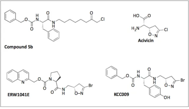
Figure 1.10. Chloromethyl ketone based inhibitor Compound 5b, dhydroisooxazole-based inhibitor KCC009, ERW1041E. Avicidin modified from [170].
A quinolyl carbamate functionalized (S)-proline compound was also developed, based on a TG2 specific gluten derived peptide sequence (PQPQPLY) [176] and found to inhibit small intestine TG2 activity when tested in a mouse model [177]. Michael acceptors are α,β-unsaturated carbonyl derivates that undergo 1,4,-addition reactions. Michael acceptors have been used to inhibit cysteine protease [178], some going for human clinical trials for cancer [179]. Irreversible inhibitors with acrylamide warhead [171] or acrylamide group on the side chain [180] of the dipeptide scaffold were generated and proved to be excellent inhibitors of TG2. Sulfonium inhibitors are bearing a dimethyl sulfonium warhead and reported to inactivate epidermal transglutaminase and TG2 [181]. The design of these inhibitors was based on the familiar Cbz-Phe scaffold, to which electrophilic warhead was attached via a spacer of varied length. A derivate was prepared, bearing an extra carboxyl group for enhanced aqueous solubility [144]. These inhibitors which are predominantly negatively charged at physiological pH (due to the added carboxylate group), are less favored to enter the cell membrane and, therefore rather target extracellular TG2.
Chapter 2. Celiac disease
Celiac disease (CD) is a gluten-sensitive enteropathy that develops in genetically susceptible individuals by exposure to cereal gluten proteins. The first description of CD was found in the first and second centuries where CD was described as an intestinal disorder associated with diarrhea and malabsorption occurring in children and adults. The idea that the disease was linked to food ingestion was brought forward in 1888, confirmed in 1950s when Dicke and colleagues established that consumption of wheat and rye brought CD and removing these grains from the diet resulted in the improvement of the patients condition. In 1954 Paulley [182] reported that the clinical manifestation of CD is linked to the destruction of the lining of small intestine.
It has been revealed that the expression of CD is strictly dependent on dietary exposure to gluten and similar cereal proteins [183]. Gluten is the name of wheat proteins only, but it is increasingly used to describe proteins of wheat, rye and barley that are rich in proline (Pro) and glutamine (Gln) residues. Of gluten proteins, both gliadins (alcohol soluble) and glutenins (alcohol insoluble) are harmful [184]. CD can occur at all ages following the introduction of gluten to the diet. Similarly to most autoimmune disorders, CD is more frequently (twice as often) found in women than in men [185]. CD is primarily the disease of caucasians, affecting 1% of the population [186], most frequently recognized among Europeans. The disease exhibits a very strong HLA association, in which the relative risk to the disease development for carriers of certain alleles is increased 30 fold [187]. Susceptibility is strongly associated with MHC class II molecules HLA-DQ2 and HLA-DQ8 [188, 189] and the immune response directed against specific gluten antigens leads to the destruction of intestinal epithelial cells. CD commonly present in early childhood with classic symptoms including chronic diarrhea, abdominal distension and failure to thrive [190]. The disease may also present later in life with symptoms that tend to be more vague, including anemia, fatigue, weight loss, diarrhea, constipation and neurological symptoms [191]. The celiac lesion is localized in the proximal part of the small intestine. The alterations such as villous atrophy, crypt cell hyperplasia, lymphocytic infiltration of the epithelium and increased density of various leukocytes in the lamina propria characterize one end of a spectrum that has classified into three stages: the infiltrative, hyperplastic and the destructive lesions [170]. The infiltrative lesion is characterized by the infiltration of small non mitotic lymphocytes in the villous epithelium without any signs of mucosal pathology.
The hyperplastic lesion is similar to the infiltrate lesion but in addition has hypertrophic crypts whose epithelium may be infiltrated with lymphocytes. The destructive lesion is synonymous to the classic lesion of CD. Oral challenge experiments with gluten revealed that these stages are dynamically related [192]. Patients with CD typically also develop antibodies against gluten and for the endogenous enzyme tissue transglutaminase. Despite the fact that gluten is the single causative agent, CD can be viewed as an organ-specific autoimmune disease.
2. 1. Antigen presentation in Celiac disease
Both genetic and environmental factors contribute to the development of the disease. CD is a polygenic disorder with involvement of many genes but the HLA locus encoding human major histocompatibility complex (MHC) molecules is far the most important genetic factor contributing about 40% of the genetic variance of the disease [193-196]. The primary HLA association for CD is conferred by class II HLA-DQ genes. Approximately 90% of CD patients express HLA-DQ2.5 molecule encoded by DQA1*05/DQB1*02 genes, and the majority of the remaining patients express HLA-DQ8, encoded by DQA1*03/DQB1*03:02 genes [188, 197]. If genes encoding the α- and the β-chains of the HLA-DQ2.5 heterodimer are carried on the same chromosome (in cis position), they are most often found as part of the highly conserved A1-B8-DR3-DQ2 haplotype. This particular haplotype is associated with several autoimmune diseases such as type 1 diabetes and myasthenia gravis and because of this, sometimes referred to as the “autoimmune haplotype” [187]. Patients that are DR5-DQ7/DR7-DQ2 heterozygous also express the HLA-DQ2.5 molecules, but then the two encoding genes are carried on different chromosomes (in trans position). The relevant DQA1 allele (DQA1*05) is encoded by the DR5- DQ7 haplotype and the relevant DQB1 allele (DQB1*02) is encoded by the DR7-DQ2 haplotype. There have been 39 non-HLA loci associated with CD so far, estimated to contribute about 14% of the genetic variance in the disease [194, 195]. The relative contribution of each of these genes is minor compared to the HLA genes. Many of the non-HLA polymorphisms seem to act by influencing gene expression [195]. Furthermore, many non-HLA CD risk loci are shared with other immune-related diseases, such as type 1 diabetes and rheumatoid arthritis [198-201]. The shared genetic background among these diseases points to common pathogenic pathways in them [202]. In CD different HLA types associated with the disease have been shown to bind proline rich gluten peptides that harbor glutamic acid, however all HLA types have distinct peptide binding preferences (Figure 1.11.).
Figure 1.11. HLA-DQ2.5 and HLA-DQ2.8 present different type of gluten peptides. HLA molecules
bind proline-rich gluten peptides that harbor glutamic acid residues (E). A. HLA-DQ2.5 presents a high risk for CD and bind peptides deamidated at positions P4 and P6. B. HLA-DQ8 low risk variant prefers binding gluten peptides with deamidation at position P1 or P9. [203]
Gluten peptides have preferred binding to DQ2.5 molecules compared to other HLA class II molecules [204]. Peptide binding studies have shown that contact between DQ2.5/DQ8 and the side chains of the peptides, so-called anchor residue and pocket interactions, takes place at the P1, P4, P6, P7, and P9 position [205-208]. Peptides that bind HLA-DQ2.5 preferentially have deamidation at positions P4, P6, and occasionally P7, whereas peptides binding HLA-DQ8 have negatively charged residues at P1and/or P9 [209]. To date, more than 15 different gluten-derived T-cell epitopes have been reported in CD [210-212] and all contain a deamidated residue in at least one of these pocket positions. The great majority of these T-cell epitopes contain glutamine residues that are targeted by TG2 and converted into glutamate residues by a deamidation reaction. In general T cells of celiac disease patients recognize deamidated epitopes with greater efficiency than native ones. This effect of PTM appears to be stronger for DQ2.5 restricted epitopes than for HLA-DQ8 restricted epitopes, as many of the HLA-DQ2.5 restricted epitopes are not recognized in their native form whereas some HLA-DQ8 restricted epitopes are equally well recognized as native peptides [213].
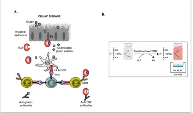
There is a correlation between how frequently T-cell epitopes are recognized by CD patients and their propensity to be targeted as substrates for TG2 [214] (Figure 1.12.). The idea that TG2 is one of the most important factor selecting gluten T-cell epitopes is supported by an experiment where TG2 was used to select peptides from a proteolytic digest of gluten containing several thousand different peptides [215]. Peptides, which were targeted by TG2, were purified and sequenced. Strikingly, of 31 selected peptides, more than 75% contained celiac disease related T-cell epitopes. The negative charges introduced by the TG2-mediated deamidation increase the binding affinity of gluten peptides to HLA-DQ2.5 and HLA-DQ8. Majority of extracellular TG2 is inactive, however it can be transiently activated by certain types of inflammation or injury signals [216].
Figure 1.12. Pathogenesis in celiac disease. A, TG2 mediates the post-translational modifications of gluten
peptides which in turn are presented on the surface of HLA molecules to T cells. In turn, the generation of antibodies start against gluten and TG2. B, TG2 mediated deamidation turns the protein bound, neutral glutamine into negatively charged glutamate which can be presented on the surface of HLA molecules. modified from [217]
The enzymatic activity of TG2 is found to be tightly regulated by the redox potential of the environment, mediated by a redox-sensitive cysteine triad consisting of Cys230, Cys370, and Cys371
[218]. TG2 has been shown to reversibly inactivated by oxidation, however Ca2+ can protect the
In a model of TG2 activation, TG2 is released extracellular matrix upon cell wounding and remains catalytically active for a short period of time but then becomes silenced through oxidation. A change in the reductive environment may rescue TG2 from inactive state as it happens when ongoing immune reactions alter the redox state of lymphoid tissue. The thiol content is increased after antigen stimulation, particularly in mesenteric lymph nodes after intraperitoneal immunization [219]. An ongoing immune response, including those directed against infectious agents, may thus facilitate TG2 activity and an immune response to deamidated gluten. Because of its high Pro content gliadin is remarkably resistant to luminal and brush border proteolysis and large fragments remain intact after digestion. The most illustrative fragment is the 33-mer produced by the digestions of certain α-gliadin proteins. The 33mer fragment remains intact even after extended incubation with gastric, pancreatic, and intestinal brush-border membrane enzymes [220]. It contains six overlapping copies of two different DQ2.5-restricted T-cell epitopes and is recognized by T-cell lines from nearly all adult CD patients. The two epitopes contained within this 33mer peptide, the DQ2. 5-glia-α1 and DQ2.5-glia-α2 epitopes, are often referred to as the dominant T-cell epitopes in CD. Intriguingly, the 33mer peptide can bind DQ2.5 molecules directly on the surface of APCs and can thus be presented to T cells without further intracellular processing [221]. The gliadin-derived epitopes cluster in the Pro-rich regions of gliadin, a property that both confers relative proteolytic resistance and favors TG2-mediated deamidation [210].
The adaptive immune response is initiated by APCs, primarily dendritic cells (DCs) but also macrophages and B cell subsets, which present to T-cell antigenic fragments in complex with cell surface MHC class II molecules. This antigen priming usually takes place in organized lymphoid tissues. APCs travel from peripheral tissues to lymph nodes where they generate effector or tolerogenic T cells that operate in the peripheral tissues. The re-activation of effector and tolerogenic T cells in peripheral tissues will again require interaction with APCs. Antigen presentation is a key initial step during the pathogenesis of CD both in mesenteric lymph nodes and the intestinal lamina propria.
Macrophages are tissue-resident cells that differentiate from circulating monocytes [222, 223]. These cells are long-lived with low turnover in the tissue. They occupy the cellular zone beneath the enterocytes and also can be found scattered in the lamina propria. Human jejunal macrophages have been reported to have a decreased capacity to produce cytokines and do not express receptors typical for innate immune responses.
The role of activated macrophages in CD is elusive, since they were not found to be effective presenters of gluten peptides to gluten reactive T-cell clones [224]. However activated macrophages may play role in the pathogenesis by producing cytokines. In contrast to macrophages DCs are messenger cells with a rapid turnover. Precursors of DCs have not yet been described in the human blood, but three distinct cell subsets may generate tissue DCs: monocytes, classical myeloid DCs, and plasmacytoid DCs.
The two main subgroups of DCs, myeloid and plasmacytoid DCs, are characterized respectively by their abilities to prime naïve T cells and to secrete large amounts of IFN-α in response to viral infections. In the lamina propria of patients with CD, the density of classical myeloid DCs is reduced [225]. Lower tissue density may be due to inflammation-induced expansion of the tissue volume. Another plausible scenario is that genuine cell depletion is due to migration to lymph nodes, diminished recruitment, cell death, or a combination of these events. Human plasmacytoid DCs (PDCs) typically reside in lymphoid tissues and rarely infiltrate inflamed tissues. Plasmacytoid DCs residing in the mesenteric lymph shown to induce mucosal T-cell-independent IgA synthesis through the cytokines APRIL and BAFF [226]. Plasmacytoid DCs probably do not migrate in intestinal or hepatic lymph, distinguishing these cells from myeloid DCs that carry intestinal antigens to mesenteric lymph nodes [227]. Plasmacytoid DCs are producers of IFN-α which has been reported to induce development of CD. This finding supports their role in the pathogenesis of the disease. Intermediate dendritic cells are a subset of antigen presenting cells in the normal duodenum [225]. These cells have an intermediate phenotype and express markers typical for both DCs and macrophages. In mice intermediate DCs appear to originate from monocytes rather than DC precursors and believed to be tissue resident cells playing an important role in modifying local immune responses. Several lines of evidence suggest that intermediate DCs play a direct role in the pathology of CD. Unlike classical DCs and macrophages that show a decrease in the tissue concentration, the density of these cells increases after 3-day gluten challenge of CD patients. Beyond this, the increased density of intermediate DCs proceeds the typical inflammation related changes of the intestine and the surge of intraepithelial lymphocytes and eosinophils, suggesting that they have a role in the initiation of CD [228]. These findings indicate a plausible scenario where myeloid DCs traveling from the tissue to the mesenteric lymph nodes, carry gluten antigens and inflammatory signals for priming the naive T-cells into effector T-cells.
This causes a depletion of myeloid DCs in the lamina propria. In the other hand an influx of blood monocytes that differentiate into intermediate DCs can be observed in the. These cells mediate the reactivation of memory T cells in the intestinal mucosa. B cells likely play a role as antigen presenting cells in the mesenteric lymph nodes for the amplification of gluten T cell response.
2. 2.
T cell response in Celiac Disease
CD4+T cells that recognize gluten peptides can readily be isolated from biopsies of CD patients, but
not from individuals without the disease [229, 230]. These T cells recognize epitopes of gluten presented by disease associated HLA-DQ molecules [231, 232]. The MHC association in CD is linked to the preferential binding by HLA-DQ2 and HLA-DQ8 molecules to the proteolysis resistant gluten peptides that have negatively charged glutamate residues introduced by TG2 [220, 233-235]. Moreover, it has been proposed that gluten peptide-MHC complexes, expressed on the surface of antigen presenting cells are defining the magnitude of the gluten-specific CD4+ T cell
response and consequent induction of intestinal tissue damage [236]. This was based on a finding that susceptibility to CD is higher for those individuals who are homozygous for HLA-DQ2 or
HLA-DQ8 alleles [237]. Unlike in healthy individuals, CD4+T cell responses to dietary gluten in
the small intestinal mucosa can be observed for CD patients [230] (Figure 1.13). In CD an alteration in the intestinal environment can be observed which affects the differentiation/function of forkhead box P3 FOXP3+ T
Reg cells. FOXP3 cells are responsible for oral tolerance [238] by secretion of
anti-inflammatory cytokines such as transforming growth factor-β (TGFβ), IL-10 and IL-4 and promote the production of IgA antibodies [239].
Figure 1.13. In the pathogenesis of CD gluten-specific CD4+ T helper 1 (TH1) cells secrete
pro-inflammatory mediators such as interferon-γ (IFNγ) or interleukin-21 (IL-21), which promote activation of intraepithelial cytotoxic T lymphocytes (CTLs) and block the inhibitory effects of forkhead box P3 (FOXP3)+ T Reg cells. In addition, gluten-specific TH1 cells help B cells to produce gluten- and TG2-specific
IgG and IgA antibodies [240].
The intestinal mucosa in CD is characterized by presence of high levels of pro-inflammatory cytokines such as IL-15 [241-243] and IFNα [244-246]. It has been observed that in HLA-DQ8-transgenic mice IL-15 is able to alter the phenotype of intestinal DCs and prevent the activation of FOXP3+Treg cells after oral challenge with gluten. Based on these observations it has been proposed
that intestinal DCs when stimulated by pro-inflammatory factors as IL-15 or IFNα might loose their tolerogenic phenotype, promoting the differentiation of pro-inflammatory T cells. Beyond this effect IL-15 was shown to prevent the inhibitory effect of TGFβ [247] supporting that effector T cells in the gut mucosa of CD patients might be insensitive to the regulatory effect of TGFβ and FOXP3+ T
Reg cells. CD4+ T cells have been implicated to help to set up the inflammatory
environment which allows the intraepithelial cytotoxic T lymphocytes (CTL) to induce tissue damage. By the secretion of pro-inflammatory cytokines such as IFNγand IL-21, which may promote epithelial cell destruction by intraepithelial CTL activation. After activation by IL-15, intraepithelial CTLs were reported to expand early in the disease process [248], leading to the destruction of intestinal epithelial cells and villous atrophy [249, 250]. In humans CTLs has been shown to express the activating receptor NKG2D [251].
![Figure 2.1. Schematic representation of the cloning vector (A) modified from[393] and expressed scFv-Fc](https://thumb-eu.123doks.com/thumbv2/123dokorg/4815152.50083/66.892.79.815.99.359/figure-schematic-representation-cloning-vector-modified-expressed-scfv.webp)