3
’
’
Non c e niente che sia per sempre
il tuo diploma in fallimento
’
e una laurea per reagire
è
2
SUMMARY
INTRODUTION4HUMAN PAPILLOMA VIRUS 4
Characteristics 4
HPV infection and Life Cycle 9
Natural history of HPV infection 10
Epidemiology 13
Immunopathogenesis of HPV infection……….………... 14
HPV Strategies for Evading Host Immune Response ………. 16
HPV and Cervical Cancer ……….17
CHLAMYDIA TRACHOMATIS 19 Characteristics ………19
CT infection and Cell Cycle……… 21
Epidemiology……… 23
Immunopathogenesis of CT infection ………...24
CT Strategies for Evading Host Immune Response………. 26
CT Persistence and Chronic Infection……… 28
CT and Apoptosis ……….. 30
Interaction between CT and HPV……… 32
OBJECTIVES OF STUDY 34
MATERIALS AND METHODS 35
Clinical Specimens ……….. 35
Isolation of DNA from cervical cells ……….. 35
HPV detection and characterization ………... 36
CT detection ………. 36
CT HSp60 Real Time PCR detection……… 37
CT genotyping ………. 37
Statistical Analysis ………. 38
RESULTS 39
HPV infection ……… 39
CT infection ……… 39
CT and HPV co-infection and HPV genotypes distribution ……… 40
HPV genotypes distribution in single and multiple infections from CT/HPV and HPV women……….40
3
CT Hsp60 chronic infection and HPV infections ……… 41
DISCUSSION 47
CONCLUSIONS 51
REFERENCES 52
4
INTRODUCTION
In the worldwide, sexual transmitted infections (STIs) represent an important public health problem with an estimated million of new cases of infections occurring each year. Young people account for only 25% of the sexually active population which accounts for almost 50% of newly acquired STIs (Siracusano et al., 2014). These consist in bacterial, fungal and protozoal infections that can be treated with appropriate chemotherapeutic agents or, as reported, can trigger viruses often leading incurable STIs (Gewirtzman et al., 2011). Microorganisms causing chronic inflammatory diseases have become to be increasingly investigated in the last decade as possible cancer initiators/promoters (Idahl et al., 2011). So far, the most common STIs are those caused by Human papillomavirus (HPV) and
Chlamydia trachomatis (CT). However, epidemiological data on CT prevalence and
CT/HPV co-infection are not yet well defined in Italy (Bellaminutti et al. 2014, Panatto et al 2014; Seraceni et al. 2014).
HUMAN PAPILLOMAVIRUS
Characteristics
Papillomavirus belong to the Papovaviridae family, which includes 16 different genera, five in humans (Alphapapillomavirus, Betapapillomavirus, Gammapapillomavirus,
Mupapillomavirus, Nupapillomavirus), that have different types, life-cycle characteristics
and disease associations, based on DNA sequence analysis. Of these, The Alpha genus contains the mucosal type viruses that cause genital warts or lesions associated with the development of cervical neoplasia and cancer at low-risk (LR-HPV) and high risk (HR-HPV), respectively. The Beta genus, instead, contains those genotypes that are associated with the development of cutaneous cancers. Their possible role in cancer progression in the general population is currently unresolved (Bernard et al., 2013) (Figure 1).
5
Fig. 1: Evolutionary Relationship between Human Papillomaviruses.
Human Papillomaviruses comprise five evolutionary groups with different epithelial tropisms and disease associations. (Modified and adapted from de Villers et al. 2004). http://www.bioafrica.net/rega-genotype/html/subtypeprocesshpv.html
These viruses contain a double strand DNA with 8000 pb approximately arranged in an 8 well defined genes. All members of the HPV family have a typical genomic organization with 8 or 9 open reading frames (ORFs) on the same DNA strand. The HPV genome is divided into three regions: six early genes (E) are involved in virus expression, replication and survival and two other genes called late genes (L), are involved in virus assembly; finally, the long control region (LCR), which is localized between ORFs L1 and E6, contains most of the regulatory elements involved in viral DNA replication and transcription. The designations E and L refer to the phase in the viral life cycle when these proteins are first expressed (Figure 2). E2 regulates early gene promoter and together E1 forms a heterodimer complex to control virus DNA replication. E4 may mediate the release of viral particles by destabilization of the cytokeratin network, whereas E5 stimulates mitogenic signals of growth factors. E6 and E7 are oncoproteines capable of deregulate
Cutaneous Benign lesions. Some types
detected at oral sites and
rarely found in skin cancers. Cutaneous Includes LR and HR-HPV. In immunocompetent unapparent infections. Persistent infection can predispose to skin cancer Mucosal and cutaneous
Include HR-HPV associated to cervical cancer and LR-HPV associated benign lesions
cutaneous Cutaneous
6
fundamental cellular events, such as cell cycle, DNA repair, senescence, apoptosis and differentiation, facilitating the accumulation of DNA damage and the progression towards malignancy. If the immune system can eliminate quickly the HPV-infected cells, E6 and E7 do not manage to accumulate chromosomal abnormalities and acquire a malignant phenotype, despite their transforming properties. Therefore, the establishment of a chronic infection is a fundamental and crucial event for the development of HPV-associated malignant diseases. Host and environmental factors significantly contribute to the chronicity of HPV infection, despite HR-HPV E6 and E7 target cellular pathways related to innate and adaptive immunity. L1 and L2 are major and minor capsid protein, respectively and L1 is the component of the HPV prophylactic vaccine.
HPV infects the epithelium of the cervix and their replication is closely linked to the differentiation of the epithelium (Doorbar et al., 2012; Tommasino, 2014).
Fig. 2: The genome organization of HPV16 is typical of the high-risk Alphapapillomaviruses (Agnes and Gunnar, 2008).http://.medscape.com.
7
These virus, of which have been identified more than 150 HPV genotypes, have selective tropism for cutaneous or mucosal epithelia and are divided into two groups: LR-HPV and HR-HPV, as previously described. LR-HPV that cause benign lesions asymptomatic that may resolve spontaneously in 3-4 months, are named papillomas (small wart-like neoplasias), which are due to sexual transmission of the virus, occur in male and female genitals, urethra, anus and perianal area and rarely lead to cancer. HPV6 and HPV11, two among the most common LR-HPV, are associated with 90% of genital warts and recurrent respiratory papillomas. HR-HPV cause malignant cellular transformation and develop into large tumors, characterized by squamous intraepithelial lesion (SIL) that occur with rounded cells with nuclear and perinuclear (koilocytosis) atypia. Nuclear abnormalities, such as enlarged nuclei, hyperchromasia and mitotic cell features can also be found.
HPV infections are also common in intraepithelial cervical neoplasia (CIN); these are characterized by the presence of koilocytosis and are divided into low-grade (LSIL) and high-grade (HSIL). According to the histological classification there are three degrees of CIN: CIN 1 (mild), corresponds to LSIL, CIN2 and CIN3 correspond to moderate and severe HSIL, respectively. HPV infection is the major cause of the CIN development. Despite women’s frequent exposure to HPV, the development of cervical cancer (CC) is relatively rare. Most low-grade cervical abnormalities, such as CIN1, are associated with benign viral replication, and spontaneously regress without requiring treatment (Martin et al., 2011). Studies in women have shown CIN1 regression rates of up to 70–80%; however, in adolescents and young women under 25 years, more than 90% show regression (Cox et al., 2003; Moscicki et al., 2004; 2010). In contrast, HSIL, specifically CIN3, has a much greater potential to progress to invasive cancer (progression rates of between 0.2–4% within 12 months) (Fearly et al., 2010). HR-HPV genotypes identified as causing CC belong to groups based on epidemiologic and mechanistic evidence of their carcinogenicity (Rosales and Rosales, 2014). Twelve HPV genotypes (HPV16,-18,-31,-33,-35,-39,-45,-51,-52,-56,-58 and-59) are classified as “carcinogenic to humans” (Group 1), HPV68 as “probably carcinogenic” (Group 2A) whilst others seven HPV genotypes, as “possibly carcinogenic” HPV26,-53,-66,-67,-70,-73,-82 (Group 2B) (IARC, 2012) (Figure 3).
8
Fig. 3: The HR Alpha types have been clearly linked with the development of squamous cell carcinoma
(SCC) and adenocarcinoma (AC) of the cervix. IARC category 1 and 2A HPV genotypes are classified (respectively) as carcinogenic and possibly-carcinogenic. Despite limited epidemiological data, the 2B classification is proposed for genotypes that are probably carcinogenic because of their close phylogenetic relationship with the established carcinogenic types. HPV genotypes in category 3 are considered non-carcinogenic (Doorbar et al., 2012).
9 HPV infection and Life Cycle
So far, most of the biological studies concerning the clear association of mucosal HR-HPV genotypes with human carcinogenesis (Zur Hausen, 2002), have been focused on these types. HPV infects the cells of the basal layer, where it is present at a relatively low copy number. The HR-HPV life cycle is tightly linked to the differentiation programme of stratified epithelia. HPV initiates the productive phase of its life cycle, that is characterized by vegetative viral DNA replication, when cells leave the basal layer of the epithelium. During this phase, the HPV genome is amplified to more than 1000 copies per cell and subsequently, the expression of late genes starts and viral particles are produced and released. In contrast to mucosal HPV types, nothing is known about the life cycle of the majority of HPV types that belong to the Beta and Gamma genera. Studies of mucosal HPV types have shown that the first step in HPV infection is the interaction of the viral capsid with the cytoplasmic membrane of cells at the basal layer of the epithelium (Tommasino, 2013). This event is mainly mediated by the major capsid protein, L1, which interacts with the cell surface via heparan sulfonated proteoglycan (HSPG) (Doobar et al., 2012). It is also possible that the viral particles bind to another component of the cellular membrane, the integrin α6, proposed as a secondary cellular receptor for HPV particles (Evander et al., 1997) even if, the precise nature of the entry receptor remains somewhat controversial (Doobar et a., 2012; Tommasino, 2013).
The internalization of HPV16 particles, after binding to the cellular membrane, is mediated by a clathrin-dependent endocytic pathway (Day et al., 2003). Additional findings indicate that other mucosal HPV types may use different endocytosis pathways (Bousarghin et al.,2003). It is also highly likely that the minor capsid protein, L2, plays a role in membrane binding and cellular internalization. In fact, in vitro assays anti-L2antibodies against specific linear epitopes are able to block the internalization of L1/L2 virus-like particles (Kawana et al., 2001; Gambhira et al., 2007) and the annexin A2 hetero tetramer contributes to HPV16 infection in an L2-dependent manner (Woodham et al., 2012). Recently, Surviladze and colleagues have presented evidence of a novel mechanism of viral entry, observing that HPV16 particles, after binding to the cell surface, are released as a soluble complex with HSPGs and growth factors. The growth factors mediate the interaction of the soluble complex with their cognate receptors, facilitating the internalization of the viral particles (Surviladze et al., 2012).
10 Natural history of HPV infection
The HPV lifecycle is closely linked to stratified epithelium differentiation (Pyeon et al., 2009). HPV virions infect the basal epithelium through micro-abrasions in the epidermis. The modality of virus invasion is still not fully understood, but several receptors, including heparan sulphate proteoglycans and alpha-6 integrin, have been associated with this process (Doobar, 2006). Upon migration to the basal cell nucleus, the viral genomes are established as episomes, the early promoter are activated and finally resulting in low levels of viral synthesis. During normal epithelium differentiation, the daughter cells migrate from the basal layer upwards and undergo terminal differentiation. Viruses finally reach the epithelial surface where they form a cornified layer of dead cells, which are eventually eliminated (Figure 4).
Fig 4: The location in the squamous epithelium of the main stages of the papillomavirus life cycle. Cervical
stratified squamous epithelial cell architecture and the expression of HPV proteins after infection. Daughter cells of epithelial stem cells divide along the basement membrane and then mature vertically through the epithelium without further division (right side). (Muñoz et al, 2006) http://www.nature.com/nri/journal/v4/n1/fig_tab/nri1260_F2.html .
11
In HPV-infected differentiating cells, the late promoter is activated, leading to the vegetative state of the HPV lifecycle (Longworth et al., 2004). During this phase, high levels of viral DNA are replicated, enveloped into capsids and released from the cell. To maintain viral synthesis in the epithelium, the virus takes advantage of the host cell replication system. Consequently, the HPV oncoproteins E6 and E7 come into play, maintaining the cell cycle and preventing terminal differentiation. The HPV infected cells move up through the epithelium and the viral infected basal cell layer is maintained with a low level of viral DNA synthesis. This typically occurs in LSIL disease. HSIL lesions, such as CIN3, are typically associated with HPV DNA that has integrated into the host genome. Viral integration often occurs in the E1 and E2 regions downstream of the late genes. This can result in disruption and loss of these late genes, with subsequent loss of control of oncogene expression by the E2 viral gene (Woodman et al., 2007). To maintain the HPV infection, HR-HPV genotypes produce E6 and E7 oncogenes, which interfere with critical cell–cycle checkpoint pathways and proteins, namely p53 and retinoblastoma (Martin et al., 2011) (Fig. 5).
12
Fig. 5: A: The cervical squamocolumnar junction. The basal cells rest on the basement membrane, which is supported by the dermis. Normal squamous epithelium differentiates as shown. The transformation zone is the most common site for the development of CC. Prophylactic vaccines induce L1- or L2-specific antibodies that neutralize the virus. B: After the HPV infection of basal keratinocytes, the early HPV genes E1, E2, E5, E6 and E7 are expressed (red nuclei) and the viral DNA replicates. LSILs support productive viral replication. In the upper layers of epithelium the viral genome is replicated further, and E4 (green cytoplasm), L1 and L2 (orange nuclei) are expressed. L1 and L2 encapsidate the viral genomes to form progeny virions in the nucleus. The shed virus then re-initiates infection. C: A significant fraction of HR-HPV infections progress to HSILs, which show a lesser degree of differentiation. HSILs are effectively treated by loop electrosurgical excision (LEEP). Pap screening and HPV tests can be used to detect SILs.
D:The progression of untreated lesions to micro invasive and frankly invasive cancer is associated with the
integration of the HPV genome into the host chromosomes, loss of E2 and up-regulation of viral oncogene expression and genomic instability. These cancers are treated with surgery, chemotherapy or radiotherapy with limited success. Therapeutic vaccines and immune stimulants such as imiquimod can potentially induce an infiltration of T cells specific for the early viral antigens and clearance. (Roden and Wu, 2006).
13 Epidemiology
The worldwide prevalence of HPV infection in women with normal cytology is around 11– 12%, with the majority prevalence in sub- Saharan Africa (24%), Eastern Europe (21%) and Latin America (16%) (Bruni et al, 2010). In women less than 25y, HPV prevalence has been observed highest, decreasing average with progression of age in many populations, some of which have a secondary peak in peri-menopausal or early menopausal time (Denis et al., 2008; Ali et al., 2013; Melo et al., 2014; Cuzicke et al., 2014). In China, the prevalence is instead relatively age independent. The explanation of these difference prevalence pattern and the clinical significance is not understood. Globally, the five most prevalent types are HPV16 (3.2%), HPV18 (1.4%), HPV52 (0.9%), HPV31 (0.8%) and HPV58 (0.7%) (Bruni et al, 2010). Prevalence increases in women with cytologic cervical pathology in direct proportion to the severity of the lesion, reaching around 90% in women with CIN3 and invasive CC. In fact, 100% of all CC has been found to be HPV positive. Of note, the proportion of HPV positive women in whom HPV16 is greatly detected, increases with lesion severity (Bosch et al., 2013).
Infection with HR-HPV types is recognized as one of the major causes of infection-related cancer worldwide. A strong evidence for a causal etiology with HPV has been stated by the IARC for cancers of the cervix uteri, penis, vulva, vagina, anus and oropharynx (including base of the tongue and tonsils), which has estimated the overall number of cancers attributed to HPV and classified by geographic region.
The prevalence of HPV infection with specific genotypes differ by age and area. In 2008, there were estimated in the world 12.7 million new cancers, of which 700,000 with an HPV-associated cancer site. 610,000 of these were attributable to HPV only (Ferlay et al., 2010) which alone represents 4.8% of the total burden of cancer worldwide that varies widely by geographic region, ranging from 1.2% in Australia and New Zealand to 14.2% in sub-Saharan Africa and 15.5% in India.
Of note, 80.6% of the total number of cases attributable to HPV occurred in underdeveloped countries (6.9%) compared with 2.1% in more developed countries. In the world, CC resulted the third most common female cancer, (approximately 86% of these cases occurred in underdeveloped country), with a strong association between CC incidence and level of development.
In underdeveloped countries, the incidence and mortality rates tend to be higher compared to developed and after 5-year relative survival, in these different conditions is observed a similar pattern 20% vs 65%, respectively. Global maps of CC rates show patterns of
14
variation largely consistent with level of develop. According to a meta-analysis regarding the prevalence of HPV genotypes in women with normal cytology, HR-HPV16, although high and variable across world regions, resulted the most prevalent genotype (22.5%). Others HPV genotypes highly prevalent resulted, HR-HPV18,-52-,31-58,-39,-51,-56 and LR-HPV6 (Bruni et al., 2010).
Italy, mirrors the world trend, with a strong variability of HPV prevalence by age and area (Agarossi et al., 2009; Giorgi Rossi et al., 2010, Giuffrè et al., 2010; Bellaminutti et al., 2014; Carrozzi et al., 2014). HPV16 resulted the most common genotype in our country (Giorgi et al., 2011; Bianchi et al., 2013; Carozzi et al., 2014).
Immunopathogenesis of HPV
The most frequent HPV infections recover spontaneously, within two years from infection and without any clinical manifestation by immune-competent individuals; consequently the immune system is able to effectively eliminate virus-infected cells. However, the natural immune response may be insufficient, not allowing the infection resolution. The virus does not cause viremia, or systemic infection. The virus absence in the blood and its characteristic intracellular replication and not accompanied by cell lysis, involves the inability of the immune system to mount a strong antibody response or to induce inflammation (Rosales and Rosales, 2014).
The initial inflammatory response induced by tissue damage leads to infiltration of immune cells mainly neutrophils, followed by macrophages and later lymphocytes, that recognize “danger” viral molecules detected by pattern recognition receptor (PRR), such as Toll like receptors (TLR) (Kawai and Akira, 2011). The immune response innate induces the lysis of the infected cells and the production of cytokines pro-inflammatory such as 1β, IL-6,IL-8, IL-12 and INF-α,-β,-γ, to activate natural killer cells (NK) and other immune cells (Woodworth, 2002).
T cells, activated by recognition of viral proteins, induce the growth and maturation of B cells. These cells, together with the cell-mediated response, are necessary to ensure an effective protection against the virus. The induction of neutralizing antibodies specific for L1 and L2 proteins viral capsid, is critical to prevent the onset of symptoms and the
entrance of the virus into cell.
Infiltrations of CD4+ (Helper) and CD8+ (Cytotoxic) T cells that are absent in persistent lesions, has been detected in spontaneously regressing HPV-related lesions, indicating that the adaptive immune response against the virus is important and for the most cases
15
effective. This adaptive response comprises elements of humoral and cellular immunity (Stanley, 2006).
The humoral immune response, in the majority of patients with HPV infections, is characterized, in the first stage of infection, by antibodies against different HPV proteins such as L1, E2, and E4 proteins. Antibodies against E6 and E7 oncoproteins can be detected in low- and high-grade lesions, when viral DNA gets integrated into cellular genome. However, this antibody response is usually weak and variable. It does not seem to protect from future re-infections (Tjiong et al., 2001) and the seropositivity seems depending on the site of the cancer around the anogenital area, indicating that cancer development may lead to changes in antibody responses in a site-specific fashion (Carter, 2001). Thus, humoral responses are not enough efficient to eliminate established HPV lesions. In addition, antibody titers can persist for many years even after the virus is cleared, so seropositivity is a useful marker for past infection rather than current infection. Cell-mediated immune responses are more important in clearing HPV-related lesions. T cells HPV-specific are generated to fight and eliminate infected cells; thus both, CD4+ and CD8+ T cells and also responses that include HPV specific regulatory T cells (Treg) that inhibit cytotoxic activity (Welters et al., 2003), are important for elimination of HPV infection. In fact, T cell responses to viral proteins are present in patients who successfully eliminated previous HPV16 infections. Moreover, T cells, with a predominance of Th1 cytokines, were observed in high ratio during regressing lesions. In contrast, in patients presenting CIN or CC, a deficient T cell response with a strong shift to Th-2 cytokine profile, are observed in persistent lesions. Thus, an efficient cytotoxic cell-mediated immune response is critical for elimination of HPV-related lesions. Unfortunately the virus has also evolved mechanisms to interfere with the immune response (Rosales and Rosales, 2014).
16
HPV Strategies for Evading Host Immune Response
HPV has evolved several mechanisms to evade the immune system. In the areas where HPV replication takes place, the immune surveillance is poor. In the stratified squamous epithelium of the uterine cervix, surveillance by dendritic cells (DCs) greatly declines towards the keratinized layers.
The keratinocytes infected by HPV express a low load of viral proteins and thus, do not induce their lysis. Expression of viral gene products up-regulates progressively with differentiation and upward migration of keratinocytes Moreover, HPV down-regulates, by E6 and E7 oncoproteins, the expression of major histocompatibility complex (MHC) class I molecules, TLR9, and cytokines such as, interferon and interleukin (IL-8) (Stanley, 2006).
In this way, HPV late proteins, which are the most immunogenic, are expressed at areas of poor immune surveillance (Figure 5). In addition, new virions are released through the normal rupture of surface epithelium, reducing any possible inflammatory response and avoiding uptake by DCs. Therefore, HPV replication is a local phenomenon with minimal activation of the immune system. Moreover, a reduced inflammation state is found in persistent lesions and in tumors. This condition correlates with a change in the cytokine profile produced at the site of infection.
A shift to Th2 cytokines is also common in persistent lesions (Bais et al., 2007; Rosenthal et al., 2012). This leads to an inhibitory state for helper CD4+ T cells. In addition, Tregs
have been found infiltrating tumors, especially in the early stage of tumor progression (Piersma et al., 2008). Thus, any therapeutic approach must be able to induce a strong HPV-specific immune cell response that involves CD4+, CD8+ cells, and Th1 type
cytokines.
However, HPV has also evolved mechanisms to avoid both initial detection and to interfere with adaptive response, that allows the virus to persist and lesions to progress into cancer (Figure 6).
17
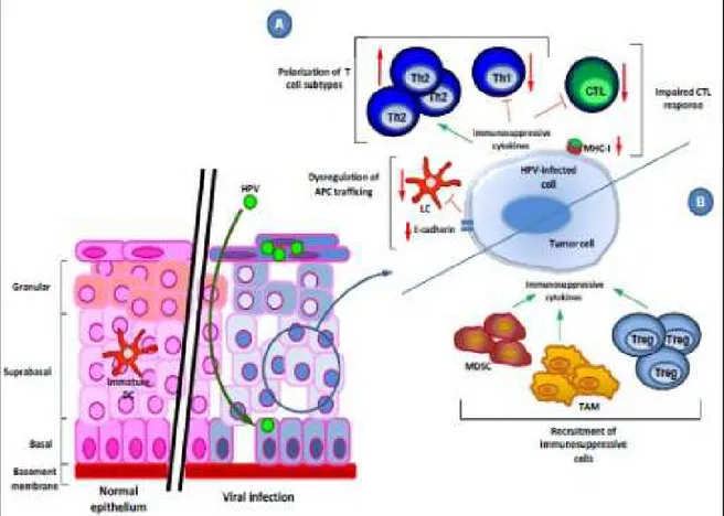
Fig. 6: HPV-mediated effects on the host immune response.
A: Immune evasion mechanisms employed by a HPV-infected cell are polarization of T cell subtypes, inhibition of the CTL (C8+) response and modulation of APC trafficking.
B: Immune evasion mechanisms of HPV-driven malignantly transformed cells include recruitment of immunosuppressive cells, leading to immunosuppressive cytokine production. (Grabowska et a., 2012). http://openi.nlm.nih.gov/
HPV infection and Cervical Cancer
HPV is a sexually transmitted agent deemed a cause of CIN and invasive CC in worldwide. Currently it is widely accepted that specific genotypes of HPV are potentially oncogenic and are associated with virtually all cases of CC and, to a lesser extent, with cancers of the vagina, vulva, anus, penis, skin and oropharynx (Trottier et al., 2006; Parkin et al., 2006; Shukla et al., 2009; Moody et al., 2010).
Over than 100 HPV types classified into HR-HPV are linked to both to tumor precursor lesions and to the progression of invasive CC (Simonetti et al., 2009), the second most common cancer among women (Arbyn et al., 2011) and the seventh in the world (Forman et al.,2012). Persistent infection with one of the oncogenic HPV genotypes is required to cause CC (Walboomers et al. 1999; Bosch et al., 2002).
18
HPV must be persistent within the host epithelial cells as a preliminary step toward advanced neoplastic changes. This process takes years, if not decades, to occur after initial HPV infection. Persistent infection, with HPV genotypes with high oncogenic potential, increases the probability of ICC following an extended period of latency. The integrity of the cell-mediated immune response against HPV is an important factor for healing and prevention of the reactivation of the latent infection (Fernandes et al., 2013; Stanley, 2006).
Recent studies seem to suggest that these changes may develop more quickly than previously thought. Winer et al., followed women after initial HPV infection for the development of CIN 2/3 and approximately 27% of women with an initial HPV16 or 18 infection progressed to CIN 2/3 within 36 months (Winer et al., 2005). A second study, performed on a large health maintenance cohort, found that approximately 20% of women 30y or older, who were initially infected with HPV16, developed CIN3 or CC within 120 months, while women who had an initial HPV18 infection, had approximately a 15% risk (Khan et al., 2005).
The strong correlation between infection with HR-HPV and LSIL, HSIL, and CC suggests that HPV DNA testing would be an useful tool for the management of women with abnormal Pap test results, especially in the case of those with equivocal test results. In the case of an equivocal Pap test result, HPV DNA testing can help determine whether the individual should be referred for colposcopic assessment (Cox et a., 2003).
Accumulating evidence suggests that a combination of screening strategies is needed to detect as early as possible HPV induced lesions CC.
Epidemiological studies in HPV infected females have provided important clues about a spectrum of cofactors that can increase the carcinogenic HPV potential. Presumably, cervical infections with other pathogens, exposure to physical and chemical agents, hormonal factors and Chlamydia trachomatis (CT) itself, as previously demonstrated (Smith et al, 2002; de Abreu et al., 2012). Notably, the infections caused by these two pathogens are often associated with an intense chronic inflammatory response and ulcerations in the cervical epithelium (Scott et al., 1999; Adefuye and Sales, 2012, Silva et al., 2014).
19 CHLAMYDIA TRACHOMATIS
Characteristics
The Chlamydiaceae are a family of ubiquitous gram-negative, aerobic, obligate intracellular bacteria present in the environment including aquatic.
Once considered viruses, they grow in eukaryotic cells and are responsible of a wide range of diseases in human and animals. A recent revision has taxonomically re-classified the group in 4 distinct families (Chlamydiaceae, Simkaniaceae, Parachlamydiaceae and
Waddliaceae) based on > 90% 16S rRNA identity and a single genus which include the
different species. C. pneumoniae (CP) and C. trachomatis (CT) are common pathogens in humans, but the routes of transmission, susceptible populations and clinical presentations, differ markedly, although have a common developmental cycle (Contini and Seraceni, 2012). Currently, there have been described 11 species of Chlamydia. Most are able to infect several host species and anatomical sites. CT, the most medically significant chlamydial species, is a human-specific microorganism capable of produce pulmonary, ocular and genital pathologies in either neonates or adults (Bachmann et al., 2014) (Figure 7).
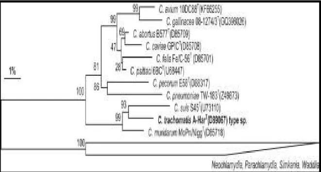
Fig. 7: Phylogenetic reconstruction based on almost complete 16S rRNA genes from type strains of established Chlamydiaceae spp., including the recently proposed new species C. avium and C. gallinacea (Sachse et al. Systematic and Applied Microbiology, 2015).
20
Ocular infections caused by CT are the leading cause of preventable blindness (trachoma) (Lu et al., 2013) or lymphomas (Contini et al., 2009; Contini et al., 2013) whereas genital infections are known to have adverse effects on female reproduction (Shao et al., 2013). In women, due to the high frequency of the asymptomatic phase, CT can lead to pelvic inflammatory disease (PID), infertility, ectopic pregnancy and chronic pelvic pain. (Haggerty et al., 2010). Infections by CT are also known to facilitate rheumatic disease (Gerard et al., 2010; Carter et al., 2012; Zeidler and Hudson, 2014).
CP is the other human chlamydial pathogen prevalently associated to respiratory infections as community-acquired pneumonia worldwide (10–20%) and asthma (Blasi et al., 2004; Stocks et al., 2004; Hahn et al. 2012; Asner et al., 2014). Moreover, evidence seems to suggest that CP is also linked to cardiovascular, rheumatic and neurologic disease (Witkiewicz et al., 2005; Fernandez et al., 2005; Contini et al.; 2010; Contini et al., 2011). The least five other Chlamydia species have a broad host-range in animals. Chlamydia
psittaci is primarily a pathogen of avian species that causes respiratory diseases (interstitial
pneumonia transmitted by birds) which usually occur through inhalation of the organism when it is dispersed in the air as fine droplets (aerosol) or dust particles (Moroney et al., 1998; Van Droogenbroeck et al., 2009).
CT have extremely small circular genome (1042 kbp), which contains also a cryptic plasmid (length 7500 bp) linked to virulence that can contribute, to the regulation of chlamydial chromosomal gene expression, by its transcriptional activity (Carlson et al., 2008). A detection of cryptic plasmid’s nucleic acid, is utilized for diagnostic purposes (Ljubin-Sternak and Meštrović, 2014) and usually can detect all variants discovered such as Sweden mutation (Paavonen, 2012).
CT has tropism for conjunctival and genitals mucous membranes, where can lead to diseases with chronic inflammation.
According to different immunoreactivity, 19 human serotypes and related variants (A, B/Ba, C, D/Da, E, F, G, Ga, H, I/Ia, J, K, L1, L2, L2a and L3) have been identified by using mono or polyclonal antibodies directed against epitopes of the major outer protein membrane (MOMP). These serotypes are closely related to the genotype, which is based on the ompA gene (encoding the protein MOMP) (Wang et al., 1985; 1991; Hsu et al., 2006) and can be divided into three serogroups: the B group (serotypes B, Ba, D, Da, E, L1, L2 and L2a); the intermediate (I) group (serotypes F, G and Ga); and the C group (serotypes I, Ia, J, K, C, A, H and L3) (Bax et al., 2013). In contrast with the individual serotypes, that possess a certain correlation with the disease and the affected tissues, the serogroups do not correlate with either tissue tropism or the biological properties of the
21
organism. Therefore, based on the pathogenic potential, serotypes A, B, Ba and C are commonly associated with the development of trachoma, chronic eye condition, while D to K serotypes, are responsible of urogenital infections as well as neonatal conjunctivitis and pneumonia. The serotypes of lymphogranuloma venereum (LGV), L1 to L3 and L2a, are responsible of most invasive urogenital diseases (Dean, 1997; Gomes et al., 2006).
Additionally, it seems that the infections with CT serovars G, I and D are associated with cervical squamous cell carcinoma and chronic infections with serotype K, in women, have been recognized as a cause of infertility (Marrazzo & Stamm, 1998; Koskela et al., 2000; Morre et al., 2000; Anttila et al., 2001). The CT serotypes most commonly isolated from patients are: E (50%), F (20%) and D (10%). According to recent findings, F serotype seems be responsible of more severe infections, whilst E of asymptomatic infections (Choroszy-Król et al, 2012).
CT infection and Cell Cycle
The intracellular growth cycle of the Chlamydiae is complex and several growth options are possible, depending on the host-cell type, the particular environmental conditions in the host cell and the nature of tissue that is being affected.
Chlamydiae, have a characteristic biphasic growth cycle within a eukaryotic host cell,
during which infectious, elementary bodies (EBs, 0.3–0.6 mm diameter) differentiate into the metabolically active but non infective reticulate bodies (RBs, 0.6–1 mm diameter), that divide by binary fission within the host, derived vacuoles named Chlamydial inclusions. After 48–72 h, RBs multiply by binary fission and reorganize into EB, which are released after host cell lysis. In vitro, this orderly alternation between EB and RB in life cycle development usually take place in 72 h, ranging from 36 to 96 h to complete, depending on each species and in the number of inclusions per host cell (from one in CT infected cell, to several inclusions for the others Chlamydiae). Under in vitro conditions with adverse factors, e.g., penicillins or INF-γ, RBs block division and maintain a stable association with the infected cell and become the aberrant or persistent bodies with enlarged forms, altered gene expression profile and multiple nucleoids, instead of undergoing rapid replication and differentiating into infectious EBs (Figure 8) (Contini and Seraceni, 2012). Although the life cycle of Chlamydiae is well characterized by microscopy, the signals that trigger interconversion of the morphologically distinct forms are not completely known (Beatty et al., 1994; Dautry-Varsat et al., 2005). However, EBs are no longer considered as inert organisms. The discovery that EBs can translocate stored proteins into the host under
22
distinct signaling pathways is further evidence that the entry process results from a dialogue between the bacteria and the host, although many features including EB protein attachment to target cells, remain to be clarified or discovered (Dautry-Varsat et al., 2005; Wuppermann et al., 2008). During chlamydial cell cycle, a stop in development may lead chronic infection, characterized by high transcriptional activity with aberrant bodies formation (Gerard et al., 2013). These events, reversible, constitute the basis of clinical persistence leading to chronic sequelae.
Moreover, aberrant forms of RBs, with reduced MOMP and lipopolysaccharide (LPS) antigens, persist with high production of chlamydial heat shock protein 60 (Hsp60) can induce inflammation and scarring, classic characteristics of chronic infection (Malhotra et al., 2013). The virulence of these microorganisms is principally due to components of the outer membrane, inclusion proteins and polymorphic membrane proteins, (pmp) related to the third type secretion system (TTS), to different secretory proteins (e.g. glycosyltransferase), chromosomally encoded and especially to extrachromosomal factors, specifically plasmids (Pawlikowska-Warych et al., 2015), capable to define the outcome of infection and disease severity. Multiple types of genetic variation are found in CT that impact variability and expression of virulence factors, such as high degree of variability in the exposed portions of MOMP, polymorphic TTS effectors, and amino acid substitutions in pmp autotransporters (Abdelsamed et al., 2103). These strategies have been demonstrated to foster chlamydial intracellular survival, aid in the evasion of the host immune system, and form the basis for distinct chlamydial disease variations in host tissue tropism (Byrne, 2010). Host genetics also play a role in the disease severity. For example, women who carry specific HLA DQ and IL-10 promoter alleles that modify host immune response were found to develop TFI more frequent than control group (Kinnunen et al., 2002).
23
Fig. 8: Chlamydia life cycle with main morphological and metabolic features of normal productive vs.
persistent CT infection.
A: schematic description of persistent life Chlamydia arrested at partially known state of the normal life cycle.
B: the morphologies, the results of culture-based detection of Chlamydia, their metabolic state, their gene expression profiles, and energy supply of Chlamydia during productive infection and in a persistent state. + indicates detection; −indicates lack of the corresponding messenger RNA (Zeidler and Hudson, 2013)
Epidemiology
The WHO estimates that, each year, CT infections are diagnosed over 90 million new cases (Kucinskiene et al., 2006). A 2012 report has revealed that in United States occur over 1 million new cases (rate 456.7 per 100,000 people) for year (CDC, 2012). In the developed countries, CT prevalence is high (3-6%) especially in health young heterosexual adults under 25y, especially those who are sexually active (Goulet et al., 2010; Eggleston et al., 2011). Urogenital chlamydial diseases are common in young population and tightly associated with sexual behavior. Other than age, other risk factors such as STDs co-infection, new o multiple sexual partners, oral contraceptive use, are important in the pathogenesis of this pathology. CT prevalence in the world varies among different types of persons and depending on laboratory techniques used for microorganism detection. These differences could represent real differences in sexual behaviours patterns and CT control efforts, but might also result from different study design and participation rates. Paavonen (2012), in a review reported that European prevalence among asymptomatic women varies
A
24
from 1.7% to 17%, while Ljubin-Sternak and Meštrović revealed a higher prevalence (35.3%), in symptomatic patients (18-25y) (Ljubin-Sternak and Meštrović .2014). A large meta-analysis from 11 EU/EEA countries and 14 studies from five other high income countries, reported on young women sexually active people (18–26y), a CT prevalence from 3.0–5.3% (Redmond et al., 2015).
In Italy, Italian Institute of Health reported an overall prevalence in women of 2.3% which was estimated approximately highest in subjects less 25y compared to over 25 (7.9% vs 2.5 %) (Salfa et al., 2014).
Immunopathogenesis of CT infection
CT causes clinically unapparent infections of the upper genital tract that may result in significant damage to the reproductive organs, such urethritis, mucopurulent cervicitis, plasma cell endometritis, salpingitis (Paaovonen, 2012). CT infections increase the risk for tubal factor infertility and can lead to pelvic inflammatory disease (PID), infertility, ectopic pregnancy and chronic pelvic pain (Haggerty et al., 2010) and have been also linked to other adverse pregnancy outcomes, including chorioamnionitis, placemtitis, premature rupture of membranes and preterm birth (Rours et al., 2011). Vertical transmission from the genital tract can cause conjunctivitis and pneumonitis in new-borns (Paaovonen, 2012). The clinical course is usually subacute and poorly symptomatic, but the microorganisms are rarely detected in patients without clinical signs of infection. In fact, most CT infections are symptom-free or paucisymptomatic, remaining undetected and thus untreated for a prolonged period with the possibility of developing chronic infections because of spreading via monocytes and can cause local and systemic infections. CT is also a potent immunogen, stimulating the immune processes of microorganisms.
In the course of CT infection, the response mechanisms involved are: non-specific, specific, humoral and cellular. Chronic infection is characterized by maintenance of microorganisms in the host cell. Inflammation occurs in a less time period and with increased intensity and evokes a rapid immune response of lymphocytes, previously sensitized (Choroszy-Król et al., 2012) (Figure 9).
25
Fig 9: Immune protection against chlamydial infection in the female genital tract. Innate, humoral, and
cell-mediated immunity act in concert to protect against CT infection of non-immune host genital epithelial cells and local innate immune cells in the female genital tract (FGT).
Innate: The epithelial barrier is relatively ineffective at protecting against Chlamydia as this mucosal
pathogen has a myriad of mechanisms to evade barrier protection. A mucus layer containing a variety of antimicrobial factors and endogenous microbiota contributes towards regulating the pH of the FGT to protect against genital tract pathogens. Innate immune cells constitutively secrete an array of soluble antimicrobials, including secretory leukocyte protease inhibitor (SLPI), human β-defensin 2 (HBD2), lysozyme, lactoferrin, Elafin, cathelicidins. Chlamydial infection of columnar epithelial cells and local genital tract immune cells, including neutrophils, macrophages, and NK cells, produces soluble antimicrobials chemokines, and pro-inflammatory cytokines that selectively prevent bacterial infection of target host cells. (e.g., IL-1 released from infected epithelial cells promotes Th17 differentiation). Recruitment and activation of adaptive immune cells (T and B cells) are also orchestrated by the release of these secreted soluble antimicrobial factors from epithelial cells, dendritic cells, and macrophages.
Humoral: Antibodies potentially can prevent infection by Chlamydia. Immunoglobulin G (IgG) is the
predominant antibody in the FGT. Antibodies released from plasma cells (IgG and IgA) inactivate extracellular chlamydial EB.
Cell-mediated: CD4+, by IFN-γ production, contributes to host defence by inhibiting intracellular chlamydial
replication, while CD8+ induces apoptosis of infected cells.
CTL:CD8+; GM-CSF: granulocyte-macrophages colony-stimulating factor; MMP: matrix metalloproteinase; GRO-α: growth related
oncogene-α; TNF: tumor necrosis factor (Hafner et al., 2013).
26 CT Strategies for Evading Host Immune Response
CT pathogenesis depends on the cell population invaded, the initiation of the replicative genetic state of the pathogen and the efficiency of the release of effector molecules into the host cell. A number of mechanisms can be considered to explain the evasion of host immune response. As many other intracellular bacteria (Brinkmann et al., 1987), endocystosed Chlamydiae are in fact sequestered within a host derived phagosome during the intracellular phase of developmental cycle. Their intracellular location largely protects them from antibody and complement attack. Cell mediated immunity is the predominant component in controlling CT infection, even if Chlamydia antibodies may play a significant role in controlling the infection at a later stage of the disease (Zhong, 2009). Moreover, studies using animal models have shown that both the IgA secreting B cells and IFN-γ producing CD4+. Th1 T cells are the most important adaptive immunity mechanisms in course of infection, although other immune components also play some roles (Malhotra et al., 2013). Despite these powerful host defense mechanisms, acute infection (if not treated) can activate inflammation, inducing the production of a wide variety of inflammatory cytokines, (IL-1, IL-6, IL-8 and TN-α) and can persist in some infected hosts (Gottlieb et al, 2010). In fact, Chlamydia species have shown a tendency to cause persistent infections that may also play a role in oncogenesis. In this regard, the induced inflammatory responses cannot only fail to effectively clear the infection but also contribute to inflammatory pathologies (Stephens, 2003). The failure by the host to eradicate the disease involves the establishment of a state of chronic infection in which CT after internalization into mononuclear cells, enter into a state of quiescence (cryptic body) with intermittent periods of replication and characterized by antigenic variation, production of Hsps and pro-inflammatory cytokines (capable of evading host defenses) which trigger tissue damage (Stratton and Mitchell, 1997). In this regard, Hsp60, an ubiquitous and evolutionarily conserved chaperonin, normally sequestered inside the cell, particularly into mitochondria, can elicit an immune response in humans which although directed against the microbial molecule but also reacts with endogenous Hsp60 (Pockley, 2003). During cell stress conditions, as well as during carcinogenesis, this chaperonin becomes exposed on the cell surface and/or is secreted from cells into the extracellular space and circulation (Figure 10).
27
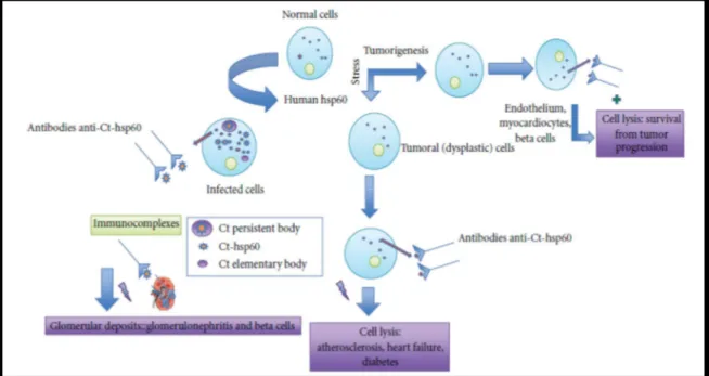
Fig. 10: Potential effects of anti-CT-Hsp60 antibodies. These antibodies recognize surface-Hsp60 onstressed
or tumor cells, and consequently, they can lead to either damage and persistence of infection or cell lysis producing a regression of certain types of cancer. Immunocomplexes (CT-Hsp60 and anti-CT-Hsp60) can cause disease if they form deposits in the renal glomerulus (Mascellino et al., 2011). http://www.hindawi.com/journals/isrn/2011/436936/fig3/.
Quantification of circulating Hsp60 has recently become a potential useful marker of infection for clinicians in patients affected by a variety of diseases. However, interpretation of its values should be carefully evaluated, as a correlation between chaperonin levels and disease is difficult to establish. Hsp60 is also a ligand of TLR and its expression on cell membrane surface’s correlates with apoptotic phenomena (Cappello et al., 2009). During CT invasion and intracellular growth, sensors of the host innate immunity (PRR) can detect the infection by recognizing microbial components (pathogen associated molecular patterns, PAMPs). Chlamydia PAMPs such as Hsp60 and Macrophage Infectivity Potentiator lipoprotein (MIP) are recognized by host PRR TLR4 and TLR2 respectively. These host receptors selectively recognize a broad spectrum of microbial components and endogenous molecules released by injured tissue (Bulut et al., 2002; Bas et al., 2008). In human cells, TLR4 recognizes CT LPS and Hsp60 and it is mainly expressed in the tubes and endometrium and little in endocervix, while TLR2 recognizes peptidoglycan and it is mainly expressed in the tubes and cervix (Mascellino et al., 2011) and appears to be the predominant receptor required for an inflammatory response to infection. Interestingly, TLR2 and its adaptor MyD88 localize to the periphery of the chlamydial inclusion during active infection, suggesting that may signal intracellularly during infection (Bastidas et al.,
28
2013). MIP or other lipoproteins could be released from EB surface and RBs, and retain inside tissues where they might activate resident cells and perpetuate inflammatory response even after the eradication of live bacteria with antibiotic therapy (Bas et al., 2008). In general, PRRs, upon ligand binding, can lead to activation of various inflammatory signaling pathways including NF-kB, NF-IL-6 and MAP kinases.
TTS apparatus is another mechanism which seems central to the biology of the
Chlamydiae, as it mediates the translocation of bacterial toxins to the cytosol of infected
cells. It is present in several important gram-negative bacterial pathogens (Peters et al., 2007). It consists in a molecular injection system protruding from the outer membrane, that appears to be expressed and functional in acute as well as in chronic infection and may represent a prominent virulence factor. A major role of T3S may also be involved, ensuring growth and development of the pathogen by modifying apoptosis signals or some other transcriptional regulation important for Chlamydia survival.
CT Persistence and Chronic Infection
The exact role of persistent stage in the CT developmental cycle as well as the molecular mechanisms allowing persistence remains to be elucidated. In vivo studies of microorganism persistence are hampered by genotype definition and viability of organism. However, characterization of in vitro persistent phase of pathogen and multiple lines of in
vivo evidence, suggest that CT persists in an altered form during chronic disease (Hogan et
al., 2004).
Persistence has long been recognized as a major factor in the pathogenesis of CT disease. It has been described as a viable but non-cultivable growth stage resulting in a long-term relationship with the infected host cell that may not necessarily manifest as clinically recognizable disease.
It is distinct from unapparent infections, which may or may not involve evident CT growth and refers to an atypical, intracellular and metabolically less active state that is difficult to resolve not only by the host-defense system, but also by antibiotic therapy.
Unlike the re-infections believed to be the result of exposure to a CT serotype different from the initial, persistent infections are due to the same type of pathogen genotype entered into a metabolic quiescent and non-infectious form and responsible of three to ten recurrences which can last many years (Dean and Powers, 2001).
29
In vitro studies have shown that several factors including nutrient depletion, cytokines, iron
restriction, amino acids, Ca++ and certain antibiotics can favor the Chlamydial persistent
stage (Beatty et al., 1994 ; Raulston ,1997; Dreses- Werringloer et al., 2000).
IFN-γ is considered a primary host protective cytokine against endocervical CT infections. This directly inhibits bacterial growth through the depletion of cellular tryptophan (TRP) by indolamine-2,3-dioxygenase (IDO) (Aiyar, 2014), which can stop the expression of late proteins, such as MOMP, that in turn stop the progress of RB division and RB conversion into EBs leading to aberrant Chlamydial RBs (Sardinia et al., 1988; Beatty et al., 1994). A failed or weak Th1 response will allow CT RBs to respond to immune challenge by converting into a non-replicating but revivable persistent state (Debattista et al., 2003; Leonhardt et al., 2007). CT, in this persistent state, is able to survive and still allows for antigen-presentation (Rey-Ladino et al., 2007). A direct consequence of this prolonged infection is antibody or Th2-mediated hypersensitivity (Debattista et al., 2003).Moreover, an over-stimulated Th1 response will lead to delayed-type hypersensitivity and an increased risk of IFN-γ-mediated tissue damage, that is likely a consequence of an initially dominant Th2 response. Infected cells increase through new infections and decrease by cell death and clearance by host immune responses; nevertheless, antibiotic treatment, reduces the number of infected cells by eradicating CT; depletion of infected cells further, removes the antigen that is reflected on the immune system. As a direct result, the arrested immunity hypothesis underscores the importance of gaining a better understanding of the interplay between the immune biology of infection and the use of antibiotics (Gottlieb et al., 2010).This hypothesis suggests that early antibiotic treatment effectively attenuates the optimal development of protective immunity, leaving individuals as susceptible as before to reinfection with the same or a new serovar. Therefore the treatment may attenuate protective immunity in some persons and conversely that natural immunity may protect against reinfection (Marrazzo and Suchland, 2014).
In this context, a complete transcriptome analysis of CT serovar D growth in HeLa cells exposed to IFN-γ, demonstrated the up-regulation of many genes involved in active metabolic processes in the aberrant RBs, including those involved in DNA repair and recombination, protein translation, and phospholipid utilization (Belland et al. 2003). Moreover, separate studies at transcriptional level have demonstrated a down-regulation of CT MOMP in HeLa cells and an up-regulation of CP MOMP in response to IFN-γ stimulation (Mathews et al. 2001; Molestina et al. 2002). This underlines the different roles played by MOMP in the two species. Also, Hsp60–1/groEL was found to be expressed predominantly during acute phase growth of CT serovar K and that the
Hsp60-copy2/Ct-30
604 gene transcript/protein was increased in iron-induced persistent cultures (Gérard et al. 2004).
Antibiotics such as penicillin and quinolones (such as ciprofloxacin and ofloxacin) have shown to favor persistence instead of resolving infection because of inducing aberrant but viable particles which may explain therapy failure (Dreses-Werringloer et al. 2000). CT exposure to penicillin leads to enlarged and aberrant RBs, the so called “penicillin forms” that return to normal growth after penicillin removal. On the other hand, although Chlamydial RBs are killed by macrolide treatment (azithromycin), residual antigens can persist for more than 28 days continuing to harbour inflammatory responses (Wyrick and Knight 2004).
In vivo studies have shown that the presence of Chlamydial antigens and nucleic acids even
in absence of cultivable organisms is indicative of persisting organisms probably as result of immunologic stimulation during chronic disease. Chlamydia rRNA demonstration may provide evidence for in-apparent Chlamydial infections (Beagley et al., 2009).
All different species of Chlamydia have tendency to cause persistent infections that may play a role in chronic diseases (inflammation and scarring with significant damage to the host) and oncogenesis.
Previous studies have also revealed that in CT infection, the cytosolic levels of Hsp60 in
vivo gradually increase during carcinogenetic steps, from normal tissue to dysplasia to
fully developed carcinoma in various organs (Cappello et al., 2009).
CT and Apoptosis
Cell death by apoptosis is an active and important defense mechanism against invading pathogens. Apoptosis has a direct role in many infectious diseases, especially those caused by viruses, intracellular protozoans and intracellular bacteria (Byrne and Ojcius, 2004). For many of these pathogens, the apoptotic signaling starts from the pathogen and not by the host cell. In this regard, Chlamydia inhibits apoptotic signalling cascades during productive growth as part of its intracellular survival strategy (Miyairi and Byrne, 2006) in order to maintain the integrity of the host cell for the completion of its intracellular growth (Zhong, 2009). This is in part due to the proteolysis of host proteins for ensuring its own intracellular replication while maintaining the integrity of the infected host cells for long periods of time. CT also inhibits apoptosis during persistent growth or in phagocytes, but induces apoptosis in T cells, which suggests that apoptosis has an immunomodulatory role in Chlamydial infections. The anti-apoptotic activity has shown to be prolonged during CT
31
persistence. This strengthens the hypothesis that active CT metabolism maintains host cell integrity and contributes to intracellular survival (Bastidas et al., 2013).
The circumstances that dictate whether the Chlamydiae inhibit or activate host cell death reflect important pathogenic considerations, including whether if an acute or chronic infection is in progress and whether intracellular Chlamydia growth is programmed to go through a productive infectious cycle or is stalled under non-productive growth conditions. It is possible that apoptotic activity is controlled to some extent by the intracellular growth status of the Chlamydiae, which can be influenced by any or all of these considerations (Byrne and Ojcius 2004) and by strain. While for CP, active inhibition of apoptosis occurs in epithelial cells, macrophages and neutrophils, for CT and C. psittaci, the anti-apoptotic activity has been demonstrated mainly in epithelial cells later in their developmental cycle (Miyairi and Byrne, 2006; van Zandbergen et al., 2004 ). It is not known exactly how pro-apoptotic and antipro-apoptotic effects correlate with the wide spectrum of clinical manifestations and Chlamydial diseases. A Chlamydia induced apoptotic activity has been hypothesized during acute manifestation of disease, whereas inhibition of apoptosis, in chronic disease states (Byrne and Ojcius 2004). Chronic infection and clinical persistence are closely related. Inhibition of apoptosis could represent a mechanism that has evolved to establish a chronic infection. Several lines of evidence suggest that to provoke chronic infection, CT could adopt several strategies. One of these consists of being silent, resulting in asymptomatic infections that cannot be diagnosed at that time. This promotes bacterial progression, even to the most internal tissues. In addition, CT MOMP displays variable immunodominant antigenic epitopes. Variations in these epitopes explain the absence of strain specific immunity and multiple re-infections by different serovars or by the same mutated serovar are still possible (Millman et al. 2001). For these reasons, even if the initial infection is resolved, re-infections are possible and can lead to auto-pathological immune response induction (Beatty et al.1994). Although re-infections occur, the refinement of Chlamydial diagnostic methods will allow us to establish whether CT can persist.
32 Interaction between CT and HPV
Infectious agents play an important role in the aetiology of certain human malignancies, and are thought to be responsible for around 20% of the worldwide cancer burden (Parkin, 2001). Much of the burden of cancer incidence, morbidity, and mortality occurs in the developing world (up to 27%), with a large body of evidence regarding the role of viruses such as human papilloma virus (HPV), hepatitis B virus (HBV) and Epstein-Barr virus (EBV) in the complex processes of carcinogenesis of the cervix, stomach and liver (Jemal et al., 2010). In addition to viral agents implicated in carcinogenesis, a theory of possible association between bacterial infection and cancer has been proposed in early nineteenth century (Lax, 2005). Moreover, many bacteria that cause persistent infections produce toxins that disrupt cellular signaling, alter the regulation of cell growth, induce inflammation or directly damage DNA. Toxins may also mimic carcinogens and tumor promoters and might represent a paradigm for bacterially induced carcinogenesis. The question however remains quite controversial especially with regard to certain species of bacteria for oncogenic properties.
Some authors have suggested that exposure to CT-Hsp60 may be a risk factor for development of cancer (Di Felice et al., 2005), while the development of anti-CT-Hsp60 is also proposed to protect against malignancy (Cappello et al., 2009). Chronic persistent infection with CT to the upper genital tract is able to incur significant damage to the reproductive tract and proposed to induce ovarian cancer (Quirk & Kupinski, 2001).
Although epidemiological data have not yet provided consistent evidence about a real implication of CT in cervical cancer, the co-infection with Human papillomavirus (HPV), sharing the transmission route and the same risk factors, have been recently highlighted (Simonetti et al., 2009; Vaccarella et al.,2010; Paavonen, 2012). A role for CT as cofactor was suggested, since it seems to facilitate the penetration of HPV and the progress of cervical lesions interfering in the immunological response (Deluca et al., 2011). Moreover, some authors recently detected a high-risk for the development of cervical cancer in patients with HPV infection and history of CT (Jensen et al., 2014). Nevertheless, the prevalence and distribution of HPV genotypes associated to CT infection and its clinical persistence are poorly explored. On this basis, the characterization of HPV infection in women suffering from CT could be important in generating hypotheses regarding the possible synergism of these pathogens in cervical malignancy (Bathla et al., 2013; Silva et al., 2014; Tavares et al., 2014; Shew et al., 2013). Recently, a case-control study showed that CT is not able to modify the risk of progression to a high-grade lesion of HPV-positive
33
women but can increase the susceptibility of the cervical epithelium to further HPV infection and its persistence (Safaeian et al., 2010) Specifically young age (less than 25y) is related to an increased risk of both HPV and CT infection (Silva et al., 2013). Furthermore, the role of CT chronic infection in promoting HPV susceptibility has been evaluated. In some cases this infection has been associated with cervical atypia and/or metaplasia, which in turn, may increase the risk of neoplasia (Luostarinen et al., 2013). In this setting, to investigate a pathogen as CT potentially implicated as oncogenic for its tendency to cause chronic and persistent infections, together with HPV co-infection, could have a great importance for the public health.
34
OBJECTIVES OF THE STUDY
As described previously, HPV and CT are STIs with significant implications for global health. In a large series (2009-2014) of women from the North-Eastern Italian area, we aimed to:
• highlight, by molecular techniques, the overall prevalence of HPV and CT infections in cervical swabs (CS) specimen of women that perform these types of investigations.
• highlight the overall prevalence of CT/HPV co-infections.
• highlight the prevalence of CT chronic infections and its performance in the context of HPV co-infections
• evaluate the distribution of HPV genotypes in CT/HPV co-infections, compared to women infected with HPV only
• examine the prevalence of single or multiple HPV infections in the setting of CT/HPV co-infections compared to women infected with HPV only.
35
MATERIALS AND METHODS
Clinical Specimens
A retrospective study was conducted on a large cohort of 7135 consecutive cytology samples collected starting January 2009 to December 2014 during gynecological health checks from immunocompetent women in Trieste, Italy.
Of this series, 6214 cervical swabs (CS) were collected from women at risk for C.
trachomatis infection of which 5481 from Outpatients (mean age 35 ± 10y) asymptomatic
women, 733 were from symptomatic women attending Sexually Transmitted Infection (STI) clinic (mean age 33 ± 10y) while 921 (mean age 43 ± 10y) samples were from women at risk for HPV infection attending as outpatients a second level centre for Cervical Cancer prevention.
CS were collected using a 200 mm polyethylene Cervex brush device (Rovers Medical Devices B.V., The Netherlands) and suspended in 1.5 ml of TE buffer. The top portion uses a soft, flexible brush to obtain cell samples, while the shape allows the top edges to follow the contours of the cervix. The longer middle bristles reach deep into the endocervical canal while the shorter bristles touch both the ectocervical area (external os) and the transformation zone (T-zone). The sample was divided into 3 aliquots of 500 µl each and stored at -80°C until analysis.
No informed consent or any action of the patient was required for this study because the anonymity of the patients was guaranteed. The analysis on this series of samples was conducted blinded.
Isolation of DNA from cervical cells
DNA isolation was performed within 24 hours after the collection of the samples. After specimen centrifugation, 500 µl of each sample was extracted using the NucliSENS® EasyMAG® automated system for total nucleic acid extraction (Biomérieux S.p.a. Florence, Italy), according to the manufacturer’s instructions.