Review Article
Recurrent Painful Ophthalmoplegic Neuropathy and Oculomotor
Nerve Schwannoma: A Pediatric Case Report with Long-Term
MRI Follow-Up and Literature Review
Maria Giuseppina Petruzzelli , Mariella Margari, Flora Furente,
Maria Carmela Costanza, Anna Rosi Legrottaglie, Franca Dicuonzo, and Lucia Margari
Department of Basic Medical Sciences, Neurosciences and Sense Organs Department, University of Bari “Aldo Moro”, Bari, ItalyCorrespondence should be addressed to Maria Giuseppina Petruzzelli; [email protected] Received 5 July 2019; Accepted 9 September 2019; Published 25 September 2019
Academic Editor: Parisa Gazerani
Copyright © 2019 Maria Giuseppina Petruzzelli et al. This is an open access article distributed under the Creative Commons Attribution License, which permits unrestricted use, distribution, and reproduction in any medium, provided the original work is properly cited.
Background. Recurrent painful ophthalmoplegic neuropathy (RPON), previously known as ophthalmoplegic migraine (OM), is
an uncommon disorder with repeated episodes of ocular cranial nerve neuropathy associated with ipsilateral headache. The age of presentation is most often during childhood or adolescence. MRI has a central role in the assessment of the RPON, especially to distinguish orbital, parasellar, or posterior fossa lesions that mimic symptoms of RPON. Actually, oculomotor nerve tumors may be masquerade as RPON so that MRI follow-ups are required to detect the possibility of tumor etiology. Case presentation. We report a 16-year-old boy with a 7-year follow-up and multiple brain MRI data, previously diagnosed as OM. The last brain MRI, performed during an acute phase of oculomotor paresis with ipsilateral headache, showed a nodular lesion described as schwannoma of III cranial nerve. Then, we reviewed the literature on OM and RPON in pediatric age with a focus on brain MRI findings. Conclusions. This review highlights the important role of serial brain MRIs in the long-term follow-up of RPON, especially in the cases with childhood onset, in order to not delay the diagnosis of a possible oculomotor nerve schwannoma.
1. Introduction
In the 3rdedition of International Classification of Headache
Disorders (ICHD) (2018) has been introduced a chapter
named “painful lesions of the cranial nerves and other facial pains” after a consensus between the International Headache Society (IHS) and the International Association for the Study of Pain (IASP) [1]. The IASP defines neuropathic pain (NP) as “pain arising as a direct consequence of a lesion or disease affecting the somatosensory system” [2]. There is increasing awareness about NP in pediatric chronic pain because there are fewer information available regarding the prevalence of NP in the pediatric population than adults [3], and the affected children experience significant physical, psychological, and social sequelae that affect not only themselves but also family and friends [4]. The chapter “painful lesions of the cranial nerves and other facial pains”
includes the recurrent painful ophthalmoplegic neuropathy (RPON), a condition characterized by repeated attacks of paresis of one or more ocular cranial nerves (commonly the III nerve), with ipsilateral headache [1]. This disorder is extremely rare and can occur at any age, but the highest prevalence is in children under the age of 12 years, with the median age at onset about 8 years [5]. The diagnostical definition of RPON changed throughout the history of headache classifications. The 1stedition (1987) of the ICHD had considered this condition as a migraine variant [6], but in the ICHD-II (2004), the disorder, called “oph-thalmoplegic migraine” (with the quotes around “migraine” included), was classified in the group of cranial neuralgias and central causes of facial pain [7]. However, the In-ternational Headache Society again revised the classification in 2013, renaming the disorder “RPON” [8]. Brain magnetic resonance imaging (MRI) is essential in the assessment of
RPON to exclude orbital, parasellar, or posterior fossa le-sions. Gadolinium enhancement of the affected nerve or nerve thickening can be evident using MRI during an acute episode of RPON, whereas negative findings are common during the period of full recovery [1, 9, 10]. Moreover, few MRI studies show the presence of a schwannoma in some patients with RPON disease. Isolated schwannomas are extremely rare. They are benign peripheral nerve sheath tumors, with slowlygrowing, accounting for 6% to 8% of all intracranial tumors [11, 12]. They usually arise from the Schwann cell layer of the vestibular branch of the VIII nerve; otherwise oculomotor nerve schwannomas without neuro-fibromatosis are very rare, and only few cases with pediatric onset have been described in literature [11–13]. In the contest of this ongoing debate [14] about the classification of RPON, we presented a pediatric case report with a 7-year follow-up and multiple brain MRI data, previously di-agnosed as OM. The last brain MRI during an acute phase of oculomotor paresis with ipsilateral headache showed a nodular lesion described as schwannoma of III cranial nerve. Then, we reviewed the literature on OM and RPON with a focus on brain MRI findings.
2. Methods of Review
An electronic literature search was conducted for all articles published up to December 2018, with a range of databases searched including PubMed, Scopus, and Web of Science. The following combinations of keywords were searched: “opthalmoplegic migraine AND magnetic resonance im-aging” and “recurrent painful ophthalmoplegic neuropathy AND magnetic resonance imaging.” The initial search returned 227 records; we considered 117 publications after screening to remove duplication of papers from different sources of research. Research articles, case reports, and reviews describing patients under the age of 18 years have been included. As inclusion criteria papers needed to pro-vide enough information to extract clinical features of headache and oculomotor involvement and a description of MRI findings, 28 publications for a total of 43 clinical cases were selected for inclusion in this review.
3. Case Report
We reported the case of a male patient, 16 years old, with a history of frontal headaches accompanied by photophobia, nausea, and vomiting since the age of 4. He had his first episode of migraine associated with ocular pain and ptosis in the right eyelid, lasting 24 hours, in Jun 2010 at the age of 6. Six months later (Jan 2011), he had a heavier and persistent episode of migraine associated with right ocular pain (partial responsiveness to administration of paracetamol), right ptosis, and diplopia, so he was admitted to the Child Neuropsychiatry Unit, Department of Basic Medical Sci-ences, Neuroscience and Sense Organs, University of Bari “Aldo Moro,” Italy. Family history of migraine in both parents, multiple sclerosis in the father’s family, and gastric neoplastic disease in the mother’s family was referred. General physical examination was normal; neurologic
examination showed paresis of the third and sixth cranial nerves. Laboratory investigation, including virological and organ-specific serum antibodies, and instrumental exami-nation including awake/sleep electroencephalograph pro-duced normal results. Brain and cervical-spine magnetic resonance imaging (MRI) enhanced after contrast admin-istration and magnetic resonance angiography (MRA) produced normal findings. Diagnosis of “ophthalmoplegic migraine” was made according to ICHD 2, as discussed in a previous publication [10]. During the following years, about one time a month, the subject suffered from a moderate to severe intensity headache in the frontal and supraorbital region on the right side with a transient remission with paracetamol. Since his first hospitalization, he had 2 more episodes, in Jul 2013 and Jun 2016, of OM, with ocular pain ptosis in the right eyelid and diplopia. In both episodes brain MRI confirmed normal images. The diagnosis was revised as RPON according to the new diagnostic criteria of ICHD-3 beta (2013) [15]. At the age of 15 years (Sep 2018), the patient was again admitted to our Child Neuropsychiatry Unit manifesting moderate and persistent migraine, nausea, vomiting, phonophobia, and photophobia associated with right ocular pain, diplopia, and ptosis. Neurologic exami-nation showed right eyelid ptosis, and ophthalmologic evaluation showed paresis of the III cranial nerve. Brain MRI with MRA revealed a 5-6 mm nodular enhancement mass, located within the fork of the right basilar artery near to perimesencephalic and interpeduncular cisterns, suggesting an oculomotor nerve schwannoma without any vascular malformation (Figures 1 and 2). The subject was treated with corticosteroid therapy because of the partial and transient responsiveness with paracetamol during acute episodes of right eye pain. He showed gradual and progressive im-provement of headache symptoms in about one week.
4. Discussion
To our knowledge, this is the first case reported in the lit-erature of an adolescent suffering from headache and ophthalmoplegia with a 7-year brain MRI follow-up, raising the question on the relationship between RPON and schwannoma. The evolutive changes in the presentation of symptoms and the different findings over time in brain MRI images observed in this patient repurpose some open issues on the classification and pathophysiological mechanisms of RPON. The patient had his first clinical manifestations of recurrent headaches at the age of 4 years agreeing to di-agnostic criteria of migraine without aura. Since he was 7 years old, the patient started to have migraine episodes with the same clinical features but lasting more time and with partial responsiveness to administration of paracetamol with association of right ocular pain, ipsilateral ptosis, and dip-lopia. After two episodes with these features and with MRI that showed normal findings in the image scans without any appreciable enhancement or thickening of the nerve, he was diagnosed with “ophthalmoplegic migraine” according to ICHD-II [7]. Clinical and instrumental data at next follow-ups were in agreement with the reclassification of RPON proposed in the ICHD-3 [1]. Brain MRI follow-ups of 2013
and 2016 both during an acute episode of headache with ophthalmoplegia confirmed normal findings in the image scans in absence of any brain MRI data supporting a sec-ondary cause. At this regard, according to our review, pa-tients diagnosed with OM/RPON have MRI exams that show thickening and enhancing of the oculomotor nerve [9, 16–24] or normal findings in image scans [18, 20, 22, 23, 25–30]. Nevertheless, the last brain MRI examination of our patient showed the presence of 5-6 mm nodular enhanced mass located within the fork of the right basilar artery near to perimesencephalic and inter-peduncular cisterns, enhanced after contrast, which could be described as III cranial nerve schwannoma (Figures 1 and 2). This case highlights the important role of longitudinal fol-low-up of brain MRI in RPON and raises the question on the
relationship between RPON and schwannoma. The exact pathophysiology of RPON is unknown, but the most ac-cepted postulated mechanisms include microvascular is-chemia, demyelization, and inflammation involving the oculomotor nerve [31]. Despite the reclassification as a neuropathy, a balanced debate on arguments pro and contra a migrainous background of RPON have been published. Friedman DI proposed to consider OM as a syndrome that may be “primary” as a variant of migraine with aura for cases with normal imaging and spontaneous resolution and “secondary” as a cranial neuropathy with focal nerve en-hancement on neuroimaging [31]. The case we described is far from this classification hypothesis, considering that three different brain MRIs during an acute attack of OM showed normal findings inducing to consider the clinical
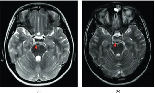
(a) (b)
Figure 1: Axial T2-weighted MRI images comparison: (a) no focal or wide thickening of the oculomotor nerve during an acute attack of OM (Jan 2011); (b) 5-6 mm nodular mass suggesting an oculomotor nerve schwannoma (Oct 2018).
(a) (b)
Figure 2: Axial T1-weighted (a) and axial T2-weighted (b) MRI images showing 5-6 mm nodular mass located within the fork of the right basilar artery near to perimesencephalic and interpeduncular cisterns (Oct 2018).
T able 1: A literature review of symptoms and magnetic resonance imaging findings in 43 patients under the age of 18 years with ophthalmoplegic migraine or recurrent painful ophthalmoplegic neuropathy. Author Reference Case M/ F Age of onset Age of observation Headache Oculomotor involvement MRI MRI follow-up Diagnosis Aers et al. [16] 1 F 8 12 Severe right unilateral headache with photophobia and vomiting Diplopia and ptosis of the right upper eyelid MRI: marked thickening and gadolinium enhancement of the right oculomotor nerve along its subarachnoid course in the interpeduncular fossa Six months later: slightly thickened but no longer enhancing cisternal portion of the right oculomotor nerve OM Wong and Wong [32] 2 M 6 6 Acute onset of headache with photophobia and vomiting Diplopia, acute periorbital pain with droopy eyelids No contrast MRI scan: slight asymmetry in size of the oculomotor nerve NA OM Mark et al. [17] 3 F Not reported 8 Headache Oculomotor nerve palsy Acute MRI: focal thickening and enhancement 7 to 9 weeks later: resolution of the enhancement OM Mark et al. [17] 4 M Not reported 12 Headache Oculomotor nerve palsy Acute MRI: focal thickening 7 to 9 weeks later: resolution of the enhancement OM Mark et al. [17] 5 F Not reported 5 Headache Oculomotor nerve palsy Acute MRI: focal thickening and enhancement 7 to 9 weeks later: resolution of the enhancement OM Mark et al. [17] 6 M Not reported 3 Headache Oculomotor nerve palsy Acute MRI: whole thickening and enhancement 7 to 9 weeks later: resolution of the enhancement OM Prats et al. [18] 7 F 11 12 Throbbing headache Defective elevation and adduction of the left eye and mild dilation of the left pupil and diplopia MRI: enlargement of the cisternal portion of the left III cranial nerve along the first 3 mm of its course, unilateral enhancement after intravenous gadolinium administration A second MRI with contrast administration: normal OM Prats et al. [18] 8 M 3.5 5 Throbbing headache Complete III cranial nerve palsy of the left side MRI: enlargement and enhancement of the cisternal portion of the left III cranial nerve 40 days later: similar findings; 4.5 years later: normal OM Prats et al. [18] 9 F 6 6 Headache in the left orbital region Moderate left ptosis of the eyelid, mild mydriasis, and displacement of the eye upward and outward MRI: normal 5 years later: normal OM
Table 1: Continued. Author Reference Case M/ F Age of onset Age of observation Headache Oculomotor involvement MRI MRI follow-up Diagnosis Del T oro et al. [33] 10 M 10 10 Ocular pain Complete right third nerve palsy MRI: right cavernous sinus enlargement, more obvious after intravenous gadolinium administration 1 month later: the normal caliber of the right carotid artery similar to the left with normal appearance of the ipsilateral cavernous sinus T olosa–Hunt syndrome O’hara et al. [2 1] 11 F 4 7 Acute onset of severe headache, nausea, and vomiting Complete right III nerve palsy Right parasagittal T 1-weighted MRI scan postcontrast: enhancing cisternal portion of the right CN III with nodular mass-like thickening of the proximal portion adjacent to the brainstem 6 weeks later, follow-up postcontrast: marked improvement in enhancement and thickening of the right CN III OM O’hara et al. [2 1] 12 M 2 3 Headache Complete left CN III palsy Left parasagittal T 1-weighted MRI scan postcontrast: thickened enhancing cisternal portion of CN III 8 months later: coronal T 1-weighted scan postcontrast again: mass-like thickening and enhancement of the proximal cisternal portion of the left CN III OM Lance and Zagami [22] 13 F 3 16 Sharp pain behind the left eye, followed by drooping of left eyelid; nausea, vomiting, and sensitivity to light, sound, and smell Left ptosis with paresis of upward deviation in the left eye which turned outwards as she attempted to look up Acute MRI: enhancement of the intracisternal portion of the oculomotor nerve with gadolinium Repeat MRI: enhancement of the oculomotor nerve present but less intense OM Lance and Zagami [22] 14 M 5 5 Right-sided headache accompanied by vomiting Drooping of right eyelid and difficulty in looking upwards and inwards with his right eye Unenhanced MRI normal NA OM Lance and Zagami [22] 15 M 3 13 Left frontal headache, vomiting, and photophobia Ptosis Unenhanced MRI normal NA OM Shin et al. [25] 16 M 7 8 Severe, throbbing, left periorbital headache with associated nausea, vomiting, and photophobia Diplopia and drooping of the upper left eyelid Contrasted MRI: no abnormal findings NA OM Shin et al. [25] 17 F 6 11 Right periorbital headache Drooping of the right eyelid and diplopia Contrasted MRI: no abnormal findings NA OM
Table 1: Continued. Author Reference Case M/ F Age of onset Age of observation Headache Oculomotor involvement MRI MRI follow-up Diagnosis Weiss and Phillips [26] 18 M 2 7 Left supraorbital pain, nausea, and vomiting Left ptosis, exodeviation of the left eye with horizontal and vertical diplopia Brain MRI scan: normal, no contrast enhancement of the oculomotor nerve at its exit from the midbrain NA OM Farage et al. [34] 19 M 16 16 Pulsatile migraine associated with nausea Ptosis, mydriasis, and divergent strabismus MRI weighted in T 1 with contrast: enhanced signal at left oculomotor nerve in cisternal portion 18 months later: no remarkable lesion OM Huang et al. [19] 20 F 9 9 Acute-onset right frontal and periorbital eye pain with migrainous characteristics Ptosis of right eyelid, a dilated pupil with slow pupillary response to light and a extraocular motor weakness compatible with partial oculomotor palsy MRI: thickening of the oculomotor nerve with abnormal enhancement at the exit of brainstem on the right side NA OM Murakami et al. [35] 21 F 4 6 Severe throbbing headache Diplopia, left blepharoptosis, and nonreactive left midriasis MRI after contrast: a thickened oculomotor nerve in its course from the brainstem through the prepontine cistern continuously NA Schwannoma Choi et al. [36] 22 F 13 13 Blurred vision, photophobia, and pulsating headache Complete right side internal ophthalmoplegia MRI: gadolinium enhancement on the cisternal portion of right oculomotor cranial nerve 8 months later: gadolinium enhancement at the proximal part of the left oculomotor nerve OM Bharucha et al. [9] 23 F 1.5 16 Right-sided headache Complete palsy of the right cranial nerve III Acute MRI: Thickening and enhancement of cranial nerve III on the right side 3 and 7 months later: reversal of abnormalities OM Mcmillan et al. [20] 24 F 9 12 Right eye pain Abrupt onset right pupil dilatation, diplopia, and ptosis Acute MRI: an area of prominent thickening and contrast enhancement at the cisternal portion of the right oculomotor nerve 10 weeks later: resolution of abnormal enhancement of the right third cranial nerve, but persistent thickening of the previously enhancing segment OM Mcmillan et al. [20] 25 M 1 1 Not described Abrupt onset left ptosis and eye deviation MRI: a small area of increased enhancement on the anterior surface of the left peduncle at the site of exit of the left oculomotor nerve 17 months later: enhancement and thickened of the cistern portion of left oculomotor nerve OM
Table 1: Continued. Author Reference Case M/ F Age of onset Age of observation Headache Oculomotor involvement MRI MRI follow-up Diagnosis Mcmillan et al. [20] 26 F 16 16 Throbbing, right-sided headache with photophobia Right pupil dilatation, no change in visual acuity MRI: normal 2 weeks later: definite enhancement of the cistern portion of the right oculomotor nerve OM Orssaud et al. [28] 27 F 14 14 Supraorbital and left ocular pain in upward movements Complete III nerve palsy MRI: normal N A T olosa–Hunt syndrome Orssaud et al. [28] 28 M 5 5 Migraine Complete III nerve palsy MRI: normal Repeat MRI 2 years later (relapse): normal OM Vieria et al. [37] 29 M 0.8 7 Frontotemporal and orbital pain always on the right side, photophobia, and phonophobia Ptosis, external ophthalmoplegia and mydriasis Infundibular dilatation of a perforating branch of the posterior cerebral artery adiacent to the III nerve NA OM Arasho [38] 30 M 5 15 Left hemicranial, more retro-orbital, throbbing/ aching headache; photophobia; and phonophobia Complete III nerve palsy Acute MRI: no mass lesion NA OM Borade et al. [29] 31 F 5 6 Bilateral throbbing headache with photophobia and intollerance to loud sounds; vomiting Ptosis on the right side and diplopia Acute MRI: thickened and enhancing right oculomotor nerve in the suprasellar cistern region NA OM Vecino- Lopez et al. [30] 32 F 0.5 3 2 months later: headache, moderate intensity; episodes < 24 hours Right eye ptosis with mydriasis, complete III nerve palsy MRI: enlargement and enhancement with contrast of the cisternal portion of the oculomotor nerve Follow-up MRI: reduced enlargement and enhancement with contrast of the cisternal portion of the oculomotor nerve OM Miglio et al. [39] 33 M 8 8 Headache on the right supraorbital side; photophobia; and vomiting Ptosis, outward deviation of the right eye and diplopia MRI: focal enlargement and marked enhancement in the cisternal portion of the third right cranial nerve at the root exit zone 3 months later: reduced thickening on the third right cranial nerve and resolution of the enhancement OM Lierly et al. [40] 34 F 3 10 Ipsilateral, throbbing headache Acute onset of right ptosis with lateral/inferior eye deviation MRI: isolated enhancement of the cisternal segment of the right oculomotor nerve NA OM
Table 1: Continued. Author Reference Case M/ F Age of onset Age of observation Headache Oculomotor involvement MRI MRI follow-up Diagnosis Da Rocha et al. [4 1] 35 M — 5 Severe frontal headache Right cranial nerve paresis T 1-weighted postcontrast MRI: typical focal thickening and enhancement of the proximal cisternal segment of the 3 cranial nerve 3 years later: persistent focal thickening without evident enhancement OM Gelfand et al. [23] 36 M 5 19 Left periorbital, sharp, throbbing headache, photophobia, and nausea Not reported Acute MRI: enhancement of the cisternal portion of the third nerve 1 year later: normal OM Gelfand et al. [23] 37 M 4 13 Right, diffuse, throbbing headache, photophobia, phonophobia, nausea, and vomiting Not reported Normal Normal OM Gelfand et al. [23] 38 F 9 16 Right frontal throbbing headache Not reported — Normal OM Gelfand et al. [23] 39 M 3 10 Left, periorbital, sharp headache, nausea, and vomiting Not reported Acute MRI thickening and enhancement of cisternal portion of third nerve Nonacute MRI: persistent thickening but no enhancement OM Verma et al. [24] 40 F 6 9 Right hemicranial, largely retro-orbital throbbing/ pulsatile headache with photophobia, phonophobia, and vomiting Drooping of right eyelid with difficulty in moving the eyeball and diplopia Acute MRI: thickened, enhancing right oculomotor nerve in the cisternal segment 3 months later: resolution of oculomotor nerve enhancement OM Riahi et al. [42] 41 F 3 episodes: 2008 20 10 20 11 12 Left side headache with vomiting Left eye ptosis and diplopia T wo cerebral MRI (2008–20 10) normal 3 years later (third episode): tissular mass in the cavernous sinus, suggesting a third nerve schawannoma Schwannoma Jibia et al. [43] 42 F 7 13 Migraine with shimmering scotomas Incomplete right ptosis with semi-midriasis, diplopia MRI: a right nodular schwannoma located within the cisternal segment of the oculomotor nerve 2 months later: no changes (absence of neuroradiological variation) Schwannoma Hurd and Sabo [27] 43 M 4 12 Severe, ipsilateral, pulsatile headache with photophobia, phonophobia, and nausea Left eye ptosis, corneal injection, mydriasis, lacrimation, exotropia, and diplopia Initial MRI without contrast: no abnormalities MRI with contrast: enhancing lesion along the cisternal segment of the left oculomotor nerve RPON
the last brain MRI showed a focal enhancement of contrast, described as a schwannoma. The review of literature on MRI findings in child and adolescent patients affected by OM/ RPON (according to ICHD classifications over time) showed only three cases with isolated schwannoma of the oculo-motor nerve associated with migraine and oculooculo-motor nerve palsy after a long time history of III nerve paresis and/or migraine (Table 1). In 2005, Murakami et al. published the first histologically proved case of schwannoma in an 11-year-old girl with a previous history of OM. The initial diagnosis of OM was corroborated by brain MRI. Five years later, the OM attacks became more frequent and not con-trolled by prophylaxis or acute episodes’ drugs, and brain MRI with three-dimensional reconstruction shows a 5 mm nodular lesion highly suggestive of a neoplastic process involving the oculomotor nerve in its course from the brainstem through the prepontine cistern continuously. Histopathological finding after surgical treatment confirmed the diagnosis of schwannoma [35]. Riahi et al. in 2014 published the case of a 12-year-old girl who presented three episodes of left eye painful ophthalmoplegia during a period of 3 years. Two previous brain MRIs produced normal image scans, while the brain MRI made after the third episode showed a mass of tissue in the cavernous sinus suggesting a third nerve schwannoma [42]. Jibia et al. in 2015 published the case of a 13-year-old girl with a previous medical history of migraine since she was 6 years old. In coincidence of hospitalization because of an attack of disabling ophthalmic migraine, the brain MRI revealed a right nodular schwan-noma located within the cisternal segment of the oculo-motor nerve (patient was treated with analgesic and corticosteroid therapy with complete regression of symp-toms three weeks later and a normal MRI follow-up) [43]. The most part of other cases included in our review, with OM/RPON diagnosis have short-term MRI follow-ups performed less than two years after the first evaluation that can show persistent thickening and enhancement [16, 17, 20–24] or their negativization [9, 18, 23]. Therefore, it is important to consider the long-term evolution of RPON and its relationship with oculomotor nerve schwannoma. Two different speculative explanations of the eventual pathophysiological relationship between RPON and schwannoma have been proposed. The first one is related to the intermittent release of a chemical substance from the tumor, which results in the stimulation of the trigeminal nerve receptors leading to a migrainous headache. According to this hypothesis, the oculomotor nerve schwannoma mimics RPON, so the painful ophthalmoplegia may be considered as the initial manifestation of a tumor and may be too small to be appreciated in MRI scans [35, 44]. A review of pediatric isolated oculomotor nerve schwannoma showed OM as one among the clinical signs of presentation of this tumor [11]. The alternative hypothesis is based on the idea that the underlying pathophysiology of RPON is an inflammatory cranial neuralgia and not mi-graine, so that repeated episodes of inflammation leading to demyelination and remyelination may be associated with a potential transformation into schwannoma from Schwann
complication of RPON.
5. Conclusions
In conclusion, both case reports we presented and the lit-erature review of other pediatric cases of OM/RPON, sug-gest an existing relationship between RPON and schwannoma. Further longitudinal studies on a larger number of cases are needed to clarify whether in some patients an ocular motor nerve schwannoma may closely mimic RPON or, alternatively, if the RPON may be con-sidered a risk factor to develop over time a schwannoma. Anyway, in both these situations, negative findings at MRI during an acute attack of RPON (without any thickening or gadolinium enhancement) do not rule out the possibility of a future evidence of the tumor, considering that schwannomas are generally slowly progressive. Serial brain MRIs during acute exacerbations and even between attacks are essential in the long-term follow-up of RPON, especially in the cases with childhood onset, in order to not delay the diagnosis of a possible oculomotor nerve schwannoma.
Additional Points
Clinical Implications. (i) Put the focus on the still open
debate on arguments pro and contra a migrainous back-ground of RPON; (ii) underlying the important role of serial brain MRIs in the long-term follow-up of RPON; (iii) further longitudinal studies are needed to clarify pathophysiological relationship between RPON and schwannoma.
Consent
The mother of the young patient gave informed written consent before taking part in the study.
Conflicts of Interest
The authors declare they have no conflicts of interest.
References
[1] IHS, “The International classification of headache disorders, 3rd edition,” Cephalalgia, vol. 38, no. 1, pp. 1–211, 2018. [2] R.-D. Treede, T. S. Jensen, J. N. Campbell et al., “Neuropathic
pain: redefinition and a grading system for clinical and re-search purposes,” Neurology, vol. 70, no. 18, pp. 1630–1635, 2008.
[3] K. J. Morgan and D. L. Anghelescu, “A review of adult and pediatric neuropathic pain assessment tools,” The Clinical
Journal of Pain, vol. 33, no. 9, pp. 844–852, 2017.
[4] B. W. Landry, P. R. Fischer, S. W. Driscoll et al., “Managing chronic pain in children and adolescents: a clinical review,”
PM&R, vol. 7, no. 11, pp. S295–S315, 2015.
[5] S. Evers, “Facial pain: overlapping syndromes,” Cephalalgia, vol. 37, no. 7, pp. 705–713, 2017.
[6] IHS, “Classification and diagnostic criteria for headache disorsers, cranial neuralgias and facial pain,” Cephalalgia, vol. 8, no. 7, pp. 1–96, 1988.
[7] IHS, “ICHD-II classification: parts 1–3: primary, secondary and other,” Cephalalgia, vol. 24, no. 1, pp. 23–136, 2004. [8] S. V. Smith and N. M. Schuster, “Relapsing painful
oph-thalmoplegic neuropathy: no longer a “migraine,” but still a headache,” Current Pain and Headache Reports, vol. 22, no. 7, p. 50, 2018.
[9] D. X. Bharucha, T. B. Campbell, I. Valencia, H. H. Hardison, and S. V. Kothare, “MRI findings in pediatric oph-thalmoplegic migraine: a case report and literature review,”
Pediatric Neurology, vol. 37, no. 1, pp. 59–63, 2007.
[10] L. Margari, A. R. Legrottaglie, F. Craig, M. G. Petruzzelli, U. Procoli, and F. Dicuonzo, “Ophthalmoplegic migraine: migraine or oculomotor neuropathy?,” Cephalalgia, vol. 32, no. 16, pp. 1208–1215, 2012.
[11] S.-S. Yang, Z.-J. Li, X. Liu, Y. Li, S.-F. Li, and H.-D. Zhang, “Pediatric isolated oculomotor nerve schwannoma: a new case report and literature review,” Pediatric Neurology, vol. 48, no. 4, pp. 321–324, 2013.
[12] Y.-H. Cho, K.-S. Sung, Y.-J. Song, D.-C. Kim, S. Choi, and K.-U. Kim, “Oculomotor nerve schwannoma: a case report,”
Brain Tumor Research and Treatment, vol. 2, no. 1, pp. 43–47,
2014.
[13] A. A. Norman, B. K. Farris, and R. M. Siatkowski, “Neuroma as a cause of oculomotor palsy in infancy and early child-hood,” Journal of American Association for Pediatric
Oph-thalmology and Strabismus, vol. 5, no. 1, pp. 9–12, 2001.
[14] I. H. Paheva and I. S. Ivanov, “Migraine variants: occurrence in pediatric neurology practice,” Clinical Neurology and
Neurosurgery, vol. 115, no. 9, pp. 1775–1783, 2013.
[15] IHS, “The international classification of headache disorders, 3rd edition (beta version),” Cephalalgia, vol. 33, no. 9, pp. 629–808, 2013.
[16] I. Aers, M. Van Zandijcke, I. Dehaene, and J. Casselman, “Magnetic resonance imaging in a case of migraine with ophthalmoplegia,” European Journal of Neurology, vol. 4, no. 1, pp. 85–89, 1997.
[17] A. S. Mark, J Casselman, D. Brown et al., “Ophthalmoplegic migraine: reversible enhancement and thickening of the cisternal segment of the oculomotor nerve on contrast-en-hanced MR images,” American Journal of Neuroradiology, vol. 19, no. 10, pp. 1887–1891, 1998.
[18] J. M. Prats, B. Mateos, and C. Garaizar, “Resolution of MRI abnormalities of the oculomotor nerve in childhood oph-thalmoplegic migraine,” Cephalalgia, vol. 19, no. 7, pp. 655– 659, 1999.
[19] T.-H. Huang, W.-C. Hsu, J.-H. Yeh, H.-C. Chiu, and W.-H. Chen, “A case of ophthalmoplegic migraine mimicking ocular myasthenia gravis,” European Neurology, vol. 53, no. 4, pp. 215–217, 2005.
[20] H. J. McMillan, D. L. Keene, P. Jacob, and P. Humphreys, “Ophthalmoplegic migraine: inflammatory neuropathy with secondary migraine?,” Canadian Journal of Neurological
Sciences/Journal Canadien des Sciences Neurologiques, vol. 34,
no. 3, pp. 349–355, 2007.
[21] M. A. O’Hara, R. T. Anderson, and D. Brown, “Magnetic resonance imaging in ophthalmoplegic migraine of children,”
Journal of American Association for Pediatric Ophthalmology and Strabismus, vol. 5, no. 5, pp. 307–310, 2001.
[22] J. W. Lance and A. S. Zagami, “Ophthalmoplegic migraine: a recurrent demyelinating neuropathy?,” Cephalalgia, vol. 21, no. 2, pp. 84–89, 2001.
[23] A. A. Gelfand, J. M. Gelfand, P. Prabakhar, and P. J. Goadsby, “Ophthalmoplegic “migraine” or recurrent ophthalmoplegic
cranial neuropathy,” Journal of Child Neurology, vol. 27, no. 6, pp. 759–766, 2012.
[24] A. Verma, A. Kumar, and V. Singh, “Ophthalmoplegic mi-graine in a child, an accelerated clinical and radiologic re-sponse to steroid therapy,” Neurology Asia, vol. 17, no. 4, pp. 357–359, 2012.
[25] D.-J. Shin, J.-H. Kim, and S.-S. Kang, “Ophthalmoplegic migraine with reversible thalamic ischemia. Shown by brain SPECT,” Headache: The Journal of Head and Face Pain, vol. 42, no. 2, pp. 132–135, 2002.
[26] A. H. Weiss and J. O. Phillips, “Ophthalmoplegic migraine,”
Pediatric Neurology, vol. 30, no. 1, pp. 64–66, 2004.
[27] A. Hurd and T. Sabo, “Episodic ophthalmoplegia and headache with cranial nerve III enhancement on MRI,”
Headache: The Journal of Head and Face Pain, vol. 58, no. 10,
pp. 1685-1686, 2018.
[28] C. Orssaud, O. Roche, H. El Dirani, J. Allali, and J.-L. Dufie, “Les ophtalmopl´egies douloureuses de l’enfant: place du syndrome de Tolosa-Hunt et de la migraine ophtal-mopl´egique,” Archives de P´ediatrie, vol. 14, no. 8, pp. 996–999, 2007.
[29] A. Borade, A. S. Prabhu, S. Kumar, V. Prasad, and L. Rajam, “Magnetic resonance imaging findings in ophthalmoplegic migraine,” Journal of Postgraduate Medicine, vol. 55, no. 2, pp. 137-138, 2009.
[30] R. Vecino Lopez, J. Casas Rivero, J. Alvarez-Linera, and S. Noval Mart´ın, “Migraña oftalmopl´ejica: valor de la reso-nancia magn´etica,” Anales de Pediatr´ıa.vol. 71, no. 1, pp. 72–75, 2009.
[31] D. I. Friedman, “The ophthalmoplegic migraines: a proposed classification,” Cephalalgia, vol. 30, no. 6, pp. 646-647, 2010. [32] V. Wong and W. C. Wong, “Enhancement of oculomotor nerve: a diagnostic criterion for ophthalmoplegic migraine?,”
Pediatric Neurology, vol. 17, no. 1, pp. 70–73, 1997.
[33] M. Del Toro, A. Macaya, E. Vazquez, and M. Roig, “Painful ophthalmoplegia with reversible carotid stenosis in a child,”
Pediatric Neurology, vol. 24, no. 4, pp. 317–319, 2001.
[34] L. Farage, M. A. Castro, T. A. Macedo, P. C. Borges, L. P. Souza, and L. O. Freitas, “Ophthalmoplegic migraine: MRI findings. Case report,” Arquivos de Neuro-Psiquiatria, vol. 63, no. 1, pp. 173–175, 2005.
[35] T. Murakami, M. Funatsuka, M. Komine et al., “Oculomotor nerve schwannoma mimicking ophthalmoplegic migraine,”
Neuropediatrics, vol. 36, no. 6, pp. 395–398, 2005.
[36] J. Y. Choi, S. H. Jang, M. H. Park, B. J. Kim, and D. H. Lee, “Ophthalmoplegic migraine with alternating unilateral and bilateral internal ophthalmoplegia,” Headache: The Journal of
Head and Face Pain, vol. 47, no. 5, pp. 726–728, 2007.
[37] J. P. Vieira, J. Castro, L. B. Gomes, S. Jacinto, and A. Dias, “Ophthalmoplegic migraine and infundibular dilatation of a cerebral artery,” Headache: The Journal of Head and Face
Pain, vol. 48, no. 9, pp. 1372–1374, 2008.
[38] B. D. Arasho, “Ophthalmoplegic migraine in a 15-year-old Ethiopian: case report and literature review,” Headache: The
Journal of Head and Face Pain, vol. 10, no. 1, pp. 45–49, 2009.
[39] L. Miglio, P. Feraco, G. Tani, and P. Ambrosetto, “Computed tomography and magnetic resonance imaging findings in ophthalmoplegic migraine,” Pediatric Neurology, vol. 42, no. 6, pp. 434–436, 2010.
[40] M. J. Lyerly, B. W. Peterson, A. K. Lara, and T. M. McGrath, “Ophthalmoplegic migraine,” Headache: The Journal of Head
and Face Pain, vol. 51, no. 7, pp. 1167-1168, 2011.
[41] A. J. Da Rocha, P. Breinis, and M. L. Monteiro, “Magnetic resonance appearance of recurrent ophthalmoplegic
[42] A. Rihai, I. Ben Youssef-Turki, K. Walha et al., “Ophtal-mopl´egie douloureuse r´ecidivante: migraine ophtal-mopl´egique ou schwannome du nerf oculomoteur ?,” Pratique
Neurologique-FMC, vol. 5, no. 3, pp. 205–208, 2014.
[43] A. Jibia, L. Chenin, M. Lefranc, and J. Peltier, “Oculomotor nerve schwannoma in a child: case report and literature re-view,” Neurochirurgie, vol. 61, no. 4, pp. 283–286, 2015. [44] R. Kim, J. H. Kim, E. Kim et al., “Oculomotor nerve tumors
masquerading as recurrent painful ophthalmoplegic neu-ropathy: report of two cases and review of the literature,”