L E T T E R T O T H E E D I T O R
Open Access
Genetic dynamics in untreated CLL patients
with either stable or progressive disease: a
longitudinal study
Alice Ramassone
1,2†, Andrea D
’Argenio
1†, Angelo Veronese
1,2†, Alessio Basti
3,4, Shimaa Hassan AbdelAziz Soliman
1,2,
Stefano Volinia
5, Cristian Bassi
5, Sara Pagotto
1,6, Manuela Ferracin
7, Laura Lupini
5, Elena Saccenti
5, Veronica Balatti
8,
Felice Pepe
1,6, Laura Z. Rassenti
9,10, Idanna Innocenti
11, Francesco Autore
11, Laura Marzetti
3,4,
Renato Mariani-Costantini
1,6, Thomas J. Kipps
9,10, Massimo Negrini
5, Luca Laurenti
11and Rosa Visone
1,6*Abstract
Clonal evolution of chronic lymphocytic leukemia (CLL) often follows chemotherapy and is associated with adverse
outcome, but also occurs in untreated patients, in which case its predictive role is debated. We investigated
whether the selection and expansion of CLL clone(s) precede an aggressive disease shift. We found that clonal
evolution occurs in all CLL patients, irrespective of the clinical outcome, but is faster during disease progression. In
particular, changes in the frequency of nucleotide variants (NVs) in specific CLL-related genes may represent an
indicator of poor clinical outcome.
Keywords: Chronic lymphocytic leukemia, Copy number variation, Nucleotide variation, Clonal evolution
To the Editor
In chronic lymphocytic leukemia (CLL), the clonal
expansion acquired relevance with the NGS era, which
allowed its use for clinical monitoring. Research was
mainly performed on large CLL cohorts sampled
be-fore and after therapy [
1
] and only a few studies
inves-tigated clonal evolution longitudinally in stable versus
progressive untreated patients [
2
–
4
]. The key results
indicate expansion of specific clones upon therapy and
heterogeneity of mutated genes among patients, but
the extent to which the genetic dynamics differs
be-tween stable and progressive untreated CLLs is still
controversial.
To address this point, we used a CLL cohort
includ-ing untreated sequential samples from patients with
either progressive (P-CLL) or stable (S-CLL) disease.
Patients’ features are in Additional file
1
: Table S1. At
each time point, the diagnosis of stable or progressive
CLL was established by the clinicians according to the
cri-teria defined during the International Workshop on
Chronic Lymphocytic Leukemia [
5
]. Using genome-wide
copy number variation (CNV) analysis, we investigated
copy number fluctuations in 11 stable CLLs (S-CLLs) and
15 progressive CLLs (P-CLLs). Data were processed using
the Rawcopy package [
6
], and paired segments were
de-fined for each patient (Additional file
2
: Figure S1). Since
the percentage of CLL cells (f) in PBMCs was not always
known, analyses were performed varying
f from 1 to 100%.
To define aberrant loci, we used two sets of thresholds on
log ratio (LogR) value, depending on
f and on copy
num-ber (k) in CLL cells (Additional file
2
: Figure S2). We did
not find significant differences in percentages of aberrant
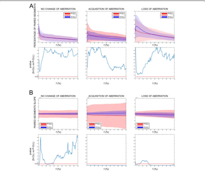
loci between S-CLLs and P-CLLs (Fig.
1
a), but the rate of
change (or slope), reflecting the rate of aberrant clones
evolving over time, was significantly higher in P-CLLs
(p ≤ 0.05, Mann-Whitney U test) (Fig.
1
b). Thus, S-CLLs
and P-CLLs seemed to have the same probability of
ac-quiring or losing clones, but this phenomenon was faster
in P-CLLs. The results were validated on 6 S-CLLs and 5
P-CLLs with known percentage of CLL cells in PBMCs
(Additional file
2
: Figure S3-S4), suggesting that tracking
© The Author(s). 2019 Open Access This article is distributed under the terms of the Creative Commons Attribution 4.0International License (http://creativecommons.org/licenses/by/4.0/), which permits unrestricted use, distribution, and reproduction in any medium, provided you give appropriate credit to the original author(s) and the source, provide a link to the Creative Commons license, and indicate if changes were made. The Creative Commons Public Domain Dedication waiver (http://creativecommons.org/publicdomain/zero/1.0/) applies to the data made available in this article, unless otherwise stated.
* Correspondence:[email protected]
†Alice Ramassone, Andrea D’Argenio and Angelo Veronese contributed
equally to this work.
1
Unit of General Pathology, Center for Advanced Studies and Technology (CAST), University G. d’Annunzio Chieti-Pescara, Chieti, Italy
6Department of Medical, Oral and Biotechnological Sciences, University G.
d’Annunzio Chieti-Pescara, Chieti, Italy
copy number changes does not mandatorily require
know-ledge of cancer cell percentage.
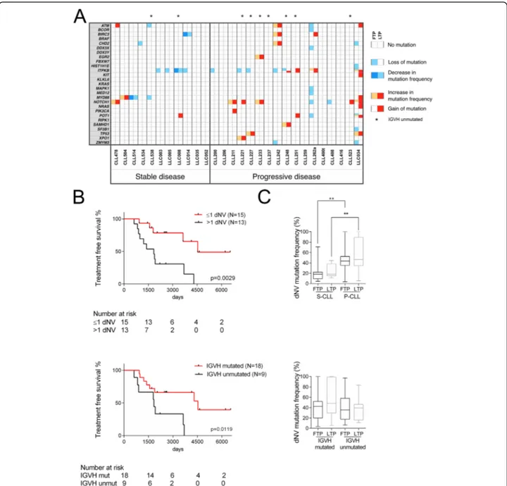
To identify genetic events associated with faster
clonal expansion, we characterized the CLL-specific
genetic features of our cohort. Analyses by qPCR of
three chromosomal abnormalities of prognostic value,
del (11q), tri (12), and del (17p) [
7
], did not reveal
sig-nificant differences between S-CLLs and P-CLLs
(Additional file
2
: Figure S5). Subsequently, we
charac-terized 11 S-CLLs and 17 P-CLLs for point mutations
or indels in regions of 27 genes reported as mutated in
CLL (Additional file
3
: Table S2). We did not register
any significant difference between S-CLLs and P-CLLs
with regard to frequency and number of nucleotide
var-iants (NVs) (data not shown). Next, we focused only on
NVs with variant allele frequencies (VAF) changing
more than 20% between longitudinal samples (dynamic
NV: dNVs, synonymous or non-synonymous). We
de-tected on average 1.18 and 3.35 dNVs per sample in
S-CLLs and P-S-CLLs, respectively (Additional file
4
: Table
S3). P-CLLs showed higher gains/increases of dNVs
(p = 0.0008, Fisher’s test) (Fig.
2
a). Patients with dNV >
Fig. 1 Longitudinal analysis of copy number aberrations in stable and progressive CLL patients. Genome-Wide Human SNP Arrays 6.0 (Affymetrix) was used to genotype patient DNAs at the two sequential sampling points, first time point (FTP), and last time point (LTP). Data were processed using the Rawcopy package, and paired segments (PSs) were identified between FTP and LTP of each patient (see Methods in Additional file2). Aberrant loci were identified by varying the percentage of cancer cells (f) using two sets of threshold on log ratio (LogR), one for DNA amplification (LogRA) and one for DNA deletion (LogRD). a The percentage of PSs and b their slopes (ΔLogRLTP, FTP/ΔtLTP, FTP) are shown as mean (solid line) and standard deviation (shades) based on no change, acquisition, or loss of aberration as a function off. No change of aberration: the considered locus was aberrant both in FTP and in LTP; acquisition of aberrations: the considered locus was aberrant only in LTP; loss of aberrations: the considered locus was aberrant only in FTP. Red colors indicate the P-CLLs; blue the S-CLLs.p values’ graphs (lower panel) report the Mann-WhitneyU test for each f, significance was defined as p < 0.050
1 had shorter treatment-free survival (TFS), considering
as starting point the date at first sampling (p = 0.0029)
or at diagnosis (p = 0.0004, log rank test) (Fig.
2
b and
Additional file
2
: Figure S6). A dNV > 1 was also
associated with poor prognostic factors, including
unmutated
IGVH and trisomy 12 (p = 0.0461 and p =
0.0407, respectively, Fisher’s test) (Additional file
5
:
Table S4). Patients with unmutated
IGVH showed
Fig. 2 Mutational status of CLL samples. a Next-generation sequencing of 27 CLL-associated genes in 11 and 17 patients with stable and progressive disease, respectively. Sequence variants were identified using Torrent Suite 3.4 and Variant Caller plugin 3.4.4. NVs with a coverage < 100 were not considered; NVs with a mutation frequency < 5% were not considered; NVs residing in homopolymer DNA sequences (≥ 4 nucleotides) were also not considered. Each square report either gain or loss of mutation or change in mutation frequency (> 20%) between FTP (left side) and LTP (right side); the mutation frequency of dynamic nucleotide variants (dNVs) was reported in Additional file4: Table S3. Fisher’s exact tests were used to compare groups. Statistical tests were two-sided, and significance was defined asp < 0.050. b Kaplan-Meier curve of treatment-free survival in CLL patients dichotomized based on the number of NVs that change more than 20% between FTP and LTP (dNVs) (upper panel) and on the mutational status of the poor prognostic factorIGVH (lower panel). The median of dNVs changed across all the samples was used as cut-off. Time to treatment was calculated from the first sampling; the last follow-up was considered for patients, which did not undergo treatment. The Log-rank test was used to test for significance. c Mutation frequency of dNVs in stable and progressive CLL groups, and in mutated or unmutated IGVH CLL groups; data are reported as median and interquartile range (box); whiskers range from min to max. Mann-Whitney test was used to compare groups; **denotes ap value ≤ 0.01
shorter TFS, supporting the reliability of our cohort
(Fig.
2
b). Finally, we found that in P-CLLs the average
of dNV frequencies was higher in the first sample (p =
0.0074, Mann-Whitney
U test), where it was not
associ-ated with
IGVH mutational status (Fig.
2
c). These
find-ings suggest that dNVs could have an exploitable
clinical relevance. However, since dNVs include
synonym-ous/non-synonymous mutations and NVs in non-coding
regions, we cannot speculate on the molecular role of the
targeted genes most frequently mutated, such as
ITPKB
and
NOTCH1 (Fig.
2
a). Indeed, these dNVs were only used
here to track genetic evolution.
In conclusion, differently from previous studies, we
calculated VAFs on PBMCs, demonstrating that this
is reliable to track CLL evolution. In fact, an increase
of a single VAF over time indicates expansion of the
clone carrying that NV, regardless of variation in
can-cer cell fraction. Overall, our study points to a higher
genetic dynamics in P-CLLs and suggests that
moni-toring VAFs of a specific gene panel in PBMCs from
sequential samples of a CLL patient may predict
dis-ease progression.
Supplementary information
Supplementary information accompanies this paper athttps://doi.org/10. 1186/s13045-019-0802-x.
Additional file 1: Table S1 Molecular and clinical features of all patients included in the study.
Additional file 2. Supplemental Data (Additional files6and7). Additional file 3: Table S2 HaloPlex SureDesign Report. Additional file 4: Table S3 Genetic variants represented in Fig.2. Additional file 5: Table S4. Characteristics of patients having few (dNV≤1) versus many (dNV>1) nucleotide variants.
Additional file 6: Table S6. SNPs and mutations described by dbSNP and COSMIC databases, respectively, and localized in the regions analyzed by Haloplex SureDesign.
Additional file 7: Table S7. SNPs and mutations detected by Haloplex SureDesign Panel.
Abbreviations
CLL:Chronic lymphocytic leukemia; CN: Copy number; CNV: Copy number variation; dNV: Dynamic nucleotide variant; FTP: First time point; LogR: Log ratio; LTP: Last time point; NV: Nucleotide variant; PBMC: Peripheral blood mononuclear cell; P-CLL: Progressive chronic lymphocytic leukemia; PS: Paired segment; S-CLL: Stable chronic lymphocytic leukemia; VAF: Variant allele frequency
Acknowledgements
We thank Ms. Lia De Amicis for the administrative work. We thank Valerie Matarese for the manuscript editing and Prof. Carlo Maria Croce for providing DNA samples.
Authors’ contributions
RV and AV designed the study. ADA and AR performed the experiments with contributions of CB, LL, ES, VB, FP, SS, SP, and MF. RV, AV, AR, AB, and ADA analyzed the experiments with contribution of CB, MF, SV, and LM. LZR, TJK, FA, II, and LL provided CLL samples and/or clinical data. RV, AV, AR, ADA, RMC, and MN discussed the data analysis. AR, ADA, AV, and RV wrote the
manuscript. All the authors critically reviewed and edited the paper. All authors read and approved the final manuscript.
Funding
This study is supported by the Italian Association for Cancer Research (AIRC) with Start Up grant 2010 (10054) to RV and partially by the Italian Association for Cancer Research (AIRC IG-17063) to SV. RV was supported by her own Marie Curie Career Integration Grant (GA-2011-303735).
Availability of data and materials
DNA CNVs and mutational data are freely available to ArrayExpress database (accession number E-MTAB-8020) and European Nucleotide Archive database (accession number ERP115524). All the other raw data are freely available at the code-hosting platform GitHub (https://github.com/VeroneseVisoneLabs/ Genetic-dynamics-in-untreated-CLL-patients-with-either-stable-or-progressive-disease-a-longitudinal).
Ethics approval and consent to participate
The institutional review board of the University of California, San Diego (171884CX), and of the Fondazione Policlinico Agostino Gemelli (P/948/CE/ 2011) approved the research protocol. Samples were provided upon written informed consent.
Consent for publication Not applicable. Competing interests
The authors declare that they have no competing interests. Author details
1Unit of General Pathology, Center for Advanced Studies and Technology
(CAST), University G. d’Annunzio Chieti-Pescara, Chieti, Italy.2Department of
Medicine and Aging Sciences, University G. d’Annunzio Chieti-Pescara, Chieti, Italy.3Department of Neuroscience, Imaging and Clinical Sciences, University
G. d’Annunzio Chieti-Pescara, Chieti, Italy.4Institute for Advanced Biomedical
Technologies (ITAB), University G. d’Annunzio Chieti-Pescara, Chieti, Italy.
5
Department of Morphology, Surgery and Experimental Medicine, University of Ferrara, Ferrara, Italy.6Department of Medical, Oral and Biotechnological
Sciences, University G. d’Annunzio Chieti-Pescara, Chieti, Italy.7Department of
Experimental, Diagnostic and Specialty Medicine, University of Bologna, Bologna, Italy.8Department of Cancer Biology and Genetics and
Comprehensive Cancer Center at the Wexner Medical Center, The Ohio State University, Columbus, OH, USA.9Department of Medicine, Moores Cancer
Center, University of California at San Diego, La Jolla, CA, USA.10Chronic
Lymphocytic Leukemia Research Consortium, San Diego, CA, USA.
11Fondazione Policlinico Universitario A Gemelli IRCCS, Rome, Italy.
Received: 2 August 2019 Accepted: 9 October 2019
References
1. Sutton LA, Rosenquist R. Deciphering the molecular landscape in chronic lymphocytic leukemia: time frame of disease evolution. Haematologica. 2015;100(1):7–16.
2. Hernandez-Sanchez M, Kotaskova J, Rodriguez AE, Radova L, Tamborero D, Abaigar M, et al. CLL cells cumulate genetic aberrations prior to the first therapy even in outwardly inactive disease phase. Leukemia. 2019;33(2):518–58. 3. Leeksma AC, Taylor J, Wu B, Gardner JR, He J, Nahas M, et al. Clonal diversity predicts adverse outcome in chronic lymphocytic leukemia. Leukemia. 2019; 33(2):390–402.
4. Rose-Zerilli MJ, Gibson J, Wang J, Tapper W, Davis Z, Parker H, et al. Longitudinal copy number, whole exome and targeted deep sequencing of 'good risk' IGHV-mutated CLL patients with progressive disease. Leukemia. 2016;30(6):1301–10.
5. Hallek M, Cheson BD, Catovsky D, Caligaris-Cappio F, Dighiero G, Dohner H, et al. Guidelines for the diagnosis and treatment of chronic lymphocytic leukemia: a report from the International Workshop on Chronic Lymphocytic Leukemia updating the National Cancer Institute-Working Group 1996 guidelines. Blood. 2008;111(12):5446–56.
6. Mayrhofer M, Viklund B, Isaksson A. Rawcopy: improved copy number analysis with Affymetrix arrays. Sci Rep. 2016;6:36158.
7. Dohner H, Stilgenbauer S, Benner A, Leupolt E, Krober A, Bullinger L, et al. Genomic aberrations and survival in chronic lymphocytic leukemia. N Engl J Med. 2000;343(26):1910–6.
Publisher
’s Note
Springer Nature remains neutral with regard to jurisdictional claims in published maps and institutional affiliations.