Gingival Endothelial and Inducible Nitric Oxide Synthase
Levels During Orthodontic Treatment: A Cross-Sectional Study
Michele D’Attillio DDSa; Franca Di Maio, DDSb; Camillo D’Arcangela, DDSa;
Maria Rita Filippi, MD, DDSb; Mario Felaco, MDc; Zsolt Lohinai, PhDd;
Felice Festa, MD, DDSe; Giuseppe Perinetti, DDSf
Abstract: This study uses a cross-sectional design to examine the endothelial and inducible nitric oxide
synthase (eNOS and iNOS, respectively) levels of gingival tissue. Fifteen subjects, 10 female and 5 male individuals (aged 14.6–21.2 years; mean 17.4 6 1.8 years), who needed extraction of the four first pre-molars for orthodontic reasons and who had indications for a gingivectomy were enrolled in the study. In each patient, two maxillary/mandibular premolars were extracted, and two months later an orthodontic appliance was placed in the same arch. A canine undergoing treatment for distal movement served as the test tooth (TT), whereas its contralateral canine was used as the control tooth (CT). The CT was included in the orthodontic appliance but was not subjected to the orthodontic force. Two weeks after the orthodontic appliance placement, clinical data consisting of the presence of supragingival plaque, bleeding on probing, and probing depth were collected from each experimental tooth. Immediately after, gingival tissue was collected from the distal aspect of each TT and CT for immunohistochemistry, messenger RNA reverse transcription by polymerase chain reaction, and Western blot analysis for both eNOS and iNOS. The results showed that no differences in clinical conditions occurred between the experimental teeth. On the contrary, both the eNOS and iNOS levels and the expression of the TTs were significantly greater than those of the CTs (all comparisons significant to P , .01). Our results indicate a role for gingival eNOS and iNOS during the early phases of orthodontic treatment in humans. (Angle Orthod 2004;74:849–856.)
Key Words: Orthodontic tooth movement; Nitric oxide synthase; Tissue remodeling/inflammation
INTRODUCTION
Tooth movement induced by orthodontic forces is con-sequent to remodeling of the periodontal tissues.1–3There
have been studies that have evaluated the hard- and soft-tissue responses as biological features during orthodontic treatment in animal4–7as well as in human8,9models.
How-aResearcher, University G D’Annunzio, Department of Oral
Sci-ences, Chieti, Italy.
bResearch Fellow, University G. D’Annunzio, Department of Oral
Sciences, Chieti, Italy.
cProfessor, University G. D’Annunzio, Department of
Biomor-phology, Chieti, Italy.
dProfessor, Institute of Human Physiology and Clinical
Experi-mental Research, University of Budapest, Budapest, Hungary.
eProfessor and Chairman, University G. D’Annunzio, Department
of Oral Sciences, Chieti, Italy.
fResearch Fellow, Present address, Department of Cell Biology and
Oncology, Istituto di Ricerche Farmacologiche Mario Negri, Consor-zio Mario Negri Sud, Chieti, Italy.
Corresponding author: Giuseppe Perinetti, DDS, Via San Lorenzo 69/1, 65010 Nocciano (PE), Italy
(e-mail: [email protected]).
Accepted: November 2003. Submitted: July 2003.
q 2004 by The EH Angle Education and Research Foundation, Inc.
ever, most of these have focused their attention on bone metabolism5,6or periodontal ligament (PDL) changes2–4that
occur during tooth movement. Gingival modifications in-cidental to tooth movement have been reported as occurring in both histological and ultrastructural analyses and at a clinical level in certain cases.10
Nitric oxide (NO) is a short-lived, highly reactive free radical involved in a variety of cellular pathways. Indeed NO is an intercellular messenger molecule released from various host cells and has important cardiovascular, neu-rological, and immune functions.11 NO released from
os-teoclasts is also involved in bone resorption.12The synthesis
of NO is catalyzed by the NO synthase (NOS) family of enzymes that contain three mammalian gene products. NOS isoforms are functionally distinguished by their modalities of regulation. Two Ca21-dependent isoforms of NOS,
neu-ronal (nNOS) and endothelial (eNOS), remain dormant un-til Ca21-calmodulin binding is elicited by a transient
ele-vation in the intracellular Ca21 concentration ([Ca21]). In
contrast, Ca21-independent NOS (iNOS) is constitutively
active, even at low resting levels of intracellular [Ca21],
because of a remarkably high affinity for calmodulin. The three isoforms are further differentiated by their maximal
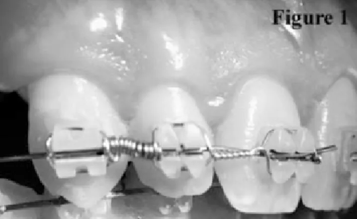
FIGURE 1. Illustration of the full orthodontic appliance used in the
present study.
rates of NO synthesis; nNOS and iNOS exhibit several-fold greater activities than eNOS.11 Moreover, iNOS is able to
produce NO for a longer period of time than either eNOS or nNOS, both of which produce NO in a pulsatile fash-ion.11Finally, both eNOS and nNOS are ubiquitously
pres-ent in tissues, whereas iNOS is detectable only in inflamed tissues.11
The presence of eNOS and iNOS has been shown in dental tissues.13,14 Moreover, increased levels of gingival
iNOS during periodontal inflammation have been reported, as compared with noninflamed gingival tissue.15,16 Indeed,
although iNOS is not ubiquitously present in tissues,11
im-munohistochemistry analyses15,16 have revealed that
clini-cally healthy gingival tissue has detectable amounts of iNOS. Evidence of the expression of iNOS in epithelial17
and inflammatory18cells of the gingiva has also been seen.
Other in vitro investigations19,20 have reported noteworthy
changes in NO levels in PDL fibroblasts when they are mechanically stressed.
To date, studies have neither shown the localization of eNOS in human gingival tissue nor investigated the possi-ble role of gingival iNOS in the early events of orthodontic treatment. Thus, the present study was designed as a cross-sectional assessment to determine whether eNOS levels are detectable in gingival tissue and whether eNOS and iNOS are sensitive markers for gingival tissue changes during the early phases of such treatment in humans.
MATERIALS AND METHODS Study population
Fifteen patients, 10 female and 5 male individuals (aged: 14.6–21.2 years; mean 17.4 6 1.8 years), who had pre-sented at the Unit of Orthodontics, Department of Oral Sci-ences, University G. D’Annunzio (Chieti) and who were diagnosed for the extraction of their first premolars because of dental crowding participated in this study. The following inclusion criteria were observed: (1) the need for fixed-appliance therapy involving distal retraction of one maxil-lary/mandibular canine, with an indication for gingivecto-my to overcome tooth relapse after closure of the extraction sites,21(2) a healthy systemic condition, (3) no use of
anti-inflammatory drugs in the month preceding the beginning of the study, (4) probing depth values (measured as the distance from the bottom of the sulcus to the most apical portion of the gingival margin) not exceeding four mm in the whole dentition, (5) no loss of periodontal attachment (measured as the distance from the bottom of the sulcus to the cemento-enamel junction) exceeding two mm in any interproximal site, (6) no radiographic evidence of peri-odontal bone loss after a full-mouth radiographic periapical examination, and (7) a full-mouth plaque score (FMPS) and a full-mouth bleeding score (FMBS) #20%. FMPS and FMBS were recorded as the percentage of tooth surfaces with the presence of supragingival plaque or bleeding
with-in 15 seconds of probwith-ing with a 20-g controlled-force probe. Informed consent was obtained from the patients and the parents of patients under 18 years of age before the commencement of the study, and the protocol was reviewed and approved by the Ethical Committee of the Medical Fac-ulty of the University G. D’Annunzio.
Clinical procedures
Two maxillary/mandibular first premolars were extracted for each participant, and during the following two months, all subjects received repeated oral hygiene instructions (OHIs), which included the correct use of a toothbrush, dental floss, and an interdental brush. At the end of this period, one maxillary/mandibular canine (of the dental arch corresponding to the extracted premolars) was distalized and considered as the test tooth (TT), whereas the contra-lateral canine was used as the control tooth (CT). On the corresponding arch of the TT of each subject, orthodontic brackets (MBT, 3M-Unitek, Monrovia, Calif) were placed on the buccal surfaces of the incisors, canines, and pre-molars, and bands were placed on the first molars. A uni-lateral 0.018-inch nickel titanium (MBT) arch wire was then fixed from the central incisor to the first molar in the same quadrant as the TT. At the same time, a nickel tita-nium open coil spring (American Orthodontics, Sheboygan, Wis), exerting a constant force of 150 g, was included in the appliance to move the TT distally.22Finally, the central
and lateral incisors in the same quadrant as the TT were laced together with a continuous 0.010-inch steel orthodon-tic wire to provide anchorage. The entire orthodonorthodon-tic ap-pliance (Figure 1) was placed in a single clinical session.
Further OHIs were given to the subjects on how to per-form effective tooth cleaning in the presence of the ortho-dontic appliance, and they were not allowed to take anti-inflammatory drugs for the entire duration of the study. Moreover, all patients underwent one session of accurate supra- and subgingival ultrasonic scaling two weeks before the orthodontic appliance placement.
Two weeks after the orthodontic appliance placement, six sites (mesio-, mid-, and distobuccal; mesio-, mid- and dis-tolingual/palatal sites) on the test and control teeth were clinically examined for the presence of (1) supragingival plaque (PL1), assessed by visual criteria; (2) gingival bleeding within 15 seconds of probing (BOP1) with a 20-g controlled-force probe; and (3) probin20-g depth (PD). The same operator always collected the clinical data (MD’A).
Immediately after completion of the clinical data, gin-gival tissue distal to both the TTs and the CTs was collected under local anesthesia for eNOS and iNOS analyses. The tissue was transferred to plastic vials and immediately stored at2808C for 48 hours. Subsequently, multiple sev-en-mm tissue slices were obtained using a cryostat (Reich-ert-Jung Frigocut 2800, Monochengladback, Germany). These slices were placed on slides, previously treated with gelatin and chromium alum, and kept at a constant tem-perature of 208C. Randomly selected slices from each group were stained with hematoxylin-eosin for histopathological analysis.
Immunohistochemistry for eNOS and iNOS
The anti-eNOS and -iNOS immunohistochemical reac-tions were performed at room temperature in a humid chamber. The defrosted sections were rinsed with Tris-HCl buffer (pH 7.4) for five minutes in a petri dish. After block-ing the nonspecific reactivity with 3% goat serum (Santa Cruz Biotech Inc., Santa Cruz, Calif) for 30 minutes in Tris-HCl buffer, the sections were again rinsed with Tris-Tris-HCl buffer (three3 five minutes) and incubated with primary rabbit anti-human eNOS or iNOS polyclonal antibodies (Santa Cruz Biotech Inc.), at a dilution of 1:100 in phos-phate-buffered saline (PBS), for 30 minutes at room tem-perature. Subsequently, the sections were washed with Tris-HCl buffer (two3 five minutes) and incubated with a sec-ondary goat anti-rabbit antibody (Santa Cruz Biotech Inc.), at a dilution of 1:100 in PBS, for 30 minutes at room tem-perature. They were then washed with Tris-HCl buffer (three3 five minutes) and incubated with the peroxidase-antiperoxidase complex (Santa Cruz Biotech Inc.) for 10 minutes. The sections were then washed with Tris-HCl buffer (two 3 five minutes), and the peroxidase was de-veloped using diaminobenzidine, a chromogen (Santa Cruz Biotech Inc.), mixed in an imidazole buffer (pH 7.6) for 10 minutes. The sections were then dehydrated, clarified, and mounted with a Permount. The presence of eNOS or iNOS was identified by the appearance of a brick red precipitate in the gingival structure.23Immunohistochemical specificity
was tested by omitting the antisera before immunohisto-chemical staining.
Reverse transcription by polymerase chain reaction for eNOS and iNOS messenger RNA
Total RNA was extracted using one mL RNAzol (Cinna Biotex, Huston, Tex) with 20mg Escherichia coli ribosomal
RNA (rRNA) (Boehringer, Ingelheim GmbH, Germany) as carrier. Reverse transcription (RT) was performed in a vol-ume of 20 mL containing M-MLV reverse transcriptase (Perkin-Elmer, Wellesley, Mass), one mM deoxynucleoside triphosphate, 2.5 mM random primers, and one unit/mL RNAse inhibitor (Pharmacia Corp., North Peapack, NJ) for 30 minutes at 428C. Polymerase chain reaction (PCR) am-plification was performed using an Eppendorf Mastercycler 5330, operating at a 728C step temperature of 60-seconds length. The MgCl2concentration used for human eNOS and
iNOS complementary DNA amplification was 2.0 mM. The following primer pairs were used: 59-TGTCTGTCTGCTG CTAG-39 (sense) and 59-CTCTCCAGGCACTTCAGGC-39 (antisense) for human eNOS and 59-AGTGATGGC AAGCACGACTTC-39 (sense) and 59-TCTGTCACTCGC TCACCACGG-39 (antisense) for human iNOS. A PCR was performed using 18S rRNA as internal standard as previ-ously described.24
Western blot analysis for eNOS and iNOS
After homogenizing the gingival tissues in lysis buffer (10% PBS, 10% NP-40, 10% sodium deoxycholate, 10% sodium dodecyl sulfate [SDS], and a protease inhibitor cocktail containing aprotinin, leupeptin, and Na3VO4)
(Pharmacia Corp.), equal amounts of protein (50mg) from the gingival specimens were separated by electrophoresis in a 7.5% polyacrylamide-SDS gel (BioRad, Hercules, Ca-lif) and transferred at 48C to nitrocellulose membranes (BioRad) in glycine-methanol buffer.b-Actin was also run as an internal standard of 130 kDa. The nitrocellulose was then blocked in Tris-buffered saline (TBS)–milk and incu-bated overnight with the primary anti-eNOS or -iNOS an-tibodies (BioRad). The nitrocellulose was washed in TBS, incubated with the secondary antibodies conjugated with alkaline phosphatase for two hours, rewashed, and devel-oped in an alkaline buffer with NBT (alkaline phosphatase conjugate substrate kit) (BioRad).14
Image processing and analysis
The immunohistochemical sections of the gingival spec-imens were examined under a Leitz Dialux 22 microscope (Leica, Heidelberg, Germany). The quantitative evaluation of the eNOS and iNOS immune reactions and the messen-ger RNA (mRNA) and protein levels was performed by determination of the integrated optical density (IOD) scores by digital image analysis.14 The areas investigated for
im-munohistochemistry were randomly chosen and recorded from five slides in each group. For data processing, each experimental frame was digitized into 5123 512 pixels by a Sony video camera connected to a Leica Quantimet 500 plus microscope, and the IOD scores, from 1 to 4 according to the increasing optical density, were determined using ISO Transmission Density (Kodak CAT 152-3406, Eastman Kodak Company, Rochester, New York) as a standard.
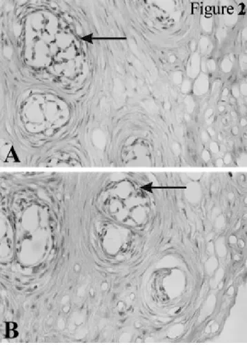
FIGURE 2. Immunohistochemical localization of eNOS in the (A) TT
and (B) CT groups (magnification 2503). Endothelial cells and fibro-blasts are predominantly positive for eNOS (arrows). eNOS indi-cates endothelial nitric oxide synthase; TT, test tooth; and CT, con-trol tooth.
Data processing
The Statistical Package for Social Sciences software (SPSSt Inc., Chicago, Ill) was used to perform the data analysis. The percentage of tooth sites positive for plaque (%PL1), bleeding on probing (%BOP1), and mean PD were calculated for the TT and CT groups, considering the tooth as the statistical unit. Wilcoxon paired signed rank tests were used to evaluate the statistical significance of the differences in the %PL1 and %BOP1 between the exper-imental categories. A paired t-test was used to assess the significance of the differences in the PD between the ex-perimental groups. Moreover, differences in the clinical data between the TTs and CTs obtained from the corre-sponding experimental collection sites were also tested. The number of PL1 and BOP1 experimental sites were pro-cessed as paired dichotomous data by using a McNemar test, whereas the PD scores were assessed performing a paired t-test.
For both the eNOS and iNOS immune reactions and the mRNA and protein levels, the Wilcoxon paired sign rank sum test was used to evaluate the significance of the dif-ferences in the IOD scores between the experimental groups. A probability of P, .05 was accepted for rejection of the null hypothesis.
RESULTS
The %PL1, %BOP1, and mean PD in the TTs and CTs were similar, with no statistically significant differences seen between the groups (all significance levels at P. .5; data not shown). Similarly, no significant differences were detected between the TTs and CTs in all the clinical param-eters of the corresponding collection sites (P. .5; data not shown). Finally, pooled data regarding %PL1, %BOP1, and mean PD from the TTs and CTs, presented as means
6 standard errors, are 18.4 6 2.5, 12.4 6 1.9, and 1.9 6
0.1, respectively.
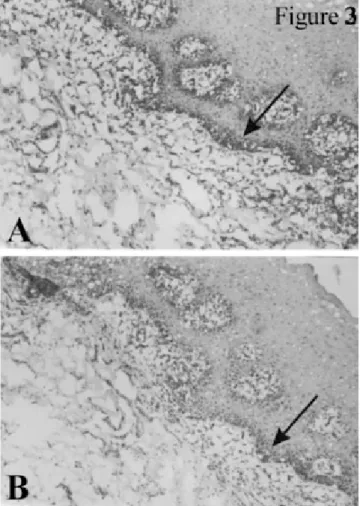
Epithelial cells, connective tissue, blood vessels, nerve fibers, and lymphocytes could all be detected in all the his-tological sections from both the TT and the CT gingival specimens. An overall impression through the analysis of the full range of samples suggests that vasodilatation and leukocyte infiltration (predominantly macrophages and plasma cells) appeared to be more evident in the TT gin-gival specimens than in those of the CTs (data not shown). However, representative immunohistochemical sections of gingival specimens from both the TT and the CT groups, illustrating the positive precipitates for eNOS and iNOS, are shown in Figures 2 and 3. All the specimens examined were positive for the presence of both these NOSs. More-over, endothelial cells and fibroblasts were predominantly positive for eNOS (Figure 2), whereas iNOS immunore-activity was found mainly along the basal membrane and in inflammatory cells within the connective tissue (Figure 3).
The results regarding the NOSs’ immune reactions and the mRNA and protein levels in each TT and CT group are summarized in Table 1. In particular, the IOD scores of both eNOS and iNOS were higher in the TTs than in the CTs in the immunohistochemistry (Figures 2 and 3), RT-PCR (Fig-ure 4), and Western blot (Fig(Fig-ure 5) investigations.
DISCUSSION
Our controlled cross-sectional study evaluated enzymatic gingival changes that occur during the early phases of or-thodontic treatment in humans. Although no differences were detected in the clinical conditions, the eNOS and iNOS immunoreactivites and the mRNA and protein levels were significantly greater in the gingival tissues compressed by the tooth movement (TTs) than in those of the untreated controls (CTs) (Table 1).
Although several experimental models have been used to evaluate periodontal and dental tissue responses to mechan-ical stimuli,7 only a limited number of studies have been
avail-FIGURE 3. Immunohistochemical localization of iNOS in the (A) TT
and (B) CT groups (magnification 1003). Basal membranes and in-flammatory cells within the connective tissue are predominantly pos-itive for iNOS (arrows). iNOS indicates inducible nitric oxide syn-thase; TT, test tooth; and CT, control tooth.
TABLE 1. Distribution of IOD scores and medians for eNOS and iNOS immunoreactivities, and mRNA and protein levels in the ex-perimental groups (n515).
Group
Number of Specimens in Each IOD Score
1 2 3 4 Median IOD Score eNOS Imm.* RT-PCR* WB* TT CT TT CT TT CT 1 7 0 8 0 10 4 6 3 7 4 3 6 2 8 0 8 2 4 0 4 0 3 0 3 2 3 1 3 1 iNOS Imm.* RT-PCR* WB* TT CT TT CT TT CT 1 11 2 11 0 13 3 3 2 2 4 1 4 0 6 2 6 1 7 1 5 0 5 0 3 1 3 1 3 1
aTT indicates test tooth; CT, control tooth; eNOS, endothelial nitric
oxide synthase; iNOS, inducible nitric oxide synthase; IOD, inte-grated optical density; Imm., immunohistochemistry; RT-PCR, re-verse transcription by polymerase chain reaction; and WB, Western blot analysis.
* Level of significance of the differences between the TT and CT groups, within either eNOS or iNOS, by analytical investigation: *p,.01.
FIGURE 4. Reverse transcription by polymerase chain reaction for
eNOS and iNOS mRNA in the TT and CT groups (arrows). The last lane shows the standard (S). Higher mRNA expression is evident for both eNOS and iNOS in the TT group as compared with the CT group. eNOS indicates endothelial nitric oxide synthase; iNOS, in-ducible nitric oxide synthase; mRNA, messenger RNA; TT, test tooth; and CT, control tooth.
able have been obtained from animal models.7In vitro
ex-periments have been performed on human periodontal cells under mechanical stress,8,19but such a study design cannot
account for the complexities of the in vivo situation. In-deed, orthodontic forces can affect all periodontal and den-tal tissues, and the pattern of reaction of one has been seen related to the responses in the other.7The present study has
the advantage of evaluating the gingival response to com-pression stress using an in vivo design where the complex-ity of the interactions among the periodontal tissues has been accounted for. Moreover, the use of a control within the same patient is effective in controlling the individual patient responses.
The study population enrolled in the present investigation showed similar gingival conditions for the TTs and CTs, probably because of the repeated OHIs imparted to each participant before the beginning of the treatment. Moreover, it has been reported that good maintenance of clinical con-ditions is possible despite orthodontic appliance place-ment.25 The short duration of the study design also may
have contributed to these results.
All the histological samples obtained in the present study showed an inflammatory infiltrate, with an overall impres-sion that vasodilatation and leukocyte infiltration (predom-inantly macrophages and plasma cells) appeared to be more evident in the TT, as opposed to the CT, gingival specimens. Because it is virtually impossible to obtain pristine healthy tissue samples from humans despite the absence of any clinically detectable signs of inflammation, and because these apparent differences in the TTs as opposed to the CTs were both subtle and not consistently seen, this aspect re-quires further specific investigation. However, all the
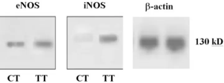
ana-FIGURE 5. Western blot analysis for eNOS and iNOS in the TT and
CT groups.b-Actin was used as an internal standard (130 kDa). Higher levels are evident for both eNOS and iNOS in the TT group as compared with the CT group. eNOS indicates endothelial nitric oxide synthase; iNOS, inducible nitric oxide synthase; TT, test tooth; and CT, control tooth.
lytical investigations demonstrated detectable levels and ex-pression of NOSs in both the TT and the CT gingival tis-sues.
High levels of eNOS have been seen in the endothelial cells of the blood vessels,26and it was not surprising to find
detectable immunoreactivity of eNOS in the endothelial cells of the gingival tissues from both the TT and the CT groups (Figure 2). The NO produced by these cells is re-sponsible for the relaxation of the smooth muscle and, hence, the blood flow to the tissues.26Previous studies have
demonstrated that the NO involved in the maintenance of resting blood flow in dental tissues derives from both eNOS and nNOS activities.13
During orthodontic tooth movement, animal research has described an intense vascular activity in the PDL and al-veolar bone that decreases one day after the orthodontic force is applied, which increases after three days.27Similar
results have been obtained in other investigations, which have also reported a remarkable dilatation of blood vessels corresponding to the areas compressed by the tooth move-ment.2,28,29These studies evaluated the vascular changes in
the PDL and alveolar bone only and not in the gingival tissues. However, considering that the pattern of reactions in any of the periodontal tissues is strongly related to those of the other compartments,7the increase in blood flow
con-sequent to an increase in blood vessel diameter might also occur in the gingival tissue. This hypothesis is supported by the increase in eNOS levels found in the TT gingival tissues as compared with those of the CT group.
A correlation between iNOS levels and inflammation in periodontal tissues has been demonstrated previously.15,16
However, we can be certain that the higher iNOS levels from the TTs, in comparison with the CTs, were not due to inflammation because all the experimental sites showed good clinical conditions throughout the duration of the study. However, during orthodontic tooth movement in hu-mans, an increase in several inflammatory mediators in the gingival crevicular fluid (GCF), ie, interleukins, has been found in spite of a good clinical condition.30Hence, these
authors concluded that subclinical inflammation may occur as one of the earliest phenomena responsive to the tissue
remodeling. Moreover, cytokines have been shown to stim-ulate iNOS production from several cells.11,26This supports
the increase in iNOS levels in the TT gingival tissue be-cause it may be a consequence of an increase in the inflam-matory mediators in the periodontal tissues before they mi-grate to the gingival crevice where they are detected.31This
hypothesis is reinforced by the evidence that constituents of GCF derive from a variety of sources, including the gin-gival tissue.31 Furthermore, inflammatory changes
detect-able at a histological level have been demonstrated to also directly affect the gingival tissue surrounding orthodonti-cally moved teeth.7
Other explanations regarding the increase in both iNOS and eNOS levels and expression in the TT gingival tissues can be derived from a consideration of the functions of their product, NO. Some years ago, the role of NO in relaxation of arteriole wall smooth muscle and in inhibition of platelet aggregation was demonstrated.32 It is also of interest that
there is evidence that NO plays a role in bone resorption,12
particularly because it is well known that during orthodon-tic tooth movement bone resorption is constantly present in the sites of compression2and even in the sites of tension.5
Thus, considering the existence of tight anastomoses in the blood vessels between the periodontal tissues,27,33,34the
in-crease in NOS levels and expression in the TT gingival tissues two weeks after the orthodontic force was applied might be further explained by the influence of bone activity on gingival composition.
During orthodontic tooth movement, a hyalinization of the most compressed area of the PDL that is induced by the compressive forces has been reported.2,35 This hyaline
zone is described as an area of focal aseptic necrosis that is resistant to degradation, persists in the pressure zone, and is dependent on the magnitude of the force.35Other
inves-tigations36,37have reported increases in the number and
ac-tivity of macrophage-like cells in the resorptive area of periodontal tissues undergoing mechanical stress, corre-sponding to the hyaline zone, and this activity is involved in the removal of the necrotic periodontal tissue. Consid-ering further that during orthodontic tooth movement an intense macrophage activity occurs consistently near blood vessels of the periodontal tissues both in the areas of ten-sion and in the areas of resorption,28 and that NO is also
released from macrophages during cell-cell interactions,32
the increase in the NOS levels and in their expression found in the TT gingival tissues might be due, in part, to the activity of these cells. This hypothesis is reinforced by a recent investigation that showed detectable levels of iNOS in macrophages of gingival tissue.18
Further consideration of the results of the present study indicates that they can be linked to previous investigations that have reported increases in NO production from PDL fibroblasts when they are mechanically stressed.19,20
Al-though the present study evaluated the NOS levels and ex-pression in gingival tissue, rather than in the PDL, it is
likely that the anatomical and functional connections among the periodontal tissues27,33,34 contribute, at least in
part, to the increases in NOS levels and expression found in the TTs. Moreover, fibroblasts are the most abundant cell type in gingival tissue, and their volume comprises 5.6% of the total gingival volume.38 It is of interest that human
fibroblasts increase their NO basal production under me-chanical stimulation and that gingival tissue surrounding a moving tooth does not undergo resorption but is com-pressed and consequently retracted.10 In connection with
this, a direct increase in NO production, through increases in NOS levels and expression, by the fibroblasts of the com-pressed gingiva could have occurred in the TT gingival tissues.
Conversely, in the CT gingival tissues where no ortho-dontic force was applied, both NOS levels and their ex-pression are lower than those of the TT gingival tissues (although always detectable) in all analytical investigations (Table 1). Moreover, the presence of detectable levels of iNOS in normal PDL cells and gingival tissues, and eNOS in clinically healthy pulp tissue has been reported in other investigations.14–16,19 Hence, this study demonstrates
gingi-val localization of eNOS in human subjects and that both eNOS and iNOS of the gingival tissues can be considered suitable markers for orthodontic treatment in humans, al-though further investigations are needed to fully elucidate these aspects.
Briefly, these results suggest roles for eNOS and iNOS in gingival tissues undergoing compression stress. More-over, the increase in their levels and expression may be due to the activity of several cell types that are directly or in-directly involved in the tissue response to mechanical stress.
ACKNOWLEDGMENTS
The authors are grateful to Dr Christopher P. Berrie for critical review of the text and to Claudia Brue` for statistical advice.
REFERENCES
1. Reitan K. Clinical and histological observations on tooth move-ment during and after orthodontic treatmove-ment. Am J Orthod. 1967; 53:721–745.
2. Rygh P. Ultrastructural changes in pressure zones of rat molar periodontium incident to orthodontic movement. Acta Odontol Scand. 1972;30:575–593.
3. Rygh P. Ultrastructural changes in tension zones of rat molar peri-odontium incident to orthodontic movement. Am J Orthod. 1976; 70:269–281.
4. Lilja E, Lindskog S, Hamm L. Histochemistry of enzymes asso-ciated with tissue degradation incident to orthodontic tooth move-ment. Am J Orthod. 1983;83:62–75.
5. King GJ, Keeling SD, Wronski TJ. Histomorphometric study of alveolar bone turnover in orthodontic tooth movement. Bone. 1991;12:401–409.
6. King GJ, Latta L, Rutemberg J, Ossi A, Keeling SD. Alveolar bone turnover and tooth movement in male rats after removal of orthodontic appliances. Am J Orthod. 1997;111:266–275.
7. Vandevska-Radunovic V. Neural modulation of inflammatory re-actions in dental tissues incident to orthodontic tooth movement. A review of the literature. Eur J Orthod. 1999;21:231–247. 8. Saito M, Saito S, Ngan PW, Shanfeld J, Davidovitch Z.
Interleu-kin 1 beta and prostaglandin E are involved in the response of periodontal cells to mechanical stress in vivo and in vitro. Am J Orthod Dentofacial Orthop. 1991;99:226–240.
9. Samuels RH, Pender N, Last KS. The effects of orthodontic tooth movement on the glycosaminoglycan components of gingival cre-vicular fluid. J Clin Periodontol. 1993;20:371–377.
10. Redlich M, Shoshan S, Palmon A. Gingival response to ortho-dontic force. Am J Orthod Dentofacial Orthop. 1999;116:152– 158.
11. Alderton WK, Copper CE, Knowles RG. Nitric oxide synthases: structure, function and inhibition. Biochem J. 2001;357:593–615. 12. van’t Hof RJ, Ralston SH. Nitric oxide and bone. Immunology.
2001;103:255–261.
13. Lohinai Z, Balla I, Marczis J, Vass Z, Kova´ch AG. Evidence for the role of nitric oxide in the circulation of the dental pulp. J Dent Res. 1995;74:1501–1506.
14. Felaco M, Di Nardo Di Maio F, De Fazio P, et al. Localization of the e-NOS enzyme in endothelial cells and odontoblasts of healthy human dental pulp. Life Sci. 2000;68:297–306.
15. Lappin DF, Kjeldsen M, Sander L, Kinane DF. Inducible nitric oxide synthase expression in periodontitis. J Periodontal Res. 2000;35:369–373.
16. Hirose M, Ishihara K, Saito A, Nakagawa T, Yamada S, Okuda K. Expression of cytokines and inducible nitric oxide synthase in inflamed gingival tissue. J Periodontol. 2001;72:590–597. 17. Yanagita M, Shimabukuro Y, Nozaki T, et al. IL-15 up-regulates
iNOS expression and NO production by gingival epithelial cells. Biochem Biophys Res Commun. 2002;297:329–334.
18. Gaspirc B, Masera A, Skaleric U. Immunolocalization of induc-ible nitric oxide synthase in localized juvenile periodontitis pa-tients. Connect Tissue Res. 2002;43:413–418.
19. Kikuiri T, Hasegawa T, Yoshimura Y, Shirakawa T, Oguchi H. Cyclic tension force activates nitric oxide production in cultured human periodontal ligament cells. J Periodontol. 2000;714:533– 539.
20. Nakago-Matsuo C, Matsuo T, Nakago T. Basal nitric oxide pro-duction is enhanced by hydraulic pressure in cultured human peri-odontal ligament fibroblasts. Am J Orthod Dentofacial Orthop. 2000;117:474–478.
21. Ewen SJ, Pasternak R. Periodontal surgery, an adjunct to ortho-dontic therapy. Perioortho-dontics. 1964;2:162–171.
22. Perinetti G, Paolantonio M, D’Attilio M, D’Archivio D, Tripodi D, Femminella B, Festa F, Spoto G. Alkaline phosphatase activity in gingival crevicular fluid during human orthodontic tooth move-ment. Am J Orthod Dentofacial Orthop. 2002;122:548–556. 23. Di Napoli P, Taccardi A, Grilli A, et al. Simvastatin reduces
re-perfusion injury by modulating nitric oxide synthase expression: an ex vivo study in isolated working rat hearts. Cardiovasc Res. 2001;51:283–293.
24. Innis MA, Gelfand DH, Sninsky J, White TJ, eds. PCR Protocols, a Guide to Methods and Applications. San Diego, Calif: Academ-ic Press; 1990;147.
25. Lundstrom F, Hamp SE, Nyman S. Systematic plaque control in children undergoing long-term orthodontic treatment. Eur J Or-thod. 1980;2:27–39.
26. Nathan C. Nitric oxide as a secretory product of mammalian cell. FASEB J. 1992;6:3051–3064.
27. Vandevska-Radunovic V, Kristiansen AB, Heyeraas KJ, Kvins-land S. Changes in blood circulation in teeth and supporting tis-sues incident to experimental tooth movement. Eur J Orthod. 1994;16:361–369.
vascular system: a main mediator of periodontal fiber remodelling in orthodontic tooth movement. Am J Orthod. 1986;89:453–468. 29. Vandevska-Radunovic V, Kvinsland S, Hals Kvinsland I. Effect of experimental tooth movement on nerve fibres immunoreactive to calcitonin gene-related peptide, protein gene product 9.5, and blood vessel density and distribution in rats. Eur J Orthod. 1997; 19:517–529.
30. Uematsu S, Mogi M, Deguchi T. Interleukin (IL)-1 beta, IL-6, tumor necrosis factor-alpha, epidermal growth factor, and beta 2-microglobulin levels are elevated in gingival crevicular fluid dur-ing human orthodontic tooth movement. J Dent Res. 1996;75: 562–567.
31. Egelberg J. Permeability of the dento-gingival blood vessels II: clinically healthy gingivae. J Periodontal Res. 1966;1:276–286. 32. Marletta MA. Nitric oxide synthase structure and mechanism.
Biol Chem. 1993;268:12231–12334.
33. Weekes WT, Sims MR. The vasculature of the rat molar peri-odontal ligament. J Periperi-odontal Res. 1986;21:184–194. 34. Schroeder HE, Page RC. The normal periodontium. In: Schluger
S, Yuodelis R, Page RC, Johnson RH, eds. Periodontal Diseases. Philadelphia, Pa: Lea & Febiger; 1990:42.
35. Reitan K. Biomechanical principles and reactions. In: Graber TM, eds. Current Orthodontic Principles and Techniques, vol 1. Phil-adelphia, Pa: WB Saunders Company; 1969:56–150.
36. Kvam E. Cellular dynamics on the pressure side of the rat peri-odontium following experimental tooth movement. Scand J Dent Res. 1972;80:369–383.
37. Brudvik P, Righ P. Non-clast cells start orthodontic root resorption in the periphery of hyalinized zones. Eur J Orthod. 1993;15:467– 480.
38. Schroeder HE, Munzel-Pedrazzoli S, Page RC. Correlated mor-phometric and biochemical analysis of early chronic gingivitis in man. Arch Oral Biol. 1973;35:337–346.