Università degli Studi del Piemonte Orientale
“Amedeo Avogadro”
Department of Pharmaceutical Sciences
Ph.D. in Chemistry and Biology
XXXII cycle 2016-2019
BIO/14
TRISOMIC NEURAL PROGENITOR CELLS
AS NOVEL PHARMACOLOGICAL TARGETS
IN DOWN SYNDROME
Maria Elisa Salvalai
Supervised by Prof. Mariagrazia Grilli
Abbreviations
Aβ Amyloid β
ACM Astrocyte conditioned medium
AD Alzheimer’s disease
APP Amyloid precursor protein
ARP 2/3 Actin-related protein complex 2/3 BDNF Brain derived neurotrophic factor BFCN Basal forebrain cholinergic neurons bFGF Basic fibroblast growth factor BrdU 5-bromo-2’-deoxyuridine CA 1, 2 Cornu Ammonis field 1, 2 CFC Contextual fear conditioning CHD Congenital heart disease CNS Central nervous system
CSA Cyclosporine A
DPN Day post natal
DCX Doublecortin
DG Dentate gyrus
DIV Days in vitro
DS Down syndrome
DSCR Down syndrome critical region
DYRK1A Dual specificity tyrosine phosphorylation regulated kinase 1A
ECD Extracellular domain
EdU 5-ethynyl-2’-deoxyuridine EGCG Epigallocatechin-3-gallate EGF Epidermal growth factor
EU Euploid
GABA Gamma-aminobutyric acid
GAPs GTPase-activating proteins
GBP Gabapentin
GC Glucocorticoids
GEFs Guanine exchange factors GFAP Glial fibrillary acidic protein
GR Glucocorticoid receptor
GSK3ß Glycogen synthase kinase 3 beta
GW Gestation week
Hsa21 Human chromosome 21
IHC Immunohistochemistry
IQ Intelligence quotient
JAK-STAT Janus kinase-signal transducer and activator
KO Knockout
LiCl Lithium chloride
LTD Long-term depression
LTP Long-term potentiation
LA Linoleic acid
MAP2 Microtubule associated protein 2 Mmu 10,16,17 Mouse chromosome 10,16,17
MWM Morris water maze
NFATc Nuclear factor of activated T cell cytoplasmatic
NMDA N-methyl-D-aspartic acid
NPC Neural progenitor cells
NSC Neural stem cells
OA Oleic acid
OLIG1-2 Oligodendrocyte transcription factor 1 and 2
Ptch1 Patched 1
PGB Pregabalin
PI3K Phosphoinositide 3-kinase
PPARs Peroxisome-proliferator activated receptors Rac1 Ras-related C3 botulinum toxin substrate 1 RCAN1 Regulator of calcineurin 1
SGZ Subgranular zone
Shh Sonic hedgehog
SOD1 Superoxide dismutase 1
Smo Smoothened
SSRI Selective serotonin reuptake inhibitor
SVZ Subventricular zone
S100ß S100 calcium binding protein B
TM Trans-membrane
TrkB Tropomyosin receptor kinase B TRKs Tyrosine receptor kinases
TS Trisomic
TSP-1 Thrombospondin-1
VWF-A Von Willebrand factor A
VZ Ventricular zone
WT Wild type
5-HT 5-hydroxytryptamine
7,8-DHF 7,8-dihydroxyflavone β2AR β2 adrenergic receptor
Contents
Chapter 1 ... 9
1.1 Down syndrome ... 10
1.1.1 Epidemiology ... 10
1.1.2 Etiology and the genetics of Down syndrome ... 11
1.1.3 Phenotypic traits and clinical features observed in Down syndrome ... 13
1.1.4 The neurological phenotype of Down syndrome ... 14
1.2 Neurodevelopmental alterations in Down syndrome brain... 15
1.2.1 Gross anatomy... 15
1.2.2 Cytoarchitecture ... 16
1.2.3 Neurogenesis alterations ... 16
1.2.4 Gliogenesis alterations ... 18
1.2.5 Dendrite, spine, synapse and neurotrophin alterations in Down syndrome brain ... 18
1.2.6 Specific triplicated genes with a role in Down syndrome neuropathology... 20
1.3 Rodent models in Down Syndrome research ... 22
1.4. Focus on cell types involved in Down syndrome pathophysiology... 27
1.4.1 Neural progenitor cells ... 27
1.4.1.1 Mechanisms underlying impaired neural progenitor proliferation 29 1.4.1.2 Mechanisms underlying impaired neural progenitor phenotype acquisition ... 31
1.4.2 Astrocytes... 32
1.4.2.1 Non-cell autonomous regulation of neural progenitor cells... 33
1.5. Potential therapeutic approaches proposed in Down syndrome ... 36
1.5.1 Lithium ... 38
1.5.2 Fluoxetine... 39
1.5.3 Epigallocathechin-3-gallate ... 40
1.5.4 Diet supplementation ... 42
1.6. Phenotypic drug screening and the drug repurposing strategy ... 43
Chapter 2 ... 55
Thesis outline ... 56
Chapter 3 ... 62
A drug repurposing strategy results in the identification of novel drug classes correcting defective properties of trisomic neural progenitor cells ... 63
Chapter 4 ... 98
Neonatal treatment with cyclosporine A restores neurogenesis and spinogenesis in the Ts65Dn model of Down syndrome ... 99
Chapter 5 ... 137
A flavonoid agonist of the TrkB receptor for BDNF improves hippocampal neurogenesis and hippocampus-dependent memory in the Ts65Dn mouse model of DS ... 138
Chapter 6 ... 193
Treatment with corn oil improves neurogenesis and cognitive performance in the Ts65Dn mouse model of Down syndrome... 194
Chapter 7 ... 235
TSP-121 mediated signalling pathway in neural progenitor cells: potential relevance in Down syndrome pathophysiology ... 236
Chapter 8 ... 271
Final discussion ... 272
List of peer reviewed publications ... 288
1.1 Down syndrome
Down syndrome (DS) is the most common genetic cause of intellectual disability. DS was discovered and, for the first time, clinically described by John Langdon Down in 1866 (Langdon-Down, 1866). Only a century later, in 1959, the triad Lejeune–Gautier–Turpin identified that trisomy 21 (T21) was the genomic abnormality underlying DS (Lejeune, Gautier, & Turpin, 1959). People with DS have experienced an increase in their life expectancy over the past few decades, at least in western countries, thanks to improvements in medical cares and social interaction, passing from 12 years (in 1940) to 60 years (Presson et al., 2013). Nowadays the most invalidating aspect of the disease remains intellectual disability. Thus, the need to identify therapies to improve intellectual disability is becoming urgent (Presson et al., 2013). Despite numerous efforts, at present no therapies are available to rescue brain developmental alterations in DS individuals (Bartesaghi et al., 2011; Kazemi et al., 2016). The following chapters summarize DS phenotypic abnormalities, focusing on neurodevelopmental alterations, preclinical models used in DS research, cell types involved in DS pathophysiology and potential therapeutic approaches.
1.1.1 Epidemiology
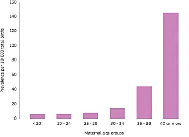
DS manifests itself in people of all races and socio-economic status, with a prevalence of 1/700-800 live births. Data showed that the probability of giving birth to a child with DS increases significantly with the mother age (above 35 years). At present the incidence of DS has not decreased despite several prenatal diagnosis, available since the middle of sixties, (Summers et al., 2007) and maternal serum screening (Smith & Visootsak, 2013). Indeed, studies have reported increasing trends in DS pregnancies in various parts of the world ascribed to increased lifespan and maternal age. As shown in Figure 1, Maternal
age is the biggest risk factor with an incidence of 1:1,500 births under age 25 and 1:100 births at age 40 (Fig. 1) (Herault et al., 2017; Loane et al., 2012; McKenzie et al., 2016).
Figure 1. Graph from the European Union showing the prevalence of Down syndrome increasing
with maternal age (Lanzoni et al., 2019).
1.1.2 Etiology and the genetics of Down syndrome
The triplication of human chromosome 21 (Hsa21) observed in DS is caused by abnormal cell division that results in an extra full or partial copy of chromosome 21. The most frequent form of DS is full Hsa21 trisomy: chromosome 21 is unable to separate during meiosis in a developing ovum, or less frequently, in sperm, culminating in an extra copy of the entire Hsa21 in all cell types (Antonarakis et al., 2004). Another cytogenetic form of DS is Robertsonian translocation. This condition is less prevalent than full trisomy 21 and occurs only in 2-4% of the cases, where a segment of the chromosome becomes fused to a different chromosome pair (such as chromosomes 13, 14, 15, 22 or 21) (Pelleri et al., 2016). Another genetic rearrangement that can occur in DS is mosaicism. This condition occurs in 3-4 % of DS population: in this case some
cells within a single tissue display a normal karyotype, while others show trisomy (Antonarakis et al., 2017; Rachidi & Lopes, 2011).
In 2000 a consortium performed the sequencing of chromosome 21 (Hsa21). [The most updated and revised version is available in the web, (www.ncbi.nlm.nih.gov/genome/gdv/) (Strippoli et al., 2019)]. Hsa21 is the smallest among human autosomes, consisting of about 46 million base pairs in its DNA, containing 325 non‐protein encoding genes and 222 protein coding genes (Gupta et al., 2016). Since 1959 literature data have showed that DS is caused by an extra copy of chromosome 21 (Lejeune et al., 1959), however the mechanisms by which the trisomy disrupts development are still not well understood (El Hajj et al., 2016). Two hypothesis have been proposed to explain the effect of trisomy on brain phenotypes: the “gene-dosage effect” and the “amplified developmental instability”. The first one proposes that increased dosage of several dosage-sensitive genes and their encoded proteins determined DS phenotypes (Bartesaghi et al., 2011; Delabar et al., 1993; Korenberg et al., 1994; Lyle et al., 2008). The second hypothesis asserts that trisomy 21 causes a general alteration in developmental homeostasis determining most manifestations of DS (Antonarakis et al., 2004; Bartesaghi et al., 2011; Roizen & Patterson, 2003). These two different theses could coexist in DS pathophysiology and the validity of one of the two hypothesis is still an open discussion (Strippoli et al., 2019).
Furthermore, literature data suggested that the triplication of some Hsa21 genes is sufficient to manifest DS (Aula et al., 1973; Ilbery et al., 1961; Wahlsten et al., 2019) supporting another hypothesis: the “Down syndrome critical region” (DSCR). The DSCR is a region of 3.8-6.5 Mb on 21q21.22, with approximately 33 genes responsible for the majority of DS phenotypes that will be summarized in the following chapters (Asim et al., 2015; Pritchard & Kola, 1999).
Studies showed that also epigenetic changes can occur both in fetal brains and blood from newborn DS infants and contribute to the pathology (Strippoli et al., 2019; Vacca et al., 2019).
1.1.3 Phenotypic traits and clinical features observed in Down syndrome
Down syndrome individuals are characterized by several phenotypic traits and clinical features that occur to some degree in every person with trisomy 21. A typical DS trait is the facial dysmorphology identified in microgenia, flat nasal bridge, oblique eye fissures, a bulging tongue and a short neck. In addition to these phenotypic traits, trisomy 21 is also a risk factor for other several diseases. One of the most invaliding is the congenital heart disease (CHD) which is the main cause of death for the two first years of life in individual with DS, whit a frequency of 40-50 % (Benhaourech et al., 2016; Ferencz et al., 1989; Roper & Reeves, 2006). Other phenotypic features that can affect DS individuals are malformations of the gastrointestinal tract, muscle hypotonia, leukemia, thyroid disorders, such as hypothyroidism, epilepsy and also Alzheimer’s disease (AD) (Amr, 2018; Barca et al., 2014; Mateos et al., 2015; Noble, 1998).
In particular, a connection between AD and DS has been long suspected since 50-70 % of DS individuals develop dementia by the age of 40. For this reason trisomy 21 is considered as the most common genetic cause of a neurodegenerative disease (Ballard et al., 2016; Dekker et al., 2018; Herault et al., 2017). An important role for this connection is partially played by the overexpression of amyloid precursor protein, encoded by the gene APP, which increases the risk of early-onset of AD. Indeed, amyloid- accumulates in the brain across the lifespan of people with DS. APP is not the only one AD-linked protein described in DS literature. Studies reported that the triplication of the gene that encodes for the Dual-specificity tyrosine-(Y) phosphorylation regulated
kinase (DYRK1A) may play an important role in this context. DYRK1A is a proline-directed serine/threonine kinase (Park et al., 2010) proposed as one of the most relevant contributors to the neurological abnormalities that will be summarized in the next paragraph (Dowjat et al., 2007).
1.1.4 The neurological phenotype of Down syndrome
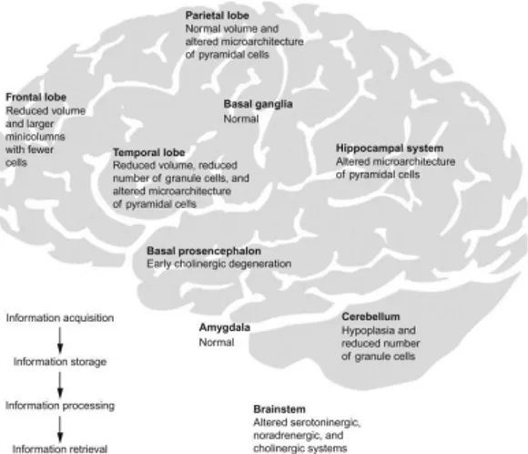
Intellectual disability (ID) is the main feature of DS resulting in a gradually declining intelligence quotient (IQ) during childhood (between 6 months and 2 years). In child IQ varies from 35 (mild) to 70 (moderate) and DS adult individuals usually show a mental age from 8 to 9 years old (Weijerman & De Winter, 2010). Beginning at an early age, DS neurological phenotype seems to be due to impairments in several brain regions such as the cerebellum, the medial temporal lobe, hippocampus and prefrontal cortex (Fig. 2) (Bartesaghi et al., 2011; Nadel, 2003).
In particular, ID affects the area involved in speech (poor and slower), in spatial memory and in long-/short-term memory performances, especially in maintaining phonological information over a short delay. By contrast, DS individuals show a relative preserved visuo-spatial memory (“where” memory) (Bostelmann et al., 2018; Godfrey & Lee, 2018; Vicari, 2006). Implicit memory is preserved in DS children, while explicit memory is impaired. In detail, implicit memory requires low attention and is supported by automatic processes. Explicit memory requires high attention, conscious learning and strategies. Infants with DS show also motor skill impairments: these children are unable to roll until 5-6.4 months and to sit independently until 8.5-11.7 months (Bartesaghi et al., 2011). Cerebellar hypoplasia and motor dysfunctions can affect learning skills in DS patients (Vicari, 2006).
Figure 2. Brain map of structures involved in the learning circuit (acquisition, processing or
information storage). Under each brain area the status in a Down syndrome brain is specified (Lott & Dierssen, 2010).
Furthermore, studies showed that both environment and genetic expression influence DS neurological phenotype that changes across the life span and, in most cases, culminate with dementia, as previously described (Bartesaghi et al., 2011).
1.2 Neurodevelopmental alterations in Down syndrome brain
Down syndrome is a neurodevelopmental disorder in which the brain develops differently from an euploid child and in particular is altered in configuration and reduced in size (Bartesaghi et al., 2011). The widespread brain hypoplasia and the consequent hypocellularity are considered to be one of the main cause of intellectual disability (Stagni et al., 2018).
1.2.1 Gross anatomy
DS Brain morphology is characterized by reduced size (20 % smaller than euploid developing brain) and weight. The reduction in size is detectable in 4-5 month fetuses and is maintained for the rest of the gestation (Engidawork &
Lubec, 2003; Lott, 2012). Numerous DS brain regions are smaller compared with control individuals (Pinter et al., 2001; Stagni et al., 2018) and volume reduction is pronounced for the hippocampus, cerebellum and brainstem (Guidi et al., 2014; Raz et al., 1995; Stagni et al., 2018).
1.2.2 Cytoarchitecture
Characteristic features of the DS brain are the diffused hypocellularity, astrocytic hypertrophy and decreased thickness of cortical layers (Bartesaghi et al., 2011). At early gestation stages cell density appears normal, but later (gestation week, GW, 19-23) less neurons are described and scarcity continues throughout early life compared to euploid fetuses (Golden & Hyman, 1994; Guidi et al., 2008). Compared to matched euploid fetuses at GW 17-21, DS hypocellularity is prominent in the dentate gyrus (DG) of hippocampus and in the parahippocampal gyrus (Guidi et al., 2008; Stagni et al., 2018). Also the granule cell density in the cerebellum is reduced in children and then adults with DS (Baxter, 2000; Guidi et al., 2008; Ross et al., 1984; Stagni et al., 2018).
In addition to neuronal alterations there are also defects of other cell types, including glial cells. Indeed, an increased astrocyte-neuron ratio has been described in hippocampal structures of DS fetuses compared to euploid ones (Bartesaghi et al., 2011; Guidi et al., 2008). An increased astrocyte number and astrocytic hypertrophy have been shown in both developing and adult DS brain (Griffin et al., 1998).
1.2.3 Neurogenesis alterations
During the development of fetal brain, neural stem cells (NSC) proliferate, maturate into neural progenitor cells (NPC) and differentiate into neurons in a process called neurogenesis (Kitamura et al., 2009). In detail, the neural tube
starts to differentiate into an outer zone and an inner proliferative layer since GW 6, at that time some neurons have already been born. At GW 7 the subventricular zone (SVZ) and the ventricular zone (VZ) appear. At GW 7-8 neurons migrate from these zones to form the cortical plate (Chan et al., 2002). By GW 24 a large reduction of VZ and SVZ occurs combined with a reduction in proliferation (Chan et al., 2002). The neurogenesis process in the dentate gyrus (DG) starts at GW 12 and is maintained within the first postnatal year (Rice & Barone Jr., 2000). Neurogenesis is mainly active during brain development, but this process is maintained also during adulthood in two specific neurogenic niches: the subventricular zone (SVZ) and the subgranular zone (SGZ) of hippocampus (Bortolotto et al., 2019; Cvijetic et al., 2017; Martínez-Cerdeño & Noctor, 2018).
The reduction of this process during early life stages is thought to be among the major neurodevelopmental defects leading to DS cognitive impairment (Stagni et al., 2018). Neurogenesis in the fetal DS brain has been little studied since the difficulties in obtaining fetal material. However, several studies showed that cell proliferation is impaired in different regions of the fetal DS brain, such as in the hippocampus germinal zones, DG and germinal matrix of the inferior horn of the lateral ventricle (Contestabile et al., 2007; Guidi et al., 2008; Stagni et al., 2018). This defective development determines the reduction in neuron number and impairment in cortical structures detected in DS fetuses and then in children (Contestabile et al., 2007; Pinter et al., 2001; Schmidt-Sidor et al., n.d.; Sylvester, 1983; Winter et al., 2000). These data have been further investigated in the chapter about neural progenitor cells in DS.
1.2.4 Gliogenesis alterations
In humans, the formation of the general architecture of brain regions and neurogenesis are mostly complete at birth, while maturation of the two major glial cell populations (astrocytes and oligodendrocytes), myelination, synaptogenesis and synapse pruning occur during postnatal brain development (Jiang & Nardelli, 2015; Lee et al., 2016), since neurogenesis precedes gliogenesis.
Several studies showed an increased neurogenic-to-gliogenic modulation in DS brain, but the mechanisms and the relative consequences of this shift are still unclear. Indeed, compared to matched euploid fetuses at GW 17-21, astrocytes are increased in the hippocampus (Guidi et al., 2008) and in the frontal lobe, where they appear also more mature than the euploid counterpart (Dossi et al., 2018)
1.2.5 Dendrite, spine, synapse and neurotrophin alterations in Down syndrome brain
Dendritic spines are structures, rich in actin, that constitutes the postsynaptic terminals of excitatory synapses (Lee, Zhang, & Webb, 2015). Beginning from infancy (3-4 months of age), typical DS hallmarks are the reduced neuronal complexity with atrophy of the dendritic tree, spine density reduction and alterations in spine shape (Bartesaghi et al., 2011). These abnormalities do not recover at subsequent life stages (Torres et al., 2018). 3-4-month-old DS children show dendritic hypotrophy in neurons of the parietal cortex (Schulz & Scholz, 1992), motor cortex (Prinz et al., 1997) and visual cortex (Becker, Armstrong & Chan, 1986). Another typical DS feature, not observed in fetuses, but appearing in newborns and older DS infants, is the reduction and alteration in spine number and morphology (Takashima et al., 1981). Indeed smaller spines, with short
stalks, have been reported in children with DS compared with age-matched euploid individuals (Marin‐Padilla, 1976).
All together dendrite and spine alterations in DS imply a reduction of the surface available to exchange synaptic inputs. Indeed, a defective synaptic function and organization are typical features of DS brain, associated with alterations of transmitter systems (Bartesaghi et al., 2011; Chakrabarti et al., 2007; Kurt et al., 2000). Monoamines, such as serotonin and dopamine, has been demonstrated to be reduced in frontal cortex of DS fetuses compared to euploid one (Risser et al., 1997; Whittle et al., 2007)
Actin cytoskeleton rearrangements are important for the formation of dendritic spines and synapses (Risher & Eroglu, 2012). Different proteins involved in the formation of the neuronal cytoskeleton are downregulated in DS brain including the beta-tubulin (Pollak et al., 2003), the microtubule associated protein, MAP2 (Ohara et al., 1999), and the actin-related protein complex 2/3 (ARP 2/3). ARP2/3 is a complex that controls actin remodeling and is required, as a downstream effector, at various stages of brain development (Chou & Wang, 2016; Weitzdoerfer et al., 2002). Another protein downregulated in DS brain is moesin (Lubec et al., 2001). Moesin is involved in plasma membrane-actin cytoskeleton cross-linking and it is critical for the morphology of neurons and the formation of long-term memory (Freymuth et al., 2017).
Furthermore, neurotrophin levels are impaired during DS brain development (Bartesaghi et al., 2011). These molecules are important to support neuronal differentiation, migration, synaptic plasticity and survival (Campenot & MacInnis, 2004; Chao et al., 1998; Chao et al., 2006; Sofroniew et al., 2001). In particular the brain derived neurotrophic factor (BDNF), a neurotrophin that binds the surface tyrosine receptor kinases (TRKs), belongs to this family.
Reduced expression of BDNF is observed both in hippocampus (Guedj et al., 2009) and cerebral cortex of DS fetuses (Toiber et al., 2010).
1.2.6 Specific triplicated genes with a role in Down syndrome neuropathology
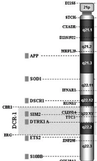
The sequencing of genes on chromosome 21 has been completed, although the functions of most of the encoded proteins is not still clear (Engidawork & Lubec, 2003). Recently, Roizer et al. showed that proteins encoded by specific triplicated genes play a role in DS neuropathology (Fig. 3) (Roizen & Patterson, 2003). Below there are several examples of genes that encode proteins involved in DS pathology.
Figure 3. A cartoon in which specific genes on the human chromosome 21 (Hsa21) triplicated in
DS fetal brain and involved in brain development are represented, including DYRK1A, APP, S100B, SOD1, DSCR-1 (also known as RCAN1). DCR-1: Down syndrome chromosomal region-1. Picture modified from Rachidi & Lopes, 201region-1.
DYRK1A (dual specificity tyrosine phosphorylation regulated kinase 1A)
encodes a member of the dual-specificity tyrosine phosphorylation regulated kinase (DYRK) family. As previously introduced it plays an important role in the
neurological abnormalities associated with DS. Indeed, during brain development DYRK1A regulates neural progenitor cell proliferation and neuronal differentiation (Dowjat et al., 2007). In the young adult brain DYRK1A can hyperphosphorylate tau, determining progressively depolymerization of actin microfilament, dendritic hypotrophy and neurofibrillary tangles formation. This aspect affects several DS brain regions, such as hippocampus, prefrontal cortex, midbrain, thalamus, hypothalamus and basal ganglia (Lott & Head, 2019; Wisniewski et al.,1985).
APP (amyloid precursor protein) encodes the amyloid precursor protein (APP),
a trans-membrane protein mostly expressed in neuronal synapses. This gene is reported to play a key role in DS neurodevelopmental alteration and, as previously mentioned, is involved in the development of Alzheimer-like pathology in DS adults. Furthermore, APP can participate to neuronal plasticity (Turner et al., 2003) and it can affect the formation and transmission of synapses in cultured hippocampal neurons (Priller et al., 2006).
S100 (S100 calcium binding protein B) encodes for a protein member of the
S100 family released by astroglial cells. It is highly expressed during development and aging. Increased levels of S100 characterize both adult DS and AD individuals.
SOD1 (superoxide dismutase 1) encodes the superoxide dismutase [Cu-Zn], an
enzyme that binds Cu and Znto breakdown superoxide radicals and convert them to H2O2 to avoid cell damage. This is a constitutive enzyme which activity is
increased by 50% in DS. Increased levels of SOD1 determine increased oxidative stress and lipid peroxidation in brain human DS cortical neurons (Bartesaghi et al., 2011).
RCAN1 (regulator of calcineurin 1) encodes the regulator of calcineurin factor
and it is overexpressed especially in fetal DS brain. RCAN1 protein inhibits calcineurin A, a serine threonine phosphatase that activates the nuclear factor of
activated T cell cytoplasmatic (NFATc). NFAT is involved in the regulation of cell proliferation, neuronal migration and survival (Serrano-Pérez et al., 2015).
1.3 Rodent models in Down Syndrome research
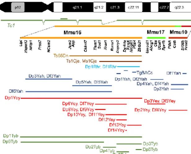
Mouse models are able to mimic, as closely as possible, human pathologies. Mice are important tools that can be investigated to understand the mechanisms underpinning a disease. In particular, in DS they can be used to: 1) investigate genotype-phenotype relationship; 2) identify dosage-sensitive genes involved in DS pathophysiology, 3) test the effect of potential drugs. Till now, a large number of mouse models that recapitulate the phenotypic features of DS have been developed in order to exploit this complex genetic disorder (Bartesaghi et al., 2011; Herault et al., 2017). Several chromosomal rearrangements and modifications have occurred over the evolutionary time that separate mice and humans. Thus, the human chromosome 21 has three orthologous regions on mouse chromosomes 16, 17 and 10, as shown in Figure 4 (Herault et al., 2017). Most of the murine genes that are homologous to humans reside on Mmu16 (102), Mmu17 (19) and the rest on Mmu 10 (37) (Gupta et al., 2016).
Figure 4. The human chromosome 21 (Hsa21) is represented at the top. Below the orthologous
green) are shown. Several genes homologous to Hsa21 in the Down syndrome critical region are indicated below each murine chromosome. The humanized Tc1 mouse model is represented in dark green below the Hsa21. Below the murine chromosomes there are several examples of DS mouse models. For each mouse model the part of Down syndrome critical region incorporated in each mouse model (different color) is reported (Herault et al., 2017).
The first effort in DS mouse modelling was attempted by Gropp et al. in 1975. Gropp et al. developed a mice with full trisomy of chr 16, Ts16, (Gropp, Kolbus & Giers, 1975). This model is limited due to its embryonic lethality and, importantly, they do not model DS phenotype because the bulk of triplicated genes derive from Mmu16 regions not homologous to Hsa21 (Webb, Brown & Anderson, 1998).
The field of DS preclinical research progressed by the discovery (1990) and phenotypic characterization (1995) of the Ts65Dn mouse (Davisson et al., 1990; Reeves et al., 1995). This model shows a segmental (partial) trisomy of Chr16 generated by a Robertsonian translocation (as a consequence of exposure to radiation). Ts65Dn mice bear the extra copy of chromosome 16 (Mmu 16) translocated onto a small Mmu17 segment. The triplicated Mmu16 includes 90 conserved protein-coding genes that are orthologous to Hsa21 (Choong et al., 2015; Gupta et al., 2016). Ts65Dn mice are trisomic for around the 55% of the orthologous genes on Hsa21, but are also trisomic for a large number of genes that are not orthologs of Hsa21 genes (Gupta et al., 2016). These mice recapitulate many features similar to DS individuals since embryonic life stages and then in adulthood (Stagni et al., 2018). For these reasons, in the last decades the Ts65Dn mouse line has been widely used to investigate DS and has provided many important understandings in this research field (Bartesaghi et al., 2015).
Ts65Dn mice phenotype is characterized by postnatal developmental delay starting from a reduced birth weight, skeletal malformations, muscular trembling, and, in adulthood, male sterility (Galdzicki & Siarey, 2003).
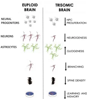
Similarly to humans, the Ts65Dn mouse line shows several neurodevelopmental alterations, the majority summarized in Figure 5 and described in the following paragraph. Indeed, Ts65Dn mice exhibit a reduced brain size and a widespread cell paucity immediately after birth (since the post-natal day 2, P2) in the hippocampus, DG, SVZ, striatum, thalamus, neocortex and cerebellum (Belichenko et al., 2004; Bianchi et al., 2010; Giacomini et al., 2015; Guidi et al., 2014; Lorenzi & Reeves, 2006).
Figure 5. Major brain alterations in euploid and trisomic brain in the Ts65Dn animal model.
These features recapitulate the human Down syndrome brain phenotype. Image modified from Bartesaghi et al., 2011.
As in humans, the hypocellularity that characterized Ts65Dn brain is associated with a widespread neurogenesis impairment ascribed to an impaired proliferation of neural progenitor cells (Stagni et al., 2018).
The neurogenic-to-gliogenic shift is observed also in Ts65Dn mice. Contestabile et al., showed an increased number of cells with an astrocytic phenotype in the DG of young Ts65Dn mice (P 30) compared to age-matched euploid mice (Contestabile et al., 2007; Lee et al., 2016). Furthermore, in these mice an increased inhibitory-excitatory neuron ratio is observed in the cortex and in the Cornu Ammonis field 1 (CA1) of Ts65Dn mice at P8 and P15 (Chakrabarti et al., 2010). Studies observed dendrite and spine abnormalities in density and shape in young Ts65Dn mice. 45-day-old Ts65Dn mice show hypotrophic dendritic trees in the DG granule cells compared to age-matched mice and 10-week-old Ts65Dn mice show fewer and shorter dendritic branches in pyramidal cells (Dierssen et al., 2003; Pollonini et al., 2008). In parallel, in adult Ts65Dn the synaptic density is significantly impaired in the DG, CA1 and CA2 of hippocampus if compared with euploid mice (Ayberk Kurt et al., 2004). As in humans, reduced BDNF levels were detected in the hippocampus of 15-day-old Ts65Dn mice (Bianchi et al., 2010).
Importantly, young adult Ts65Dn mice exhibit impairment in learning and memory. They show abnormalities in hippocampal synaptic plasticity and hyperactivity under certain experimental condition. These defects are associated with a defective long-term potentiation (LTP), ascribed to a decrease in the density of N-methyl-D-aspartate (NMDA) receptors, and depression (LTD) (Contestabile et al., 2010). Similar to DS child, studies showed sensor and motor alterations in DS mice since the birth, maintained during adulthood: 4-6-month-old Ts65Dn mice show impaired motor functions (Costa et al., 2010).
As in humans, APP protein expression is upregulation from embryonic day 15 in Ts65Dn mouse cerebral cortex. In adulthood these mice exhibit the signs that are considered the onset of Alzheimer’s disease: degeneration of cholinergic basal forebrain neurons and impaired cholinergic system (Galdzicki & Siarey, 2003).
Despite its importance, Ts65Dn mice have some limitations. At first, as previously introduced, the animal bears the triplication of several genes that are non-DS-related (Reinholdt et al., 2011). Second, since Ts65Dn males are sterile (Moore et al., 2010) the colony is usually expanded only by using Ts65Dn dams. This aspect can affect pups determining developmental abnormalities that are independently from the trisomy (Bartesaghi et al., 2015, 2011; Herault et al., 2017; Stagni et al., 2018).
Another model of partial trisomy 16 is the Ts1Cje , created by Sago et al. Ts1Cje are mice trisomic for a small region of Mmu 16, containing 79 gene orthologous to Hsa21. As for Ts65Dn mice, this model exhibits learning and behavioral disabilities, but these deficits are less severe than those of Ts65Dn. Studies showed basal forebrain cholinergic neuron degeneration also in these mice (Bhattacharyya & Svendsen, 2003; Villar et al., 2005).
In the last two decades the field of DS mouse modelling changed significantly with the advent of two lines: the chromosome engineered and the transchromosomic mice, that are transgenic animals bearing a chromosome isolated from a different species. Indeed, in 2005 O’Doherty et al. published the first transchromosomic DS model, namely Tc1 (formally called Tc(Hsa21)1TybEmcf) (O’Doherty et al., 2005). These animals contain 269 genes of the Hsa21, including those gene orthologous located on Mmu17, 16 and 10 that are not present in Ts65Dn and Ts1Cje (O’Doherty et al., 2005). However, the biggest disadvantage of Tc1 mice is that they develop mosaicism in different tissues (Herault et al., 2017).
1.4. Focus on cell types involved in Down syndrome pathophysiology 1.4.1 Neural progenitor cells
Stem cells are defined as “pluripotent cells able to self-renew and to differentiate into other cell types in tissues and organs” (Li & Zhao, 2008). The behavior and fate of stem cells can be affected by their location and by signals released in the niche, a specific microenvironment where stem cells can exist also as progenitor cells (Romito & Cobellis, 2016).
In the Central Nervous System (CNS) neural progenitor cells (NPC) are able to self-renew and to differentiate into neurons, astrocytes and oligodendrocytes. NPC can be found widespread in the developing fetal brain whereas in the neonatal, but especially in the mature adult brain, NPC are mainly restricted into the subventricular zone (SVZ) and the subgranular zone (SGZ) of the dentate gyrus of hippocampus (Bernabeu-Zornoza et al., 2019; Bortolotto et al., 2017; Meneghini et al., 2010; Valente et al., 2012).
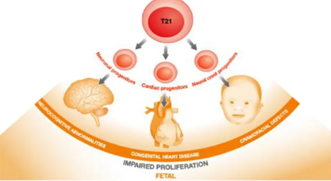
Several studies investigated whether trisomy 21 affects progenitor cells during fetal development. Liu et al. performed a systematic review of the literature, both in DS individuals and in DS mouse models. They showed that trisomy 21 impairs proliferation of different type of progenitor cells, in particular severely impaired are neural progenitor cells of trisomic individuals (Figure 6) (Liu et al., 2015).
Figure 6. Impact of trisomy 21 on the proliferation of progenitor cells from different lineages
trisomy 21 and impact on the development of craniofacial structures, hearth and, importantly, brain. Image adapted from Liu et al., 2015.
Literature data showed that DS brain hypocellularity, already mentioned in previous chapters, is associated with a widespread reduction in the NPC proliferation rate in several brain regions of DS fetuses (Stagni et al., 2018): the ventricular germinal matrix, cerebellum and hippocampus (Contestabile et al., 2007; Guidi et al., 2008). Studies performed on neural progenitor cells differentiated from trisomic induced pluripotent stem cells (iPSCs) derived from a DS individual exhibited a reduced proliferation compared to euploid (Murray et al., 2015). Moreover, Hibaoui et al. characterized iPSCs from monozygotic twins discordant for trisomy 21: also in this case a reduced number of NPC has been detected in trisomic cells compared to diploid ones (Hibaoui et al., 2014).
Mice models are helpful for investigating the distribution in space and time of the phenotypic defects observed in the trisomic brain. Indeed, similarly to human brain the bulk of neurogenesis in mice happens in the VZ and SVZ before birth, while in the SGZ it occurs in the two postnatal weeks and continues into young adulthood, then decreases with age (Altman & Bayer, 1975, 1990).
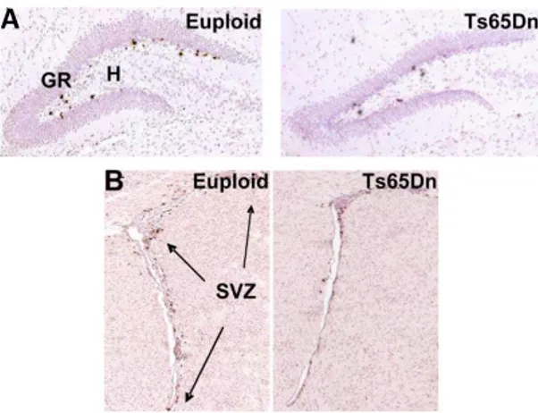
In particular, SVZ is an important postnatal niche that gives rise to granule neurons of the olfactory bulbs and cells of the neocortex in the first postnatal days (Brazel et al., 2003). In Ts65Dn pups a reduced proliferation rate have been detected both in the SVZ at the day post-natal 2 (P2), at P15 (representative picture in Figure 7B) (Bianchi et al., 2010; Guidi et al., 2014; Stagni et al., 2016) and in the DG at P2, P6 and P15 (representative picture in Figure 7A) (Contestabile et al., 2009; Giacomini et al., 2015; Stagni et al., 2018, 2019). Moreover, studies showed a strong decreased proliferation also in cerebellar precursor cells of P0, P2, P6 Ts65Dn pups (Contestabile et al., 2009; Roper et al., 2006).
Figure 7. Representative pictures of proliferation impaired in the dentate gyrus (DG, A) and
subventricular zone (SVZ, B) of euploid and Ts65Dn mice at the post-natal day 15 (P15). A and
B are immunostained sections for Bromodeoxyuridine (BrdU) (brown), commonly used to detect
proliferating cells. Sections were counterstained with hematoxylin (pink). Euploid and Ts65Dn mice received an injection of BrdU at the day fifteen after birth and then were sacrificed after 2 h. GR=granular cell layer; H=hilus, SVZ=subventricular zone (from Bianchi et al., 2010).
1.4.1.1 Mechanisms underlying impaired neural progenitor proliferation
The mechanisms underlying a reduction in the number of neural progenitor cells in DS are not totally understood (Liu et al., 2015). Evidence in literature shows that cell cycle alterations in DS brain are strictly involved in the impaired NPC proliferation (Najas et al., 2015; Salomoni & Calegari, 2010). Cell cycle is controlled by cyclin-dependent kinases (CDK), their inhibitors, cyclins and it is composed by four different phases: G1, G2, S and M phase. Impaired phases in the cell cycle can affect the balance between undifferentiated NPC and NPC addressed to a specific phenotype acquisition (Smith & Calegari, 2015). Literature data showed an extended G1 phase in DS human fibroblasts (Chen et al., 2013). Other studies detected an elongated G2 phase in DG and in ventricular germinal layer of DS fetuses compared to age-matched euploid fetuses (Contestabile et al., 2007).
In mice, NPC isolated from the hippocampus and lateral ventricle of Ts65Dn embryos exhibit an elongated S phase as well as of the entire cycle (Chakrabarti et al., 2007). G1 phase is elongated also in granule precursor cells of Ts65Dn neonates if compared with the euploid counterpart (Lorenzi & Reeves, 2006). Several data mainly ascribe these mechanisms to the triplicated genes RCAN1, DYRK1A, APP and the oligodendrocyte transcription factor 1 and 2 (OLIG1-2) (Chakrabarti et al., 2010; Najas et al., 2015; Stagni et al., 2018, 2019). As already mentioned, RCAN1 encodes for a protein able to interact with calcineurin A. This interaction inhibits the pathway calcineurin-NFATc, axis hypothesized to regulate SVZ-derived NPC proliferation and differentiation (Bianchi et al., 2010). NFATc and the related transcripted genes can be regulated also by DYRK1A (Arron et al., 2006; Jung et al., 2011). Interestingly DYRK1A can phosphorylate RCAN1 and this event primes RCAN1 phosphorylation mediated by GSK3 (Glycogen synthase kinase 3 beta), resulting in RCAN1 increased activity (Coronel et al., 2019). Since GSK3 is overactivated in DS brain, in conjugation with DYRK1A and RCAN1, it may inhibit NFAT activity (Stagni et al., 2018).
Moreover, at GW 14 and GW 18 OLIG2 is overexpressed in DS frontal cortex. Studies showed OLIG2 overexpression also in human NPC differentiated from iPSCs derived from a DS patient compared with euploid NPC (Chakrabarti et al., 2010; Lu et al., 2012). In particular, in human trisomic NPC an impaired proliferation rate matches with increased OLIG2 expression (Lu et al., 2012). To make this picture more complex, the increased APP levels detected in DS brain negatively affect cell proliferation. Data suggested that this mechanism is due to increase in APP intracellular fragment (AICD), obtained by APP processing. Excessive levels of AICD may interfere with GSK3 signaling and Sonic Hedgehog (SHH) pathway, involved in stem cell proliferation. Thus,
increased AICD levels determine the consequent increase of the protein patched homolog 1 (PTCH1), a repressor of the mitogenic SHH (Trazzi et al., 2011).
1.4.1.2 Mechanisms underlying impaired neural progenitor phenotype acquisition
Neurons, astrocytes and oligodendrocytes are cells that compose the human forebrain, derived from the VZ and SVZ, and, at first, neurons are generated, followed by astrocytes and oligodendrocytes (Sauvageot & Stiles, 2002). As previously mentioned, in Down syndrome neuronal phenotype acquisition is defective, and trisomic NPC show a shift toward an astroglial phenotype, observed since the earliest stages of life. The quantification of the number of astrocytes (GFAP+) and of the mature neurons (NeuN+) in DS fetal hippocampi showed fewer neurons and more astrocytes compared to euploid fetuses (Guidi et al., 2008). Also in vitro, trisomic NPC differentiated from iPSCs spontaneously give rise to more astrocytes (S100 +) and fewer neurons (betaIII-Tubulin+). Moreover, these neurons exhibit a decreased neurite length (Chen et al., 2014a; Hibaoui et al., 2014).
Even in DS mouse models the NPC phenotype acquisition is altered. Indeed, an imbalance between neurons and astrocytes is observed in cultures of SVZ-derived NPC from Ts65Dn pups (Stagni et al., 2019; Trazzi et al., 2011) and in the DG and cerebellum of young adult Ts65Dn mice (Ishihara et al., 2010; Stagni et al., 2016).
Different intracellular pathways may play a role in the DS dysregulated phenotype acquisition. In particular, a key role seems to be played by the Janus kinase-signal transducer and activator (JAK-STAT). JAK-STAT activator ligands and receptors are overexpressed in DS brain (Bonni et al., 1997; Trazzi et al., 2013). Studies suggested that the overstimulation of this pathway is linked to the triplicated genes DYRK1A and APP: APP enhances the activity of
JAK-STAT and increases GFAP levels in Ts65Dn NPC derived from P2 pups (Trazzi et al., 2013). Further studies showed that gliogenesis may be affected by the interaction between JAK-STAT signaling and NOTCH (Taylor et al., 2007) which is activated in DS and affected by APP (Fischer et al., 2005).
1.4.2 Astrocytes
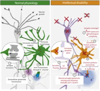
Astrocytes display several functions such as modulation of neuronal plasticity, synaptic maturation and pruning, ionic homeostasis and, importantly, a trophic function to support neuronal activity (Dossi et al., 2018; Sidoryk-Wegrzynowicz et al., 2011; Vasile et al., 2017). In recent years novel insights have been gained about the role of astrocytes in normal brain and in pathological condition, as summarized in Figure 8. Indeed, astrocytes in intellectual disabilities show an altered astrocytic morphology (increased GFAP levels), a defective secretion of trophic signals, impaired synaptic pruning, ionic homeostasis and an altered glutamate sensing and clearance (Figure 8) (Chen et al., 2014b; Cresto et al., 2019; Vacca et al., 2019)
Figure 8. Astrocytes in physiology (left, green) and in intellectual disabilities (right, orange)
In particular, DS astrocytes are not only more abundant, proliferative and mature in their morphology (Zdaniuk et al., 2011), but their functions are altered. Chen et. al studied both DS astrocytes and DS neurons obtained from iPSCs isolated from DS individuals, compared with euploid cells. They demonstrated that DS astrocytes exhibit lower levels of synaptogenic molecules and higher levels of reactive oxygen species (ROS). This reactive state has been hypothesized to disrupt astrocyte homeostasis hampering their ability to promote maturation and support to neurons (Chen et al., 2014). Furthermore, evidence in literature showed an aberrant calcium signaling (more frequent) and increased Ca2+ fluctuations both in astrocytes derived from DS iPSCs and in DS mouse models. Elevated Ca2+ levels are hypothesized to reduce neuronal excitability in DS (Cresto et al., 2019; Mizuno et al., 2018).
1.4.2.1 Non-cell autonomous regulation of neural progenitor cells
Astrocytes are secretory cells able to secrete a wide array of hormones, neurotransmitters and metabolic, trophic factors. Release occurs through several distinct pathways that include diffusion through channels, controlled exocytosis and transporter translocation (Verkhratsky et al., 2016). These molecules exhibit important functions such as the formation of functional synapses (Araujo et al., 2018) or neurogenesis, mediated, for example by BDNF, S100B and thrombospondins (Clarke & Barres, 2013; Cvijetic et al., 2017). Cvijetic et al. recently revealed first evidence of the complex signaling system implicated in the cross-talk between NPC and astrocytes, identifying novel pathways that can affect progenitors in a non-cell autonomous way (Cvijetic et al., 2017).
Taking into account this information, an important aspect that recently emerged is that DS astroglia presents functional alterations (Chen et al., 2014b) that may affect NPC and their progeny. To support this hypothesis Chen et al. showed that the media collected from human trisomic astrocytes (astrocyte conditioned
media-ACM) reduces human trisomic NPC neuronal differentiation (reduced levels of ßIII+ cells) and neurite length of trisomic neurons compared with trisomic cells treated with control media (Chen et al., 2013; Cresto et al., 2019). Furthermore, Garcia et al., demonstrated that a key astrocyte secreted molecule, thrombospondin-1 (TSP-1), is defective both in the secretome of DS human fetal astrocytes and in DS fetal brain (Garcia et al., 2010; Torres et al., 2018). Moreover, defective TSP-1 levels impair the development and morphology of dendritic spine in neurons from newborns rat. Indeed, addition of exogenous TSP-1 on neurons positively modulated astrocyte-mediated spine and corrected synaptic alterations (Garcia et al., 2010). All together these data highlight a potential role of TSP-1 in DS spine pathology (Torres et al., 2018).
TSP-1 is a calcium-binding protein that participates in cellular responses to injury, cytokines and growth factors (Chen et al., 2000). This protein plays a role in adult NPC proliferation and differentiation (Lu & Kipnis, 2010), synaptogenesis and spine formation (Eroglu et al., 2009; Garcia et al., 2010; Risher & Eroglu, 2012).
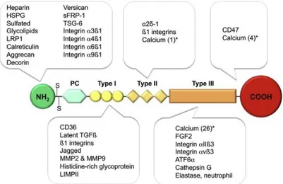
Figure 9. Representative structure of Thrombospondin 1. Type I, II, III are different binding
sites. For each domain are indicated the TSP-1 interactors. The asterisk (*) indicates the number of bounded calcium ions (Resovi et al., 2014).
As shown in Figure 9, TSP-1 contains different domains involved in the interaction with a multiplicity of receptors and ligands (Chen et al., 2000; Resovi et al., 2014). Worthy of attention is the α2δ-1 subunit of neuronal voltage-sensitive calcium channels (Dolphin, 2013). Recent evidence show that astrocytes control excitatory synaptogenesis by TSP-1 which functions via the α2δ-1 subunit independently from calcium channels (Dolphin, 2018; Eroglu et al., 2009).
Furthermore, this subunit is important since the antiepileptic and neuropathic pain drugs pregabalin (PGB) and gabapentin (GBP) bind α2δ-1 (Eroglu et al., 2009; Valente et al., 2012). A recent study showed that α2δ-1 is expressed on the surface of neural progenitor cells isolated from wild type mice and, through its binding, PGB and GBP are able to promote adult hippocampal neurogenesis, both
in vitro and in vivo (Valente et al., 2012). Thus, this study gave the first evidence
of a proneurogenic effect mediated via α2δ-1 (Valente et al., 2012).
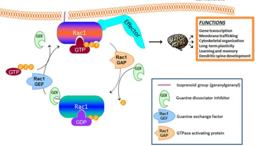
TSPs are known as molecules able to regulate the actin cytoskeleton that, as already mentioned, is involved in formation and remodeling of dendrites and synapses (Risher & Eroglu, 2012). Importantly, recent evidence showed that TSP-1/α2δ-1 interaction control synaptogenesis postsynaptically via Rac1. Rac1 is a small GTPase that belongs to the Rho family GTPases, proteins that control spine morphology and number (Risher & Eroglu, 2012). As shown in Figure 10, Rac1 exists in two forms: an inactive form (GDP-bound) and an active form (GTP-bound). The transition between the two forms is mediated by two catalytic factors, the GTPase-activating proteins (GAPs) and the guanine exchange factors (GEFs) (Tejada-Simon, 2015). The relationship between Rac1 inactivated and activated state is important for proper interaction of Rac1 with other targets downstream the signaling pathway. As summarized in Figure 11, Rac1 is important for several brain functions, including learning and memory and
dendritic spine development. Indeed, Rac1 alterations are described in different neurodegenerative diseases, including Alzheirmer’s disease (Désiré et al., 2005; Kikuchi et al., 2019; Risher et al., 2018).
Figure 10. Graphical representation of key players in activation and membrane translocation
for the small GTPase Rac1. In the orange field are listed several function that are ascribed to Rac1 activity in the brain. (Image modified from Tejada-Simon, 2015).
1.5. Potential therapeutic approaches proposed in Down syndrome
As already introduced, DS brain, both human and mouse, shows a constellation of defects involved in the cognitive impairment. The investigation of these defects is the starting point to find the rational basis to devise therapies that may correct DS brain developmental defects. Indeed, in the last decades, intense efforts have been carried out to identify pharmacotherapies and/or neurobiological factors known to positively affect brain of DS individuals and to potentially improve one or more DS-linked brain phenotype (Stagni et al., 2015). Mostly of these studies have been performed using the Ts65Dn mouse model (Gupta et al., 2016).
A key issue in the field is the optimum timing for drug administration in DS. Indeed, most of the attempts to pharmacologically correct DS-brain linked
defects have been made in adult mice. However, neurodevelopmental defects, both in human and in mice, are already detectable at fetal life stages. As represented in Figure 11 the bulk of neurogenesis occurs before birth, both in humans and mice. DG and cerebellum represent two exceptions: while in the DG neurogenesis continues throughout life, in the cerebellum stops after the first post-natal period. In this picture, adult therapies may relatively affect brain (hippocampal neurogenesis and circuitry), while perinatal (neonatal and prenatal) therapies may potential affect and correct overall brain development (Guidi et al., 2014; Stagni et al., 2015) (see chapter about NPC alterations in DS). Overall these data suggest that perinatal therapies that target neural progenitor cell alterations may be the potential optimal interventions in DS. Indeed, proof of concept studies showed that DS brain defects can be pharmacologically corrected in the animal model, if the therapy is administered in the perinatal period (Bianchi et al., 2010; Guidi et al., 2014; Nakano-Kobayashi et al., 2017; Stagni et al., 2015). These corrections can be maintained through individual life span. In the following chapters, some examples of drugs that have been tested in DS animal model will be summarized.
Figure 11. Timeline of brain development in mouse (A) and human (B). CB, cerebellum; CX,
neocortex; DG, dentate gyrus; E, embryonic; F, fetal; M, month; P, post-natal; W, week (Stagni et al., 2015)
1.5.1 Lithium
Lithium chloride (LiCl) is the first-line treatment for bipolar disorder. The molecular mechanisms of LiCl are not known in detail, studies demonstrated that lithium acts in part inhibiting the activity of GSK3 and modulating Wnt/β-catenin pathway (Pasquali et al., 2010). GSK3 antagonizes the Wnt signaling pathway and is upregulated in DS brain (Granno et al., 2019; Stagni et al., 2018; Zhang et al., 2019).
Contestabile et al. showed that administration of a diet containing LiCO3 (2.4
g/kg) for 1 or 4 weeks in 5-6-month-old Ts65Dn mice promotes the proliferation of NPC through the activation of the Wnt/-catenin pathway. The treatment is
able to restore neurogenesis in the DG of these mice to physiological levels, completely rescuing the synaptic plasticity of newborn neurons and recovering mice cognitive performances (Contestabile et al., 2013). In agreement with these findings, Bianchi et al., demonstrated that administration for 1 month of lithium (2.4 g/kg of LiCO3 in the food pellets) fully restores SVZ and DG proliferation
in 12-month-old Ts65Dn mice (Bianchi et al., 2010b). Despite these interesting data in adult trisomic mice, in clinic the treatment with lithium is associated with many side effects [including gastrointestinal side effects, polyuria, tremor, weight gain, dermatological effects (Gitlin, 2016)]. Bartesaghi et al. tried a neonatal treatment with LiCl in Ts65Dn pups. In detail, they gave lithium to trisomic mothers in order that pups should receive the drug through the milk, but lithium had a lethal effect on the offspring (Bartesaghi et al., 2011).
Despite its interesting effects in preclinical models, overall experimental data exclude the possibility of using lithium in patients.
1.5.2 Fluoxetine
Fluoxetine is an antidepressant that acts as a selective serotonin reuptake inhibitor (SSRI). This drug is largely prescribed in clinic for adult patients, but also for children and adolescents (Boylan et al., 2007).
5-hydroxytryptamine (5-HT, also named serotonin) is one of the major neurotransmitters of the CNS. Serotonin is essential for brain development since the earliest fetal stages and the serotoninergic system is shown to be altered both in DS fetuses and mice (Whittle et al., 2007).
Guidi et al. demonstrated that a prenatal treatment with fluoxetine in pregnant Ts65Dn mice (10 mg/kg, daily subcutaneous injection, from E10 to birth) is able to fully rescue DS brain abnormalities and behavioral deficits (Guidi et al., 2014). Furthermore, Bianchi et al. showed that fluoxetine modulates cell survival,
increases neurogenesis and dendritic development in the DG (Bianchi et al., 2010), restores functional connectivity of hippocampal synapses and hippocampus-dependent learning in Ts65Dn pups treated only during the two postnatal weeks (5 mg/kg from P3 to P7; 10 mg/kg from P8 to P15, daily subcutaneous injection) (Bianchi et al., 2010; Guidi et al., 2014). In addition, Stagni et al. observed that the beneficial effects of a neonatal treatment with fluoxetine endure in 45-day-old Ts65Dn mice, rescuing cognitive impairment in adulthood (Stagni et al., 2015).
These results represented a breakthrough in DS preclinical research, since they showed that DS neurodevelopmental impairments can be pharmacologically corrected by a perinatal intervention. These corrections rescue cognitive impairment in DS mice. Based on these findings, fluoxetine has been suggested as a potential prenatal therapy in rescuing fetal DS brain defects and dysfunctions (Kuehn, 2016). In 2014 the University of Texas Southwestern Medical Center approved a pilot study in pregnant mothers to investigate whether fluoxetine is effective in rescue cognitive impairments in DS fetuses, no information about this trial is yet available (Stagni et al., 2015).
Despite the really encouraging evidence obtained in the animal model, several adverse effects have been reported both in patients and mouse model of depression after long term treatment with this drug, including dizziness, nausea, headache and lipid metabolism abnormalities (Pan et al., 2018; Riediger et al., 2017). Furthermore, treatment with fluoxetine during pregnancy can affect the normal heart development of fetuses (Daud et al., 2016).
1.5.3 Epigallocathechin-3-gallate
In the past few years a lot of interest has been raised up in polyphenols. Polyphenols are phytochemicals produced by plants as secondary metabolites in
response to stress conditions. Epigallocatechin-3-gallate (EGCG) is a flavonoid of green tea extracts. The antioxidant activity and the inhibition of DYRK1A seems to be the potential mechanisms of action of EGCG in DS (Pons-Espinal et al., 2013; Thomazeau et al., 2014). Indeed, Valenti et al. showed that EGCG (20 µM) restores mitochondrial energy deficits observed in peripheral cells isolated from DS individuals (Valenti et al., 2013).
Stagni et al. demonstrated that neonatal administration of ECGC (25 mg/kg, daily subcutaneous injection from P3 to P15) corrects many DS-associated brain alterations at P15, but does not elicit durable effects in the hippocampus when measured at P45 (Stagni et al., 2016).
Catuara-Solarz et al. reported that green tea extracts containing EGCG (green tea extract, 45% EGCG) in combination with environmental enrichment for 30 days in 1-2-month-old Ts65Dn mice, enhances dendritic spine density in Cornu Ammonis field 1 (CA1) and stabilizes the proportion between excitatory and inhibitory synaptic markers in the DG and CA (Catuara-Solarz et al., 2016). In 3-month-old Ts65Dn mice EGCG (2-3 mg/day, 30 day treatment) normalizes DYRK1A activity, restores brain plasticity and partially rescues learning and memory (De la Torre et al., 2014). EGCG (20 µM) improves proliferation of adult hippocampal NPC isolated from 6/8-week-old Ts65Dn mice and restores the defective mitochondrial biogenesis (Valenti et al., 2016).
Despite this evidence, other scientific reports suggested that the administration of 10 mg/kg/day of pure stabilize ECGC failed to improve learning and memory in Ts65Dn young mice (treatment from P24 to 3-7 weeks after birth) (Stringer et al., 2015; Vacca et al., 2019b).
However, in the recent years EGCG has been proposed as a drug candidate for treatment of DS (Valenti et al., 2016). This treatment appears suitable for clinical applications due to the positive effects detected in the animal models and the low toxicity measured after long term treatments (Isbrucker et al., 2006). Based on
these data, recently, De la Torre et al. proved that the treatment with EGCG in young DS individuals (9 mg/kg/day, 6 month treatment) improves adaptive behavior and some specific memory skills compared to placebo-treated patients (De la Torre et al., 2014; De la Torre & Dierssen, 2012). At present, a new clinical trial in young DS patients (10 mg/kg/day, 6-12 years old DS and fragile X patients), started in January 2018, is ongoing. This trial aims at evaluating EGCG safety and tolerability in DS children and young DS adolescents and investigate whether EGCG affects cognitive performances. Trial information are available at clinicaltrials.gov/show/NTC03624556 (PERSEUS).
1.5.4 Diet supplementation
In the past years diet supplementations raised up interest in the scientific community since several studies indicated that DS individual are deficient of micronutrients such as vitamins, amino acids and enzymes.
As already mentioned, a common feature between DS individuals and DS mice is the degeneration of basal forebrain cholinergic neurons (BFCN) (Bartesaghi et al., 2011; Sago et al., 1998). Cholinergic neurons provide acetylcholine, a key neurotransmitter in brain (Stagni et al., 2015). Studies showed that the administration of choline in Ts65Dn mice during pregnancy (25 mM in drinking water, starting at the embryonic day, E1) and continued during lactation (up to P21) increases BFCN and cognitive performances in the offspring (evaluated at the age of 6 months) (Moon et al., 2010). Moon et al. suggested that choline mechanism of action should be mediated via epigenetic regulation or targeting the phospholipid composition of membranes (Moon et al., 2010)
However, despite clinical trials conducted in early infants using diet supplementations are available, at present no one showed an improvement either in cognitive functions or in psychomotor development (Vacca et al., 2019).
1.6. Phenotypic drug screening and the drug repurposing strategy
At present drug discovery in DS is mainly focused in early pharmaceutical interventions and development of appropriate outcome measures (Hart et al., 2017), but as already introduced, there are no approved pharmacotherapies to treat DS brain alterations so far (Kazemi et al., 2016).
The goal of drug discovery is to develop efficacious and safe therapeutics to treat human diseases, but develop new human therapeutics is a lengthy, costly process with an attrition rate > 90%. Drug screening is one of the mostly used process that allows the identification and the optimization of potential drug candidates to progress into clinical trials (Croston, 2017). In particular, there are two main ways to perform a screening campaign, through a target based or a phenotypic-based approach. In the past 25 years, molecular target-phenotypic-based drug screening has become the most commonly used technology in pharmaceutical industry and academia. This approach is based on the knowledge of a dysfunctional molecular target and/or mechanism of action (Zheng et al., 2013). However, recently the interest in phenotypic screening has been renewed. In detail, a phenotype is defined as any type of observation or biochemical/physical characteristic of an organism, such as the heart rate in a zebrafish (Williams & Hong, 2016) or in a cell system, the cell proliferation rate (Yin et al., 2017). These phenotypes can be used as read out in the process of drug discovery. Thus, phenotypic drug screening, also called ‘forward pharmacology’ or ‘classical pharmacology’ (Takenaka, 2008; Vogt & Lazo, 2005), is an appealing strategy because it does not need an a priori knowledge of a target or a molecular mechanism of action but it is usually associated with features of the disease, that may be exploited to develop a cell-based assay (Aulner et al., 2019). In particular, primary cell cultures can be used in phenotypic drug screening campaigns in order to achieve more physiologically relevant results (Yin et al., 2017; Zheng et al., 2013).
In the past years, the pharmaceutical industry invested a lot of money in the search for new molecular entities, but, as mentioned before, the process of identification of new drugs is costly, time consuming and with high attrition rate (Pushpakom et al., 2018). Since 2000, some industries changed their strategies investing energies also in drug repurposing. Drug repurposing or repositioning consists in finding new pharmacological indications for approved drugs. This approach is useful to bypass the long, risky and really expensive preclinical phase. In such regard drug repurposing is a potentially successful strategy that may help discovering effective therapies in orphan diseases, since relying on approved drugs with established bioavailability/safety profiles in humans (Clout et al., 2019).
This approach is particularly attractive and it could be a winning strategy when, in preclinical research, it is associated with a phenotypic drug screening, especially in the CNS therapeutic area that exhibits the lowest success rates in research and development (Clout et al., 2019).
The most famous example of drug repositioned was Sildenafil in 1998, approved for hypertension, then repositioned for erectile dysfunction (Kim, 2015). In the nervous system pharmacotherapies one other example is the anticonvulsant drug gabapentin repositioned as analgesic in neuropathic pain (Reaume, 2011).