INDEX
ABSTRACT 3
RIASSUNTO 4
INTRODUCTION 5
1. Herpesviruses: general features 5
1.1 Classification of herpesviruses 5 1.2 Structure of herpesviruses 7 1.3 Herpesviruses genome 8 1.4 Replication of herpesviruses 10 2. Human herpesvirus 6 14 2.1 Virion 15 2.2 Genome 16 2.3 DNA replication 19
2.4 Internal repeated items 20
2.5 HHV6 replicative cycle 21
2.6 Cell tropism 23
2.7 Effects on cells 24
2.8 Epidemiology 24
2.9 Disease association 25
3. Infections and autoimmunity: the multifaceted relationship 28
4. Autoimmune thyroid diseases 32
AIM OF RESEARCH 35
MATHERIAL AND METHOD 37
1. Clinical samples 37
2. Cell lines 38
4. DNA extraction 39
5. RNA extraction and retrotranscription 39
6. PCR analyses 40
7. Immunofluorescence assay (IFA) 42
8. HLA class I and class II analyses 43
9. Killing assay 43
RESULTS 45
1. Analysis of clinical specimens 45
2. HHV6 infection of thyroid cells 49
3. HHV6 infection alters the expression of HLA class II antigens 50 4. Killing of HHV6 infected thyroid cells by immune effectors 52
DISCUSSION 55
PUBLICATIONS & CONGRESSES 58
Introduction
INTRODUCTION
1. Herpesviruses: general features
1.1 Classification of herpesviruses
More than 100 herpesviruses have been discovered, all of them are double-stranded DNA viruses that can establish latent infections in their respective hosts. Eight herpesviruses infect humans.
Introduction
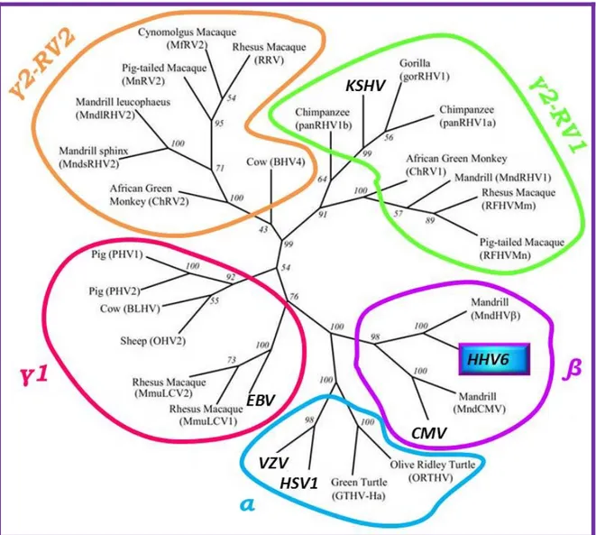
The Herpesviridae family includes three subfamilies: the Alpha-, Beta-, and
Gammaherpesvirinae 1 (Figure 1). The Alphaherpesvirinae are defined by wide cellular
host range, shorter viral reproductive cycle, rapid growth in culture, high cytotoxic effects, and the ability to establish latency in sensory ganglia. Human α-herpesviruses are herpes
simplex viruses 1 and 2 (HSV1 and HSV2) and varicella zoster virus (VZV).
The Betaherpesvirinae have a more restricted host range with a longer reproductive viral cycle and slower growth in culture. Infected cells show cytomegalia (enlargement of the infected cells). Latency is established in secretory glands, lymphoreticular cells, and in different tissues, such as the kidneys and others. In humans, these are human
cytomegalovirus (HCMV) and roseoloviruses (causing the disease roseola infantum in
children) including human herpesviruses 6A and 6B (HHV6A and 6B) and human
herpesvirus 7 (HHV7).
The Gammaherpesvirinae in vitro replication occurs in lymphoblastoid cells, but lytic infections may occur in epithelial and fibroblasts for some viral species in this subfamily. Γ-herpesviruses are specific for either B or T cells with latent virus found in lymphoid tissues. The γ-herpesviruses subfamily contains two genera that include both the γ-1 or Lymphocryptovirus (LCV) and the γ-2 or Rhadinovirus (RDV) virus genera. Only two human γ-herpesviruses are known to date, human herpesvirus 4, or Epstein-Barr virus (EBV), and human herpesvirus 8, referred to as HHV8 or Kaposi's sarcoma-associated herpesvirus (KSHV). EBV is the only Lymphocryptovirus and HHV8 is the only Rhadinovirus discovered in humans. LCV are found only in primates whereas RDV can be found in both primates and subprimate mammals.
Introduction
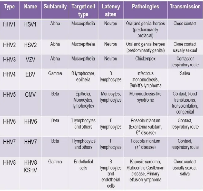
Table 1. Human herpesviruses classification and characteristics.
1.2 Structure of herpesviruses
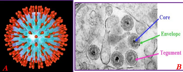
Herpesviruses have a central toroidal-shaped viral core containing a linear double stranded DNA. This DNA is embedded in a proteinaceous spindle 2. The capsid is icosadeltahedral
(16 surfaces) with 2-fold symmetry and a diameter of 100-120 nm that is partially dependent upon the thickness of the tegument. The capsid has 162 capsomeres. The herpesvirus tegument, an amorphous proteinaceous material that under EM lacks
Introduction
distribution. Thickness of the tegument is variable depending on the location in the cell and varies among different herpesviruses 3. The herpesvirus envelope contains viral
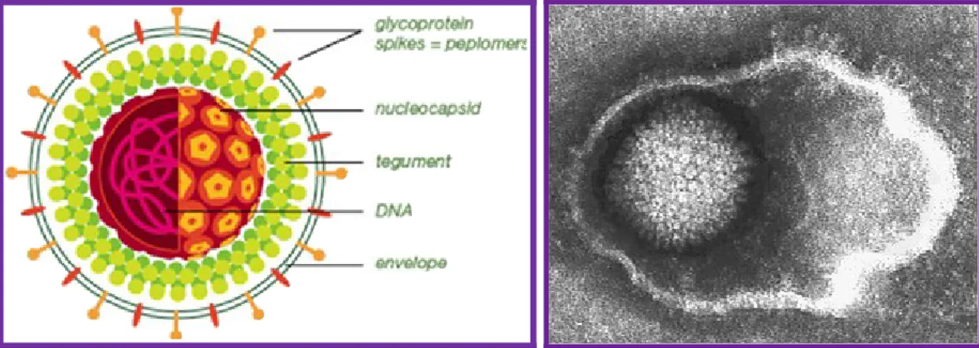
glycoprotein protrusions on the surface of the virus (Figure 2), and by EM it appears as a lipid trilaminar structure, derived from the cellular membranes.
Figure 2. Schematic representation of a herpesvirus virion (left) and a EM picture of the herpesvirus particle (right).
1.3 Herpesviruses genome
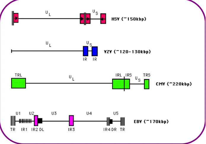
Herpesviruses genomes studied to date range in size from 130 to 235 kbp. Herpesviruses DNA is characterized by two unique components, unique long (UL) and unique small (US)
regions, each flanked by identical inverted repeat sequences (TRL and IRL: Terminal and Internal repeats) (Figure 3). Herpesvirus genome also contains multiple repeated
sequences. All known herpesviruses have capsid packaging signals at their termini 4. The
majority of herpes genes contain upstream promoter and regulatory sequences, an initiation site followed by a 5' nontranslated leader sequence, the open reading frame (ORF) itself, some 3' nontranslated sequences, and finally, a polyadenylation signal. Gene overlaps are common, whereby the promoter sequences of antisense strand (3') genes are located in the coding region of sense strand (5') genes; ORFs can be antisense to one another.
Proteins can be embedded within larger coding sequences and yet have different functions. Most genes are not spliced and therefore are without introns and sequences for noncoding RNAs are present.
Introduction
Herpesviruses code for genes that synthetize proteins involved in establishment of latency, production of DNA, and structural proteins for viral replication, nucleic acid packaging, viral entry, capsid envelopment, for blocking or modifying host immune defences, and for transition from latency to lytic growth. Although all herpesviruses establish latency, some (e.g., HSV) do not necessarily require latent protein expression to remain latent, unlike others (e.g., EBV and HHV6).
Introduction 1.4 Replication of herpesviruses
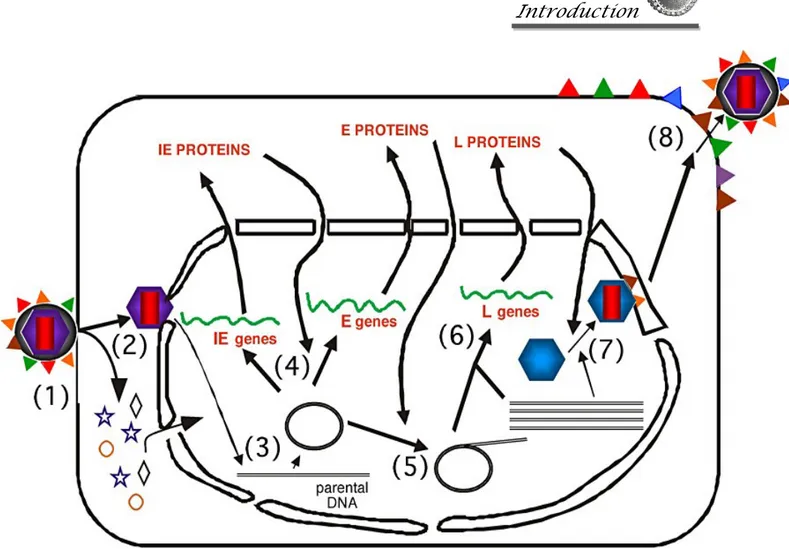
Herpesviruses can establish lytic or latent infections. The lytic stage can be divided in 6 phases:
I. Binding to the cell surface. Virus attachment and entry require sequential interactions between specific viral membrane glycoproteins and cellular receptors.
II-III. Fusion of the virion membrane with the host cell membrane; then the capsid and some tegument proteins are transported to the nucleus along cellular microtubules. Upon entry into the cytoplasm, the nucleocapsid is transported to the nuclear pores, where viral DNA is released into the nucleus.
IV. Virus transcription, replication and translation. This is a very complex process, as expected considering the large size of viral genome. Viral DNA replicates by circularization followed by production of concatemers and cleavage of unit-length genome during packaging (“rolling-circle” mechanism).
Herpesviruses transcription is temporally regulated, and can be divided into four steps: α or immediate early (IE) genes expression, requiring no prior viral protein synthesis and mRNAs are frequently detectable within 3 h after infection.. The proteins encoded in this stage are expressed first and are involved in transactivating transcription from other viral genes. Expression of IE gene products is essential for the activation of transcription from early gene promoters.
β or early (E) genes expression, which is independent of viral DNA synthesis. The early protein transcribed are involved in DNA metabolism and DNA replication. The expression of β genes is dependent on α gene expression.
γ1 or partial late genes expression, which starts in concert with the beginning of viral DNA synthesis.
Introduction
γ2 or late (L) genes expression, which is totally dependent upon synthesis of viral DNA and expression of structural genes encoding for capsid proteins and envelope glycoproteins occurs.
Herpesviruses DNA is transcribed to RNA by cellular RNA polymerase I. The neo-formed viral mRNAs block cellular protein synthesis and activate the replication of viral DNA. Herpesviruses encode their own DNA-dependent DNA polymerase and other enzymes and proteins necessary to replication, such as ori-Lyt (replication start for the lytic phase), major DNA binding protein (MDBP) and origin DNA binding protein (OBP). Herpesviruses can alter their environment by affecting host cell protein synthesis and host cell DNA replication, immortalizing the host cell, and altering the host's immune responses (e.g., blocking apoptosis, cell surface MHC I expression, modulation of the interferon pathway).
V. Assembly: capsids are assembled in the nucleus.
VI. Viral exit. The viral particles bud through the inner lamella of the nuclear membrane which has been modified by the insertion of viral glycoproteins and leave the cell via the exocytosis pathway, or by budding from the cell membrane or by lysis.
Introduction
Figure 4. The cycle of productive HSV replication in a cell. The stages of HSV infection are: (1) Receptor binding and membrane fusion; (2) Release of the viral nucleocapsid and tegument into the cell cytoplasm and transport of the nucleocapsid to the nuclear pore; (3) Release of viral DNA into the nucleus; (4) Transcription and translation of the viral immediate early (IE) and early (E) genes; (5) viral DNA synthesis; (6) Transcription and translation of the viral late (L) genes; (7) capsid assembly and DNA packaging; and (8) egress of progeny virions.
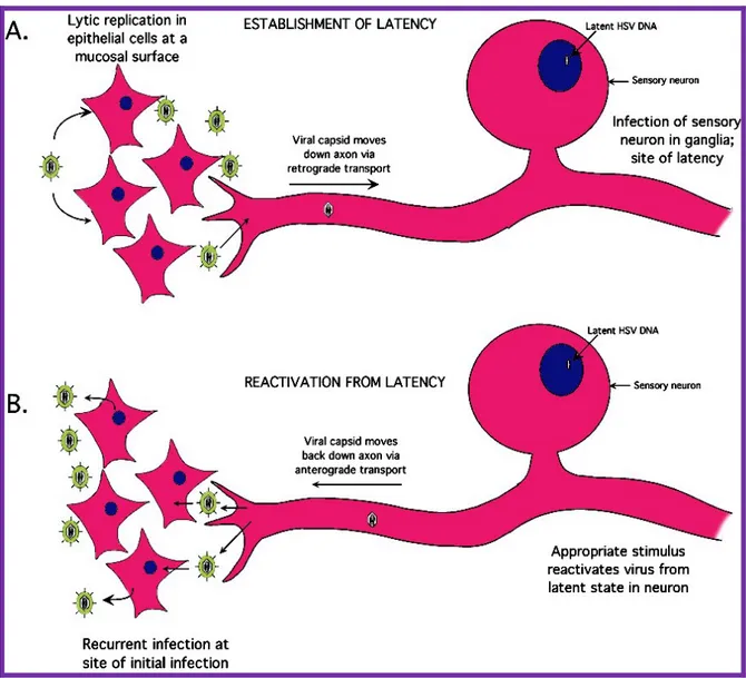
All herpesviruses can establish latent infections in their natural host. Such an infection is characterized by periods of highly restricted (or no) viral gene expression in the cells harboring the latent genomes, interspersed with periods of virus replication and infectivity (reactivation). The different type of herpesviruses carry out latency and reactivation processes in generally similar ways, but details depend on the specific virus. Neurotropic herpesviruses like α-herpesvirinae (i.e. HSV) establish a latent infection by entering a sensory nerve axon near the infection site. The virus particle then migrates to the neuron nucleus in the nerve ganglion (Figure 5). If virus gene expression occurs normally, viral DNA replication and cell death will occur; however, in most infected neurons, the earliest
Introduction
stages of gene expression appear to be blocked. Thus, these neurons are nonpermissive for replication. This results from the fact that one or several cellular transcription factors required to express the immediate-early genes are absent or present in a modified form. The viral genome of herpesviruses is not fully shut off during latent infection; one transcript termed the latency-associated transcripts (LAT) is expressed from a single latent phase active promoter.
Figura 5. Stages of HSV infection. (A) HSV replicates initially in epithelial cells at the genital or oral mucosal surface. The virus enters innervating sensory neurons where it travels up the axon to the neuronal cell body. Once in the neuron, the virus establishes a latent infection that may last the lifetime of the host. (B) Upon the proper stimulus, the virus can reactivate, travel back down the axon, and establish another round of lytic replication.
Introduction
Since viral DNA is in the neuronal nucleus and fully differentiated neurons do not replicate, the DNA can remain silent for long period of time. Then, when the host is stressed with certain agents the cellular transcriptional environment change and immediate-early as well as other viral transcripts are expressed at detectable levels. This is sufficient for the replication of virus in at least a few neurons, which results in the transitory appearance of a small amount of virus that travels down the axon and reinfects the area where the virus first infected, producing a new lytic infection in the target epithelial cells.
2. Human herpesvirus 6
Human herpesvirus type 6 (HHV6) was first isolated in 1986 from peripheral blood lymphocytes (PBL) of patients with lymphoproliferative disorders 5. Initially the virus was
named "Human B lymphotropic virus" (HBLV) because it seemed to manifest a tropism for B cells, but subsequent studies have shown that the preferential cellular target are T cells 6.
The International Committee on Taxonomy of Viruses has finally named the virus HHV6 7.
HHV6 is a members of the Betaherpesvirinae subfamily, included in the Roseolovirus genus along with HHV7. Members of this genus are characterized by elective tropism for T lymphocytes, the ability to grow with less efficiency also in other cell types, high rates of latent infection in the human population, and by association with childhood febrile syndromes, which might be accompanied by rash.
Furthermore, HHV6 strain cluster in two variants (HHV6A and HHV6B), which differ on the basis of distinct genetic, immunological and biological characteristics 8, and for the
associated diseases, as detailed in the “Disease association” chapter.
HHV6 is highly prevalent in the healthy population, establish latency in macrophages and T-lymphocytes, is frequently shed in saliva of healthy donors, and their pathogenic potential ranges from frequent asymptomatic infection to rare cases of severe diseases in immunocompromised patients 9.
B
A
Introduction 2.1 Virion
HHV6 virion, as for all herpesviruses, have four main structural elements: an electron-dense core, a capsid with icosahedral symmetry, a tegument, and an outer envelope in which virally encoded glycoproteins and integral membrane proteins are embedded 1 (Figure 6).
Figure 6. A) Schematic representation of HHV6. B) Electron microscope photograph of HHV6 virions in infected lymphoid cells. Bar is 100 nm.
The assembly process leading to mature HHV6 particles begins in the nucleus, where nucleocapsids are assembled. HHV6 capsids have a diameter of 90 to 110 nm 10,11. After
capsid assembly, the tegument is acquired while still in the nucleus by a series of envelopment and de-envelopment steps which have not been fully characterized 12,13. The
tegument may be acquired in a novel structure, the tegusome, which is a membrane-bounded spherical compartment, apparently of cytoplasmic origin, that is present in the nuclei of HHV6 infected cells 13. Fully tegumented capsids, with a diameter of
approximately 165 nm, appear to be released into the cytoplasm via fusion of the tegusome with the nuclear membrane 13,11. HHV6 morphogenesis has the unique feature of
Introduction
envelope by budding into cytoplasmic vesicles. Mature virions are approximately 200 nm in diameter 10,11 and are released by exocytosis 13,14.
2.2 Genome
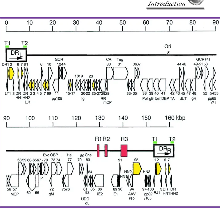
HHV6 genome is composed of a linear, double-stranded DNA molecule, that was completely sequenced in 1995 15. The genome is 160-162 kbp in size and is formed by a
central unique region (approximately 143 kbp) bound at both ends by terminal direct repeats (each 8-9 kbp long) (Figure 7). The direct repeats contain genetic elements that are necessary for viral DNA packaging and replication 16 and include a tandem repetitive
sequence that is also present in human telomers 17. The genome of HHV6B is predicted to
contain 119 open reading frames (ORFs), 9 of which are absent in HHV6A. ORFs encoded in the terminal direct repeats are identified with the prefix DR and those in the unique region are named U1-100, starting from the left end of the genome. Coding regions are located on both DNA strands, and a few genes are spliced after transcription. Overall, nucleotide sequence identity between HHV6A and HHV6B is 90%, with variability according to specific regions. Some DNA regions are conserved between variants for over 95%, and other regions show higher divergence, with homology as low as 75% 18. Instead,
variability within variants is very low, and viral strain belonging to the same variant differ by 1% or less across their genomes 19.
Introduction
Figure 7. Genomic organization of HHV6. The asterisk indicates the start of lytic genomic replication. Viral telomeric sequences are indicated by green bars T1 and T2. Open arrows represent protein coding regions.
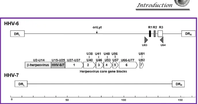
Most of HHV6 genes are conserved and present in the genome of all herpesviruses, and can be organized in seven gene blocks (Figure 8), including genes U27 – U82.
The products of HHV6 genes U27, U41, U43, U74, U77, and U73 are involved in viral replication. The protein product of gene U27 encodes the DNA polymerase processivity factor, binding to DNA polymerase and thus enhancing the rate of viral genome
Introduction
Figure 8. Schematic representation of HHV6 and HHV7 genomes, showing the direct repeats (DR), the position of the seven conserved herpesvirus gene blocks, of the β-herpesvirus specific genes, and of the two HHV6 specific genes (U83 and U94).
Gene U41 encodes a single stranded DNA binding protein, also facilitating DNA polymerase interaction with the viral genome. The protein products of genes U43, U74, and U77 produce a viral helicase-primase complex that facilitates replication of the viral genome. Additionally, the origin-binding protein produced by gene U73 is also involved in genomic DNA replication.
HHV6 attachment to host cells is facilitated by viral surface proteins gB and gH, encoded by viral genes U39 and U48, respectively 20.
One additional gene block includes 17 genes which are conserved only in β-herpesviruses, including genes U2 – U14.
There are also genes specific to Roseoloviruses, not present in the other β-herpesviruses, including U20-21, U23-24, U26, U85 and U100. Finally, two genes are unique to HHV6 and present in both variants. One is U83, which encodes a chemokine with chemotactic activity for monocytic cells, and is expressed in two forms. The spliced variant is expressed as an early protein, before DNA replication, and the full length protein is expressed only after DNA replication 21. The other gene unique to HHV6 is U94, encoding
Introduction
gene product complements the replication of Rep-defective AAV-2 mutants, it localizes in the nucleus of the infected cell and binds to the human TATA-binding protein, a transcription factor 23. It is transcribed as an IE gene and in latently infected lymphocytes 24, suggesting that it likely contributes to the maintenance of latency 9.
2.3 DNA replication
The elements required for lytic HHV6 DNA replication are the origin for lytic-phase replication (oriLyt) and 7 viral gene products: the MDBP (major DNA binding protein), the DNA polymerase, a polymerase-associated protein, an origin binding protein (OBP), and a helicase-primase complex. An interesting feature is that HHV6 encodes a sequence homolog of the HSV-1 OBP, which has been found only in α-herpesviruses 25, 26, 27, 28, 29.
The HHV6 OBP, like other α-herpesvirus encoded OBP homologs, has greater amino acid sequence similarity to other herpesvirus OBPs in the N-terminal helicase region than in the C-terminal DNA binding region 27, 30.
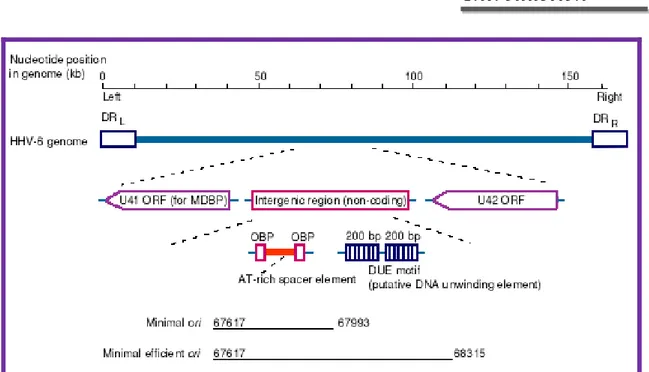
The HHV6 oriLyt (as in HSV-1, VZV, and HCMV) is located upstream of the gene encoding the MDBP homolog 31, 32, 33. The HHV6 origin lies in a unique 1,400-bp region
that has no protein-coding capacity and is located between the genes encoding the MDBP and the HCMV UL69/HSV-1 ICP27 homolog 34, 30, 35. These oriLyt sequences have been
functionally defined by using transient-transfection assays with plasmids containing different portions of the HHV6 oriLyt 34: a region of approximately 400 bp was identified
as the minimal active region. The HHV6 oriLyt is located in a genomic region that includes two binding sites, OBP-1 and OBP-2, for the HHV6 OBP. The OBP sites are arranged with dyad symmetry, flank an AT-rich spacer, and are within the minimum essential origin region. This region is adjacent to a larger AT-rich imperfect direct repeat of about 195 bp that has been proposed to act as a DNA-unwinding element 34. Both OBP
Introduction
Figure 9. Schematic diagram of human herpesvirus 6 (HHV6) oryLit.
2.4 Internal repeated items
Several repeated items are located in the UL region of the HHV6 genome, namely: R0, R1,
R2, R3. These elements are placed in correspondence with regions of the genome with a low percentage of identity between the two variants of the virus. R0 is present only in HHV6B and is located near the junction between DRL and UL and within the ORF putative
B4.
R1 is located near the 3’ end of ORF U86 and determines, once translated, the production of a series of serine and arginine (SR) repeated in the carboxy terminal portion U86 protein.
R2 is located in the region between U86 and U90. In the genome of HHV6A, R2 consists of repetitions of TG dinucleotide, in HHV6B are identified in the same region 2 distintict items repeats: R2A and R2B. R2A contains several TATA sequences; R2B is similar to R2 of HHV6A, but is much shorter and contains multiple potential binding sites for the factor HNF transcription-5 37, suggesting a possible role of these sequences in the regulation of
Introduction
R3 is located upstream of the IE-A locus, which includes ORFs from U86 up to U91, and contains regulatory elements that may play a role in the regulation of transcription of this locus. The region is highly variable among the variants of the virus but retains some sites in the consensus sequence of conserved regulatory elements 38.
2.5 HHV6 replicative cycle
The infectious cycle of HHV6 begin with the interaction between specific virus glycoproteins of the envelope (gB, gH, gL, gQ) and the specific receptor on the cellular membrane, CD46. HHV6 enters in the cell through an endocytic pathway 40 (Figure 10).
Unenveloped nucleocapsids are then released into the cytosol and migrate to the nucleus where the viral DNA is transcribed and replicated.
As already described for herpesviruses in general, during lytic infection, herpesvirus genes are expressed as three kinetic classes 39. The α, or immediate-early (IE), genes are
transcribed with no requirement for de novo protein synthesis and α proteins regulate the expression of other genes. The second class of genes, the β or early (E) genes, are involved in DNA metabolism and DNA replication. The expression of β genes is dependent on α-gene expression. The third class of α-genes is the γ, or late (L), α-genes; the products of these genes are usually involved in virion assembly and as components of the mature virus particle 14.
The newly produced capsid proteins are assembled to form the viral core. The nucleocapsid is than expelled from the nucleus in two forms: as a mature-like particle, vehicled toward the cell surface within cytoplasmic vacuoles, or as a free cytosolic particle coated by a seemingly amorphous tegument structure, which subsequently buds into a cytoplasmic vacuole, possibly of Golgi origin, thereby acquiring the viral envelope. The maturation cycle culminates with the final expulsion of the mature particles from the cellular membrane.
Like all herpesviruses, HHV6 can persist latently in the host following primary infection. Persistence may include both a true latent state, without production of infectious virus, and chronic replication with continuous or intermittent production of infectious progeny.
Introduction
Both forms of persistence develop in the same individual, with chronic infection taking place mostly in salivary glands 41,42, while true latent infection, with episodes of
reactivation, occurs mainly in macrophages and CD4+ T cells 43,44,45,46.
Introduction
PCR studies have shown that HHV6 DNA sequences can also be detected in many different tissues from asymptomatic healthy individuals, including urinary tract, genital mucosa, fibroblasts and epithelium, thyroid, pancreas, liver 47,48.
However, there is no evidence that the virus can reactivate from these sites. It has been reported that latently infected cells contain four species of latency-associated transcripts, originating from the IE1/IE2 genic regions, with a transcription pattern similar to that of human cytomegalovirus 49. Another transcript expressed during latent infection, in the
absence of transcripts from lytic genes, is U94 50. 2.6 Cell tropism
HHV6 is characterized by a broad tropism for different human cell types, and this observation is in accordance with the fact that the cellular receptor is the CD46 molecule
51, a ubiquitous type-1 glycoprotein member of a family of regulators of complement
activation. However, only few cell lines expressing CD46 support efficient viral replication, suggesting that in addition to the presence of receptor, there is the need of specific requirements still unidentified.
HHV6A and HHV6B variants differ in the respective ability with which they can replicate in specific transformed T-lymphocyte cell lines. In addition, also CD8 T lymphocytes, γδ T lymphocytes and natural killer (NK) cells support HHV6 replication 52,53,54.
HHV6 can also infect macrophages, dendritic cells, fibroblasts, epithelial cells and bone-marrow progenitors 55,56,57,58,59. Laboratory strains have been adapted to grow in specific
human T cell lines, including Jurkat, J-Jhan, HSB2 for HHV6A, Molt-3, MT4 and SupT1 for HHV6B 60 , but the efficiency of viral replication is 1-2 fold lower than in primary
cells. Both variants are neurotropic and can infect neural cells, including astrocytes and oligodendrocytes, but HHV6B supports only abortive infection, while HHV6A establishes a productive infection that subsequently develops into latency 61,62.
HHV6 can also infect endothelial cells, both in vitro and in vivo 63, establishing a low level
productive infection 64. Both HHV6A and HHV6B can infect fibroblasts, but only HHV6A
Introduction
shows that HHV6A and HHV6B have differences in their cell tropism, and possibly HHV6A is more virulent, but the reasons for this difference are still unknown 9.
2.7 Effects on cells
HHV6 infection causes significant alterations on the host cell metabolism. As is common with herpesviruses, infection induces shut off of host cell DNA synthesis 66, stimulates
protein synthesis 67, and causes T lymphocytes to develop a cytopathic effect with
characteristic ballooning (Figure 11), refractile and syncithial cells. Viral infection induces differential secretion of a wide array of cytokines and chemokines, including interferon-α, interleukin 1β, RANTES, tumor necrosis factor and many others 64,68,69,70, depending on
the virus and the cell type involved. HHV6 infection upregulates CD4 in T lymphocytes and induces its expression in CD4-negative cells 9.
Figure 11. Cytopathic effects of HHV6 (ballooning): CD4+ T lymphocytes non infected (on the left) and HHV6-infected (on the right). Pictures was obtained by phase contrast microscopy after 7 days p.i., 100X.
2.8 Epidemiology
HHV6 is ubiquitous and has a worldwide distribution. Seroprevalence is very high in the adult population, and specific IgG antibodies to HHV6 are detected in more than 90% of adults. Primary infection occurs during early childhood. The peak for HHV6 infection is observed at 6-9 months of age 71. There are no convenient serological assays to
Introduction
discriminate between HHV6 variants, however the available evidences suggest that the majority of clinical infections in immunocompetent patients are caused by HHV6B. In fact, the viral sequences detected by PCR in latently infected peripheral blood cells from healthy individuals belong almost invariably to HHV6B 72,73 , supporting the notion that
HHV6A is less prevalent or that it establishes latency in different body districts. HHV6A is detected more often in immunocompromised than in immunocompetent hosts, and appears to be potentially more neurotropic 74. The mode of transmission is unclear, but HHV6
replicates in epithelial cells and is present in saliva, suggesting that oral secretions contribute to transmission, especially for HHV6B 75,76,59. Congenital infections occur
approximately in 1% of births and are asymptomatic 77. Sequence analysis of HHV6
isolated from mother/infant pairs suggests that mother-to-infant transmission can occur 78,
whereas there is no convincing evidence of sexual spread 9. 2.9 Disease association
HHV6 can be considered an opportunistic pathogen which persists asymptomatically in immunocompetent individuals but can cause severe pathologies in the context of immunosuppression.
Primary HHV6B infection in children causes exanthem subitum (ES) (roseola infantum, sixth disease) with high fever and the development of a rash after resolution of fever 79.
However, only a subset (17%) of children with acute infection develop ES, and the majority suffer from undifferentiated short febrile illness, without appearance of rash 80,81.
Although usually mild, ES may be accompanied by convulsions 82,80. Rare complications
include hepatitis, arthritis, encephalopathy and haemophagocytosis syndrome 80,83. Primary
infection in older age groups is rare and results in an undifferentiated febrile illness or in an heterophile-negative infectious mononucleosis 84,85. Skin rash, hepatitis and atypical
lymphocytosis may occur. HHV6A primary infection is not associated to ES and is likely mostly asymptomatic.
HHV6 related diseases in adults are mostly the results of reactivation, which might be clinically significant especially in the immunocompromised host. HHV6 can reactivate in
Introduction
admission to intensive-care units 86,87. Many reports describe other possible disease
associations involving HHV6 reactivation 88. In particular, the central nervous system is a
potential target for HHV6 pathogenesis. In fact, HHV6 DNA is often detected in autoptic brain tissue from healthy individuals 89,90, indicating that the virus is able to invade and
persist asymptomatically in the CNS. The pathogenic potential of HHV6 in the CNS is witnessed by several clinical reports 91.
HHV6 has been associated with neurological complications in children with primary infection 92,93, encephalitis both in immunocompetent and immunocompromised patients 94,95, demyelinating encephalomyelitis 96,97 and chronic myelopathy 98. HHV6 DNA
sequences are often detected in patients with temporal lobe epilepsy and the high levels of viral sequences suggest a potential etiological association 99,100,101. Some reports suggest
that HHV6A may have higher neurotropism than HHV6B 102,103,104. For example, it was
shown that HHV6A has a considerably higher prevalence in cerebrospinal fluid than in peripheral blood mononuclear cells or in saliva of children 105. The difficulty with these
studies is that HHV6 infection is ubiquitous, it shows a wide tissue tropism, and therefore the detection of viral footprints in a pathological condition is not univocal proof of etiology. Viral presence might be due to latent virus, and viral reactivation may be a consequence, rather than the cause of disease. A further complication is the fact that there is not yet any biological explanation of how HHV6 variants might differentially contribute to the pathogenesis. The proposed association of HHV6 with multiple sclerosis (MS) is a paradigmatic example of how difficult it is to substantiate, or to disprove, HHV6 etiological association. In 1995, Challoner 106 proposed that HHV6 might be associated
with MS. One of the original observations was the presence of HHV6 DNA sequences and antigens in the nuclei of oligodendrocytes from patients with MS adjacent to plaques of demyelination but not in control samples. The original report raised considerable interest, and subsequently, several studies were performed. However, after more years, the role of HHV6 in MS is still controversial.
There is substantial evidence that HHV6 can be a significant pathogen in immunocompromised hosts. HHV6 infection or reactivation in AIDS patients was reported to cause severe disease, such as fatal pneumonitis, encephalitis, retinitis and HHV6 dissemination to many organs 107.HHV6 is also an important pathogen in organ transplant
Introduction
recipients 108,109,110. HHV6 infection or reactivation occurs in nearly 50% of all bone
marrow and in 20-30% of solid-organ transplant recipients, 2-3 weeks following the procedure 108,111. Prospective studies employing DNA detection by PCR or viral culture in
sequential blood samples post-transplantation provide the strongest evidence for HHV-6 infection. The majority of HHV6 infections post-transplantation are due to reactivation of HHV6B, and the peak incidence of infection is 2-4 weeks post transplantation 112,110.
However, cases of primary infection due to transmission of the virus in donor tissue have been described, and superinfection with a donor strain distinct from the strain latent in the recipient may occur 113. Viral infection and/or reactivation may result in clinical symptoms,
including fever (sometimes associated with leukopenia), skin rash, pneumonia, bone marrow suppression, encephalitis, and transplant failure 114,115,116,112,117. Idiopathic bone
marrow suppression is more common in bone marrow transplant or stem-cell transplant recipients, but can also occur following solid organ transplantation 118,116,119. Potential
causes of marrow suppression include indirect effects mediated by cytokines and HHV6 soluble products or direct infection of bone-marrow progenitors 120,121,122. In spite of the
available evidence that HHV6 can be a serious pathogen responsible for complications in transplant recipients, definite proof is still under debate, mainly because the underlying pathophysiologic mechanisms have not yet been elucidated. Considerable attention has been focused on possible associations of HHV6 and the pathogenesis of AIDS because HHV6 replicates in CD4+ T cells, the primary HIV-1 target, but studies of serologic responses to HHV6 in HIV-infected persons have yielded widely varying results 52.
Moreover, a possible role has been postulated for HHV6 in a variety of neoplastic disorders, but as yet there has been no conclusive demonstration that either HHV6A or HHV6B is involved in any causative way with any malignancy. Nevertheless, the accumulated data suggest that at least for some neoplasms, there is more than a chance association with the virus.
Finally, HHV6 infection was also associated with the development of autoimmune diseases include chronic fatigue syndrome, graft versus host disease, encephalitis, pneumonitis, graft failure. HHV6 has also been tentatively associated to several chronic autoimmune inflammatory processes, including Sjogren syndrome, multiple sclerosis, rheumatoid
Introduction
infection might be related to the onset of autoimmune pathologies such as nonbacterial purpura fulminans and severe autoimmune acquired protein S deficiency, autoimmune connective tissue diseases, severe autoimmune hepatitis.
.
3. Infections and autoimmunity: the multifaceted relationship
The autoimmune diseases are a diverse group of conditions characterized by abnormal immune reactivity in association with autoreactive B and T cell responses. There are more than 80 different autoimmune diseases 123 that affect hundreds of millions of people around
the world. These systemic or organ-specific conditions are the third leading cause of morbidity and mortality in the industrial world after cancer and heart disease 124. Multiple
factors are thought to contribute to the development of immune response to self, including differences in genotypes, hormonal milieu, and environmental factors 125,126.
The role of environmental factors is clearly apparent when considering the low disease concordance rate between monozygotic twins for major autoimmune diseases.
Of all environmental factors with a potential to trigger autoimmunity, the most important seems to be viruses, bacteria, and other infectious pathogens (Figure 12).
Introduction
The pivotal role of infection in the induction of these disorders has been supported by several evidences, such as disease seasonal onset, disease occurrence after some infections, and the isolation of viral genomic DNA/RNA from affected tissues of patients once the disease gets started 127,128. Infections and autoimmune diseases have multifaceted and
multidirectional relationships 129.
The human immune system is highly complex, having evolved to fight a broad spectrum of pathogens. The host response to infections encompasses a huge number of immunological mechanisms that can eventually lead to autoimmunity. Although the development of autoimmune phenomena linked to infections is a common finding, the onset of autoimmune diseases is a rare event, arising from a combination of genetic susceptibility and environmental factors 127,129,132,133. There are several mechanisms through which
Introduction
Figure 13. Host response and autoimmunity: mechanisms that can eventually lead to autoimmunity in a susceptible individual.
Introduction
Some of them are antigen-specific, including molecular mimicry, expression of modified, cryptic, or new antigenic determinants, and superantigens. Others are non specific and collectively known as “bystander activation.” They include enhanced processing and presentation of self-antigens, immune cell activation, cytokine release, and cell apoptosis/necrosis 134.
Molecular mimicry
In molecular mimicry, pathogens bear elements that are similar enough in amino acid sequence or structure to antigen. Immune response can eventually turn toward the self-peptide as a result of cross-reactivity, leading to the activation of naıve, autoreactive T cells specific to the corresponding self-molecule 135,130.
Protein changes, cryptic antigens
Following tissue injury, cell death, oxidative stress, free radical production, and reparative changes, as happens in several infections, proteins that are usually recognized as self can became non self and induce an autoimmune response 134. This occurs as a consequence of
protein changes as a result of altered expression, post-translational modifications, denaturation, misfolding, or mutations 136. In addition, the inflammatory environment can
induce differential processing of released self-epitopes by APCs. Proteins that are normally sequestered and shielded from immune recognition can be exposed to the immune system and become immunogenic. Therefore, cryptic antigens become accessible to self-reacting T lymphocytes that had escaped central and peripheral tolerance, as they had not been presented appropriately to induce tolerance.
Superantigens
Superantigens are proteins produced by a variety of microorganisms, especially bacteria or mycoplasmae, or virus-infected cells that can bind TCR, irrespective of its antigenic specificity, resulting in the activation of a large number of T lymphocytes of different antigenic specificity, thus behaving as a potent immune-stimulating molecule 134.
Bystander activation
It is a nonspecific mechanisms which is defined as the activation of T cells through a mechanism that is independent of specific TCR stimulation. Bystander activation can lead to enhanced processing and presentation of self-antigens, which induces the expansion or
Introduction
epitope spreading, has been widely involved in the pathogenesis of many systemic autoimmune diseases as well as in determining the different expression of such diseases
137.
4. Autoimmune thyroid diseases
Autoimmune thyroid diseases (AITDs), are caused by an immune response to self thyroid antigens. Hashimoto's thyroiditis (HT), also known as chronic lymphocytic thyroiditis, is an autoimmune disease in which the thyroid gland is gradually destroyed by a variety of cell- and antibody-mediated immune processes. It was first described by Hashimoto Hakaru in Germany in 1912. HT is a relatively common disease, affecting 5-15% of women and 1-5% of men 138,139.
The development of the autoimmune failure of the thyroid is a multistep process, requiring several genetic and environmental abnormalities to converge before full-blown disease develops. At the onset of disease, major histocompatibility complex (MHC) class II-positive antigen-presenting cells (APC), particularly dendritic cells, and different subclasses of macrophages, accumulate in the thyroid 158,159. APC present thyroid-specific
auto-antigens to the naïve T cells, leading to activation and clonal expansion of the latter. Thus, the initial stage of the disease is followed by a clonal expansion phase and maturation of autoreactive T and B lymphocytes in the draining lymph nodes.
The mechanisms, whereby autoreactive T cells escape deletion and become activated remain uncertain. There is evidence that the thyroid cell itself, by "aberrantly" expressing MHC molecules, can play the role of "non-professional " APC and present disease-initiating antigen directly to the T cells 160,161.
A central phase of HT is characterized by the recognition of presented auto-antigens by the lymphocytes, followed by an apparent uncontrolled production of autoreactive CD4+ T cells, CD8+ cytotoxic T cells and immunoglobulin G (IgG) autoantibodies. Initially, the production of self-reactive cells and autoantibodies occurs in the draining lymph nodes. Later, the lymphoid tissue often develops directly in the thyroid gland itself. This tissue is generally very well organized, with cords of anti-Thyroglobulin (Tg)-antibody- producing plasma cells in the periphery.
Introduction
In a final, destructive step of Hashimoto's thyroiditis, the autoreactive T cells diffusely accumulate in large numbers and infiltrate thyroid parenchyma, leading to destruction of thyrocytes (Figure 15).
Figure 15. Schematic model of cells death in Hashimoto’s thyroiditis.
Two classes of autoantibodies are particularly relevant in the diagnosis of HT: anti-Tg Ab and anti-thyroperossidase (TPO) Ab.
Introduction
synthesis sites from the iodinated tyrosine residues. The hormone synthesis sites and the iodine content of Tg play an important role in its autoantigenicity 162. Tg is one of the
major autoantigens in thyroid autoimmunity.
TPO is another significant autoantigen in the thyroid of patients affected by HT. This enzyme catalyses the oxidation of iodine to an iodinating species that forms iodotyrosines in a Tg molecule and subsequently iodotyronines 163.
Autoimmune responses against specific antigens are primary determinants in thyroid autoimmunity. This process progressively destroy the thyroid cell ultimately inducing a gradual reduction in thyrocyte numbers, leading to hypothyroidism 164,165.
The disease is associated with several genetic and environmental factors, and the involvement of infectious agents, including viral infections, has been repeatedly suggested
140,141. Several viruses have been incriminated, including influenza, coxackie, adenovirus,
echovirus and mumps. Herpesvirus infection has occasionally been detected in AITD
142,143, although there are no conclusive data. It has been shown that HCMV infection of
thyrocytes induces HLA-DR expression on thyroid follicular cells, which can act as antigen presenting cells 145. There are only scant data on the possible association between
HHV6 infection and HT, although the virus has been implicated in the onset and/or promotion of several autoimmune diseases.
Material & Methods
MATERIAL AND METHODS
1. Clinical samples
All clinical specimens were obtained from patients referred to the Section of Endocrinology of the University of Ferrara. Thyroid lesions were defined on the basis of ultrasound examination, and fine needle aspiration biopsy (FNAB) followed by cytological and laboratory tests.
A total of 62 patients were enrolled in the study after signing informed consent. None of them had other concomitant diseases or was taking drugs possibly affecting thyroid function. Serum TSH, free T4, thyroperoxidase antibodies (TPO Ab; normal value < 35 IU/ml) and thyroglobulin antibodies (Tg Ab; normal value < 115 IU/ml) were measured in all patients by using an immuno-electrochemiluminescence technique (Modular E 170, Roche Diagnostics GmbH). All patients underwent ultrasound (US) guided thyroid fine needle aspiration. Thyroid aspirates were used for both cytology and molecular analysis. The 34 HT patients showed TPO Ab > 35 IU/ml, ranging from 146 to 8229 IU/ml (mean value = 1312 IU/ml), and Tg Ab > 115 IU/ml, ranging from 280 to 3500 IU/ml (mean value 751 IU/ml). The 28 patients whose FNAB material was considered as control showed TPO Ab < 35 IU/ml, ranging from 7 to 11 IU/ml (mean value = 9 IU/ml), and Tg Ab < 115 IU/ml, ranging from 10 to 16 IU/ml (mean value 12 IU/ml).
All FNAB samples were immediately suspended in a small volume of sterile PBS (300 l), divided in two aliquots, frozen in liquid nitrogen, and conserved at -80°C until use. In two HT cases, separated epithelial and lymphoid FNAB fractions were obtained, by using Dynal Epithelial Enrich method (Dynal AS, Oslo, Norway), based on immunomagnetic selection of cells binding to EpCAM (Ber-EP4) antibodies. The obtained fractions (epithelial-enriched and epithelial-depleted) were then frozen in liquid nitrogen, and conserved at -80°C until use.
Material & Methods
Lympholyte-H (CEDARLANE) density centrifugation. Aliquots of 106 PBMCs were
frozen in liquid nitrogen and maintained at -80°C for DNA and RNA analyses and plasma fractions were also collected and frozen at -80°C until used.
2. Cell lines
Lymphoid CD4+ J-Jhan and SupT1 cell lines were used, respectively, for production of purified cell free virus inocula of HHV6 variants A (strain U1102) and B (strain CV). For
in vitro infection experiments, we used Nthy-ori3-1 cell line, a thyroid follicular epithelial
cell line 35. All cell lines were maintained in RPMI medium (Gibco) supplemented with 10
% inactivated fetal bovine serum (FBS), 2 mM L-glutamine, 100 U/mL of penicillin, and 100 g/mL of streptomycin (Invitrogen, Carlsbad, CA).
3. Virus production and cells infection
Cell-free HHV6A inoculum (strain U1102) was obtained from infected J-Jhan cells, while cell-free HHV6B inoculum (strain CV) was obtained from SupT1 infected cells. Briefly, infected cells were collected by centrifugation and lysed by rapid freezing and thawing followed by sonication (3 cycles of 5 seconds at medium power with 10-second intervals in a water bath sonicator). Cleared cellular content was added to culture supernatant, and virions were collected by centrifugation for 30 minutes at 20000 x g at 4°C.
Virus particles were purified by density centrifugation on Optiprep self-forming gradients (Sentinel), at 58000 x g for 3.5 hours at 4°C. Purified virions were washed in phosphate buffered saline (PBS) and collected by centrifugation at 20000 x g for 30 minutes at 4°C. Virions were suspended in sterile PBS containing 0.1% bovine serum albumin (BSA) and stored at -80°C until use.
Prior to use, virus stock was treated with DNase-I and RNase-A, to eliminate free viral nucleic acids eventually present in the preparation.
Infectivity of virus preparation was evaluated by specifically designed infection experiments performed respectively in J-Jhan or SupT1 cells for variant A or B using PCR,
Material & Methods
PCR after retrotranscription (RT-PCR) and immunofluorescence assays to evaluate virus presence, transcription, and antigen expression.
Quantification of virus genomes present in the stock preparation was obtained by real-time quantitative PCR (qPCR)144, showing that inocula contained about 1010 genome
equivalents per ml. Aliquots from the same stock were used for all infection experiments. Mock virus (used as negative control) was inactivated by UV irradiation as described 37.
Thyroid Nthy-ori3-1 cells were seeded at optimal density 24 hours before infection, then infected with a m.o.i. of 10:1 (10 genome equivalents per 1 cell). After 3 hours of adsorption followed by extensive washes, cells were incubated in complete medium and collected at specific time points for further analyses.
4. DNA extraction
Total DNA were isolated from FNABs, PBMCs and cultured Nthy-ori3-1 cells using 1 ml of lysis buffer (10 mM Tris-HCl pH 8.0, 10 mM disodium EDTA pH 8.0, 10 mM NaCl, 0.6% SDS and 100 µg/ml proteinase K) and incubated at 37°C for a minimum time of 4 hours. After three cycles of phenol:chloroform:isoamyl alcohol (25:24:1) extractions in
Phase Lock Gel (PLG), DNA was precipitate in ethanol 100% and NaAc (CH3COONa pH
5.0 300 mM) and stored at -80°C for at least 1h. After precipitation with ethanol, DNA pellets were dissolved in sterile water and stored at -20°C until PCR or qPCR analysis. DNA concentration was determined by reading optical density at 260 nm.
5. RNA extraction and retrotranscription
Total RNA was extracted with RNAzol B (Tel-Test), following manufacturer’s instructions. Briefly, samples were lysed with RNAzol, then chloroform in ratio 1:5 (v/v) was added. The mixture was centrifuged 20 minutes at 8500 x g, the aqueous phase was collected and RNA precipitated with isopropanol 100%. After two washes with 75% ethanol and DNAse treatment (4 U/mg RNA, 3 x 20 minutes at room temperature), RNA was precipitated with isopropanol 20 minutes at 8500 x g at 4°C. RNA quality was checked by electrophoretic analysis on a 1% agarose gel. The absence of contaminating DNA was checked by PCR
Material & Methods
for β-actin ensured that the RNA sample was completely free from DNA sequences. First strand cDNA synthesis was carried out with MuLV reverse transcriptase and random hexamer primers (Applied Biosystems), following the manufacturer’s instructions, retrotranscribing 2 µg of total RNA from all the samples. The mixture was incubated for 1 hour at 42°C. Efficiency of retrotranscription was assessed by analysis of dilutions of cDNA with PCR specific for human β-actin gene (RT+).
6. PCR analyses
The presence and transcription of HHV6 were analyzed by PCR and PCR after retrotranscription (RT-PCR) amplification of the U94 and U42 genes.
PCR and RT-PCR amplifications were respectively performed using 100 ng of total DNA or an amount of cDNA corresponding to 200 ng of total RNA extracted from samples. Amplification of the housekeeping β-actin gene was used as a control. Specific primers and PCR conditions are described in Table 2 and 3.
Table 2. Primer sets used for PCR and RT-PCR amplification.
As already described, prior to retrotranscription and specific PCR amplification, the absence of contaminating DNA was checked by PCR amplification of human β-actin gene
Material & Methods
(RT-). Particular care was also taken to avoid sample-to-sample contamination: different rooms and dedicated equipment were used for DNA extraction and processing, for PCR set-up and gel analyses, all pipette tips had filters for aerosol protection.
Table 3. Thermal conditions used for PCR and RT-PCR amplifications.
To quantify the amount of viral genomes or transcripts, we used a real time quantitative PCR (qPCR) or a qPCR after retrotranscription (RT-qPCR), analyzing U94 and U42 genes. RNase P housekeeping gene was used as internal control in order to normalize the results. qPCR reactions were performed using the 7300 ABI Prism (Applied Biosystems). The DNA or cDNA copy numbers were quantified by comparison with standard curves obtained by using a 10-fold serial dilutions of known concentrations of the pSR2pH plasmid (containing the full length U94 gene)50 or of the amplification product of U42
gene first round amplification (amplicon = 525bp).
Amplifications were carried out in a 50 µL total volume containing 25 µL 2X TaqMan Universal PCR Master Mix (Applied Biosystems), 5 µL of primer and probes, and 20 µL template. The target genes were amplified using TaqMan FAM-labelled probe at the 5’-end and TAMRA quencher at the 3’-5’-end. Primers and probes used are shown in Table 4. Primers and probes concentrations were respectively 900 nM for each primer and 200 nM for each probe.
Material & Methods
annealing and extension. Fluorescence data were collected in the primer elongation phase at 60°C. To normalise the data, the cycle threshold (Ct) values of the housekeeping gene were subtracted from the target gene Ct value of the sample.
Table 4. Primer and probe sets used for real time PCR quantifications. 7. Immunofluorescence assay (IFA)
Antigen expression of HHV6 in infected Nthy-ori3-1 cells was monitored by IFA, using a mouse mAb directed against gp116 of HHV6 (ABI, Columbia, MD). Cells infected with HHV6A or 6B were fixed in cold methanol:acetone 1:1 (30 min at -20°C), rehydrated in PBS and incubated for 35 minutes with the anti-gp116 mAb diluted 1:100 in PBS at 37°C in a humid chamber. Slides were then washed twice in PBS, once with tap water and once with deionized water, then incubated with a secondary goat anti-mouse IgG antibody diluted 1:100 in PBS (AlexaFluor, Invitrogen) for 35 minutes at 37°C in a humid chamber. After four additional washes as described, slides were mounted with the mounting medium Vectashield additioned with DAPI staining (Vector Laboratories, Burlingame, CA) for microscope observation. All samples were observed under a UV light microscope (Nikon Eclipse TE2000S) equipped with a digital camera.
Material & Methods
8. HLA class I and class II analyses
Surface expression of HLA class I and II (type DR) antigens was analyzed on Nthy-ori3-1 cells infected with HHV6 or treated with recombinant U94 virus protein, prepared as described 10. Thyroid cells were seeded at optimal density and infected with HHV6A or
HHV6B as described above, or treated by adding 2 μg/ml of U94 protein to the culture medium. HHV7 infected cells (at the same m.o.i.) and IFN-gamma (10 U/ml) (Roche, Mannheim, DE) treated cells were also used as internal controls, whereas uninfected cells were used as a negative control. The expression of surface HLA I or II (DR) antigens was analysed at 24, 48 and 72 hours post infection/treatment, by a cytofluorimetric approach with FACS Count flow cytometer (Becton Dickinson, San Jose, CA, USA) using standard settings and Cell Quest software (Becton Dickinson, San Jose, CA, USA) for data analysis. Briefly, cells were collected, washed in PBS and stained with anti-HLA I (W6/32, IgG2a, FITC) and anti-HLA DR mAb (L243, IgG2a, PECy5.5) (Caltag Laboratories, CA, USA). Anti-isotype controls (Exbio, Praha, Czech Republic) have been performed.
9. Killing assay
In vitro killing experiments were performed using Nthy-ori3-1 cells as the target cells and
PBMC from healthy donors as the effector cells. Thyroid cells were seeded at optimal density in 6 wells (5x10⁵ cells/well), and infected with HHV6 as described (m.o.i. 10:1).
Following 3 hours of adsorption, cells were washed and incubated in complete medium for 24, 48 and 72 hours. PBMCs were then added to infected or uninfected cells, used as negative control, using an effector: target (E:T) ratio of 2:1. K562 cells were also used as target cells for a positive control of NK activation. NK killing ability was evaluated after 4 hours, a time chosen to evidence NK innate activity 39-40, by Annexin-V quantification and
CD107a exposure. Each sample was performed in triplicate.
For Annexin-V analysis, adherent thyroid cells were collected separately from suspension cells (PBMC), stained with FITC-labelled Annexin-V (Bender MedSystem, Vienna, AU) and analysed by cytofluorimeter. Propidium Iodide staining was also used to assess cell viability, detecting late apoptosis.
Material & Methods
For quantification of degranulation, non-adherent cells were stained with PE-Cy5 conjugated anti-CD107a mAb (e-Bioscience, Frankfurt, DE). After 1 hour of incubation at 37°C and 3 hours of treatment with Golgi Stop solution (Becton Dickinson, San Jose, CA, USA), cells were stained with anti-CD3-PerCp-Cy5.5 and anti-CD56-FITC mAbs (e-Bioscience, Frankfurt, DE), and analyzed by flow cytometric analysis, acquiring 10,000 events. NK cells were defined as CD3-/CD56+ cells within the lymphocyte gate.
DISCUSSION
Viral infections have been frequently considered as critical environmental factors implicated in HT pathogenesis 147,141, but no specific virus has yet been conclusively
associated with the disease. In particular, herpesviruses have been also implicated, with contrasting evidence. Case reports suggested a potential association between herpesvirus infection and HT, 142, 148, 149,150 but when thyroid FNAB specimens were analysed, no
herpesvirus DNA was detected 157. A recent study analysed the presence of herpesvirus
DNA in post-operative thyroid specimens from tissue blocks 142, and HHV6 was detected
by single round PCR in 2 out of 15 (13.3%) HT tissue specimens, whereas no HHV6 DNA was isolated in Grave’s Disease or Multi Nodular Goiter tissues.
In the present work, employing a sensitive real time qPCR, 28/34 samples were positive to HHV6 DNA. To ensure that positivity was specific, we amplified two different regions of HHV6 DNA (U42 and U94), obtaining similar viral loads for both genes. In particular, the majority of HT specimens harboured high viral (> 103 copies/μg of human DNA), whereas
control thyroid tissues were consistently negative. Statistical analysis showed that differences in HHV6 prevalence were highly significant, considering HT vs controls (p<0.001). The possibility that positive HHV6 results in HT FNABs reflects contamination of blood harbouring latent HHV6 was ruled out by the observation that viral DNA is preferentially present in the epithelial fraction and not in lymphoid cells.
The analysis of peripheral blood samples showed that HHV6 was present exclusively in a latent state both in HT patients and in controls (patients with multinodular goiter and healthy donors), but viral loads were markedly higher in HT patients (about 5.5x10³
copies/μg cell DNA), compared to MNG (4.5x10² copies/μg DNA) and healthy subjects
(5x10² copies/μg DNA).
We also provide evidence indicating that HHV6 infects follicular thyroid cells, with the typical behaviour of herpesviruses, i.e. onset of productive infection followed by persistence in a latent form. In fact, productive infection was detected up to 7 days p.i., as shown both by transcription analysis and IFA staining. Subsequently, latent infection was established, as shown by persistence of viral DNA in the presence of the latency-associated
U94 gene. Therefore, HHV6 is capable of long-term persistence in follicular cell elements of the thyroid.
HHV6 in vitro infection of Nthy-ori3-1 cells results in the induction of HLA class II (DR) expression on thyroid follicular cells, which may thus behave as antigen presenting cells promoting the autoimmune responses underlying HT development. This observation was already reported for human cytomegalovirus infection both in primary endothelial and thyroid cells 145,151, suggesting that infection by β-herpesviruses could be associated to
immune dysfunction, such as autoimmune disorders. Furthermore, HHV6 infected thyroid cells become targets for the cytotoxic activity mediated by innate immunity effectors, as shown by killing experiments performed after 4 hours of incubation with PBMCs, suggesting that virus infection might trigger the activation of immune cells which could attack and destroy infected thyroid cells.
The detection of viral footprints in pathologic specimens does not necessarily prove that the pathogen is responsible for the disease, especially in the case of a ubiquitous agent such as HHV6. Nevertheless, the results on the high prevalence of HHV6 infection in HT samples and the fact that follicular thyroid cells support both lytic and latent HHV6 infection show for the first time that HHV6 is strictly associated with HT, and should be considered an important environmental factor implicated in the pathogenesis of disease. Further studies are however required to elucidate the mechanisms underlying the possible role of HHV6 as a trigger of HT. Indeed, there are several potential mechanisms by which HHV6 might induce autoimmune responses. On a theoretical level, viral infections might trigger autoimmunity by suddenly exposing high amounts of normally sequestered cell antigens, through lysis of infected cells. Another potential trigger is represented by molecular mimicry, with the synthesis of viral proteins that resemble cellular molecules, as a mechanism of immune escape. Viral infection often induces the synthesis of cytokines and chemokines, that might have undesired effects on physiologic immune mechanisms. Finally, the virus could induce aberrant expression of histocompatibility molecules thereby promoting the presentation of auto-antigens.
Notably, HHV6 has the ability to induce all the above mentioned mechanisms and in the recent years, several anecdotic reports have repeatedly suggested a potential role of HHV6 in autoimmunity 152,153,154,155,156. Even if this results do not prove a causal association, they
thyroid, to elucidate local interactions with the immune system, and to identify predisposing genetic factors.