S
C
U
O
L
A
N
O
R
M
A
L
E
S
U
P
E
R
I
O
R
E
P I S AClasse di Scienze Matematiche, Fisiche e Naturali
Corso di Perfezionamento in Neurobiologia
Triennio 2004-2006
Tesi di Perfezionamento
Molecular determinants of Xotx2 and Xotx5b
action in retinal cell fate specification
Candidato:
Relatori:
Marco
Onorati
Dr.
Federico
Cremisi
"I do not know what I may appear to the world, but to myself I seem to have been only like a boy playing on the sea-shore, and diverting myself in now and then finding a smoother pebble or a prettier shell than ordinary, whilst the great ocean of truth lay all undiscovered before me."
Isaac Newton
III
Index
Introduction 1
1.1 The Vertebrate retina 1
1.1.a The Xenopus retina 4
1.2 Retinal development 8
1.2.a Retinal neurogenesis 10
1.2.b The competence model of the retinal cell fate determination 12 1.2.c Regulators of competence state: extrinsic versus intrinsic signals 18
1.2.d Towards an integrated model 24
1.2.e Cell cycle progression and cell fate determination 26 1.2.f Concluding remarks: wiring cell components of the retina 29
1.3 Otx genes and retinogenesis 30
1.3.a Molecular characteristics of OTX proteins 33 1.3.b Insights on OTX interaction: molecular network underlying
photoreceptor differentiation 36
1.3.c Phylogenetic considerations on photoreceptor lineage 41
Thesis scope and design 45
Materials and Methods
48
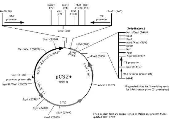
2.1 DNA constructs 48
2.1.a Purification of plasmid DNA 52
IV
2.2 Xenopus laevis embryos 53
2.2.a Lipofections 53
2.2.b In situ hybridization 55
2.2.c Immunostaining and immunofluorescence 56
2.3 GST-pull down assay 56
2.3.a GST-fusion protein production and purification 56 2.3.b Cell transfection and pull-down assay 57
2.3.c Western blotting 58
2.4 Transactivation assays 59
Results 61
3.1 Molecular dissection of XOTX2 and XOTX5b during Xenopus
retinogenesis: a 10 AA box switches XOTX2 and XOTX5b cell fate choice
activities. 61 3.2 The RS box confers specific activities to Drosophila OTD 71
3.3 The RS box is involved in the correct nuclear localization of
XOTX/OTD proteins in retinal neurons 75 3.4 XOTX2 and XOTX5b differentially synergize with XNRL to
transactivate the Xenopus rhodopsin promoter 78 3.4 In vitro interactions of XOTX/OTD proteins with XNRL 81
V
3.5 XOTX2 and XOTX5b can form homo/heterodimers that influence their
activity 85
Discussion 88
4.1 Molecular dissection of XOTX2 and XOTX5b during Xenopus
retinogenesis: a 10 AA box switches XOTX2 and XOTX5b cell fate choice
activities. 88 4.2 The RS box confers specific activities to Drosophila OTD 91
4.3 The RS box is involved in the correct nuclear localization of
XOTX/OTD proteins in retinal neurons 94 4.5 XOTX2 and XOTX5b can form homo/heterodimers 98
4.6 Evolution of eye and retinal cell types 99
Concluding remarks 103
References 105
1
Introduction
1.1 The Vertebrate retina
"No one ever have the courage to start a historical overview of any topic in neuroscience without mentioning Cajal's contribution to that given field. It is particularly true for retinal research, where he has been instrumental in defining retinal connection pattern and possible function of the main neuron classes (Cajal,
1892)."
- Robert Gabriel -
Figure 1.1. Structure of the Mammalian Retina.
Cross-sectional microscopic drawing of the nerve cells in the retina made by Santiago Ramon y Cajal (1900). http://hubel.med.harvard.edu/12.jpg
2 The retina has arguably the most intricate and aesthetically pleasing cytoarchitecture of any sensory system (Fig. 1.1). The combination of highly specialized cell types in a well-organized wiring and complex modulatory activity, results in an amazing and flexible sensory processing system.
Although our knowledge of how the retina is organized and functions is absolutely essential, understanding how it is assembled during development is a big challenge in Neurobiology. Indeed, understanding how the retina arises is attractive not only to developmental neuroscientists interested in vision, but to all neuroscientists interested in neural development, because the retina is "an approachable part of the brain" and developmental processes required to build up this exquisitely organized system are basically relevant to all other parts of the Central Nervous System.
The vertebrate retina comprises five major classes of nerve cells (see Wässle, 2004, for review). Rod and cone photoreceptors convert light information to chemical and electrical signals that are relayed to interneurons in the outer retina. Bipolar interneurons are contacted by photoreceptors and convey signals from the outer retina to the inner retina. Transmission from photoreceptors is modulated by horizontal cells that also contact the bipolar cell. In the inner retina, bipolar cells form chemical synapses with two classes of neurons, amacrine interneurons and retinal ganglion cells. Amacrine cells not only modulate signals from the bipolar cells by providing inhibition directly onto ganglion cells, but also modulate transmitter release from the bipolar cells. Light information leaves the retina and reaches the other stations in the brain via axons of the ganglion cells that collectively form the optic nerve (Wong, 2006).
Beside these five major classes of neurons, in the retina a type of macroglia exists: the Müller glia cells. Müller cells span the depth of the retina and provide important, structural and functional support for the retinal neurons.
The cell bodies and connections of retinal neurons are arranged in layers and this laminar organization of the retina is stereotypic across species.
3 Connections are restricted to two major laminae, the outer plexiform layer (OPL) and the inner plexiform layer (IPL). The nuclei of nerve and glial cells are organized in three nuclear layers. Photoreceptor cell bodies form the outer nuclear layer (ONL); horizontal, bipolar, amacrine and Müller glia cell bodies are located in the inner nuclear layer (INL) and, finally, ganglion cells form the ganglion cell layer (GCL) (Fig. 1.2).
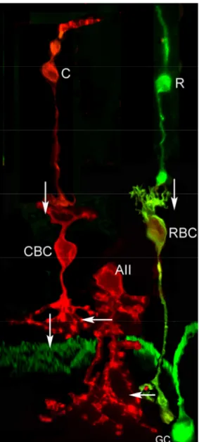
Embedded within this basic organization of the vertebrate retina many specialized subcircuits are present, working together in parallel to process different features of the visual image. For example, rods are sensitive to low-light levels and rod-driven circuit exists for visualizing objects under dim light conditions. In most vertebrates, this circuit involves connections among rod photoreceptors, rod bipolar cells and a specialized type of amacrine cells, the AII amacrine cells connecting to ganglion cells (Strettoi et al., 1990). On the other hand, under high light level conditions, cones work involving two vertical pathways, giving also a chromatic information of the visual stimulus. Cones contact a variety of cone bipolar cells, some of which are depolarized (ON) and others hyperpolarized (OFF) by increased illumination. ON and OFF-cone bipolar cells contact ganglion cells, which respond to changes in illumination according to their bipolar input. Together, the ON and OFF pathways provide contrast information. In addition to these basic features, the retina has also specialized circuits that can compute other features of the visual scene, such as the direction of motion and orientation of edges (Wong, 2006)
4
Figure 1.2. Tri-dimensional representation of the retinal structure.
The retinal structure and the principal retinal cell types are shown (modified from Balboni et al., 1993).
1.1.a The Xenopus retina
In this work great relevance has been given to Xenopus laevis as model system. The clawed frog, Xenopus laevis, is an ancient Anuran amphibian, exploited in developmental studies because of its well-characterized larval stages (Nieuwkoop and Faber, 1956). In relation to experimentation on the visual system, Xenopus has been utilized as a model system for studies of retinotectal
5 projection (Gaze and Keating, 1970) and also for biochemical studies of visual pigment, thanks to its large rod photoreceptors (Dartnall, 1954; Wald, 1955; Moritz et al., 1999). In the recent era of molecular biology, the Xenopus embryo is a favoured system for studies of gene function during neural development.
Frogs usually have a good vision and Wilhelm and Gabriel suggested that the potential spatial resolution power of the Xenopus retina is approximately as good as that of the central retina of mammals (Wilhelm and Gabriel, 1999). The basic structural and functional organization of the amphibian retina is fundamentally similar to the mammalian/primate one (Krizaj, 2000).
However, it is generally accepted that the relative simplicity of the brain visual system in lower vertebrates may require a higher degree of visual information processing at the retinal level (Vigh et al., 2000). For this reason, the retina of non-mammalian vertebrates seemed to posses more complex receptive field properties than those of mammals. Edge, dimming and convexity detectors, directional selectivity and processing of moving stimuli, all the neural circuits that analyze these properties seemed to be present in the Xenopus retina (Gabriel et al., 2000).
On account of this, the Xenopus retina shows some peculiarities in respect to the mammalian retina.
Photoreceptors. Rod photoreceptors represent 53% of total photoreceptors
(Wilhelm and Gabriel, 1999). Rods are rather uniform regarding their morphology, although a minor population (2-3%) of blue-sensitive rods, with a thinner outer segment, has been described on the basis of its visual pigment content (Witkovsky et al., 1981). Cones have also been classified on the basis of their visual pigment content and can be divided into three types: miniature, ultraviolet-wavelength-sensitive (UWS) (4% of all cones); large, short-wavelength-sensitive (SWS) (10%); and large, long-short-wavelength-sensitive (LWS) cones (86%) (Rohlich et al., 1989).
6
Horizontal cells. The Xenopus retina differs from the pattern in mammals, in
which horizontal cell dendrites contact only cones, while the axon terminal contacts rods (Steinberg, 1969; reviewed in Wassle and Boycott, 1991). There are two types of horizontal cells in Xenopus retina (Stephan and Weiler, 1981). In the axon-bearing cell of the luminosity type (i.e. lights of any spectral composition elicit a hyperpolarization), both dendrites arise in the cell body and axonal branches contact both rods and cones. The other horizontal cell type lacks an axon and is of the chromatic type (depolarized by red light, hyperpolarized by blue light) (Stone et al., 1990). Its dendrites contact what are short-wavelength-sensitive cones and rods (Witkovsky et al., 1995).
Bipolar cells. The main point in the structure of the lower vertebrates is that
retinal circuits did not evolve separate rod-dedicated and cone-dedicated pathways. So, bipolar cells receive direct rod and cone inputs and specific rod bipolar cells do not exist. In intermediate light condition (mesopic state), when both cones and rods are active, it is probably disadvantageous for an animal to receive double information concerning the same object (due to, for instance, the different latency of the two kinds of photoreceptors). However, the mutual antagonism between rod and cone signals decreases the magnitude of this problem; this mechanism of the mutual inhibition involves the neuromodulator dopamine, synthesized by the dopaminergic amacrine cells (reviewed in Krizaj, 2000).
Amacrine cells. Sporadic attempts to note down the cell types by their
morphology have been made by early researchers, particularly by Cajal (1892). He distinguished 13 amacrine cell types, mostly on the basis of ramification pattern in the IPL. In the Anuran retina, the majority of the amacrine cells takes up GABA and glycine (Voaden, 1974). Moreover, dopaminergic cells are present at low density, representing about 0.5% of the total amacrine cell number. In addition, in the Xenopus retina other amacrine cell types are present, identified by
7 different markers: serotonin-immunoreactive, nitric-oxide producing cells, neuropeptide Y, substance P, somatostatin and colecystokinin immunoreactive (reviewed in Vigh et al., 2000). The importance of diverse amacrine populations in retinal information processing can be explained by the relative simplicity of the brain visual system in lower vertebrates (Vigh et al., 2000).
Ganglion cells. It can be cautiously considered the presence of 12 types of
ganglion cells in the frog retina. There has been a long-standing debate on the existence of direct bipolar to ganglion cell contacts in the lower vertebrate retina. Definite bipolar to ganglion cell synapses have been identified in Xenopus laevis (Buzas et al., 1996), representing about 10% of all ganglion cell inputs in the IPL. This fact can be explained by considering that there is a higher divergence of bipolar cell output to amacrine cells in frog than in mammals (reviewed in Gabriel et al., 2000).
Ciliary marginal zone. A peculiarity of the amphibian and fish retina is that its
peripheral portion, termed ciliary marginal zone (CMZ), is a pseudo-stratified neuroepithelium from which retinal precursors differentiate, allowing growth of the retina throughout the whole life of the animal. In fact, after the embryonic phase of retinogenesis, new cells are added to the central retina from this peculiar proliferative region (Wetts et al., 1989). CMZ progenitors are multipotent and can give rise to all retinal cell types, including pigmented epithelial cells (Wetts and Fraser, 1988). The main feature of this region is that the retinoblasts are ordered along the CMZ, from its peripheral edge towards the centre, according to their grade of commitment.
Therefore, stem cells are located in the most peripheral region of the ciliary margin, post-mitotic precursors are adjacent to the central retina, and proliferating neuroblasts are distributed between these two regions. Because each set of precursors, in their state of commitment, is characterized by the expression of an unique combination of genes, the consequence of such a defined spatial
8 distribution of the precursors is that the CMZ recapitulates spatially the temporal sequence of gene expression during retinogenesis (Wetts and Fraser, 1988; Perron et al., 1998).
Such a spatially arrayed ciliary margin does not exist in the mammalian and bird retina, even if both types of retina contain stem cells. The mouse retinal stem cells are located in the pigmented ciliary margin (PCM) (Tropepe et al., 2000). These cells may be the evolutionarily homologs of the amphibian and fish CMZ precursors, but their location is completely different; in fact, the non-pigmented iris margin of the mammalian retina (corresponding to the amphibian and fish CMZ) is devoid of stem cells. The PCM stem cells can differentiate into various retinal neuronal types including photoreceptors, bipolar neurons and Müller glia (Tropepe et al., 2000).
More recently, retinal stem cells have been isolated from the human retina. These cells display self-renewal properties and, above all, when transplanted into mouse post-natal or embryonic chick eyes, are able to survive and differentiate in the host retina (Coles et al., 2004). Thus, the adult mammalian eye harbours stem cells, which can be induced to re-enter the cell cycle and initiate neuronal differentiation.
1.2 Retinal development
Vertebrate retina is a complex neural structure that comprises highly organized, laminated networks of nerve and glial cells. The biological question we ask is how this complexity arises during development.
The vertebrate retina shares a common origin with the rest of the central nervous system. Retinal development begins with specification of the eye primordia during early stages of embryonic life, highlighted by the appearance of a bilateral evagination of the ventro-lateral diencephalon. Upon continuous evagination of the optic primordia, two optic vesicles are generated which extend
9 towards the overlying, non-neural ectoderm which will ultimately originate the lens and the cornea. The optic vesicle invaginates and gives rise to a double-layered optic cup: the inner layer (facing the lens placode) will give rise to the neural retina, while the outer layer will differentiate into the retinal pigmented epithelium (reviewed in Chow and Lang, 2001).
The retinal progenitors in the inner layer of the optic cup are proliferating and share an uniform morphology; they are initially arranged as a pseudostratified neuroepithelium, whereby cells contact both surfaces of the layer (Fig. 1.3). This layer is apposed at its outer (scleral) surface to the retinal pigmented epithelium (RPE), but remains separated by the potential space of the obliterated neural tube lumen (optic ventricle). The nuclei of progenitors undergo S-phase distal to the neural tube lumen but enter M-phase at the luminal surface. During mitosis retinal progenitors likely retain their basal process, divide and subsequently re-establish contact with both sides of the layer (Cayouette and Raff 2003).
It is interesting to note that during retinal progenitor proliferation there is a change of cell cycle length and all studies of cell cycle timing in the retina are in accordance with the fact that it slows during development (Rapaport, 2006; Decembrini et al., 2006).
Another mechanism affecting the production of cells is the mode of division, of which three can be described. Early progenitors go through a period of symmetrical divisions, each daughter returning the cell cycle. This mode allows the pool of progenitors to expand exponentially. Later, retinal progenitors divide asymmetrically, one daughter returning the cell cycle, the other exiting, migrating and differentiating. At some late stage, progenitors go through a terminal symmetrical division in which both daughters become post-mitotic and differentiate in the correct retinal cell types (Rapaport, 2006).
In this regard, it is noteworthy that the sequence of cell genesis in the vertebrate retina is highly conserved.
10
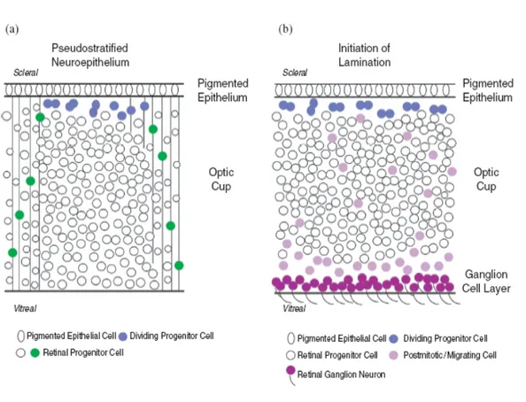
Figure 1.3. Cellular arrangement during retina development.
(A) The initial neural retina is arranged as a pseudostratified neuroepithelium. Prior to neuron formation, uncommitted progenitor cells contact both surfaces of the optic cup. At the onset of mitosis, cells move their nuclei to the scleral side, lose a contact with the optic cup and round up. This process is reversed after division is complete.
(B) When neurogenesis begins, proliferation still occurs at the outer side of the optic cup, but the migration of committed neuroblasts and the accumulation of mature ganglion cells at the vitreal side is now apparent (from Vetter and Brown, 2001).
1.2.a Retinal neurogenesis
The characteristics of the retina make it an ideal tissue to study neurogenesis. Its development proceeds through three overlapping steps starting with retinal progenitor cell proliferation, followed by birth of post-mitotic retinal
11 transition cells (also referred to as precursors), and ending with terminal differentiation of the seven major cell types (Chen et al., 2007).
Despite the differences among each vertebrate class, two common features are shared by the newly generated neurons during development of the retina. First, as shown by birthdating analyses performed in chick (Prada et al., 1991), monkey (LaVail et al., 1991), rat (Rapaport et al., 2004) and Xenopus (Stiemke et al., 1994; Decembrini et al., 2006), the seven major retinal cell types are generated in an extremely conserved histogenetic order, with ganglion cells born first and Müller cells born last (Cepko et al., 1996). In particular, in all species studied cell birth proceeds in the following retinogenetic timing: ganglion cells, horizontal cells, cones, amacrine cells, rods and bipolar cells. Finally, Müller glial cells differentiate. Moreover, the first cells to be generated, ganglion and horizontal cells, are the largest in the retina, supporting the hypothesis that large neurons are generated before small ones. Likewise, the last cells to be born are the Müller cells, supporting a trend in the CNS for glia to be generated late (Rapaport, 2006). The second important fact concerning the retinal neurogenesis is that, as demonstrated by means of lineage tracing analyses and cell ablation studies, retinal progenitors are multipotent at the different developmental stages and a single progenitor is able to produce all the different retinal cell types (Turner and Cepko, 1987; Holt et al., 1988; Wetts and Fraser, 1988; Turner et al., 1990). So, retinal neurogenesis follows a precise and evolutionarily conserved order which suggests the conservation of the underlying molecular mechanisms among the Vertebrates.
An attractive hypothesis to accommodate these findings was that, once specified as retinal progenitors, the various cell fates of postmitotic neurons are determined by environmental signals. Alternatively, these inducing signals might be present at many stages, but an autonomous clock could regulate the competence of cells to respond to them. To differentiate between these mechanisms, in vitro heterochronic transplant experiments had been performed in both chick and rodents, in which progenitors from different stages of development
12 were placed in an environment of a different age (Livesey and Cepko, 2001). For example, early chick progenitors, which normally generate ganglion cells in vivo, originate ganglion cells regardless of the age of the environment that they are placed in (Austin et al., 1995). Another strong evidence, in this regard, derives from experiment of heterochronic transplantation. Cells from young embryonic retinae were dissociated and grown together with those from older embryos, and the timing of rod determination assayed. Young cells appeared uninfluenced by older cells, expressing photoreceptor markers on the same time schedule as when cultured alone, even if there is a change in the percentage of differentiated rods (Watanabe and Raff, 1990). A similar result was obtained when the heterochronic mixing was done in vivo by grafting a small plug of optic vesicle from younger embryos into older hosts. Even the graft cells at the immediate margin of the transplant failed to express photoreceptor markers earlier than normal, despite their being in contact with older cells (Rapaport et al., 2001).
1.2.b The competence model of the retinal cell fate determination
The above-mentioned and other observations led Connie Cepko and co-workers to the elaboration of the “competence state” model (Cepko et al., 1996). The competence model states that progenitors pass through a series of competence states, during each of which the retinal progenitors are competent to produce one or a subset of retinal cell types. Within a given competence state, the generation of a particular type of cell is regulated by positive and negative extrinsic signals (Livesey and Cepko, 2001) (Fig. 1.4).
Although the competence model was formulated to explain cell fate choice in the vertebrate retina, it is clear that cell specification in many other regions of the developing nervous system - including neural crest (Selleck and Bronner-Fraser, 1996), spinal cord (Ericson et al., 1996), and cerebral cortex (McConnell, 1988; Qian et al., 2000) - involves changes in progenitor competence over time,
13 frequently resulting in altered sensitivity to extrinsic factors (Blackshaw et al., 2001). Moreover, the model of temporal changes in competence is strongly
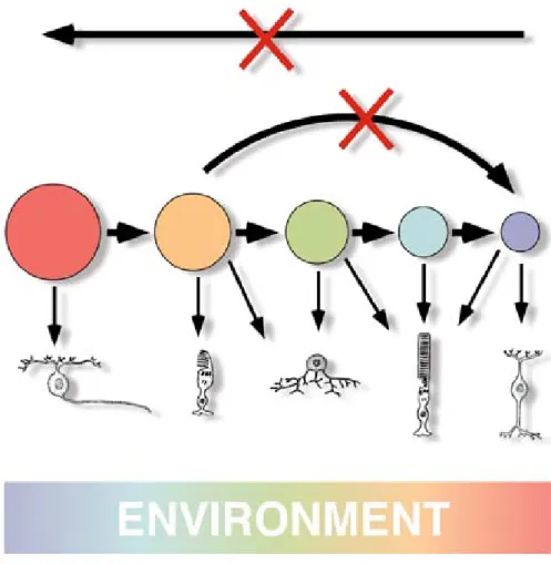
Figure 1.4. The competence model of retinal cell fate determination.
Retinal progenitors comprise a dynamic mixture of mitotic cell types that interact with the environment to make the different postmitotic cell types. Each progenitor cell is thought to be controlled by a complex of transcription factors that define its competence state. Retinal progenitors are modulated to progress from one state of competence to another in only one direction. The environment is shown to be changing over time (from Cepko et al., 1999).
14 supported by elegant studies of Drosophila CNS development (Isshiki et al., 2001; Pearson and Doe, 2003), where the sequential expression of the transcription factors Hunchback, Krüppel, Pdm and Castor was found to change cell fate competence (Isshiki et al., 2001). Noteworthy, cdc25 mutants, whose cell cycle is arrested at the G2-M transition, fail to progress through the normal sequence of gene expression (Isshiki et al., 2001), suggesting the importance of a
cell cycle-dependent clock (Cremisi et al., 2003). It is interesting to note that the situation in the retina, where early progenitor cells cannot be induced to adopt late fates and vice versa, is distinct from the progressive developmental restriction that is seen in the cerebral cortex, where early cortical progenitor cells are competent to generate cells of upper (late-born) and lower (early-(late-born) layers of the cortex, but become restricted to generate only late-born fates as development proceeds (Desai and McConnell,
2000).
The obvious questions are what defines the cellular differences among progenitors at different times, how those differences define different competences and how passage between one state and the next is regulated. The main mechanisms for control of a competence state are transcriptional program and post-transcriptional regulation, such as protein expression, modification, accumulation and degradation (Livesey and Cepko, 2001).
In order to identify genes that might regulate retinal development, Cepko and coworkers recently performed a gene expression study in the mouse developing retina using serial analysis of gene expression (SAGE). SAGE is a technique that provides a comprehensive profiling of gene expression; moreover, genes that show dynamic expression via SAGE have been analyzed by in situ hybridization. In this way, a molecular atlas of the expression patterns of 1051 genes in the developing and mature retina was thereby constructed (Blackshaw et al., 2001). The laminar structure of the retina makes it relatively simple to assign an identity to cells expressing a given gene. During early stages of retinal development, mitotic progenitors form the outer neuroblastic layer (ONBL), while
15 newborn neurons reside in the inner neuroblastic layer (INBL). As we have seen, the position of mitotic progenitors within the ONBL varies depending upon their progress through the cell cycle, with S phase cells being found on the vitreal side of the ONBL near the border with the INBL and M-phase cells being found on the scleral side of the ONBL, near the retinal pigment epithelium. Blackshaw et al. analyzed the gene expression in the scleral and vitreal portions of both the ONBL and INBL separately. Virtually every gene previously reported to regulate retinal development was detected in this analysis and showed dynamic expression during development. For instance, NeuroD1, which regulates rod photoreceptor survival, as well as possibly rod differentiation (Morrow et al. 1999; Wang et al. 2001), is
overexpressed at P4.5.
In the case of genes previously shown to be required for production of certain cell types in the developing retina, such as Ath5 and Chx10 - which are required for ganglion cell and bipolar neurons, respectively (Burmeister et al., 1996; Brown et al., 2001; Wang et al., 2001) - peak expression typically occurred around or just after the peak time of exit from mitosis for that cell type
(Blackshaw et al., 2001).
On the other hand, the authors have identified a number of genes that show temporally restricted expression in early ONBL. By analyzing the expression of a large number of genes that were highly expressed early in development they found that are expressed in broad but temporally restricted subsets of mitotic progenitor cells. For example, sFrp2 (secreted Frizzled related protein 2, a modulator of the Wnt signalling pathway) RNA was found to be broadly expressed in the ONBL until E16, after which it rapidly decreased. Expression of Fgf15 (Fibroblast growth factor 15) was seen to persist longer, but never was easily detected after P0. Lhx2, by contrast, was weakly expressed in subsets of cells in the ONBL until P0, when it was dramatically and transiently upregulated
throughout the ONBL.
From this study it has been confirmed that the population of progenitors is complex at any time, as there is evidence for progenitor heterogeneity at several
16 points during development (Livesey and Cepko, 2001). In fact, a limited number of genes have previously been reported as expressed in subsets of mitotic retinal progenitor cells, including genes such as Ath5, and have been shown to be required for retinal ganglion cell development (Brown et al. 2001; Wang et al. 2001. They identified a large number of genes that showed selective expression at certain times during development in relatively small subsets of cells in the ONBL. These include a large number of known and putative transcription factors, such as Sox2, Sox4, Tbx2, Eya2 and Mbtd1 (a novel polycomb family member), along with many genes of other functional classes.
Particularly intriguing is the early and transient expression of Pum1, a mammalian homolog of the pumilio gene, which has been shown to mediate asymmetric mRNA distribution in Drosophila (Micklem, 1995). Many of these genes showed highly dynamic expression during development - rapidly shifting their cellular expression patterns in the course of a few days. In some cases, these genes were scattered throughout the ONBL, such as Eya2 at E14, while for other genes, such as Pum1 and Pgrmc2 (a surface membrane progesterone receptors), expression was in only the scleral portion of the ONBL, suggesting that these genes may show strongest expression near M-phase in retinal progenitor cells.
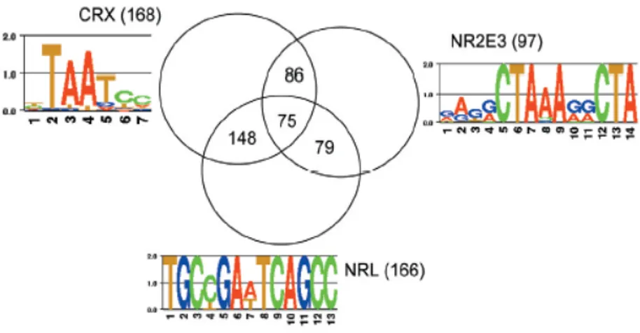
When retinal neuron differentiation begins, several genes involved in cell fate specification and/or in the early steps of the differentiation process are expressed only in newly postmitotic cells and cells actively undergoing differentiation. Some of these genes, namely Crx, Nrl, and Nr2e3 (Furukawa et al. 1997; Chen et al. 1997; Haider et al. 2001; Mears et al. 2001) have been shown to play an active role in regulating photoreceptor differentiation (Blackshaw et al.
2001).
Finally, in this work the authors found that genes selectively expressed in Müller glia share a great degree of transcriptional overlap with mitotic retinal progenitor cells. Among the genes identified as being specifically expressed in Müller glia after the first post-natal week, 68% was found to be enriched in mitotic progenitor cells based on their in situ hybridization pattern. This extensive
17 overlap raises the question of how closely these two cell types are at the functional level. Müller glia morphologically resembles mitotic progenitor cells in having apical and basal processes that span the radial dimension of the retina - a feature that is shared with retinal progenitor cells as well as radial glia of the developing brain, a cell type known to be the cortical progenitor cell (Doetsch, 2003). Müller glia is the last cell type to exit mitosis and represents the only cells in the mature retina that can reenter mitosis following retinal injury (Dyer and Cepko 2000; Vetter and Moore, 2001; Bernardos et al., 2007).
Finally, data from chicken suggest that Müller glia can be induced to divide and give rise to some types of retinal neurons for a short period of time near the end of retinal development (Fischer and Reh, 2001).
Recently, it has been shown that Müller glia-derived progenitors express Crx and are late retinal progenitors that generate the rod photoreceptor lineage in the post-embryonic zebrafish retina (Bernardos et al., 2007). Moreover, Müller glial cells also are competent to produce earlier neuronal lineages, in that they respond to injury-induced loss of photoreceptors by specifically regenerating missing cones and rods (Bernardos et al., 2007).
The question arises whether Müller glia cells of mammalian retina are fundamentally multipotent progenitor cells that are quiescent regarding cell division and the production of neurons (Morest and Silver, 2003; Walcott and Provis, 2003), conversely to the fish retina. Indeed, if they are progenitor cells, they have acquired the specialized properties needed for a support role in the mature retina, e.g., neurotransmitter reuptake and structural roles (Blackshaw et
al., 2001).
Beside mRNA expressed during retina development, a number of RNA transcripts that do not appear to encode proteins were strongly expressed in the developing retina (Blackshaw et al., 2001). These transcripts are typically spliced and polyadenylated, but do not encode evolutionarily conserved open reading frames (ORFs), or any ORFs encoding proteins longer than 100 amino acids, while often showing high similarity at the nucleotide level between mouse and
18 human (Numata et al., 2003). Putative non-coding transcripts that showed developmentally dynamic expression include retinal non-coding RNA 1 (RNCR1), which was expressed throughout the ONBL during early development and which was later restricted to Müller glia. It was transcribed in a head-to-head fashion, and largely coexpressed, with Six3. RNCR2, on the other hand, was expressed in a large subset of cells in both the ONBL and INBL prenatally, with expression restricted to the INL and GCL postnatally (Blackshaw et al., 2001).
Large-scale EST sequencing efforts from mouse have uncovered up to several thousand putative spliced transcripts that do not appear to encode for proteins (Numata et al., 2003). The functional role of these transcripts is obscure, although non-coding spliced RNAs such as Xist and H19 in mammals and Rox1 and Rox2 in Drosophila have been implicated in a variety of epigenetic processes (Mattick, 2003).
1.2.c Regulators of competence state: extrinsic versus intrinsic signals
There is a long-standing debate on the relative importance of the extrinsic signals versus intrinsic regulators on retinal cell fate during retinal development. One idea in favour of the extrinsic possibility is that the addition of new differentiated cells could feed signals back to the dividing progenitors and influence the fate of their daughters (Agathocleos and Harris, 2006).
Experimental evidence supports a feedback inhibition mechanism: by cell-mixing experiments, using amacrine-enriched or amacrine depleted cellular environments, Belliveau and Cepko demonstrated that the postnatal environment had at least two signals that affected the cell fate; one signals inhibited the production of amacrine cells and the second affected the production of cones. In particular, previously generated amacrine cells produce a feedback signal that inhibits the production of the amacrine cell themselves. At the same time, this inhibition is compensated by the production of cones and no changes in other cell
19 type frequency are observed (Belliveau and Cepko, 1999).
The authors suggested that extrinsic signals can influence progenitor decision, in order to control the number of differentiated cells, but the choice of the cell fate is restricted by the intrinsic biases of progenitor cells (Belliveau and Cepko, 1999). On the other hand, signals in the embryonic retinae inhibit rod and favour bipolar cell generation from postnatal progenitors (Belliveau et al., 2000). This alteration in cell fates appeared to be caused by a secreted factor released by embryonic cells that requires the LIFR, a receptor for LIF (Leukaemia Inhibitory Factor), CNTF (Ciliary Neurotrophic Factor) and other cytokines (Belliveau et al., 2000). Another molecule, Sonic Hedgehog (Shh), has been identified as a factor having the potential to be both a feedback inhibitory signal for the production of ganglion cells (Zhang and Yang, 2001) and a positive factor for the differentiation of other retinal cell types (Stenkamp et al., 2002; Shkumatava et al., 2004).
Taurine, an unusual amino acid, is an extrinsic factor produced from P0 rat retinal cultures and its addition to retinal explants promotes rod differentiation (Altshuler et al., 1993), acting via glycine receptor and (GABA)A receptor (Young
and Cepko, 2004). It has also been shown that isolated progenitors differentiate to rods or cones according to the relative amounts of retinoic acid and thyroid hormone (Kelley et al., 1995 and 1999).
Among the extrinsic factors important for retinal development there are neurotrophins, a family of growth factors consisting of NGF (Nerve Growth Factor), BDNF (Brain-Derived Neurotrophic Factor), NT-3 (Neurotrophin-3) and NT-4/5. Besides their critical importance for correct specification and survival of a number of classes of neurons in the central and peripheral nervous system (Lewin and Barde, 1996), neurotrophins have an important role in earlier stages of development (Pearson, 2006).
For example, NT-3 is expressed in retinal pigmented epithelium and then in neural retina (Rodriguez-Tebar et al., 1993). It has been demonstrated that NT-3 stimulates the birth of new neurons. By inhibiting the NT-NT-3 action using specific antibodies to neutralize endogenous NT-3 (Bovolenta et al., 1996) there is a
20 marked decrease in retinal neuron differentiation, ganglion cells being most affected. Additionally, the impairment of NT-3 signalling causes a decrease in clonal expansion of cells derived from a single retinal progenitor (Das et al., 2000). In contrast, NGF and BDNF have a role during programmed cell death occurring during retinogenesis (Frade et al., 1999).
Ultimately, extrinsic signals need to be translated into an internal code that will drive a cell towards one fate or another, by switching on a precise transcriptional program. The Notch-Delta pathway is a paradigmatic example of how an extracellular signal can do so (Agathocleos and Harris, 2006).
Notch is a transmembrane receptor that transduces an extrinsic cue, that of the binding of its ligand Delta or Serrate, to directly regulate the transcription of several target genes, in particular repressing proneural genes coding for basic helix-loop-helix (bHLH) transcription factors (Artavanis-Tsakonas et al., 1999) (Fig. 1.5). Studies in frog, rat, chick and mouse have shown that Notch1 is expressed by proliferating and undifferentiated cells (Dorsky et al., 1995; Bao and Cepko, 1997; Lindsell et al., 1996) and its expression is retained by Müller glial cells (Furukawa et al., 2000; Dorsky et al., 1995).
It has been demonstrated that constitutive activation of Notch pathway in fish, frog, chick and rat retina inhibits neurogenesis (Dorsky et al., 1995; Austin et al., 1995; Bao and Cepko, 1997; Scheer et al., 2001) and promotes gliogenesis (Furukawa et al., 2000; Scheer et al., 2001).
A good progress in this regard comes from a recent work where Jadhav et al. (2006) demonstrated, by means of a comprehensive molecular characterization, that activation of Notch in early progenitors allowed them to retain appropriate early progenitor gene expression. When examined at later stages of development, however, the cells exhibited expression of an inappropriate mixture of progenitor genes (like fgf15 and cyclin D1) and glial genes (Jadhav et al., 2006). Moreover, a functional assay showed that these cells could form neurospheres, similar to stem cells derived from the retinal pigmented epithelium of the mammalian peripheral retina (Jadhav et al., 2006).
21 Furthermore, selective reactivation of Notch pathway in newly generated postmitotic cells that had previously released Notch activation during development, led to their differentiation in proper Müller glial cells (Jadhav et al., 2006). In conclusion, prolonged Notch activity in progenitors permits them to progress through multiple states without perturbing temporal identity, promoting early progenitor characteristics early in development and late characteristics later in development. Remarkably, constitutive Notch activation led these cells to acquire both glial and stem cells characteristics (Jadhav et al., 2006).
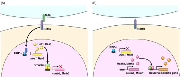
Figure 1.5. Molecular mechanism of Notch signalling.
(A) Upon activation by Notch ligands from surrounding cells, the intracellular domain of Notch (ICN) is cleaved off from the transmembrane region and translocated into the nucleus. In the nucleus, the ICN forms a complex with the DNA-binding protein RBP-J. This complex induces expression of bHLH repressors such as Hes1 and Hes5. Hes1 and Hes5 repress transcription of bHLH activators and inhibit neuronal differentiation. Thus, the Notch pathway links the extrinsic signals (Notch ligands from neighbouring cells) to the intrinsic factors (Hes1/Hes5) for regulation of cell differentiation.
(B) When Notch is not activated, Hes1/Hes5 expression is off, allowing bHLH activators to trigger neuronal-specific gene expression (from Hatakeyama and Kageyama, 2004).
22 On the other hand, the relationship between lineage and histogenesis is certainly consistent with the idea of an intrinsic developmental clock (Cayouette et al., 2006). Beside the studies of extrinsic regulation of cell fate, we have several evidences on the role of intrinsic factors during retinal neuron determination. bHLH proneural genes, mentioned above as target of Notch signalling, and homeobox genes are basically the best characterized transcription factors involved in generating the diversity of retinal cell fates (Fig. 1.6).
A prime example of a bHLH gene is Ath5. The Xenopus homologue, Xath5, promotes retinal ganglion cell genesis when overexpressed in vivo (Kanekar et al., 1997). It can induce the expression of Xbh1, a homeodomain transcription factor involved in ganglion cell differentiation (Hutcheson and Vetter, 2001; Liu et al., 2001; Poggi et al., 2004). When ath5 gene is non functional, such as in zebrafish lakritz mutants (Kay et al., 2001) or in Math5 mutant mice, there is a depletion of ganglion cells (Brown et al., 2001; Wang et al., 2001). Ath5 has an interesting effect on cell cycle because cells that express Xath5 tend to exit the cell cycle early, at the appropriate time for ganglion cell
genesis (Ohnuma et al., 2002a).
Other bHLH have different profiles of activity with respect to cell determination in the retina. NeuroD, for example, promotes amacrine over bipolar cell fate and favour photoreceptor survival (Morrow et al., 1999). Mash1 and Math3 are both expressed in bipolar cells and in their double mutation virtually all bipolar cells are abolished (Tomita et al., 2000).
The loss of Chx10 gene, coding for a homeodomain transcription factor, results in the complete loss of bipolar cells in mice, too (Burmeister et al., 1996). Recently, it has been identified a bHLH transcription factor, Bhlhb4, that is required for rod bipolar cell maturation. Bhlhb4-/- mice lack specifically rod bipolar cells, while the other retinal neurons are unaffected (Bramblett et al.,
23
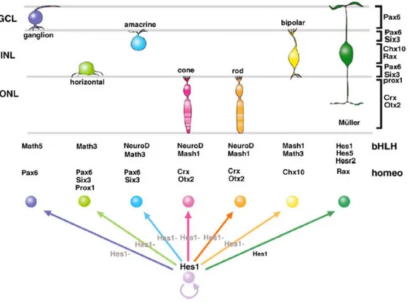
Figure 1.6. Cooperation of bHLH and homeodomain transcription factors for retinal cell type specification.
Hes1 inhibits neuronal differentiation and maintains progenitors. Differentiating neurons lose Hes1 expression. Homeodomain and bHLH factors determine the neural fate of retinal cell type. The cells that do not lose Hes1/Hes5 expression during neurogenesis stages adopt the Müller glial fate (from Hatakeyama and Kageyama, 2004).
Instead, Prox1 is involved in horizontal cell differentiation and null mice lack horizontal cells (Dyer et al., 2003).
Another gene, Foxn4, coding for a winged/helix forkhead transcription factor, has been isolated and controls the competence state of amacrine and horizontal cells. Mutation in Foxn4 result in the elimination of horizontal cells and a great reduction of amacrine cells (Li et al., 2004).
24 Finally, Crx/otx5 is involved in photoreceptor differentiation in mouse and Xenopus (Furukawa et al., 1997; Viczian et al., 2003). It is also demonstrated that CRX/OTX5 works in synergism with NRL, a leucine zipper transcription factor, in order to promote rod differentiation (Mitton et al., 2000; Mears et al., 2001).
1.2.d Towards an integrated model
On the basis of the above-mentioned observations, the competence model can explain the different roles of the extrinsic and intrinsic factors. Environmental signal can alter the relative proportion of each cell type generated at a given time but it cannot influence progenitors to make temporally inappropriate cell types. Competence states seem to be intrinsically defined and within a given competence state, the generation of a particular type of cell is regulated by positive and negative extrinsic signals (Livesey and Cepko, 2001).
Retinal histogenesis offers a good example of a complex phenomenon where multiple players are involved in establishing the generation of a specific type of neuron. The emerging scenario is that the presence of these players must be tightly controlled in space and time to generate the different retinal cell types. In other words, the molecular players must be active in the right cellular type at the appropriate time. This regulation is a multi-step process which is carried out at different levels by means of transcriptional, translational and post-translational
mechanisms.
In this point of view, the debate on the major importance of the extrinsic or intrinsic factors on cell fate determination should be downsized. For example, most of the extrinsic factors, like growth factors, neurotrophins and secreted peptides, are molecules produced by specific cells at a specific time and are the result of a strong-intrinsic-programmed cell activity.
On the other hand, these extrinsic factors do usually act on specific receptors whose expression, localization and function is highly controlled by
25 transcriptional and post-transcriptional mechanisms.
Moreover, some extrinsic factors can regulate the expression of several transcription factors. For example, it has been demonstrated that CNTF/LIF, extrinsic factors able to block rod differentiation, as we have seen, specifically activate a molecular cascade that acts on Crx promoter, inhibiting its transcription (Ozawa et al., 2004). This observation represents an evidence of the strong interplay between extrinsic signals and intrinsic factor activity during neuroretinal
histogenesis.
Transcription factor activity is well-controlled at different levels. For instance, Moore and co-workers have shown that, in Xenopus, the activity of bHLH factors can be regulated post-translationally in a temporally tight specific manner, so that each factor is active at the right time during retinogenesis (Moore et al., 2002). This is the case of NeuroD which is regulated through phosphorylation by glycogen synthase kinase-3β (GSK 3β). GSK 3β prevents XNeuroD to promote frog retinal neurogenesis at early stages, by phosphorylation at a specific site; at later stages, GSK 3β inhibition is released, allowing XNeuroD to promote later cell type differentiation. Interestingly, a mutated form of XNeuroD which cannot be phosphorylated by GSK 3β promotes ganglion cell fate like Xath5 does (Kanekar et al., 1997; Moore et al., 2002).
Importantly, during retinal development, it seems that several different transcription factors may be expressed in the same progenitor cells, a fact that suggests the possibility of a combinatorial mode of action. There are several findings that support such an idea. Double mutants of Math3 and NeuroD have no amacrine cells, whereas single mutants of either gene exhibit normal amacrine cell number. Overexpression of either Math3 or NeuroD in murine retinal explants results in an increase in rods, however both produce amacrine cells when coexpressed with either the homeodomain transcription factors Pax6 or Six3
(Inoue et al., 2002).
Finally, although the function of the transcription factors controlling retinal cell development has been revealed, little is known regarding the
26 regulation of their transcription. Chen and Cepko (2007) have investigated this aspect by analysing histone acetylation, a post-translational modification that leads to changes in chromatin structure and transcription. The acetylation level of histones is governed by opposing effects of two enzymes, histone acetyltransferase (HATs) and histone deacetylases (HDACs). HDACs lead to transcription repression by packaging chromatin structure, while HATs relax it increasing transcriptional activity.
Interestingly, the authors found that inhibition of HDACs on P2 mouse retinal explants produces a significant reduction of RNA level for genes that regulate retinal development, as well as cell cycle regulators (Chen and Cepko,
2007).
Surprisingly, several of these genes encode transcription factors essential for photoreceptor differentiation, like Otx2, Nrl, Crx, NeuroD1 and NeuroD4/Math3. Moreover, using luciferase reporter assays, the promoter activity of both Nrl and Crx was found to be compromised by HDAC inhibition, suggesting that they may directly regulate their transcription (Chen and Cepko,
2007).
The effects of HDAC inhibition was essayed on retinal development, too. Beside the reduced proliferation and increased apoptosis, the block of HDAC activity produces a complete loss of rods (and Müller glia), and an increase of bipolar cells (Chen and Cepko, 2007), suggesting that HDACs are involved in regulating key transcription factors involved in rod differentiation and that loss of their activity can drive bipolar cell differentiation at the expense of other cell types, mainly rods.
1.2.e Cell cycle progression and cell fate determination
An important question for understanding histogenesis is how cell cycle exit is coordinated with cellular determination (Cayouette et al., 2006). Several
27 observations show that cell fate determination events are linked to specific phases
of the cell cycle.
As we have seen before, Xath5 overexpression in the retina not only biases progenitors to give rise to ganglion cells, but also induces these cells to exit the cell cycle at the appropriate histogenetic window for ganglion cell genesis, indicating that this cell fate determinant modulates the cell cycle machinery
(Ohnuma et al., 2002a).
Prox1 represents another paradigmatic example of a transcription factor coupling cell fate determination and cell cycle control. Dyer et al. (2003) demonstrated that Prox1, necessary and sufficient for horizontal cell differentiation, regulates at the same time the exit of progenitor cells from the cell cycle in the embryonic mouse retina (Dyer et al., 2003).
If cell determination factors affect the cell cycle, it might not seem surprising that cell cycle factors can affect determination. Indeed, when the cyclin-dependent kinase (CDK) inhibitor p27 (Xic1) is overexpressed, it strengthens the ganglion cell-promoting activity of Xath5. In contrast, when Xath5 and cyclinE1 are cotransfected, progenitors are kept in the cell cycle and the effect of Xath5 is largely abolished (Ohnuma et al., 2002a).
In this regard, the action of Xrx1, a homeobox gene promoting proliferation of retinal progenitors, is remarkable (Casarosa et al., 2003). By comparing the effects of Xrx1 with those of cyclin-dependent kinase 2 (cdk2), a strong mitotic promoter, the authors demonstrated that despite the similar increase in clonal proliferation, the two factors act differently on retinal cell determination. Indeed, while cdk2 promotes the differentiation of late-born retinal cell types (such as bipolar cells) at the expense of a decrease in early-born cell types, Xrx1 does not produce any change in the proportions of the different cell types, suggesting a role in supporting proliferation and multipotency of retinal progenitors (Casarosa et al., 2003).
Another important aspect concerns the observation that formation of a cell lineage involves multiple rounds of cell division and that symmetric versus
28 asymmetric mode can influence progenitors in generating neural cell diversity.
In invertebrates, such as Drosophila melanogaster and Caenorhabditis elegans, asymmetric segregation of cell fate determining proteins and mRNAs to daughter cells makes an important contribution to cell diversification (Knoblich, 2001; Lu et al., 2000; Rose and Kemphues, 1998).
In Drosophila, for example, asymmetric segregation of the cell fate determinant Numb (an inhibitor of Notch signalling) to only one daughter of the sensory organ precursor cell is essential to confer distinct fates (Rhyu et al., 1994). Evidence that this mechanism may operate in mammalian central nervous system came from a pioneering study by Chenn and McConnell (1995) on the developing ferret cortex.
In the developing retina, the process of symmetric and asymmetric division has been studied in terminal divisions. A mammalian homologue of Numb (mNumb), is asymmetrically localized at the apical pole of the dividing progenitor (Cayouette et al., 2001; Dooley et al., 2003) and is asymmetrically inherited by the apical daughter cell in vertical divisions, whereas it is symmetrically inherited by both daughter cells in horizontal divisions (Cayouette
et al., 2006).
Imaging of labelled retinal progenitors has demonstrated that the two daughter cells in a horizontal terminal division tend to become the same cell type, whereas the two daughter cells in a vertical division tend to produce daughters that become different cell types (Cayouette and Raff, 2003).
Moreover, overexpression of mNumb in progenitors resulted in more daughter cells of the same cell type (rods, at the expense of interneurons and Müller glia). This finding indicates that the plane of division influences cell fate choice in the retina and that asymmetric segregation of mNumb normally influences some of this choice (Cayouette and Raff, 2003).
29
1.2.f Concluding remarks: wiring cell components of the retina
Following proliferation, differentiation and migration of the retinal neurons, the major sequence of developmental events in the retina pertains the formation of connections between its cellular components and between the retina and its brain targets. Within the retina, organization of its networks occurs progressively and with precision.
First, the various cell types need to express their appropriate neurotransmitters for intercellular communication. Second, retinal neurons need to extend processes (Wong, 2006).
One important requirement for dendritic outgrowth of retinal neurons is that their arbors overlap, leading to a complete coverage of the retinal surface, in order to avoid any perceptual blind spot in the visual field (Eglen and Galli-Resta, 2006). Different cell types show different amount of overlap. The mechanism by which these mosaics of cell territories arise is fascinating and important because they relate to spatial processing by each cell population. In fact, ganglion cells that can sample at high acuity have small dendritic arbors that hardly overlap, whereas those that detect motion show great overlap (Wong, 2006).
It is surprisingly that early circuits are functional and able to generate electrical activity before the retina is sensitive to light. Amacrine and ganglion cells form the first synaptic circuit in the retina (Wong, 2006).
In particular, it has been demonstrated that spontaneous discharges of neighbouring ganglion cells are correlated during prenatal life and this activity is believed a process to refine retinotopic maps in the brain (Maffei and Galli-Resta,
1990).
Photoreceptors develop later and bipolar cells connect the outer retina to the inner retina after the eye is wired to the visual stations in the brain. Then, light responses emerge shortly before eye opening in mammals, preparing the retina to perform its important function in visual processing (Wong, 2006).
30
1.3 Otx genes and retinogenesis
Otx/otd genes are a class of homeobox genes related to the orthodenticle (otd) gene of Drosophila, required for normal development of anterior nervous system, eye and antenna of the fly (Cohen and Jurgens, 1990; Finkelstein et al., 1990; Boncinelli et al., 1994) and for regulating the expression of rhodopsin in photoreceptors (Tahayato et al., 2003).
Two otx genes, Otx1 and Otx2, were initially isolated in mouse (Simeone et al., 1992) and shown to be essential for correct development of the rostral brain and sensory structures, including ear, nose and eye (Simeone et al., 1993; Martinez-Morales et al., 2001).
Ectopic expression studies in Xenopus suggest that OTX2 operates early in eye development, interacting with a network of eye-field transcription factors, including RX1, PAX6 and SIX3 (Zuber et al., 2003).
During eye morphogenesis, initial expression in the entire optic vesicle becomes restricted to the presumptive retinal pigmented epithelium (Simeone et al., 1993; Bovolenta et al., 1997; Martinez-Morales et al., 2003), where OTX2 protein interacts with the transcription factors MITF, leading to the activation of target genes, including tyrosinase, coding for a melanogenic enzyme (Martinez-Morales et al., 2003). Later, Otx2 is increasingly expressed in neural retinal cells, including postmitotic precursors in the ONL and INL (Bovolenta et al., 1997; Nishida et al., 2003). In mouse and rat adult retina, OTX2 is present in the cytoplasm of photoreceptors and nuclei of bipolar cells (Baas et al., 2000; Rath et al., 2007).
Homozygous Otx2 knock-out phenotype is severe, the embryos have gastrulation defects, die early in embryogenesis and lack anterior neuroectoderm fated to become forebrain, midbrain and rostral hindbrain (Acampora et al., 1995 and 1996; Ang et al., 1996; Matsuo et al., 1995). Heterozygotes show highly variable phenotypes - ranging from acephaly, micrognathia, microphthalmia, anophthalmia, to normal - depending on genetic background (Acampora et al.,
31 1995; Matsuo et al., 1995; Ang et al., 1996).
Recently, a wide spectrum of mutations in human OTX2 has been analyzed. The expression pattern of OTX2 in human embryos is consistent with the eye phenotypes observed in the patients, which range from bilateral anophthalmia, microphthalmia to retinal defects resembling Leber congenital amaurosis and pigmentary retinopathy (Ragge et al., 2005), thus confirming its importance in retinal development.
On the other hand, Otx1-/- phenotype is less severe, causing a reduction of cerebral cortex, loss of the lateral semicircular canal in the inner ear and ciliary process in the eye (Acampora et al., 1996; Morsli et al., 1999).
Several evidences demonstrated an extensive functional conservation among otx/otd genes. For instance, Otx1-/- and Otx2-/- phenotypes can be rescued by the Drosophila otd gene (Acampora et al., 1998 and 2001).
Conversely, the effects of otd mutation in Drosophila are rescued by either human OTX1 or OTX2 (Leuzinger et al., 1998; Nagao et al., 1998). Finally, Otx1 and Otx2 seem interchangeable with respect to many aspects of mouse anterior development (Acampora et al., 2003).
Crx (Cone-rod homeobox) is an otx gene important for the differentiation and maintenance of photoreceptors and pinealocytes, where it is specifically expressed (Freund et al., 1997; Furukawa et al., 1997). Phylogenetic analysis confirmed the relationship between the otx5/5b genes characterised in amphibians and condrichthyans and the crx member of mammals and fishes (Plouhinec et al., 2003). However, in contrast to otx5/5b genes, crx underwent a relaxation of the constraints during evolution, leading to a loss of its early expression and of its role in pineal photoreceptor specification (Plouhinec et al., 2003). Probably the loss of a direct sensitivity to light in the epiphysis of adult mammals could account for this divergence of Crx in mammals (Plouhinec et al., 2003).
The CRX protein is able to bind and activate photoreceptor specific genes such as interphotoreceptor retinoid-binding protein (IRBP), β-phosphodiesterase, arrestin and opsin (Chen et al., 1997; Furukawa et al., 1997).
32 CRX biological activity greatly depends on molecular interactions with partners such as NRL, an essential cofactor for vertebrate rod development (Mears
et al., 2001).
Mutations in CRX are associated to diverse human retinal diseases: dominant cone-rod dystrophy (CORD2) (Freund et al., 1997, Swain et al., 1997), Leber congenital amaurosis (Freund et al., 1998; Swaroop et al., 1999; Rivolta et al., 2001; Tzekov et al., 2001) and late-onset dominant retinitis pigmentosa (Sohocki et al., 1998).
In mouse, Crx function seems essential for terminal differentiation: in Crx-/- mice, outer segment morphogenesis of photoreceptors is blocked at the elongation stage, leading to the failure in production of the phototransduction apparatus (Furukawa et al, 1999; Morrow et al., 2005). Further, photoreceptors demonstrated severely abnormal synaptic endings in the outer plexiform layer
(Morrow et al., 2005).
However, though defective, photoreceptors do initially develop in Crx -/-mice, suggesting that their commitment rely on other players. In particular, results of conditional Otx2 loss-of-function in the mouse retina suggest that Otx2 controls photoreceptor (and pinealocyte) initial specification by activating Crx expression in committed precursors (Nishida et al., 2003).
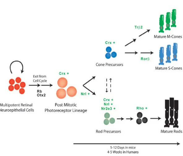
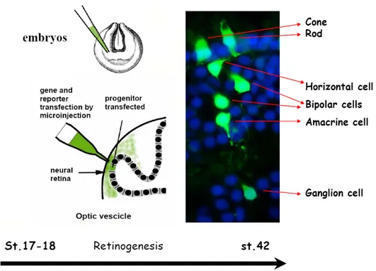
In Xenopus, Xotx2 and Xotx5b (the homolog of Crx) are expressed in different patterns during retinal histogenesis: transcription of both genes starts at tailbud stage in a diffused fashion throughout the retina, but then their expression is progressively restricted and, in the mature retina, Xotx2 mRNA is found only in bipolar cells, while Xotx5b is transcribed in both photoreceptors and a subset of bipolar cells (Viczian et al., 2003). Even more dramatic is the difference in the protein expression pattern: XOTX2 protein is detected only in bipolar cells, while XOTX5b is produced only in photoreceptors at larval stage, due to precise translational control through the 3’UTR regions of their mRNAs (Decembrini et al., 2006).
33 Consistent with the pattern of protein distribution, lipofection of progenitors with constitutively expressed Xotx2 and Xotx5b cDNAs of their coding sequences showed dramatic differences of effects, with Xotx2 driving cells toward bipolar cell fate, and Xotx5b toward photoreceptor cell fate (Viczian et al., 2003; Wang and Harris, 2005; Decembrini et al., 2006).
Interestingly, swapping domain experiments showed that the differential activities of XOTX2 and XOTX5b are due to their carboxy-terminal (C-terminal) parts (Viczian et al., 2003). Significantly, lipofection of chimeric constructs (Xotx2engR and Xotx5bengR), in which the transactivation domain of either XOTX2 or XOTX5b is replaced with the repressor domain of the Drosophila Engrailed protein, had specific effects on either bipolar cells or photoreceptor cells, respectively, this time leading to a decrease, instead of an increase, in their frequency (Viczian et al., 2003).
This suggested that these XOTX-EngR chimeric proteins retain a region of the XOTX2 or XOTX5b proteins crucial for their differential activities. Interestingly, swap-experiments performed by replacing the C-terminal of XOTX5b with the one of XOTX2 (and vice versa) showed that the obtained chimeric construct XOTX5b/2 (or XOTX2/5b) is capable of producing the same phenotype as XOTX2 (or XOTX5b, respectively), according to the C-terminal fused to (Viczian et al., 2003).
1.3.a Molecular characteristics of OTX proteins
Otx genes code for transcription factors with a homeodomain of the K50
Paired-like class, characterised by a lysine residue at position 50 of the homeodomain. The homeodomain is a 60 amino acid module representing a variation on a helix-turn-helix motif of prokaryotic repressor. Three α-helical regions are separated by turns in the protein backbone. Helix 3 (recognition helix) of the homeodomain binds to the major groove of DNA, while helices 1 and 2 lie
34 outside the double helix. Helix 3 contacts both the phosphate backbone and specific bases. An N-terminal arm lies in the minor groove, and makes additional contacts (Lewin, 2003).
The homeodomain is followed by a glutamine-rich region, a basic region (rich in lysine and arginin) and a WSP domain, a highly conserved region of unknown function (Fig. 1.7). The OTX proteins have an OTX-tail, at first identified in CRX (Furukawa et al., 1997) but usually present in tandem repetition. By deletion analysis it has been demonstrated that multiple regions in the C-terminal portion of CRX contribute to its transactivating activity. AD-1 region (formed by two subregions, a and b) plays a major role in transactivation. In contrast, AD-2 region (that comprises the basic region and the WSP domain) plays a minor role in transactivation (Chau et al., 2000a; Chen et al., 2002).
Figure 1.7. Schematic representation of the CRX structure.
The drawing shows the main regions of the primary structure of the CRX protein (from Chen et al., 2002).
OTX nuclear trafficking is highly regulated. The pathway of transport to the nucleus is mediated by nuclear localization signal (NLS) sequences that are characterized by one or more clusters of basic amino acids (Fei and Hughes, 2000). By deletion analysis it has been demonstrated that CRX NLS resides in the
35 C-terminal of the homeodomain, between residue 88 and 107 (Fei and Hughes, 2000). Moreover, nuclear translocation of CRX is mediated by Karyopherin 13 (also referred as Importin 13), that directly binds to the CRX homeodomain and to its flanking regions, mediating the nuclear translocation (Ploski et al., 2004).
Recently, a structural characterization of OTX2 was carried out. As for CRX, OTX2 nuclear localization is controlled by a nuclear localization sequence located within the homeodomain. Moreover, it works in conjunction with a novel nuclear retention domain, located downstream of the homeodomain (Chatelain et
al., 2006).
In the context of protein trafficking, it is interesting to mention the presence in homeoproteins of a peptide, called penetratin, firstly identified in Drosophila Antennapedia (Dom et al., 2003). Penetratin is a 16-amino acid long peptide corresponding to the third α-helix of the homeodomain. Penetratin allows a translocation through biological membrane by means of a receptor-, endocytotis- and energy-independent mechanism, in which a tryptophan residue has an instrumental role (Christiaens et al., 2004). OTX2 protein also contains a penetratin sequence that allows it to translocate transynaptically from bipolar cells to target visual stations in the brain (Prochiantz, unpublished). This finding may explain the presence of OTX2 protein in the cytoplasm of ganglion cells in mouse and rat retina, although there is no Otx2 transcript in those cells (Baas et al., 2000;
Rath et al., 2007).
Besides DNA binding and protein trafficking, the homeodomain is involved in protein-protein interactions. Several cofactors have been identified that interact with OTX proteins. For example, it has been demonstrated that CRX binds to NRL (Mitton et al., 2000) and to NR2E3, forming a trimeric complex able to induce photoreceptor differentiation (Peng and Chen, 2005).
CRX interacts and synergizes also with SP4 (Lerner et al., 2005); p300/CBP (Yanagi et al., 2000); HMGA1 (Arlotta et al., 1997; Chau et al., 2000b) and QRX (Wang et al., 2004).
36 CRX transactivation ability. For example, Phosducin (Phd) and Phd-like orphan protein1 (PhLOP1) - two G protein interactors - directly bind to CRX and inhibit its transactivation ability (Zhu and Craft, 2000); interestingly, the authors speculate that light-activated phototransduction events produce a Phd peptide that interacts with CRX preventing CRX-regulated gene expression in a light-dark dependent manner (Zhu and Craft, 2000).
Other transcriptional corepressors have been isolated, such as BAF (Barrier to Autointegration Factor) (Wang et al., 2002) and Ataxin-7, in which polyQ expansion, responsible for spinocerebellar ataxia 7, antagonized CRX function producing retinal degeneration (La Spada et al., 2001; Chen et al., 2004). Moreover, another mechanism of CRX inhibition is performed by MOK2, that represses its transcription by competing for DNA-binding (Arranz et al., 2001).
1.3.b Insights on OTX interaction: molecular network underlying photoreceptor differentiation
During retinal development in mouse, cells of the photoreceptor lineage turn on the expression of Otx2, which is essential but not sufficient for the photoreceptor differentiation. This has been established by means of an Otx2 conditional knock-out (CKO), in which Otx2 was inactivated under control of the Crx promoter (Nishida et al., 2003): CKO mice showed a complete loss of retinal photoreceptors. Moreover, it was found that Otx2 is a direct upstream regulator of Crx (Nishida et al., 2003). On the other hand, Crx is able to regulate its own expression and its promoter contains four CRX-binding sites (Furukawa et al., 2002). So, the upregulation of Crx may be a necessary step for the expression of both rod and cone genes (Chen et al., 1997).
Several evidences have demonstrated that some extrinsic factors are able to influence photoreceptor differentiation (see Paragraph 1.2.c). In particular, two related cytokines, CNTF and LIF, inhibit the function of photoreceptors by