A mutation in caspase-9 decreases the expression of BAFFR and ICOS in patients with immunodeficiency and
lymphoproliferation / Clemente, N; Boggio, E.; Gigliotti, C.L.; Orilieri, E.; Cappellano, G.; Toth, E.; Valletti, P.A.; Santoro,
C.; Quinti, I.; Pignata, C.; Notarangelo, L.D.; Dianzani, C.; Dianzani, I.; Ramenghi, U.; Dianzani, U.; Chiocchetti, A..
-16:2(2015), pp. 151-161.
Original Citation:
A mutation in caspase-9 decreases the expression of BAFFR and ICOS in patients with immunodeficiency and
lymphoproliferation
Published version:
DOI:10.1038/gene.2014.74
Terms of use:
Open Access
(Article begins on next page)
Anyone can freely access the full text of works made available as "Open Access". Works made available under a
Creative Commons license can be used according to the terms and conditions of said license. Use of all other works
requires consent of the right holder (author or publisher) if not exempted from copyright protection by the applicable law.
Availability:
This is the author's manuscript
ORIGINAL ARTICLE
A mutation in caspase-9 decreases the expression of
BAFFR and ICOS in patients with immunodeficiency
and lymphoproliferation
N Clemente1, E Boggio1, CL Gigliotti1, E Orilieri1, G Cappellano1, E Toth1, PA Valletti1, C Santoro1, I Quinti2, C Pignata3, LD Notarangelo4, C Dianzani5, I Dianzani1, U Ramenghi6, U Dianzani1and A Chiocchetti1
Lymphocyte apoptosis is mainly induced by either death receptor-dependent activation of caspase-8 or mitochondria-dependent activation of caspase-9. Mutations in caspase-8 lead to autoimmunity/lymphoproliferation and immunodeficiency. This work describes a heterozygous H237P mutation in caspase-9 that can lead to similar disorders. H237P mutation was detected in two patients: Pt1 with autoimmunity/lymphoproliferation, severe hypogammaglobulinemia and Pt2 with mild
hypogammaglobulinemia and Burkitt lymphoma. Their lymphocytes displayed defective caspase-9 activity and decreased apoptotic and activation responses. Transfection experiments showed that mutant caspase-9 display defective enzyme and proapoptotic activities and a dominant-negative effect on wild-type caspase-9. Ex vivo analysis of the patients’ lymphocytes and in vitro transfection experiments showed that the expression of mutant caspase-9 correlated with a downregulation of B-cell-activating factor (BAFF) receptor (BAFFR) in B cells and induci
Q2 ble T-cell costimulator (ICOS) in T cells. Both patients carried a second inherited heterozygous mutation missing in the relatives carrying H237P: Pt1 in the transmembrane activator and CAML interactor (TACI) gene (S144X) and Pt2 in the perforin g
Q3 ene (N252S). Both mutations have been previously associated with
immunodeficiencies in homozygosis or compound heterozygosis. Taken together, these data suggest that caspase-9 mutations may predispose to immunodeficiency by cooperating with other genetic factors, possibly by downregulating the expression of BAFFR and ICOS.
Genes and Immunity (2014) 00, 1–11. doi:10.1038/gene.2014.74
INTRODUCTION
Caspases are a family of cysteine-dependent aspa
Q4 rtate-directed
proteases present in the cytoplasm as inactive proenzymes; some are involved in apoptotic induction, whereas others are involved in cell activation.1–3In the immune system, apoptosis has a crucial role in the deletion of autoreactive lymphocytes and in cell-mediated cytotoxicity. Apoptosis acts via two pathways: the extrinsic pathway triggered by surface death receptors that belong to the tumor necrosis factor receptor family, which induce activation of caspase-8 and -10, and the intrinsic pathway that proceeds through the mitochondrial release of cytochrome c and activation of caspase-9.4Both pathways converge in the activation of effector caspases (caspase-3, -6 and -7) and are partly interconnected because caspase-8 cleaves BH3 interacting-domain death agonist (Bid) and this induces cytochrome c release from the mitochondria.5–7
Caspases may also exert non-apoptotic functions, such as influencing cell proliferation, migration and the immune response.8,9They are involved in the differentiation of several cell types, including macrophages, and may have a role in the secretion of cytokines and growth factors.10–13Moreover, activa-tion of caspases in a temporally and spatially restricted manner may mediate localized cellular remodeling.13
Deleterious caspase-8 gene mutations cause a peculiar immu-nological phenotype, in which B- and T-cell accumulation in the lymph nodes and spleen is associated with severe hypogamma-globulinemia, increased susceptibility to infections and defective apoptosis and lymphocyte activation in vitro.14The involvement of
caspase-8 in lymphocyte activation was confirmed by several reports showing that its inhibitors decrease lymphocyte activation in vitro and its inactivation causes immunodeficiency in mice.13,15 It has been suggested that this effect may depend on the inhibition of a caspase-dependent cleavage of proteins involved in either cell cycle control (such as p27KIP1) or signaling (such as
FADD-like interleukin-1β-converting enzyme-like inhibitory pro-tein, FLIP). Moreover, caspase-8 is involved in the activation of the Q5 nuclear factor-κ-light-chain-enhancer of activated B cells (NF-κB), a transcription factor that is crucial for lymphocyte activation.16–18
Caspase-3 has also been suggested to influence the immune system; its loss in mice results in B- and T-cell accumulation in the lymph nodes and spleen, ascribed to defective apoptosis for T cells and to altered regulation of cell cycle for B cells.19
Loss-of-function mutations of the caspase-10 gene (CASP10) can cause the autoimmune lymphoproliferative syndrome (ALPS; OMIM no. 601859) due to defective function of the Fas death receptor.20 In most ALPS patients, the Fas defect is due to Q6 1
Interdisciplinary Research Center of Autoimmune Diseases (IRCAD) and Department of Health Sciences, University of Eastern Piedmont‘A Avogadro’, Novara, Italy;2
Department of Clinical Immunology, University of Rome‘La Sapienza’, Rome, Italy;3
Department of Pediatrics,‘Federico II’ University, Naples, Italy;4
Division of Immunology, Children’s Hospital, Harvard Medical School, Boston, MA, USA;5
Department of Drug Science and Technology, University of Torino, Torino, Italy and6
Department of Pediatrics, University of Turin, Turin, Italy. Correspondence: Professor U Dianzani, Interdisciplinary Research Center of Autoimmune Diseases (IRCAD) and Department of Health Sciences, University of Eastern Piedmont‘A Avogadro’, via Solaroli 17, Novara 28100 Italy.
E-mail: [email protected]
Received 23 May 2014; revised 9 October 2014; accepted 17 November 2014
Genes and Immunity (2014), 1–11
© 2014 Macmillan Publishers Limited All rights reserved 1466-4879/14 www.nature.com/gene
mutations of the Fas gene, although mutations of the Fas ligand (FasL) or caspase-10 genes have been occasionaly detected, whereas the mutated gene is unknown in some patients.20–24 ALPS is characterized by polyclonal accumulation of lymphocytes in the spleen and lymph nodes, with lymphadenomegaly and/or splenomegaly and expansion of T-cell receptorαβ+T cells lacking CD4 and CD8, namely double-negative T cells. These patients frequently display autoimmune manifestations during childhood and may develop lymphomas in adulthood.24 We have also previously described an incomplete form of ALPS that lacks the expansion of double-negative T cells, a required criterion for ALPS diagnosis; this variant form has been named Dianzani auto-immune lymphoproliferative disease (DALD) by Victor McKusick (OMIM no. 605233).25,26
Typically, ALPS patients display hypergammaglobulinemia, but we previously described a subgroup that developed hypogam-maglobulinemia during the course of disease.24,27This phenotype resembles that of common variable immunodeficiency (CVID; OMIM no. 240500), a primary immunodeficiency characterized by defective antibody production and hypogammaglobulinemia.28,29 CVID usually manifests during the second and third decade by recurrent bacterial infections of the upper and lower respiratory tract; up to 20% of CVID patients also display signs of autoimmunity and lymphoproliferation (splenomegaly). Moreover, both ALPS and CVID patients may display a skewed distribution of B-cell subsets.30,31
CVID may be caused by variations in several genes involved in B-cell activation and differentiation, including the CD19/CD21/ CD81, transmembrane activator and calcium modulator and cyclophilin ligand interactor (TACI or TNFRSF13B), B-cell-activating factor (BAFF) receptor (BAFFR) and inducible T-cell costimulator (ICOS) genes.29,32CD19, CD21 and CD81 are components of the coreceptor of the B-cell receptor and are involved in B-cell activation. TACI and BAFFR (or TNFRSF13C) are expressed by B cells and bind the BAFF cytokine, thereby having a role in B-cell activation and survival. ICOS is expressed by activated T cells and has a role in the formation of lymphoid follicl
Q7 es by binding the
ICOS ligand (B7h or B7H2) expressed by B cells.32
Other diseases that may display mixed features of lymphopro-liferation and immunodeficiency are familial hemophagocytic lymphohistiocytosis (FHL; OMIM no. 603553), due to defective function of perforin, which leads to ineffective immune hyperactivation upon viral infection, with tissue damage and a fatal outcome;33,34 and X-linked lymphoproliferative syndrome (XLP; OMIM no. 308240), due to the deficiency of
Q8 either SAP
(SLAM-associated protein) or XIAP (X-linked inhibitor of apoptosis protein), thereby causing increased susceptibility to Epstein–Barr virus infection.35All these cases are intriguing because they show that autoimmunity/lymphoproliferation and immunodeficiency overlap in several diseases. The aim of this study was to assess the involvement of mutations in the caspase-9 gene (CASP9) in the development of hypogammaglobulinemia and/or lymphoproli-feration. We began our analysis with patients displaying both clinical phenotypes and then considered those with either ALPS or hypogammaglobulinemia. We detected a heterozygous H237P mutation in two patients who also carried a second heterozygous mutation: one in the TNFRSF13B gene, which encodes TACI, and the other in PRF1 gene, which encodes perforin. Analysis of the patients’ lymphocytes and the transfection experiments of the mutated CASP9 cDNA in lymphocytes showed that expression of the mutated caspase-9 protein correlated with the downregula-tion of BAFFR in B cells and ICOS in T cells, which may be involved in the patients’ low immunoglobulin (Ig) levels.
RESULTS Patient analyses
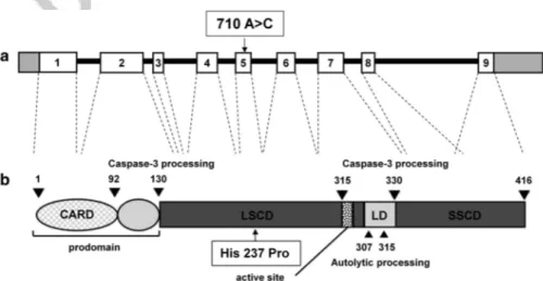
CASP9 variations were searched in the genomic DNA from seven patients displaying autoimmunity, chronic lymphadeno-megaly and/or splenolymphadeno-megaly and hypogammaglobulinemia. We sequenced exons 2–9 and their intron/exon boundaries but not Q9 exon 1, as no amplification was obtained with four different sets of primers because of its high GC content. We detected a heterozygous 710A4C nucleotide substitution (nucleotide position given from the ATG translation initiation site) in exon 5 in one patient (Pt1) (Figures 1 and 2a, b). This mutation results in the amino-acid substitution H237P (CASP9H237P) and it was not detected in any of the 140 healthy donors. This variation has been described in the dbSNP database as a rare variant with an allelic frequency o0.01% (rs 146054764).
Pt1 was an 18-year-old male, displaying features of both ALPS and CVID since the age of 3 years. The signs of ALPS were chronic thrombocytopenia, lymphadenomegaly, splenomegaly and expansion of double-negative T cells (8–10%) in the peripheral blood, whereas those of CVID were severe hypo-gammaglobulinemia (low IgM, IgG and IgA levels), recurrent pulmonary infections with bronchiectasia and retarded physical growth. In his family, the mutation was carried by the father (F1) and the elder sister (S1a), but not by the mother (M1) or the younger sister (S1b) (Figure 2b).
Figure 1. CASP9 variation carried by the patients. Graphical representation (not in scale) of caspase-9 gene: (a) boxes represent the exons; arrows indicate the mutation and protein (b); and numbers indicate the amino-acid positions. CARD, caspase recruitment domain; LD: linker between the two subunits; LSCD, large subunit catalytic domain; SSCD: small subunit catalytic domain.
The discovery of a mutation in CASP9 in a subject with signs of both ALPS and CVID prompted us to extend the analysis to 78 patients with ALPS or DALD, lacking Fas, FasL, caspase-10 and caspase-8 mutations, and 51 patients with primary hypogamma-globulinemia, including 45 who fulfilled the diagnostic criteria for CVID, but who displayed no signs of ALPS or DALD. We found the same mutation (710A4C) in one adult patient (Pt2), who displayed recurrent otitis media and respiratory infections during childhood and low serum IgM levels, whereas his IgA and IgG levels were, respectively, borderline and normal. He never showed signs of ALPS or DALD and he remained healthy in adulthood until he developed Burkitt lymphoma at the age of 30 years. In Pt2’s family, the mutation was carried by the father (F2) and sister (S2) but not by the mother (M2) (Figure 2b). Both Pt1 and Pt2 were Italian but were of different geographic origins. No other mutations in CASP9 were detected in any patient.
Because both Pt1 and Pt2 displayed hypogammaglobulinemia, we assessed the Ig levels in the available sera of the patients’ healthy family members. The results showed that all the relatives displayed normal Ig levels, except for individuals S1a and S2, who carried CASP9H237Pand displayed slightly decreased IgA and IgM levels, respectively (Figure 2c). This analysis was not performed in individuals F2 and M2 as their sera were not available.
Then, the genetic analysis was extended to the ICOS, TACI and BAFFR genes, involved in CVID;29,32FAS, FASL and CASP10 involved in ALPS;24 PRF1, UNC13D and STX11 (sintaxin 11) involved in FLH;33,34and SAP and XIAP involved in XLP.35
Results showed that Pt1 carried two mutations in TACI, a 431C4G and a 577T4C substitution that result in the S144X and C193R changes, respectively (Figures 2a and b). Homozygosity for a different base change causing S144X (431C4A) has previously been associated with CVID and this mutant allele is not expressed.36 Both mutations were detected in M1 and S1b, but
neither were detected in F1 and S1a, which shows that they were located at the same allele (Figure 2b). Furthermore, the S144X mutation was not detected in any of the 100 healthy individuals. By contrast, Pt2 carried a variation in PRF1, an A4G substitution at position 755 causing an N252S change (rs150053969) that has been associated with FLH, ALPS, type 1 diabetes and B-cell lymphomas.33,37–39This mutation was detected in M2, but not S2 (Figure 2b). In healthy donors, its allelic frequency was 40.02% (2/816).
As both patients displayed decreased Ig levels in vivo, we assessed surface expression of CD19, TACI, BAFFR and ICOS (involved in CVID) in the patients’ peripheral blood mononuclear cells (PBMCs) by immunofluorescence and flow cytometry (Figure 3). In resting PBMCs, we assessed the expression of TACI, BAFFR and HLA-DR in CD19+ B cells, whereas the expression of ICOS, HLA-DR and CD25 was assessed in CD3+ T cells from phytohaemagglutinin (PHA)-activated PBMCs. The results showed that both patients displayed decreased expression of BAFFR in B cells and decreased expression of ICOS in activated T cells. By contrast, expression of TACI was in the normal range in the B cells of both patients, but it was borderline in Pt1, congruent with his mutation of the TACI gene. Moreover, B-cell expression of CD19 and HLA-DR, and T-cell expression of CD3, CD8, CD4, HLA-DR and CD25 were in the normal range in both patients (data not shown). Lymphocyte activation was assessed in the patients’ PBMCs by evaluating the response to anti-CD3 monoclonal antibody (mAb) in terms of proliferation and interleukin (IL)-2 secretion (Figure 4a) and the response to pokeweed mitogen (PWM) in terms of proliferation and secretion of IgM and IgG (Figure 4b). The results showed that all these lymphocyte activation responses were defective in both patients. Moreover, we evaluated whether the defective proliferative response to CD3 could be overcome by CD28 costimulation, or by the addition of exogenous IL-2 Figure 2. Pedigrees of family 1 and family 2. (a) Electropherograms of CASP9, TNFRSF13B (TACI) and PRF1 sequences performed on the genomic DNA of Pt1 for CASP9 and TNFRSF13B and of Pt2 for PRF1. Black arrows show the heterozygous position. (b) Pedigrees of family 1 and family 2 showing the inheritance of the H237P CASP9 mutation; inheritance of the S144X TNFRSF13B and N252S PRF1 mutations are also shown. Circles represent females; squares, males; patients are shown in black. (c) Levels of blood double-negative (DN) T cells and serum Ig in the patients’ families.
Caspase-9 mutation causes lymphoproliferation and immunodeficiency Q1 N Clemente et al
Figure 3. Surface expression of TACI, BAFFR and ICOS in PBMCs from patients and healthy donors. Cytofluorimetric histograms showing the surface expression of TACI and BAFFR assessed in CD19+cells and of ICOS in PHA-activated CD3+T cells by two-color immunofluorescence:
unstained cells (white) and stained cells (black). The graphs on the right show the MFI ratios of the patients and the controls; the gray boxes indicate the 5th–95th percentile and the dashed lines show the median of 17 controls.
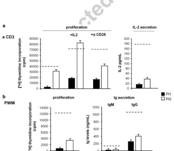
Figure 4. Proliferation and effector properties after stimulation with anti-CD3 mAb or PWM in Pt1 and Pt2. (a) Cell proliferation and IL-2 secretion induced by stimulation of PBMCs with anti-CD3 mAb (1μg ml− 1) and cell proliferation induced by anti-CD3+exogenous IL-2 (10 U ml− 1) and anti-CD3+anti-CD28 (10μg ml− 1) mAb. (b) Cell proliferation and secretion of IgM and IgG induced by stimulation of PBMCs with PWM (10μg ml− 1). The dotted lines indicate the 5th percentile of the activity displayed by all normal controls (n= 19). Each patient was analyzed four times using different blood samples and the results are shown as the mean± s.e.
(10 U ml− 1) because both patients displayed defective IL-2 secretion. The results showed that both exogenous IL-2 and CD28 costimulation had some effect in both patients and exogenous IL-2 rescued the defect in Pt2 (Figure 4a).
Functional characterization of CASP9H237P
To assess the function of the mutated protein (CASP9H237P), either anti-Fas mAb or etoposide was used to activate the extrinsic or the mitochondrial pathway, respectively, and trigger apoptosis in PBMCs from both patients. Then, we analyzed the apoptotic response in terms of cell loss and activation of caspase-8 (extrinsic pathway), caspase-9 (mitochondrial pathway) and caspase-3 (effector pathway). Figure 5 shows that both patients displayed defective activation of caspase-9 and normal activation of caspase-8 in response to either anti-Fas or etoposide. Moreover, they displayed defective cell loss and defective activation of caspase-3 in response to etoposide but not to anti-Fas.
To further assess CASP9H237P function, cDNAs coding for
CASP9H237P or the wild-type protein (caspase-9WT) were cloned into the pcDNA3.1 vector (MOCK), fused to hemagglutinin (HA)- or FLAG-tag sequences, respectively (H237PHAand WTFLAGplasmids). Then, 293T cells, expressing minimal levels of endogenous caspase-9, were transiently transfected with either MOCK alone (293TMOCK), WTFLAG plus MOCK (293TWT), H237PHA plus MOCK
(293TH237P) or WTFLAGplus H237PHA(293TWT/H237P). Western blot analysis showed that spontaneous cleavage of caspase-9, which is a sign of its autoactivation, was detectable in 293TWT, but not in 293TH237P or 293TWT/H237P, expressing cells (Figure 6a). Then, we assessed cell recovery and activation of caspase-9 in these cells cultured in the presence and absence of etoposide. Results showed that cell recovery was significantly lower and caspase-9 activity was higher in 293TWTthan in 293TH237Pand 293TWT/H237P either in the presence or in the absence of etoposide
(Figures 6b and c). These data indicate that CASP9H237P is less active than caspase-9WT and that it exerts a dominant-negative
effect on caspase-9WT. As caspase-9 activity involves caspase-9 interaction with apoptotic protease-activating factor (APAF)-1 to form the apoptosome, we assessed this interaction by immuno-precipitating caspase-9 and assessing APAF-1 co-immunoprecipi-tation by western blot in 293TWTand 293TH237P. Results detected the co-immunoprecipitation in 293TWT but not in 293TH237P (Figure 6d), which shows that CASPH237P displays defective interaction with APAF-1.
To confirm these data, PBMCs from healthy donors were transfected with a MOCK plasmid carrying green fluorescence
protein (GFP) in the absence (PBMCsGFP) or presence (PBMCsH237P) of H237PHAand cultured for 5 days in the presence of PWM or PHA. Then, cell proliferation was assessed by the incorporation of [3H]thymidine (Tdr). The results showed that cell proliferation was lower in PBMCsH237Pthan in PBMCsGFP(Figure 7a). The effect on Q10 the apoptotic response was assessed on PHA-activated PBMCs (PHA-PBMCs) transfected at day 5 of culture to obtain PBMCsGFP
and PBMCsH237P. Twenty-four hours later, cells were treated with anti-Fas mAb or etoposide to evaluate cell loss and activation of caspase-9. Results showed that PHA-PBMCsH237Pdisplayed lower cell loss and caspase-9 activation than PHA-PBMCsGFPin response
to both treatments (Figures 7b and c). Finally, the effect on cell cycle was analyzed by assessing the expression of p27KIP1in PHA-PBMCs transfected at day 5 to obtain PHA-PBMCsGFP and PHA-PBMCsH237P, or co-transfected with WTFLAG plus H237PHA
(PHA-PBMCsWT/H237P). Twenty-four hours later, western blot analysis detected higher levels of p27KIP1 in PHA-PBMCsH237P and PHA-PBMCsWT/H237Pthan in PHA-PBMCsGFP(Figure 7d).
To assess whether CASP9H237P influences the expression of BAFFR, we compared the expression of BAFFR, TACI, HLA-DR and B7h in CD19+B cells from resting PBMCsH237Pand PBMCsGFPby Figure 5. Cell loss and caspase-8, -9 and -3 activation induced by anti-Fas mAb or etoposide in Pt1 and Pt2 T cells. PBMCs were activated with PHA at days 0 (1μg ml− 1) and 8 (0.1μg ml− 1) and cultured in RPMI 1640+10% fetal calf serum (FCS)+recombinant (r)IL-2 (10 U ml− 1). Days after the second stimulation, cells were treated with either anti-Fas mAb or etoposide. (a) Cell loss was assessed by counting live cells (evaluated by the trypan blue exclusion test) in each well and (b) caspases activation by fluorimetric assays in cells treated or untreated with etoposide. The dotted lines indicate the 5th percentile of the activity displayed by normal controls (n= 19). Each patient was analyzed four times using different blood samples and the results are shown as the mean± s.e.
Caspase-9 mutation causes lymphoproliferation and immunodeficiency Q1
N Clemente et al
two-color immunofluorescence 24 h after the transfection. More-over, the effect on the expression of ICOS was assessed on PHA-PBMCs transfected at day 3 of culture to obtain PHA-PHA-PBMCsGFP and PHA-PBMCsH237P. Twenty-four hours later, expression of ICOS,
CD25 and HLA-DR was assessed in CD3+, CD4+and CD8+T cells by
two-color immunofluorescence. The results showed that CD19+
PBMCH237Pexpressed lower levels of BAFFR than CD19+PBMCsGFP (Figure 8a), whereas the expression of TACI, HLA-DR, B7h and CD19 was similar in the two cell preparations (data not shown). Moreover, CD3+ PHA-PBMCH237P expressed lower levels of ICOS than CD3+PHA-PBMCsGFP(Figure 8b), which was detected in both the CD4+ and CD8+ T-cell subsets (data not shown), whereas expression of CD3, CD4, CD8, CD25 and HLA-DR was similar in the two cell preparations (data not shown). To confirm these results, the transfection experiments were repeated in TonsBCs (B cells from tonsils) and Raji cells, expressing high levels of BAFFR, and in PHA-TH (PHA-activated purified CD4+ T helper) cells, expressing high levels of ICOS. The results demonstrated that the expression of BAFFR was lower in TonsBCH237P and RajiH237P than in TonsBCGFPand RajiGFP(Figure 8a) and that the expression of ICOS was lower in PHA-THH237Pthan in PHA-THGFP(Figure 8b).
To confirm that defective caspase-9 activity may modulate the expression of BAFFR and ICOS and cell proliferation, we treated PHA-activated PBMCs from healthy donors with Z-VAD-FMK (30μM), inhibiting caspase-9 and other caspases, at day 3
from activation and assessed BAFFR and ICOS expression, cell proliferation and the apoptotic response to etoposide at day 5. Results showed that Z-VAD-FMK significantly decreased BAFFR and ICOS expression, cell proliferation and cell apoptosis (Supplementary Figure 1).
DISCUSSION
This work shows that the H237P substitution decreases caspase-9 activity and impairs lymphocyte apoptosis and activation. The defective enzyme activity caused by this mutation is in line with the notion that the hymidazolic group of histidine 237 may coordinate the key cysteine of the catalytic site.40
The defects in caspase-9 activity and apoptotic response were demonstrated in primary patient lymphocytes, as well as in cell lines transfected with H237PHA. Transient transfection of 293T cells showed that CASP9H237Phad lower enzymatic and proapoptotic activity than caspase-9WT and also inhibited the function of caspase-9WT through a dominant-negative effect. This was surprising because caspase-9 activation requires aggregation of the apoptosome, a molecular platform composed of seven molecules of APAF-1, cytochrome c and ATP. Each APAF-1 molecule binds to one procaspase-9 molecule, which then recruits another APAF-1 molecule.41 Therefore, recruitment of inactive
forms of caspase-9 or a reduced association with APAF-1 may affect the overall function of the apoptosome, as has been previously shown by other authors using deletion mutants of caspase-9.42 Transient transfection of 293T cells showed that CASP9H237P, differently from caspase-9WT, is not associated to APAF-1 at detectable levels.
The defect in lymphocyte activation was shown in the patients’ lymphocytes that displayed defective CD3-induced proliferation and IL-2 secretion and defective PWM-induced proliferation and Ig secretion. The effect of defective caspase-9 activity on lymphocyte activation was confirmed by the decreased proliferative response displayed by normal PBMCs transfected with CASP9H237Por treated with the caspase inhibitor Z-VAD-FMK.
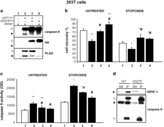
Figure 6. CASP9H237Pimpairs the apoptotic response and caspase-9 activation in 293T-transfected cells. (a) Western blot analysis of cell lysates
obtained from 293T cells transfected with the empty pcDNA3.1 vector or WTFLAGor H237PHAor both are shown. The white arrow shows the procaspase-9 and the black arrow indicates the cleaved form. (b) Cell recovery and (c) caspase-9 activity evaluated 18 h after transfection, as indicated in the panel a, and cultured in the presence and absence of etoposide (5μg ml− 1); data are expressed as the mean± s.e. of the results from six experiments. *Differences vs empty vector;#differences vs WTFLAG(Po0.05). (d) Co-immunoprecipitation of caspase-9 and APAF-1 in cell lysates from 293T cells transfected with WTFLAGor H237PHA. Upper panel shows lysates immunoprecipitated with anti-caspase-9
and blotted with anti-APAF-1; lower panel is the same blot decored with an anti-caspas
Q12 e-9 antibody; the black arrows mark the specific signals and the white arrows mark the heavy and light Ig chains.
The dual effect of the caspase-9 defect on lymphocyte apoptosis and activation mimics the defects observed in two siblings carrying homozygous mutations of the caspase-8 gene.14 They displayed defective lymphocyte apoptosis, as well as impaired activation of T, B and natural killer cells, defective IL-2 secretion and developed a disease with ALPS, hypogammaglobu-linemia and increased susceptibility to infections. The hetero-zygous carriers were healthy.
In Pt1 and Pt2, as in the caspase-8 deficient patients, the lymphocyte apoptosis and activation defects paralleled a clinical phenotype with impaired immune function and lymphoprolifera-tion. The most severe phenotype was displayed by Pt1 with overt CVID and ALPS. Pt2 displayed milder symptoms displayed by decreased serum IgM, increased susceptibility to infections during childhood and development of Burkitt lymphoma in adulthood. The lymphoma could be a consequence of immunodeficiency, but might also be favored by an unapparent preneoplastic lympho-proliferation, which is regarded as a cause of lymphomas.43,44It is noteworthy that even the ALPS displayed by Pt1 can be regarded as a preneoplastic lymphoproliferation because it predisposes patients to lymphoma development. The involvement of caspase-9 in lymphoma development is also suggested by thefinding that high expression of Bcl-2 and XIAP, which inhibit caspase-9 activation, and defective apoptotic response to etoposide
correlate with an unfavorable prognostic value in diffuse large B-cell lymphomas.45
An unexpected effect of the CASP9 mutation was its involve-ment in decreasing the expression of BAFFR and ICOS. This is shown by the finding that both patients displayed decreased expression of these receptors in B cells and activated T cells, respectively. Moreover, transfection of CASP9H237P or treatment with Z-VAD-FMK decreased the expression of BAFFR in B cells and ICOS in activated T cells from normal donors. This effect was specific because expression of several other cell markers was unchanged.
The inhibitory effect of CASP9H237P on cell proliferation and
apoptosis was independent of its effect on the expression of BAFFR and ICOS, as it was also detected in 293T cells, which do not express those receptors. Moreover, transfection of ICOS in PBMCs transfected with CASP9H237rescued the decreased expression of ICOS but not the decreased proliferative and apoptotic responses of these cells (data not shown). As suggested for caspase-8, this effect might be due to inhibition of a caspase-dependent cleavage of proteins involved in the control of cell cycle, as indicated by the increased expression of p27KIP1, which is expected to block cell entry into the S phase, displayed by PBMCs transfected with CASP9H237P.
BAFFR and ICOS have a role in follicle formation because BAFFR is expressed by follicular B cells and supports their survival,46 Figure 7. CASP9H237P
impairs the proliferative and apoptotic responses and caspase-9 activation in transfected PBMCs. (a) Resting PBMCs from normal donors were transiently transfected with vectorGFPalone or plus H237PHA (C9). Cell proliferation in response to PWM or PHA was
assessed by incorporation of [3H]thymidine. (b) Cell loss and (c) caspase-9 activity in response to treatment with anti-Fas mAb or etoposide. Data are expressed as the mean± s.e. (n = 9). (d) Western blot and respective relative expression levels (± s.e.) of p27KIP1normalized to actin expression from four independent experiments in transfected PHA-PBMCs. The black arrow indicates the p27KIP1relative band. Asterisks mark differences from GFP-transfected cells. The percentage of GFP-positive cells was 52±5% (mean ± s.e.) for PBMCsGFP and 54± 4% for PBMCsH237P.
Caspase-9 mutation causes lymphoproliferation and immunodeficiency Q1 N Clemente et al
whereas ICOS is expressed by T helper follicular cells and interacts with both cognate and bystander B cells.47 Therefore, the low expression of BAFFR and ICOS may explain the low Ig levels of our patients, which seems to be a hallmark of the CASP9 mutation because it was detected in all of the patients’ family members carrying the CASP9H237P but not in the others. Unfortunately, we could not assess BAFFR and ICOS expression in these family members because their cells were not available.
Penetrance of CASP9H237Pseems to be incomplete as the four
carriers in the patients’ families were healthy, although two of them (S1a and S2) displayed a subclinical defect with a mild decrease of serum IgM and IgA, respectively. The severity of the clinical outcome may thus depend on the influence of concurrent genetic and environmental factors. Congruent with the role of concurrent genetic factors, Pt1 and Pt2 each carried heterozygous mutations of a second gene. This was inherited from their CASP9H237P-free mothers and may have contributed to their clinical outcome.
Pt1 carried a S144X mutation in TNFRSF13B coding for TACI, a receptor that binds BAFF and APRIL (a proliferation-inducing ligand). TACI is expressed on peripheral blood B cells, particularly on the CD27+ subset. TACI-deficient mice display humoral immunodeficiency together with autoimmunity and lymphoproliferation.36,48,49 In humans, several TNFRSF13B
mutations have been associated with both CVID and IgA deficiency. In particular, Salzer et al.36 detected homozygous S144X mutations in a patient with hypogammaglobulinemia and hepatosplenomegaly; his brother, who was healthy, carried the same mutations and displayed lymphocytosis. The mutation inserts a stop codon in exon 3 and impairs the expression of the TACI protein and mRNA.36 In line with this, Pt1 displayed a borderline expression of TACI in B cells. Pt1 also carried a second mutation (C193R) in exon 4 of the same allele, but this should be irrelevant as it is located downstream of the inserted stop codon. S144X seems non-penetrant in heterozygosity as shown both by Salzer et al.36and by our analysis of heterozygous patients M1 and S1b. Therefore, the finding that Pt1 is the only member of the family with both S144X and CASP9H237P suggests that the two mutations may have cooperated in the disease development of Pt1, possibly through the borderline expression of TACI and the decreased expression of BAFFR and ICOS, which are all involved in B-cell function.
Pt2 carried a heterozygous mutation of PRF1 coding for perforin, which is stored in the lytic granules of cytotoxic cells and involved in cell-mediated cytotoxicity.33,34 Biallelic PRF1 mutations cause FHL, an immunodeficiency characterized by hemophagocytosis, fever, hepatosplenomegaly, cytopenia, hypertriglyceridemia, hypofibrinogenemia, frequent nervous system involvement and Figure 8. Surface expression of BAFFR and ICOS in lymphocytes of healthy donors after transient transfection of CASP9H237
. (a) Expression of BAFFR in CD19+PBMCs, Raji cells and total TonsBCs transiently transfected with vectorGFPalone or plus H237PHA(C9). (b) Expression of ICOS in CD3+PBMCs (PHA-PBMCs) or purified CD4+T cells (PHA-TH) transiently transfected with vectorGFPalone or with H237PHA(C9). Upper panels show representative cytofluorimetrc histograms of the unstained cells (black) and the stained cells (white); numbers indicate the mean fluorescence intensity ratio (MFI-R). Lower panels show the mean ± s.e. of MFI-R of BAFFR or ICOS expression from nine experiments; results are relative to the BAFFR or ICOS expression displayed by GFP-transfected set at 100% (dotted line) in each experiment. Asterisks mark differences from GFP-transfected cells. The 100% value corresponded to MFI ratio= 65 ± 8 for BAFFR and 11 ± 2 for ICOS.
increased frequency of lymphomas.33The disease is ascribed to
the decreased capacity of cytotoxic cells to clear viral infections, whose persistence may support the lymphoproliferative picture. FHL is a recessive disease and subjects carrying heterozygous PRF1 mutations are generally healthy; however, inherited PRF1 muta-tions have also been associated with the development of lymphomas and ALPS.37 In lymphomas, they can be either heterozygous or homozygous, whereas they are heterozygous in ALPS but are associated with inherited defects that affect Fas function. A similar role as a risk factor for ALPS has also been described for the UNC13D gene encoding for MUNC13-4, which is involved in perforin secretion and the development of a genetic variant of FHL.50,51The N252S mutation has been detected in FHL, ALPS and lymphomas.33,37–39 It occurs within the membrane attack complex, a region critically involved in the pore-forming activity of perforin, but its functional significance has been debated because it has been associated with normal natural killer activity.52In Pt2, a double caspase-9 and perforin defect may be involved in the development of his Burkitt lymphoma as both mutations may impair the immune response and favor lymphoproliferation.
The clinical phenotype of our heterozygous patients is very different from the high perinatal lethality, severe alteration of central nervous tissue development, and minor alterations of the immune system impairment displayed by the caspase-9− / − mice. These differences, however, may reflect the residual caspase-9 activity displayed by our heterozygous patients and the cooperation of their second mutations.53
In conclusion, our data show that caspase-9 is involved in both lymphocyte apoptosis and activation and that its defective activity can induce a clinical pattern of immunodeficiency and lympho-proliferation qualitatively similar to that induced by defective caspase-8 activity.
MATERIALS AND METHODS Patients
We analyzed 136 unrelated Italian patients: 7 with autoimmunity, chronic lymphadenomegaly and/or splenomegaly and hypogammaglobulinemia, 78 with ALPS or DALD, lacking Fas, FasL, caspase-10 and caspase-8 mutations and 51 with primary hypogammaglobulinemia. ALPS/DALD patients were diagnosed using the criteria established at the 2009 ALPS NIH International Workshop.24A total of 140 healthy individuals were used as controls.
The study was planned according to the guidelines of the local ethical committee, Azienda Ospedaliera della Carità, of Novara that approved the study (Protocol 106/CE). Written, informed consent was signed by the patients or by their parents if they were minors.
Sequence analyses
Specific primers were used to amplify the exons and intron boundaries of CASP9 (gene ID: 842), FAS (ID: 355), FASL (ID: 356), CASP10 (ID: 843), TNFRS13B (TACI) (ID: 23495), TNFRSF13C (BAFFR) (ID: 115650), ICOS (ID: 29851), PRF1 (ID: 5551), UNC13D (ID: 201294), STX11 (ID: 8676), SH2D1A (SAP) (ID: 4068) and XIAP (ID: 331) genes. The PCR products were sequenced with the ABI PRISMR BigDyeTM Terminator Kit (Applied Biosystems, Foster City, CA, USA) on an automatic sequencer, Applied Biosystems 3100 Genetic Analyzer (Applied Biosystems), using the same primers, except for exon 3 of PRF1 for which we used also an internal primer.
Cells
PBMCs were obtained by density gradient centrifugation from patients’ blood and buffy coats, provided by the local Blood Transfusion Service (Novara, Italy). Total CD4+T cells from healthy controls were purified using
the CD4+T Cell Isolation Kit II (Miltenyi Biotec, Teterow, Germany). This approach provided 497% cells displaying the CD3+CD4+CD45RA+
CD45RO+CD14−CD16−phenotype, as assessed by direct immuno
fluores-cence andflow cytometry. B cells were purified, after homogenization, by
density gradient centrifugation from the tonsils of patients undergoing tonsillectomy in the Otolaryngology Clinic of Azienda Ospedaliera della Carità, Novara, Italy. Cell surface markers were assessed by immuno fluor-escence and flow cytometry using the appropriate mAb to BAFFR (BioLegend, San Diego, CA, USA), ICOS (eBioscience, San Diego, CA, USA), B7h (R&D Systems, Minneapolis, MN, USA), TACI (BioLegend), CD19 (ImmunoTools, Friesoythe, Germany), CD3 (Becton Dickinson, Franklin Lakes, NJ, USA), CD4 (Sigma, St Louis, MO, Canada), CD8 (Invitrogen, Burlington, ON, Canada), CD25 (BioLegend) and HLA-DR (Becton Dickinson). In some experiments, cells were treated with Z-VAD-FMK (30μm) (Enzo Life Sciences, Florence, Italy).
Antigenic density was expressed as the mean fluorescence intensity ratio, using the following formula: mean fluorescence intensity of sample histogram (arbitrary units)/meanfluorescence intensity of control histogram (arbitrary units).
Cell death assays
PBMCs (1 × 10⁵) were incubated inflat-bottom 96-well plates with control medium (200μl) with or without anti-Fas mAb (0.5 μg ml− 1) (CH11; UPSTATE Waltham, MA, USA) or with etoposide (5μg ml− 1) (Sigma) in the presence of rIL-2 (1 U ml− 1) to minimize spontaneous cell death. Eighteen hours later, the live cells were counted by the trypan blue exclusion test and live cells were detected usingflow cytometry by excluding those cells that were propidium iodide (Becton Dickinson) and annexin V-FITC positive (Becton Dickinson); the two methods gave overlapping results. These assays were performed in duplicate. In some experiments, PBMCs were treated with Z-VAD-FMK (30μM) (Enzo Life Sciences). In transfected human
embryonic kidney 293T cells (ATCC; no. CRL-11268), cell death assay was Q11 assessed 48 h after transfection, and in the last 18 h, etoposide (5μg ml− 1) or no stimulus was added to the media of culture.
The specific cell loss % was calculated as follows: 100 − (total live cell count in the assay well/total live cell count in the control well) × 100.
Caspase-9, -3 and -8 activity was assessed in cell lysates using a fluorimetric assay (MBL, Watertown, MA, USA). At least three control samples, using PBMCs from different healthy donors, were run in parallel. In transfected human embryonic kidney 293T cells, caspase-9 activity was assessed 24 or 48 h after transfection to evaluate activation after treatment with etoposide (5μg ml− 1) or spontaneous autoactivation, respectively.
PBMCs activation assays
PBMCs (1 × 10⁵ perwell) were cultured inflat-bottom 96-well microplates
with RPMI 1640+10% fetal calf serum in the presence or absence of the appropriate stimulus. The anti-CD3 mAb (OKT3 1μg ml− 1in phosphate-buffered saline) (ATCC; no. CRL-8001) was precoated into the plates by overnight incubation at 4 °C. PHA and PWM (Sigma) were used at 1 and 10μg ml− 1, respectively. PBMC proliferation was assessed in triplicate by evaluating the uptake of [3H]TdR (1μCi per well) (Perkin-Elmer, Waltham, MA, USA) in the last 6 h of culture on days 3 and 5 for OKT3- or PHA- and PWM-treated cells, respectively; the cells were harvested and radioactivity was detected with aβ-counter (Perkin-Elmer). IL-2 secretion was evaluated by performing an enzyme-linked immunosorbent assay on the day 3 culture supernatants (Beckton Dickinson). In PWM- treated cells, IgG, IgM and IgA secretion was evaluated by enzyme-linked immunosorbent assay of the day 5 supernatants (Rockland, Gilbertsville, PA, USA). In some experiments, cells were treated with Z-VAD-FMK (30μM) (Enzo Life Sciences).
Caspase-9 cloning and transfection
Total RNA was extracted from Pt1 PBMCs using the NucleoSpin RNA II Kit (Macherey-Nagel, Düren, Germany) and reverse transcribed with the ThermoScript RT-PCR System (Invitrogen, Milan, Italy). CASP9 was amplified with the same reverse primer and with two alternative forward primers, one adding the HA-tag and the other the FLAG-tag. Because Pt1 was heterozygous for CASP9H237P, both the wild-type (FLAG-tagged) and the
mutated (HA-tagged) amplimers were obtained. These cDNAs were cloned into the pcDNA3.1 vector (Invitrogen) and sequenced.
The 293T cells were cultured in Dulbecco’s modified essential medium (Invitrogen) supplemented with 10% fetal calf serum at 37 °C. A total of 3 × 10⁶cells were plated in 90 mm dishes and transfected with 15μg of the empty vector, the WTFLAGvector, the H237PHAvector or a combination of
them using the Lipofectamine 2000 Kit (Invitrogen).
PBMCs, CD4+ T cells, TonsBCs and Raji cells (ATCC; no. CCL-86) were
cultured in RPMI 1640 medium+10% fetal calf serum and transfected using Caspase-9 mutation causes lymphoproliferation and immunodeficiency Q1
N Clemente et al
the Amaxa Cell Line Nucleofactor Kit V (Lonza, Basel, Switzerland), according to the manufacturer’s instructions. Briefly, 4 μg of each construct were co-transfected with 1μg of the pEGFP vector (Invitrogen). Transfection efficiency was analyzed by cytofluorimetric evaluation of the proportion of GFP-expressing cells.
After 30 h, adherent andfloating cells were harvested into ice-cold AKT buffer (20 mMTris, pH 7.5, 5 mMethylenediaminetetraacetic acid, 150 mM
NaCl, 1% Triton X-100, 10% glycerol, 0.5 mM dithiothreitol, 1 mM
phenylmethanesulfonylfluoride), 1 μg ml− 1leupeptin, 1μg ml− 1aprotinin, 1μg ml− 1pepstatin) for 20 min and sonicated three times. Cell debris was removed by centrifugation and equal amounts of the cleared lysates were heated for 5 min at 95 °C.
Western blotting and immunoprecipitation
Protein extracts from 293T cells and PBMCs were separated by sodium dodecyl sulfate-polyacrylamide gel electrophoresis, transferred to Hybond-C extra membranes (Amersham Pharmacia Biotech, Piscataway, NJ, USA), blotted with anti-caspase-9 (1μg ml− 1) (Sigma), anti-HA (0.5μg ml− 1) (Millipore, Billerica, MA, USA), anti-FLAG (1μg ml− 1) (Sigma), anti-APAF-1 (1μg ml− 1) (Sigma), anti-p27KIP1(1μg ml− 1) (Sigma) or anti-actin
(0.5μg ml− 1) (Sigma). A peroxidase-conjugated anti-mouse or anti-rabbit (GE Healthcare, Piscataway, NJ, USA) were used as a secondary antibody (GE Healthcare) and revealed by chemiluminescence.
In the immunoprecipitation experiments, transfected 293T cells were lysed in IPB buffer (10 mMTris-HCl, pH 7.5, 150 mMNaCl, 1% NP40, 0.5%
sodium deoxycholate, 1 mM phenylmethanesulfonyl fluoride, 1 μg ml− 1
leupeptin, 1μg ml− 1aprotinin, 1μg ml− 1pepstatin). Five micrograms of lysate was precleared with Sepharose-ProtG beads (GE Healthcare) and then incubated with 2μg anti-caspase-9 mAb (Sigma), anti-HA mAb (Sigma) or the polyclonal anti-FLAG (Sigma) for 1 h at 4 °C and with 20μl of protein G beads (GE Healthcare) for a further hour at 4 °C. Beads were washed three times, boiled in sodium dodecyl sulfate-polyacrylamide gel electrophoresis sample buffer, separated by sodium dodecyl sulfate-polyacrylamide gel electrophoresis and blotted with anti-APAF-1 (Sigma) and anti-caspase-9 (Sigma) antibodies.
Statistical analysis
Statistical analysis was performed using the analysis oif variance followed by Dunnett’s multiple comparison test, and paired t-test for analysis of transfected lymphocytes. The results are shown as the mean ± s.e. Genotype distributions were analyzed using the Fisher’s exact test. All P-values are two-tailed, and the significance cutoff was *Po0.05 and **Po0.01. Statistical analysis was performed with GraphPad Instat (GraphPad Software, San Diego, CA, USA) software.
CONFLICT OF INTEREST
The authors declare no conflict of interest.
ACKNOWLEDGEMENTS
This work was partially supported by Associazione Italiana Ricerca sul Cancro (AIRC, Milan), Fondazione Cariplo (Milan), Fondazione Italiana Sclerosi Multipla (FISM, Rome), Compagnia di San Paolo (Turin), Fondazione Cassa di Risparmio di Cuneo (Cuneo), Regione Piemonte (Turin) and Associazione ‘Amici di Jean’ (Turin). We thank Dr Michael Lenardo and Dr Frederic Rieux-Laucat for providing some patients’ DNA.
REFERENCES
1 Nagata S. Apoptosis by death factor. Cell 1997; 88: 355–365.
2 Cohen GM. Caspases: the executioners of apoptosis. J Biochem 1997; 326: 1–16. 3 Dianzani U, Chiocchetti A, Ramenghi UX. Role of inherited defects decreasing Fas
function in autoimmunity. Life Sci 2003; 72: 2803–2824.
4 Zou H, Li Y, Liu X, Wang X. An APAF-1-cytochrome c multimeric complex is a functional apoptosome that activates procaspase-9. J Biol Chem 1999; 274: 11549–11556.
5 Riedl SJ, Shi Y. Molecular mechanisms of caspase regulation during apoptosis. Nat Rev Mol Cell Biol 2004; 5: 897–907.
6 Li H, Zhu H, Xu CJ, Yuan J. Cleavage of BID by caspase 8 mediates the mitochondrial damage in the Fas pathway of apoptosis. Cell 1998; 94: 491–501.
7 Luo X, Budihardjo I, Zou H, Slaughter C, Wang X. Bid, a Bcl2 interacting protein, mediates cytochrome c release from mitochondria in response to activation of cell surface death receptors. Cell 1998; 94: 481–490.
8 Budd RC. Death receptors couple to both cell proliferation and apoptosis. J Clin Invest 2002; 109: 437–441.
9 Alam A, Cohen LY, Aouad S, Sekaly RP. Early activation of caspases during T lymphocyte stimulation results in selective substrate cleavage in nonapoptotic cells. J Exp Med 1999; 190: 1879–1890.
10 Netea MG, Lewis EC, Azam T, Joosten LA, Jaekal J, Bae SY et al. Interleukin-32 induces the differentiation of monocytes into macrophage-like cells. Proc Natl Acad Sci USA 2008; 105: 3515–3520.
11 Sordet O, Rébé C, Plenchette S, Zermati Y, Hermine O, Vainchenker W et al. Specific involvement of caspases in the differentiation of monocytes into mac-rophages. Blood 2002; 100: 4446–4453.
12 Kang TB, Ben-Moshe T, Varfolomeev EE, Pewzner-Jung Y, Yogev N, Jurewicz A et al. Caspase-8 serves both apoptotic and nonapoptotic roles. J Immunol 2004; 173: 2976–2984.
13 Yi CH, Yuan J. The Jekyll and Hyde functions of caspases. Dev Cell 2009; 16: 21–34. 14 Chun HJ, Zheng L, Ahmad M, Wang J, Speirs CK, Siegel RM et al. Pleiotropic defects in lymphocyte activation caused by caspase-8 mutations lead to human immunodeficiency. Nature 2002; 419: 395–399.
15 Salmena L, Lemmers B, Hakem A, Matysiak-Zablocki E, Murakami K, Au PY et al. Essential role for caspase 8 in T-cell homeostasis and T-cell-mediated immunity. Genes Dev 2003; 17: 883–895.
16 Levkau B, Koyama H, Raines EW, Clurman BE, Herren B, Orth K et al. Cleavage of p21Cip1/Waf1 and p27Kip1 mediates apoptosis in endothelial cells through activation of Cdk2: role of a caspase cascade. Mol Cell 1998; 1: 553–563. 17 Dohrman A, Kataoka T, Cuenin S, Russell JQ, Tschopp J, Budd RC. Cellular FLIP
(long form) regulates CD8+ T cell activation through caspase-8-dependent NF-kappa B activation. J Immunol 2005; 174: 5270–5278.
18 Su H, Bidere N, Zheng L, Cubre A, Sakai K, Dale J et al. Requirement for caspase-8 in NF-kappaB activation by antigen receptor. Science 2005; 307: 1465–1468. 19 Woo M, Hakem R, Furlonger C, Hakem A, Duncan GS, Sasaki T et al. Caspase-3
regulates cell cycle in B cells: a consequence of substrate specificity. Nat Immunol 2003; 4: 1016–1022.
20 Wang J, Zheng L, Lobito A, Chan FK, Dale J, Sneller M et al. Inherited human caspase 10 mutations underlie defective lymphocyte and dendritic cell apoptosis in autoimmune lymphoproliferative syndrome type II. Cell 1999; 98: 47–58.
21 Fisher GN, Rosenberg FJ, Straus SE, Dale JK, Middleton LA, Lin AY et al. Dominant interfering Fas gene mutations impair apoptosis in a human lymphoproliferative syndrome. Cell 1995; 81: 935–946.
22 Rieux-Laucat F, Le Deist F, Hivroz C, Roberts IA, Debatin KM, Fischer A et al. Mutations in Fas associated with human lymphoproliferative syndrome and autoimmunity. Science 1995; 268: 1347–1349.
23 Wu J, Wilson J, He J, Xiang L, Schur PH, Mountz JD.. Fas ligand mutation in a patient with systemic lupus erythematosus and lymphoproliferative disease. J Clin Invest 1996; 98: 1107–1113.
24 Oliveira JB, Bleesing JJ, Dianzani U, Fleisher TA, Jaffe ES, Lenardo MJ et al. Revised diagnostic criteria and classification for the autoimmune lymphoproliferative syndrome (ALPS): report from the 2009 NIH International Workshop. Blood 2010; 116: e35–e40.
25 Dianzani U, Bragardo M, Di Franco D, Alliaudi C, Scagni P, Buonfiglio D et al. Deficiency of the Fas apoptosis pathway without Fas gene mutations in pediatric patients with autoimmunity/lymphoproliferation. Blood 1997; 9: 2871–2879. 26 Ramenghi U, Bonissoni S, Migliaretti G, DeFranco S, Bottarel F, Gambaruto C et al.
Deficiency of the Fas apoptosis pathway without Fas gene mutations is a familial trait predisposing to development of autoimmune diseases and cancer. Blood 2000; 95: 3176–3182.
27 Campagnoli MF, Gambarini L, Quarello P, Garelli E, Carando A, Baravalle V et al. The broad spectrum of autoimmune lymphoproliferative disease: molecular bases, clinical features and long-term follow-up in 31 patients. Haematologica 2006; 91: 538–541.
28 Cunningham-Rundles C, Bodian C. Common variable immunodeficiency: clinical and immunological features of 248 patients. Clin Immunol 1999; 92: 34–48. 29 Park MA, Li JT, Hagan JB, Maddox DE, Abraham RS. Common variable
immuno-deficiency: a new look at an old disease. Lancet 2008; 372: 489–502.
30 Warnatz K, Schlesier M. Flow cytometric phenotyping of common variable immunodeficiency. Cytometry B 2008; 74: 261–271.
31 Rensing-Ehl A, Warnatz K, Fuchs S, Schlesier M, Salzer U, Draeger R et al. Clinical and immunological overlap between autoimmune lymphoproliferative syndrome and common variable immunodeficiency. Clin Immunol 2010; 137: 357–365.
32 Park JH, Resnick ES, Cunningham-Rundles C. Perspectives on common variable immune deficiency. Ann NY Acad Sci 2011; 1246: 41–49.
33 Gholam C, Grigoriadou S, Gilmour KC, Gaspar HB. Familial haemophagocytic lymphohistiocytosis: advances in the genetic basis, diagnosis and management. Clin Exp Immunol 2011; 163: 271–283.
34 Voskoboinik I, Dunstone MA, Baran K, Whisstock JC, Trapani JA. Perforin: structure, function, and role in human immunopathology. Immunol Rev 2010; 235: 35–54.
35 Rezaei N, Mahmoudi E, Aghamohammadi A, Das R, Nichols KE.. X-linked lymphoproliferative syndrome: a genetic condition typified by the triad of infection, immunodeficiency and lymphoma. Br J Haematol 2011; 152: 13–30. 36 Salzer U, Chapel HM, Webster AD, Pan-Hammarstrom Q, Schmitt-Graeff A,
Schlesier M et al. Mutations in TNFRSF13B encoding TACI are associated with common variable immunodeficiency in humans. Nat Genet 2005; 37: 820–828. 37 Clementi R, Chiocchetti A, Cappellano G, Cerutti E, Ferretti M, Orilieri E et al.
Variations of the perforin gene in patients with autoimmunity/lymphoprolifera-tion and defective Fas funcautoimmunity/lymphoprolifera-tion. Blood 2006; 10: 3079–3084.
38 Clementi R, Locatelli F, Dupre L, Garaventa A, Emmi L, Bregni M et al. A proportion of patients with lymphoma may harbor mutations of the perforin gene. Blood 2005; 105: 4424–4428.
39 Orilieri E, Cappellano G, Clementi R, Cometa A, Ferretti M, Cerutti E et al. Variations of the perforin gene in patients with type 1 diabetes. Diabetes 2008; 57: 1078–1083.
40 Renatus M, Stennicke HR, Scott FL, Liddington RC, Salvesen GS. Dimer formation drives the activation of the cell death protease caspase 9. Proc Natl Acad Sci USA 2001; 98: 14250–14255.
41 Adams JM, Cory S. Apoptosomes: engines for caspase activation. Curr Opin Cell Biol 2002; 14: 715–720.
42 Pop C, Timmer J, Sperandio S, Salvesen GS. The apoptosome activates caspase-9 by dimerization. Mol Cell 2006; 22: 269–275.
43 Ehrenfeld M, Abu-Shakra M, Buskila D, Shoenfeld Y. The dual association between lymphoma and autoimmunity. Blood Cells Mol Dis 2001; 27: 750–756.
44 Oertel SH, Riess H. Immunosurveillance, immunodeficiency and lymphoproli-ferations. Recent Results Cancer Res 2002; 159: 1–8.
45 Muris JJ, Cillessen SA, Vos W, van Houdt IS, Kummer JA, van Krieken JH et al. Immunohistochemical profiling of caspase signaling pathways predicts clinical response to chemotherapy in primary nodal diffuse large B-cell lymphomas. Blood 2005; 105: 2916–2923.
46 Shulga-Morskaya S, Dobles M, Walsh ME, Ng LG, MacKay F, Rao SP et al. B cell-activating factor belonging to the TNF family acts through separate receptors to support B cell survival and T cell-independent antibody formation. J Immunol 2004; 173: 2331–2341.
47 Xu H, Li X, Liu D, Li J, Zhang X, Chen X et al. Follicular T-helper cell recruitment governed by bystander B cells and ICOS-driven motility. Nature 2013; 496: 523–527.
48 Gross JA, Johnston J, Mudri S, Enselman R, Dillon SR, Madden K et al. TACI and BCMA are receptors for a TNF homologue implicated in B-cell autoimmune disease. Nature 2000; 404: 995–999.
49 Seshasayee D, Valdez P, Yan M, Dixit VM, Tumas D, Grewal IS. Loss of TACI causes fatal lymphoproliferation and autoimmunity, establishing TACI as an inhibitory BLyS receptor. Immunity 2003; 18: 279–288.
50 Boggio E, Aricò M, Melensi M, Dianzani I, Ramenghi U, Dianzani U et al. Mutation of FAS, XIAP, and UNC13D genes in a patient with a complex lymphoproliferative phenotype. Pediatrics 2013; 132: e1052–e1058.
51 Aricò M, Boggio E, Cetica V, Melensi M, Orilieri E, Clemente N et al. Variations of the UNC13D gene in patients with autoimmune lymphoproliferative syndrome. PLoS One 2013; 8: e68045.
52 Voskoboinik I, Thia MC, Trapani JA. A functional analysis of the putative poly-morphisms A91V and N252S and 22 missense perforin mutations associated with familial hemophagocytic lymphohistiocytosis. Blood 2005; 105: 4700–4706. 53 Zheng TS, Hunot S, Kuida K, Flavell RA. Caspase knockouts: matters of life
and death. Cell Death Differ 1999; 6: 1043–1053.
Supplementary Information accompanies this paper on Genes and Immunity website (http://www.nature.com/gene)
Caspase-9 mutation causes lymphoproliferation and immunodeficiency Q1 N Clemente et al