Scuola di Dottorato in Scienze Mediche, Cliniche e
Sperimentali
Dottorato di ricerca in
“Immunoreumatologia e Oncologia Clinica e
Sperimentale, Bioetica ed Epidemiologia dei tumori”
XXVII ciclo
Electrophysiologic and Morphologic Assessment of the Substrate in
Arrhythmogenic Cardiomyopathy and Nonischemic Cardiomyopathy:
Risk Stratification for Sudden Cardiac Death to Guide Implantation of
Defibrillators and Response to Catheter Ablation
Tutor Dottorando/a
Prof. Matteo Di Biase Dr. Pasquale Santangeli
_________________________________________________________________ Esame finale Anno Accademico 2014-2015
Table of Contents:
1. Introduction Page 2
2. Study Objective Page 2
3. Risk Stratification for SCD of Patients with ARVC: Role of Electroanatomic Voltage Mapping Page 3 4. Scar Progression in Patients with ARVC and VT: a Longitudinal Study With Electroanatomic Voltage
Mapping Page 14
5. Long-Term Outcome with Catheter Ablation of Ventricular Tachycardia in Patients with
Arrhythmogenic Right Ventricular Cardiomyopathy Page 19
6. Electroanatomic Substrate Features and Long-Term Outcome of Endo-Epicardial Catheter Ablation of Refractory VT in Patients with Non-Ischemic Cardiomyopathy Page 34
1. Introduction
Sudden cardiac death (SCD) accounts for 450,000 deaths yearly in the U.S.1 Overall, event rates in Europe are similar to those in the United States. In the past years, multiple clinical trials
documented the effectiveness of the implantable cardioverter-defibrillator (ICD) to reduce SCD in high-risk patients. In particular, guideline-concluding trials have focused on left ventricular ejection fraction (EF), because of its demonstrated association with mortality risk in patients with recent myocardial infarction,2 and current guidelines give prophylactic ICD indication on the basis of EF only.3, 4 However, the absolute number of SCDs prevented applying current guidelines in the clinical practice is unacceptably low when compared with the total number of SCDs that occur yearly.5, 6 In particular, it is worrisome that the majority of SCDs occur in the general population, as the first and last manifestation of a subclinical cardiac disease.5, 7 Moreover, a substantial proportion of patients who receive an ICD for primary prevention according to current recommendations will never experience arrhythmic events, thus negating the potential benefit of ICDs while unnecessarily exposing to a risky and costly procedure.8, 9 It bears emphasis that the only explanation to these issues is that EF does not work well as a risk stratification tool for SCD. In the following section I will focus on EF as a risk factor for SCD, discussing its sensitivity and specificity in identifying patients at risk.
Patients with non-ischemic cardiomyopathy (NICM) have been largely underrepresented in clinical trials evaluating the benefit of ICDs for the primary prevention of SCD, and whether these patients derive the same benefit from ICD as compared patients with ischemic substrates is
unclear.7, 10-13 Among patients with NICM, arrhythmogenic right ventricular cardiomyopathy (ARVC) represents a particularly malignant inherited cardiomyopathy, characterized by a high propensity to ventricular arrhythmias potentially leading to SCD. A correct identification of ARVC patients at high risk of sudden cardiac death is crucial for a rational clinical management including indications for prophylactic implantable cardioverter defibrillators (ICDs), antiarrhythmic drug therapy and invasive catheter ablation procedures. At present time there are no reliable ways to identify individual patients at higher risk of sudden cardiac death.
Recent data suggest that several substrate markers, either assessed morphologically with magnetic resonance imaging, or electrophysiologically with invasive mapping procedures, may be helpful to identify subgroup of patients at higher arrhythmic risk.14, 15 However, previous studies have been largely conducted on few patients, most were retrospective or with short follow-up. In addition, there are no data evaluating the prevalence and mechanisms of disease progression in these patients.
2. Study Objectives
The main objectives of this study are two: the first aim is to identify the morphologic and electrophysiologic substrate markers of increased arrhythmic risk in patients with ARVC who have not experienced prior episodes of malignant ventricular arrhythmias. This peculiar population was chosen given the high propensity to develop malignant ventricular arrhythmias. The second aim is to evaluate the underlying electrophysiologic substrate and outcomes of catheter ablation in a more
heterogeneous population of patients with NICM, which includes dilated NICM and cardiac sarcoidosis.
3. Risk Stratification for SCD of Patients with ARVC: Role of Electroanatomic Voltage Mapping
ARVC is characterized by a progressive fibro-fatty substitution of the ventricular myocardium leading to islets of residual myocytes interspersed among adipocytes and fibrous tissue.16 This
pathological substrate provides an ideal milieu for reentrant life-threatening ventricular arrhythmias. However, it is still obscure why only a subset of patients with ARVC will develop sustained VAs, despite the presence of potentially pro-arrhythmogenic fibro-fatty tissue in all of these patients. High-density electroanatomic mapping (EAM) has been used to characterize the electrical correlates of arrhythmogenic substrates in different clinical settings.17 Regions with delayed and fragmented conduction bordering on scar tissue and islets of surviving myocytes within otherwise dense scar have all been demonstrated to be essential components of reentrant circuits underlying VAs.17, 18 In the present study, we prospectively tested the association between late and fragmented electrograms within fibro-fatty scar and the occurrence of arrhythmic events in patients with ARVC.19
3.1 Methods
3.2 Patient population
We included in the present series 32 patients (age 47 ± 13 years, 20 males) who had a definite diagnosis of ARVC according to the Task Force diagnostic criteria,20 and consented to participate to the study. All patients underwent a complete cardiovascular evaluation, including 12-lead ECG, 24-hour Holter ECG monitoring, two-dimensional echocardiography, and contrast-enhanced cardiac magnetic resonance.
3.3 Cardiac magnetic resonance
Cardiac magnetic resonance was performed with a 1.5-T scanner using a cardiac 8-channel phased-array coil, with vector ECG gating at end-expiration. Morphological evaluation of the cardiac chambers and presence of intra-myocardial fatty infiltration were obtained by black-blood double- and triple-inversion recovery fast spin-echo sequences (repetition time 2 RR intervals, echo time 34 ms, slice thickness 8 mm, image matrix 256 to 256, and field of view 30 to 36 cm) along axial, short-axis, and horizontal long-axis planes. Functional assessment was carried out using bright-blood high-resolution steady-state free precession sequence (repetition time 3.4 ms, echo time 1.5 ms, flip angle 50°, image matrix 224 to 288, field of view 30 to 36 cm) in axial, vertical long-axis, horizontal long-axis, and short-axis stack. Finally, late gadolinium enhancement images were acquired using an inversion recovery prepared breath-hold gradient-echo sequence obtained 20 min after intravenous administration of 0.2 mmol/kg gadodiamide (Omniscan, Amersham
Health, Princeton, New Jersey). Late gadolinium enhancement was reported when it was detected in more than one imaging plane, using cross-plane localizers to confirm the position.
Post-processing was performed on an Advantage Windows Workstation using MASS software (Medis, Leiden, the Netherlands). This software was used to view images using standardized window width and level settings. The same software was also used for measurement of RV end-diastolic and end-systolic diameter. Cardiac magnetic resonance analysis was performed by an expert radiologist who was blinded to the clinical, and electroanatomic mapping data.
3.4 Electroanatomic mapping
High-density endocardial right ventricular (RV) three-dimensional EAM was in sinus rhythm through a percutaneous right femoral venous access using the CARTOTM system
(Biosense-Webster, Diamond Bar, California) was performed. A 3.5-mm open-irrigated tip catheter (NaviStar ThermoCoolTM Biosense-Webster, Diamond Bar, California) was used to generate an accurate high-density three-dimensional EAM, by sampling at least 300 uniformly distributed points and striving to maintain a fixed distance between different mapping points with higher density of mapping points within the scar. The voltage maps were edited by setting the point density (fill threshold) to 15 mm and manually eliminating intracavitary points.21 Adequate catheter contact was confirmed by concordant motion of the catheter tip with the cardiac silhouettes on fluoroscopy. To avoid low voltage recordings due to poor contact, the following tools were used: 1) the signal had to satisfy 3 stability criteria automatically detected by the CARTOTM system in terms of cycle length, local activation time, and beat-to-beat difference of the location of the catheter (<2%, <3 ms, and <4 mm, respectively); 2) both bipolar and unipolar signals were simultaneously acquired to confirm true catheter contact through the analysis of the local electrogram (in particular the shape of the unipolar electrogram); and 3) in the presence of a low voltage area, at least 3 additional points were acquired in the same site to confirm the reproducibility of the voltage measurement. Bipolar electrograms were filtered at 0.5 to 400 Hz and displayed at 200 mm/s on the CARTOTM mapping system and were analyzed for stability, amplitude, duration, morphology, and timing relative to the surface QRS complex (reference lead V1).
Electroanatomic scar was defined as an area ≥1 cm2 including at least 3 adjacent points with a bipolar signal amplitude <0.5 mV, surrounded by a border zone with reduced signal amplitudes (0.5 to 1.5 mV); the reference value for normal endocardium was set at 1.5 mV. A CARTOTM
-incorporated software was used to measure the extension of electroanatomic scars, which was reported both as total RV area displaying electroanatomic scar, and also as percentage of total RV area with electroanatomic scar. Peri-tricuspid and peri-pulmonary valve areas with low voltages were included in the analysis only if they constituted part of a larger scar extending to the rest of the inferior/free RV wall or RV outflow tract.
Bipolar electrograms were classified by independent observers into one of the following
groups:15, 17, 22 1) normal electrograms with 3 or fewer sharp and discrete deflections from baseline, amplitude >1.5 mV, duration <70 ms, and/or ratio amplitude/duration > 0.046; 2) fragmented electrograms characterized by multiple (>3) discrete deflections, amplitude ≤1.5 mV and a duration >100 ms; 3) isolated late potentials defined as any discrete deflection, either single or multiple,
separated by an isoelectric signal of >20 ms from the local ventricular electrogram (bipolar); 4) very late potentials (i.e., diastolic potentials) as any electrogram with an isolated component occurring ≥100 ms after the QRS complex of the surface ECG (lead V1). Any electrogram not fitting into 1 of these categories was classified simply as a low-voltage (scar) electrogram. When an abnormal (i.e., fragmented, isolated late, or very late) electrogram was recorded, the reliability of the recording was established by manually re-navigating the mapping catheter in the same site; typically, abnormal electrograms were clustered in discrete areas within the scar, and in no case abnormal electrograms were separated by normal electrograms. In order to prevent repeat recording of the same site, adjacent electrograms had to differ in at least one characteristic (i.e. voltage, timing, number of deflections from baseline, amplitude of each deflection, and timing of each deflection), and the tip of the mapping catheter had to be in a different location, as visually assessed through both
CARTOTM mapping and fluoroscopy. Disagreements between observers in classifying different types of electrograms were resolved by consensus, although the overall agreement was >95% for all the different types of abnormal electrograms. The quantitative criteria for classifying abnormal electrograms were validated against a control population of 5 patients undergoing RV mapping for idiopathic RV outflow tract tachycardia.
Patients who were treated with antiarrhythmic drugs had to discontinue to drug for at least 4 half-lives before the mapping procedure.
3.5 Programmed ventricular stimulation
Programmed ventricular stimulation was performed from the RV apex and outflow tract at 2 drives (600 and 400 ms) and up to 3 extrastimuli by decreasing the coupling interval until inducing sustained VAs, reaching chamber refractoriness, or a minimal coupling interval of 200 ms. The stimulation protocol was interrupted if ventricular fibrillation (VF) or sustained (≥ 30 s) or syncopal polymorphic ventricular tachycardia (VT) were induced.
3.6 Study endpoint and follow-up
All patients received a dual-chamber implantable cardioverter-defibrillator (ICD) for primary prevention of sudden cardiac death (see results). ICDs were uniformly programmed in all patients (VF zone = 220 bpm; VT zone = 180 bpm with antitachycardia pacing followed by shock). Patients who were on antiarrhythmic drugs at the time of the enrollment, resumed the drug the night of the invasive study and were discharged the next day. The study endpoint was freedom from malignant arrhythmic events, defined as VAs receiving device-based treatments (antitachycardia pacing or shock). Two electrophysiologists reviewed all stored ICD electrograms of arrhythmic events, with an interobserver agreement of 100%. Patients were followed-up with office visits and device interrogation at 3, 6, and 12 months, and then every 6 months.
3.7 Statistical analysis
Continuous data are described as mean ± standard deviation (SD). Categorical data are
presented as frequency and percentage. The Student t-test, Mann-Whitney U test, and Fisher exact test when appropriate were used to compare differences across groups. Presence of abnormal
electrograms within the scar was expressed both as a categorical variable (i.e., presence vs. absence) and as a continuous variable taking into account the absolute number of abnormal electrograms, the percentage of abnormal electrograms relative to the total number of mapping points, and the
percentage of each abnormal electrograms within the region of low-voltage electrograms. Furthermore, a separate analysis was performed combining both fragmented and isolated late electrograms into a single variable, as a reflection of the degree of non-uniform scar. In order to evaluate the presence of a threshold level of abnormal electrograms that best predict occurrence of arrhythmic events at follow-up, a receiver operating characteristic (ROC) analysis was performed. Univariate and multivariable Cox proportional hazard regression was applied for assessment of the association of baseline clinical, non-invasive, and EAM variables with occurrence of arrhythmic events. Only variables with P ≤ 0.20 at univariate analysis were included in multivariable survival models. Survival curves were constructed using the Kaplan-Meier method and compared using log-rank test. Potential risk predictors assessed in survival analyses included age, gender, family history of sudden cardiac death, ECG depolarization/repolarization abnormalities, syncope (defined as sudden transient loss of consciousness not requiring electric cardioversion for recovery and unrelated to VAs, extracardiac causes, and/or circumstances associated with reflex-mediated changes in vascular tone or heart rate), non-sustained VT (≥ 3 consecutive ventricular beats with a heart rate > 100 beats per min and lasting < 30 s), VAs burden at 24-h Holter monitoring, cardiac magnetic resonance data, and use of antiarrhythmic drugs. The hazard ratio (HR) and 95%
confidence interval (CI) of appropriate ICD interventions at follow-up were computed. All reported P values are two-tailed, and a P value < 0.05 was considered for statistical significance.
3.8 Results
3.9 Baseline characteristics
The baseline characteristics and non-invasive instrumental findings of study patients are summarized in Table 1. Family history of premature (< 40 years) sudden cardiac death due to ARVC was present in 7 (22%) patients, and 11 (34%) had experienced previous unexplained syncope (mean 1.3 episodes, range 1 to 2). ECG depolarization/repolarization abnormalities were present in 28 (88%) patients, global RV dilatation at cardiac magnetic resonance was reported in 18 (56%) patients, and 11 (34%) had evidence of global RV dysfunction. Right ventricular delayed gadolinium enhancement was reported in 15 (47%) patients, and left ventricular delayed gadolinium enhancement was present in 7 (22%) patients. A total of 13 (41%) patients had frequent premature ventricular contraction (i.e., >1000/24 h) and 12 (38%) had non-sustained VT at baseline 24-h Holter monitoring. One patient (3%) had atrial fibrillation. Among these patients, 10 (31%) were treated with antiarrhythmic drugs at the time of enrollment (6 [19%] with sotalol, 4 [13%] with class 1C antiarrhythmic agents, and none with amiodarone), which were discontinued for at least 4
half-lives before the EAM procedure and programmed ventricular stimulation, and resumed the night of the procedure. Antiarrhythmic drug therapy did not change over study follow-up.
As mentioned, all patients underwent prophylactic ICD implantation. Induction of sustained monomorphic VT at programmed electrical stimulation (see below) was the reason for prophylactic ICD implantation in 24 (75%) patients, of whom 9/24 (38%) had also repetitive runs of
non-sustained VT at 24-h Holter monitoring, 8/24 (33%) previous history of unexplained syncope, and 3/24 (13%) family history of premature sudden death due to ARVC. In the remaining 8 (25%) patients, prophylactic ICD was implanted either because previous history of unexplained syncope (3/8 [38%], of whom 2 had also family history of ARVC), family history of premature sudden cardiac death due to ARVC (2/8 [26%]), and repetitive runs of non-sustained VT at 24-h Holter monitoring (3/8 [38%]).
3.10 Electroanatomic mapping and programmed ventricular stimulation
The results of electroanatomic mapping and programmed ventricular stimulation are presented in Tables 2 and 3. The mean number of RV sites sampled was 362 ± 49. Electroanatomic scars were present in all patients, and were localized most commonly in the inferior/posterior RV wall (69%), followed by the outflow tract (53%) and the free wall (28%). Overall, the mean RV area presenting low voltages was 35.4 ± 22.1 cm², corresponding to an average 22.2 ± 14.3 % of the total RV area. With regard to electrograms characteristics, fragmented electrograms were present in 15 (47%) patients (mean number 23 ± 14 electrograms; mean percentage in the region of low voltage electrograms 15.7% ± 7.6%), isolated late potentials in 13 (41%) (mean number 24 ±14
electrograms; mean percentage in the region of low voltage electrograms 17.7% ± 8.2%), and very late potentials in 13 (41%) patients (mean number 34 ± 21 electrograms; mean percentage in the
region of low voltage electrograms 24.3% ± 13.9%). A total of 24 (75%) patients had sustained VT inducibility at programmed ventricular stimulation. Patients with VT inducibility had higher rates of fragmented (50% vs. 37%, P = 0.69) and isolated late electrograms (46% vs. 25%, P = 0.42), albeit such differences did not reach the statistical significance.
3.11 Outcomes
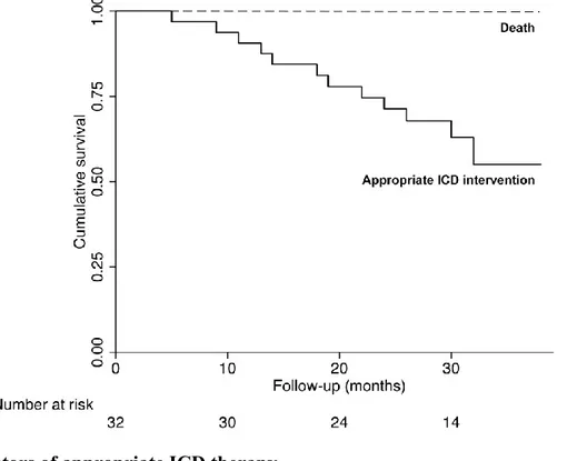
During a mean follow-up of 25 ± 8 months, no patient died. A total of 12 (38%) patients experienced appropriate ICD intervention (11/12 [92%] ICD shocks for VT [10 cases] or VF [1 case]; 1/12 [8%] two effective antitachycardia pacing interventions on VT) (Figure 1).Table 3. Electrophysiological and mapping data of patients with appropriate ICD therapy at follow-up.
Figure 1. Kaplan-Meier survival curves showing cumulative survival free from death (dashed line)
and appropriate ICD intervention (solid line) in the overall study population. During the follow-up no patient died, while 12 (38%) patients experienced appropriate ICD intervention.
3.12 Predictors of appropriate ICD therapy
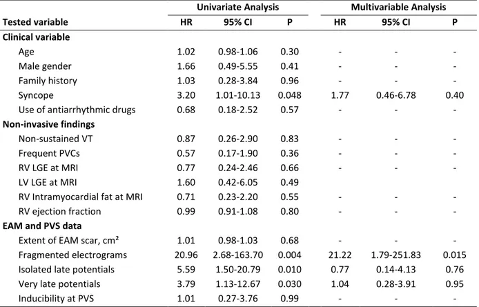
Univariate and multivariable predictors of appropriate ICD therapy are listed in Table 5. Among the baseline clinical variables, only history of syncope was significantly more prevalent in patients experiencing appropriate ICD intervention at follow-up (58% vs. 20%, P = 0.053), and was associated with arrhythmic events at univariate analysis (HR = 3.20, 95% CI 1.01-10.13, P = 0.048) (Figure 2). All other tested non-invasive variables failed to show an association with arrhythmic events at follow-up. With regard to the EAM findings, abnormal electrograms within the scar (i.e., fragmented, isolated late, and very late potentials) were all associated with arrhythmic events at follow-up, with the strongest univariate association being found for fragmented electrograms (HR = 20.96, 95% CI 2.68-163.70, P = 0.004) (Figure 2). Patients who experienced arrhythmic events at follow-up had also a greater percentage of fragmented (19.1% ± 5.9% vs. 6.6% ± 2.4%, P = 0.009) and isolated late electrograms (19.9% ± 8.5% vs. 12.8% ± 5.5%, P = 0.089). Of note, such
differences were confirmed also when combining fragmented and isolated late electrograms into a single variable (35.1% ± 12.4% vs. 12.9% ± 8.2%, P = 0.005), and a threshold percentage of fragmented and isolated late electrograms in the region of low-voltage electrograms of >15% was found the best predictor of arrhythmic events at ROC analysis (sensitivity = 92%, specificity = 90%, positive predictive value = 85%, negative predictive value = 95%). On the other hand, no significant difference in the percentage of very late potentials was found between the two patient groups (27.5% ± 13.9% vs. 19.3% ± 10.8%, respectively, P = 0.19).
The extent of RV involvement by electroanatomic scar was not associated with arrhythmic events at follow up (HR = 1.01, P = 0.68). At multivariable analysis, after adjusting for syncope, isolated late potentials and very late potentials, fragmented electrograms remained the only independent predictor of appropriate ICD interventions (HR = 21.22, 95% CI = 1.79-251.83, P = 0.015).
In a separate multivariable model combining all abnormal electrograms into one covariate, presence of abnormal electrograms within the scar was confirmed the only independent predictor of arrhythmic events at follow-up (HR = 8.91, 95% CI 1.10-71.68, P = 0.040).
Table 5. Predictors of appropriate ICD interventions.
Univariate Analysis Multivariable Analysis Tested variable HR 95% CI P HR 95% CI P Clinical variable Age 1.02 0.98-1.06 0.30 - - - Male gender 1.66 0.49-5.55 0.41 - - - Family history 1.03 0.28-3.84 0.96 - - - Syncope 3.20 1.01-10.13 0.048 1.77 0.46-6.78 0.40
Use of antiarrhythmic drugs 0.68 0.18-2.52 0.57 - - -
Non-invasive findings
Non-sustained VT 0.87 0.26-2.90 0.83 - - -
Frequent PVCs 0.57 0.17-1.90 0.36 - - -
RV LGE at MRI 0.77 0.24-2.46 0.66 - - -
LV LGE at MRI 1.60 0.42-6.05 0.49
RV Intramyocardial fat at MRI 0.71 0.23-2.20 0.55 - - -
RV ejection fraction 0.99 0.91-1.08 0.80 - - -
EAM and PVS data
Extent of EAM scar, cm² 1.01 0.98-1.03 0.68 - - -
Fragmented electrograms 20.96 2.68-163.70 0.004 21.22 1.79-251.83 0.015 Isolated late potentials 5.59 1.50-20.79 0.010 0.77 0.14-4.13 0.76 Very late potentials 3.79 1.13-12.67 0.030 1.04 0.28-3.91 0.95
Inducibility at PVS 1.01 0.27-3.76 0.99 - - -
VT = ventricular tachycardia; PVCs = premature ventricular contractions; LGE = late gadolinium enhancement; MRI = magnetic resonance imaging; RV = right ventricle; EAM = electroanatomic mapping; PVS = programmed ventricular stimulation; RV = right ventricle; LV = left ventricle.
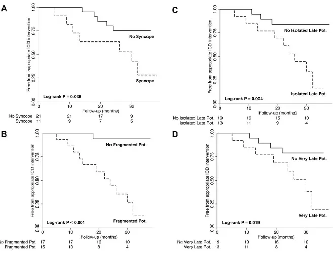
Figure 2. Kaplan-Meier survival curves showing survival free from appropriate ICD intervention
stratified for history of syncope (Panel A), presence of fragmented potentials (Panel B), isolate late potentials (Panel C), or very late potentials (Panel C). Pot. = potentials.
3.13 Commentary
The study tested the hypothesis that different arrhythmic risks among patients with ARVC might be due to different electrophysiologic substrate characteristics, with slow and delayed conduction within the scar representing the missing link between the presence of potentially arrhythmogenic fibro-fatty scars and the occurrence of malignant VAs.
In particular, fragmented and late electrograms within the scar, as assessed by high-density EAM, were the strongest predictor of appropriate ICD interventions on rapid sustained VAs at follow-up.
3.14 Significant findings and implications
Arrhythmic events are the most important manifestation of ARVC, and account for the greater part of ARVC-related mortality.23 A proper identification of ARVC patients at higher risk of arrhythmic events constitutes the main goal of risk stratification, as it would allow to adopt more aggressive therapeutic strategies, including ICD or substrate modification by catheter ablation in selected patients.24, 25
Thus far the identification of markers of increased arrhythmic risk has been elusive, and the most widespread practice is to implant an ICD in all patients with a definite diagnosis of ARVC, reserving substrate-based catheter ablation for patients with a clinical history of sustained VAs.24, 25 The limitations of such an approach have been recently highlighted.25
Data from large ICD registries of ARVC support the notion that the risk of arrhythmic events in these patients is highly variable, and among clinical and instrumental findings only history of syncope appears to independently predict the risk of subsequent arrhythmic events.25 Accordingly, in our series, history of syncope was a predictor of arrhythmic events at follow-up, although 5/12 (42%) patients who experienced arrhythmic events actually had no history of syncope. Notably, with the exception of syncope, patients who experienced arrhythmic events at follow-up and those who remained free from arrhythmic events were otherwise similar with respect to all other clinical and non-invasive characteristics, including cardiac magnetic resonance data.
The present study provides novel insights into the arrhythmic risk stratification of ARVC, showing that presence of fragmented and delayed electrograms at EAM might both identify patients at higher risk of major arrhythmic and give a pathophysiologic explanation to the different
arrhythmic risks across the spectrum of ARVC patients. In line with cardiac magnetic resonance data, the degree or RV involvement by scar at EAM also failed to show an association with arrhythmic events at follow-up. This finding supports the concept that only some fibro-fatty scars, namely, those presenting fragmented and delayed electrograms, are actually arrhythmogenic, while dense scars lacking areas with slow and fragmented conduction areas are not, regardless of their extension. Although fragmented electrograms were found the most powerful predictor of arrhythmic events at follow-up, it is important to emphasize that the presence of any abnormal electrograms remained longitudinally associated with ICD shocks at follow-up, which increases the
external reproducibility of our results. On the other hand, when analyzed as percentage of abnormal electrograms within the region of low voltage electrograms, only fragmented and isolated late electrograms showed an association with arrhythmic events at follow-up, especially when they reached a threshold value of >15% of total low-voltage electrograms.
From a therapeutic standpoint, patients with ARVC and evidence of fragmented and delayed electrograms might be those who most benefit from highly effective but more invasive therapies, such as prophylactic ICD implantation and/or substrate modification by catheter ablation.24 The latter might be of particular value when fragmented and delayed electrograms constitute more than 15% of the total low-voltage electrograms, although the validity of such cut-off value warrants confirmation in further prospective studies.
Finally, the lack of significant endocardial substrate evolution at the time of repeated mapping in the subset of patients undergoing radiofrequency catheter ablation further strengthens the clinical relevance of our findings. Such hypotheses, although intriguing, warrant to be tested in adequately designed prospective randomized trials.
3.15 Pathophysiological interpretation
In the majority of patients with ARVC the mechanism of VAs is fibro-fatty scar-related reentry.24 Inhomogeneous fibro-fatty scarring with islets of residual myocytes interspersed among adipocytes and fibrous tissue provide the substrate for slow conduction, non-uniform anisotropy, and the potential for channels within the scar tissue, which are crucial features for the development of reentry.15, 17 Three-dimensional EAM has been reported to characterize reliably the
electrophysiologic correlates of pathologic substrates underlying VT in different clinical settings, including ischemic and non-ischemic cardiomyopathy.15, 17, 18 Slow-conduction channels within scar tissue (i.e., isolated late and very late potentials) and putative VT exit-sites at the scar
border-regions (i.e., fragmented potentials) have all been identified as critical components of the reentrant VT circuit.15, 17, 18 Our findings definitely expand these concepts also to patients with ARVC, suggesting that the presence of delayed and fragmented electrograms, likely due to surviving myocyte bundles within fibro-fatty scar, may constitute a reliable substrate marker of arrhythmic risk in these patients.
The presented study identified fundamental electrophysiologic substrate characteristics that might explain why individuals with ARVC might be more or less likely to suffer from major arrhythmic events. In particular, ARVC patients with fragmented and late electrograms within the fibro-fatty scar are those at highest risk of major arrhythmic events at follow-up. Our findings provide a new tool for arrhythmic risk stratification, help identifying patients who may earn the greatest benefit from prophylactic ICD implantation, and define promising targets for prophylactic substrate modification by catheter ablation.
One of the possible limitations to the adoption of prophylactic substrate modification in ARVC patients is the presumed progressive nature of the disease with possible progression of substrate
over time. The progression of the arrhythmogenic substrate in patients with ARVC was explored in a subsequent study.
4.0 Scar Progression in Patients with ARVC and VT: a Longitudinal Study With Electroanatomic Voltage Mapping
The inherited desmosomal dysfunction of ARVC is believed to result in progressive loss of right ventricular myocytes that evolves from the epicardium towards the endocardium with replacement by fibrofatty tissue,16, 26 providing an ideal milieu for reentrant ventricular
arrhythmias.19 Thus far, the clinical management of patients with ARVC and VT has been largely based on this paradigm; the believed uniformly progressive nature of the disease has led
investigators to consider therapies only for palliative purposes. Disturbingly, the natural history of patients with ARVC and VT is still incompletely defined; the clinical manifestations of the disease are highly variable and span from no symptoms, recurrent VT, and heart failure.23 Similarly, the degree of disease progression across the spectrum of patients with ARVC is unknown; registry data reporting the clinical indications for cardiac transplantation in advanced forms of ARVC showed that patients with VT represent the large minority, with heart failure being the most common indication for transplantation.27 In this context, preliminary data from our group have shown the
lack of significant endocardial disease progression, as defined by bipolar electroanatomic voltage mapping, in the majority of cases. However, the typical epi-endocardial scar involvement of ARVC limits the value of bipolar endocardial mapping to rule out significant epicardial or midmyocardial scar progression. Recently, endocardial unipolar voltage mapping has been shown to reliably
identify the epi-endocardial pathological substrate of ARVC by detecting myocardial areas with low voltages that reflect epicardial and endocardial scar defined by bipolar voltage mapping. The
purpose of the present study is to evaluate the prevalence and mechanisms of disease progression in patients with ARVC and VT using longitudinal unipolar voltage mapping studies.28
4.1 Patient population
We studied 16 ARVC patients (age 38±13 years, 11 males) and history of recurrent drug-refractory sustained VT who were referred to the Hospital of the University of Pennsylvania for radiofrequency catheter ablation, and underwent two separate detailed endocardial unipolar voltage maps separated by at least 9 months. At the time of enrollment, the diagnosis of ARVC was
established according to the diagnostic criteria of the European Society of Cardiology and International Society and Federation of Cardiology (ESC/ISFC). All patients satisfied also the revised Task Force criteria for ARVC diagnosis.
4.2 Methods
All procedures were performed following the institutional guidelines of the University of Pennsylvania Health System (Philadelphia, PA) after obtaining informed consent. All patients underwent electrophysiological guided 3D electroanatomic mapping and ablation for sustained VT as defined by ECG, implantable cardioverter defibrillator shocks, or both. Sinus rhythm 3D bipolar
electroanatomic voltage maps of the entire RV endocardium were created in all patients at each procedure using either a 3.5- or 4-mm bipolar mapping catheter, both with a 2-mm ring. The bipolar signals were filtered at 30 to 400 Hz and displayed at 200 mm/s speed on the CARTO system. The peak-to-peak unipolar signal amplitude was measured automatically. A 3D anatomic shell of the RV endocardium was constructed, and the electrogram signals were coupled and displayed as color gradients on a bipolar voltage map. Those areas with contiguous low unipolar voltage (<5.5 mV, based on previously published critera)29 were defined as being consistent with scar.
4.3 Results
At baseline, all patients had evidence of epi-endo RV scar (mean 153±80 cm²; 60±26% of the RV surface area). After a mean follow-up of 51±38 months, no significant progression of unipolar voltage scar was observed (mean 170±80 cm², P=0.17; 67±25% of the RV surface area, P=0.07). Specifically, only 3 (19%) patients presented with progression of the RV scar >5%.
0
10
20
30
40
50
60
70
80
90
100
1 2 3 4 5 6 7 8 9 10 11 12 13 14 15 16 17 18 19
Baseline
Follow-up
*
*
*
*
No significant interaction between follow-up duration and increase in RV scar was found.
-20
-10
0
10
20
30
40
50
60
0
50
100
150
200
R = -0.005
P = 0.983
Figure: Example of lack of scar progression with significant increase in volumes over
follow-up in a 46 year-old male patient with arrhythmogenic right ventricular cardiomyopathy and recurrent ventricular tachycardia
The RV volumes increased during follow-up (from 213±73 mL to 262±79 mL, P=0.0016), with the majority of patients (13/16, 81%) having a significant increase in the RV volume (mean increase = 42.8%). Only 3 (19%) patients had no change in both RV scar size and volume over time.
4.4 Commentary
This study examined the extent to which the arrhythmogenic substrate as measured by unipolar voltage mapping in patients with ARVC is progressive. The results of this study question the widespread impression that ARVC is universally a progressive disease process. The findings observed in this group of patients draw attention to the most appropriate treatment strategy for dealing with recurrent VT in patients with ARVC. Based on the results presented here, progression of the arrhythmogenic substrate appears to be limited to a subset of patients. To the extent that these patients are not suffering from a progressive process, control of ventricular arrhythmias should be attainable. In this view, a sufficiently aggressive approach using electroanatomic mapping to
precisely define the abnormal substrate coupled with extensive ablation may sufficiently modify the substrate in many patients so as to make future VT unlikely.
The lack of uniform scar progression is in contrast to the nearly uniform evidence for
progressive and statistically significant RV chamber dilatation in this patient population. Indeed, some of the patients experienced a very significant increase in chamber size. This finding is very important and draws attention to the fact that many of these patients will experience worsening RV function over time and develop signs and symptoms of right-sided heart failure as a result of progressive RV dilatation.
In conclusions, we found that in patients with ARVC and VT, progressive RV dilatation is almost uniformly observed, while rapid epi-endo scar progression is rare. These findings suggest that aggressive epi-endocardial substrate ablation should provide long-term VT control, and further
0
50
100
150
200
250
300
350
400
450
1 2 3 4 5 6 7 8 9 10 11 12 13 14 15 16 17 18 19
Baseline
Follow-up
*
*
*
*
*
*
*
* *
*
*
*
*
* *
*
research is needed to identify the mechanism(s) for and to prevent ongoing RV dilatation in these patients.
The results of this study provided an occasion to evaluate the role of aggressive endo-epicardial substrate ablation in patients with ARVC and VT.
5.0 Long-Term Outcome with Catheter Ablation of Ventricular Tachycardia in Patients with Arrhythmogenic Right Ventricular Cardiomyopathy
Arrhythmogenic right ventricular cardiomyopathy (ARVC) is characterized by diffuse or segmental loss of right ventricular (RV) myocytes with replacement by fibrous and fatty tissue which characteristically involves more extensively the epicardium (EPI) than the endocardium (ENDO).16, 26 This peculiar pathologic process leads to islets of residual myocytes interspersed among adipocytes and fibrous tissue, providing an ideal milieu for reentrant ventricular tachycardia (VT).19 The management of recurrent VT in ARVC is challenging with antiarrhythmic drug (AAD) therapy having limited efficacy.30 Initial experiences with catheter ablation using an ENDO-only approach led to disappointing results.31-33 Given the more extensive epicardial pathological substrate,34, 35 catheter ablation approaches using a combination of ENDO-EPI ablation have been recently shown to significantly improve VT-free survival at the short to mid-term follow-up.24, 35-37 Few data are available on the long-term outcome associated after ENDO-EPI ablative therapy in patients with ARVC and recurrent VT. In the present study we report our institutional experience on catheter ablation of VT in ARVC, and document the long-term outcomes associated with extensive ENDO and/or ENDO-EPI VT ablation and substrate modification in these patients as it relates to VT recurrence and requirement for continued antiarrhythmic drug therapy.38
5.1 Methods
5.2 Study Population
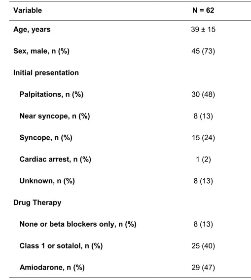
Sixty-two consecutive patients with ARVC and recurrent VT referred to the Hospital of the University of Pennsylvania for radiofrequency catheter ablation between 1998 and 2013 were included in the study. During the same study period a total of 2,716 VT ablation procedures were performed at our institution, of which 325 (12%) were epicardial. All patients met the International Task Force criteria (2 major criteria or 1 major criterion plus 2 minor criteria or 4 minor criteria) for diagnosis of ARVC (Table 1).16, 20 All patients had a minimum one year of follow-up after their last ablation procedure and no patient was lost at follow-up. The study conformed to the institutional guidelines of the University of Pennsylvania Health System, and all patients gave written informed consent to have the data collected and prospectively entered in a database for research purposes.
Table 1. Main clinical characteristics of patients included in the study. Variable N = 62 Age, years 39 ± 15 Sex, male, n (%) 45 (73) Initial presentation Palpitations, n (%) 30 (48) Near syncope, n (%) 8 (13) Syncope, n (%) 15 (24) Cardiac arrest, n (%) 1 (2) Unknown, n (%) 8 (13) Drug Therapy
None or beta blockers only, n (%) 8 (13)
Class 1 or sotalol, n (%) 25 (40)
Amiodarone, n (%) 29 (47)
5.3 Electrophysiologic Evaluation and Instrumentation
Antiarrhythmic drugs were routinely discontinued ≥5 half-lives before the procedure;
amiodarone was discontinued at least 3 days before the procedure. Recurrent unstable arrhythmias necessitated continued antiarrhythmic drug therapy in selected patients at the time of the procedure. All patients underwent the procedure in the fasting state. Conscious sedation was used whenever possible. General anesthesia was used when necessary at the discretion of the operator and/or anesthesiologist involved in the procedure for ventilation, oxygenation, or patient comfort, and during epicardial mapping and ablation procedures. Catheters were placed into position in the heart using fluoroscopic guidance. A standard transvenous 6-Fr quadripolar catheter with 5-mm
interelectrode distance (Bard Inc., Delran, New Jersey) was placed at the RV apex. An 8-Fr 64-element phased-array intracardiac echocardiography (ICE) catheter (AcuNav, Acuson, Mountain View, California) was used routinely for cases after year 2005 to assist catheter manipulation, to assess tissue-catheter contact, and to monitor for complications. In 52 (84%) patients, a deflectable 3.5 mm open irrigated-tip catheter (NaviStar ThermoCool, Biosense Webster, Diamond Bar, California) was used for mapping and ablation; a bidirectional closed-irrigated ablation catheter (Chilli, Boston Scientific, Natick, Massachusetts) was used in 2 (3%) patients. In the remaining 8
(13%) patients (all before 2002), a non-irrigated 4 mm tip ablation catheter (NaviStar, Biosense Webster, Diamond Bar, California) was used. The programmed ventricular stimulation protocol to induce VT included up to triple extrastimuli from at least 2 right ventricular sites with at least 2 drive cycle lengths.
5.4 Endocardial Voltage Mapping
A detailed electroanatomic map of the endocardial RV surface was performed during sinus or paced rhythm maintaining a fill threshold of 20 mm to ensure adequate sampling and representation of the entire endocardial surface area. The bipolar signals were filtered at 30 to 400 Hz (CARTO V.9 and V.7 systems, Biosense Webster, Inc) or 16 to 500 Hz (CARTO-3 system, Biosense Webster, Inc) and were displayed at 100-mm/s speed. The peak-to-peak signal amplitude of the bipolar electrogram was measured automatically and confirmed during manual review. The electrogram signals were displayed as color gradients on a 3-dimensional computerized bipolar voltage map. Tricuspid valvular sites were identified by the fluoroscopic catheter tip positions at the ventricular base with discrete bipolar recordings that demonstrated both sharp atrial and ventricular signals of approximately equal amplitude and confirmed with the use of direct valve visualization with ICE (for cases after year 2005). The pulmonic valve was carefully identified by passing the mapping catheter into the pulmonary artery and slowly withdrawing it until an RV electrogram was identified and RV capture was possible and confirmed with the use of direct visualization of the valve with ICE (for cases after year 2005). Valvular sites were given a “location only” tag to preclude their influence on the voltage map color. Careful attention was paid to record multiple endocardial electrograms around valvular structures. Intracavitary points were identified as abrupt indentations on the endocardial shell contour with associated sudden reductions in signal slew rate and were appropriately redacted from the voltage maps. Reference values for identifying abnormal endocardial bipolar electrogram signal amplitude in the RV were defined according to previously established criteria.17 A signal amplitude of >1.5 mV was categorized as normal and was
represented in the electroanatomic map (CARTO) by the color purple. Abnormal endocardium was represented by the nonpurple range of colors, with the most abnormal signal amplitude, arbitrarily defined as “dense scar” (consistent with signal amplitude <0.5 mV), represented by the color red.
5.5 Epicardial Mapping
Epicardial access was obtained with the techniques described by Sosa and colleagues.39 An inferior approach to the pericardial sac was used to prevent puncture or laceration of the dilated RV. Particular caution was exercised to avoid inadvertent puncture of the liver, which may be enlarged in ARVC patients due to increased right heart pressures. The access was performed on the left of the xiphoid process maintaining an entry angle as shallow as possible (tangential to the lower border of the ribs), and compressing the epigastric region to minimize the risk of intra-abdominal organ perforation. An 8F sheath (or a deflectable sheath) was introduced into the pericardial space, and the mapping/ablation catheter was advanced through the sheath. Detailed voltage mapping focused on the entire RV and extended over the left ventricular (LV) surface. The fill threshold for
until all areas were sampled. The epicardial boundaries of the RV were defined as being opposite the endocardial anatomic shell. More rigid voltage cutoff criteria were used when bipolar signals on the RV epicardium were analyzed to limit the influence of epicardial fat and coronary vasculature. The reference value for defining abnormal electrograms in the epicardium was <1.0 mV, as previously reported.35 Dense scar was also arbitrarily defined as <0.5 mV for display purposes for the epicardial electroanatomic maps.To further limit the influence of epicardial fat and small-vessel coronary vasculature (i.e., vessels that cannot be directly appreciated by coronary angiogram) on the low-voltage region, the contiguous low-voltage electrograms had to demonstrate not only a low amplitude but also signals with discrete late potentials (recorded after the QRS of the surface ECG) and/or demonstrate broad multicomponent or split signals within the boundary of the defined contiguous low-voltage abnormality. Signals larger than 1.0 mV but that also demonstrated
abnormal, multicomponent, split or late signal were also tagged and if adjacent to confluent areas of low voltage typically included in substrate based ablation targets. Beginning in 2008 endocardial unipolar voltage mapping was used to identify a high likelihood of epicardial substrate abnormality in patients with no or modest endocardial bipolar voltage abnormalities as previously described.40 Briefly a unipolar signal cutoff of <5.5 mV for recordings from the RV free wall identified a high likelihood of adjacent confluent epicardial bipolar signal abnormalities.
5.6 Catheter Ablation
The 12-lead ECG morphology of all spontaneous VTs (when available) and/or the intracardiac near-field and far-field electrograms of the implantable cardioverter defibrillator (ICD) were collected and compared to the inducible VT(s) during the procedure. For hemodynamically tolerated VT, activation and entrainment mapping were used for tachycardia localization. The defined “site of origin” and target for more directed ablation demonstrated presystolic activity and entrainment with concealed fusion and a return cycle length within 30 ms of the VT cycle length. Termination with focal radiofrequency energy application was associated with these criteria and confirmed the origin of the tachycardia. For VTs that were not mappable, the site of origin was approximated by using pace mapping to reproduce the VT QRS complex and identify sites with a long stimulus to QRS interval. Limited activation and entrainment information was used to corroborate the pace map information when possible. Radiofrequency ablation was guided on the basis of all mapping data including the location of the best pace map, the location of valvular anatomic boundaries, and detailed characterization of the substrate defined by voltage mapping, including definition of all signals with discrete split or late potentials recorded during sinus rhythm or with RV apical pacing and the identification of discrete higher voltage channels in sinus rhythm in the low-voltage region.35 Characteristically, linear lesions were placed through the site of the best pace map with long stimulus to QRS (>30ms) and/or through channels and transected the abnormal myocardium, extending from the valve annulus to normal myocardium (>1.5 mV in the
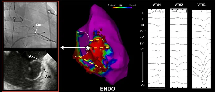
Figure 1. Typical distribution of endocardial (ENDO) ablation lesions across abnormal right
ventricle to valve annuli in a patient with multiple unmappable VTs. Linear lesions transecting the putative VT isthmuses were placed through the site of best pace map with long stimulus to QRS and were anchored to the valve annulus, as verified by signal characteristics, fluoroscopy and
intracardiac echocardiography. Abl = ablation catheter. TA = tricuspid annulus.
The ablation strategy included targeting markedly abnormal fractionated split and late
potentials, with a specific emphasis on abnormal potentials recorded within a 2- to 3-cm radius of the site of origin, defined by entrainment mapping or the best pace map as typically producing clusters of radiofrequency lesions targeting these potentials with the endpoint of signal modification and/or elimination in addition to any linear lesion. This more extensive targeting of the substrate was the routine on the epicardium where linear lesions anchored to the valve annulus were limited by proximity to the right coronary artery (Figure 2). Epicardial radiofrequency lesions always avoided large coronary vessels by at least 1 cm based on cine angiography. If monomorphic VT inducibility persisted after targeting all spontaneous and initially inducible VT, residual VT morphologies were remapped using the techniques described above. More extensive substrate ablation was typical when multiple VT morphologies were inducible as was characteristic of most patients (Figure 3).
Figure 2. Example of endo-epicardial (ENDO-EPI) substrate ablation set in a patient with
ARVC and 4 distinct inducible VT morphologies. At the end of ablation, VT was non-inducible with programmed stimulation from 2 different RV sites up to three extrastimuli.
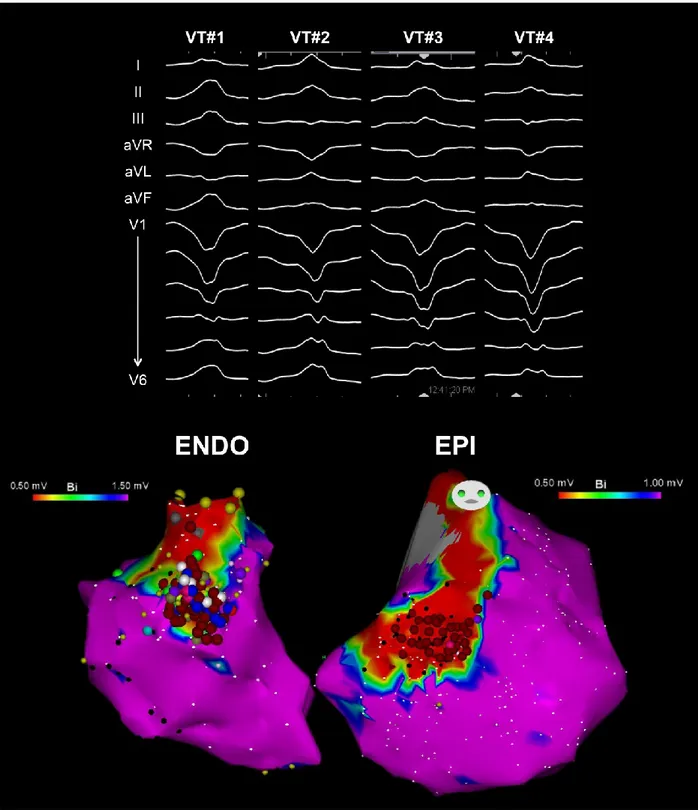
Figure 3. Example of endocardial (left panel) and epicardial (right panel) substrate ablation RF
lesion set (red circles) in a patient with ARVC and 10 distinct inducible VT morphologies. Extensive substrate ablation targeting both mappable and unmappable VT rendered VT non-inducible with programmed stimulation from 2 different RV sites using up to three extrastimuli.
Radiofrequency energy application with the 4-mm standard catheter was routinely set at 50 W and 55°. Open irrigated ablation targeted a maximum temperature of 42° and a maximum
impedance drop of 12 to 15 Ohms with an output of 20 to 50 W. Closed irrigated ablation was set to deliver for 20 to 50 W, targeting a maximum temperature of 45° and a maximum impedance drop of 12 to 15 Ohms. Lesion duration was typically set for 60 – 90 seconds but was further increased to up to 3 minutes in duration at sites associated with transient suppression of VT with monitoring to confirm stable impedance drop.
The amount of fluid in the epicardial space associated with the open irrigated catheter mapping and ablation was monitored with ICE and continuous intra-arterial blood pressure monitoring for evidence of hypotension. At the end of the ablation procedure, 2-3 mg/kg of triamcinolone was routinely administered intrapericardially. A pigtail catheter was routinely left in place in the pericardial sac and removed within 24 hours after the absence of continued pericardial drainage or fluid accumulation was confirmed on transthoracic echocardiography.
5.7 Endpoints and Follow-up
The study endpoint was freedom from any recurrent sustained ventricular arrhythmias (i.e. monomorphic VT, polymorphic/pleomorphic VT and/or ventricular fibrillation [VF]) after the last procedure. The acute procedural endpoint evolved over the years, and consisted of non-inducibility of any clinical VT(s) and of any inducible VT(s) (excluding non-clinical VTs with a cycle length <280 ms) for the earlier experience with ENDO-only ablation, and of non-inducibility of any VT
and no discrete isoelectric baseline) for patients who underwent extensive ENDO-EPI substrate ablation. The acute efficacy was assessed on the basis of inducibility of VT at the end of the ablation procedure with a consistent stimulation protocol (up to triple extrastimuli from up to 2 ventricular sites with at least 2 drive cycle lengths), and at the time of repeat programmed stimulation before hospital discharge typically non-invasively at a single RV site via the ICD system (48 patients) routinely after year 2000. In two patients a repeat invasive electrophysiological study using multisite stimulation was performed. Long-term clinical follow-up included surface ECG recordings at the time of symptoms and routine, every 4-6 months, ICD interrogation to document arrhythmia recurrences. Device programming after VT ablation has been consistent throughout the years and always included a VT zone able to detect (and treat if still inducible) the
slowest clinical and/or induced VT. The effect of additional antiarrhythmic drug therapy on cycle
length slowing was also taken into account to avoid under-detection of VT. The device programming strategy was uniform for both the ENDO-only and ENDO-EPI group. When
longitudinal office follow-up visits were not performed after the 1-year visit, telephone interviews were performed with patients, referring physicians and family members at 6- and 12-month intervals to confirm the absence of arrhythmias with review of stored device electrogram information whenever reported therapy was indicated.
5.8 Statistical analysis
Descriptive statistics are reported as mean ± standard deviation for continuous variables, median and quartiles for skewed distributions, and absolute frequencies and percentages for categorical variables. Long-term arrhythmia-free survival was reported as crude event rates, and assessed through a time-to-event analysis by the Kaplan-Meier method. Univariate and multivariable Cox proportional hazard regression was used to identify baseline clinical variables predictive of VT recurrence over follow-up. All statistical tests were 2-sided, and a P value < 0.05 was considered statistically significant. Data were analyzed by the STATA 12.1 statistical software (Stata
Corporation, Texas, USA).
5.9 Results
5.10 Baseline Characteristics
The study group included 62 patients (age 39 ± 15 years, 45 males, 58 with an ICD; 47 of which were implanted before the catheter ablation) (Table 1). All patients with an ICD had history of recurrent VT leading to either multiple ICD interventions and/or recurrent symptomatic episodes of non-sustained VT/frequent PVCs. All patients met task force criteria for ARVC after extensive clinical evaluation. The 4 patients who were discharged after ablation without an ICD initially presented with recurrent episodes of sustained monomorphic VT associated with palpitations and/or dizziness; an ICD had been previously recommended in all 4 patients.
Upon clinical presentation, none of the patients had a known family history of proven ARVC. Genetic testing was available in 20/62 (32%) patients. Genetic testing was positive for a class I or II variant (most commonly involving the plakophilin and desmoglein genes) in 7 (35%) cases. On
transthoracic echocardiography (62/62, 100%) and CMR (39/62, 63%), most (57/62, 92%) patients had evidence of classical structural and functional abnormalities of the RV. In total, 34/39 (87%) patients had CMR abnormalities diagnostic for ARVC including: 1) significant RV
dilatation/dysfunction (34/34 [100%]), 2) segmental wall motion abnormalities (32/34 [94%]) and 3) late gadolinium enhancement (23/34 [68%]). Evidence of LV disease involvement at CMR was found in 18/39 (46%) patients, with an additional 2/39 (5%) patients showing mild global
hypokinesis and no other abnormalities. LV abnormalities included significant LV
dilatation/dysfunction (5/18 [27%]), segmental wall motion abnormalities (13/18 [72%]), and/or area(s) of midmyocardial late gadolinium enhancement (16/18 [89%]). Patients were excluded if there was cardiac or non-cardiac evidence of sarcoidosis.
The ECG documented clinical ventricular arrhythmias with a left bundle branch block morphology in all patients. Prior to ablation, 54/62 (87%) patients had failed antiarrhythmic medications (mean 2.4 AADs, median 2 AADs) with recurrent VT episodes. This included 25 patients who failed class 1 and/or 3 AADs (excluding amiodarone), 29 (47%) who failed
amiodarone, and 8 beta blockers only or no antiarrhythmic medication. A total of 29 patients had a prior ablation at outside Institutions. All but one of these ablations were ENDO-only.
5.11 Procedural Findings
During the study period, a total of 121 procedures were performed in 62 patients (median 2 [range 1 to 5] procedures per patient) with 23 (37%) receiving ENDO-only procedures and 39 (63%) more extensive substrate ablation which included ENDO and adjunctive EPI procedures due to either VT recurrence after ENDO-only ablation (n=13) or when the acute procedural endpoint (non-inducibility at programmed stimulation) was not met. Of the 62 patients, 28 (45%) underwent a single procedure. Of the 34 patients with repeat procedures, 9 had all of their procedures during a single hospitalization and, as mentioned, 13 patients had an ENDO-EPI ablation procedure
following recurrence after ENDO ablation. Of the remaining 12 patients, the last ablation procedure was performed an average of 25.5 ± 29.8 months (range 1-99 months) after the prior ablation and was followed by an average of 86.8 ± 61.1 months (range 19 -187 months) of additional follow-up.
The number of distinct inducible and/or clinical VTs that were recorded and targeted for ablation throughout the study duration was a median of 4 per patient (range 1 to 14). In 10 (16%) patients, frequent PVCs were also targeted during the ablation procedure(s) due to history of symptoms associated with PVCs and/or morphology mimicking the clinical sustained VT(s).
All patients had evidence of ENDO and/or EPI electroanatomic low voltage areas consistent with scar, with predominant locations in the perivalvular regions (peritricuspid and RV
abnormal electroanatomic substrate nor the site of VT origin in any patient, as previously reported.33, 35
To achieve the acute procedural endpoint, patients required extensive ablation with a cumulative radiofrequency time throughout the duration of the study of 148.7 ± 94.6 min (range 25.1 to 370.4 min) in the ENDO-EPI group versus 80.6 ± 88.3 min (range 11.2 to 342 min) in the ENDO-only group (P = 0.008 for comparison). At the end of the last procedure VT non-inducibility was
achieved in 44/57 (77%); in 5 patients programmed ventricular stimulation was not performed at the end of the procedure.
A total of 50 patients underwent repeat programmed ventricular stimulation from the ICD (n = 48) or with a repeat invasive electrophysiologic study (n = 2) a median of 3 (25th-75th quartiles = 2-4) days after the procedure. Non-inducibility at repeat programmed ventricular stimulation study was achieved in 43/50 (86%) patients.
5.12 Long-term Follow-up After the Last Ablation Procedure
5.12.1 Arrhythmia-free survival
After a mean follow-up of 56 ± 44 months (median 40 months [range 1 to 154 months]), cumulative VT-free survival was 71%. In the 18 patients with recurrence, 9 had only a single episode of VT at a mean 38 ± 41 months with no further episodes with an additional 31 ± 23 months of follow up (Figures 4 and 5). Of the 9 patients with a single recurrence, 5 were continued on their current regimen (4 on beta blockers, 1 sotalol) and 4 either started (2) sotalol, increased (1) sotalol dose, or switched (1) from quinidine to sotalol. There was no significant association between successful ablation outcome and total number of procedures (median 1.5 [1 to 2] in the successful group vs. 2 [1 to 2.5] in the unsuccessful group, P = 0.31) or the cumulative radiofrequency time throughout the study duration (median 104 min [43 to 220 min] in the successful group vs. 95 min [80 to 107 min] in the unsuccessful group, P = 0.90).
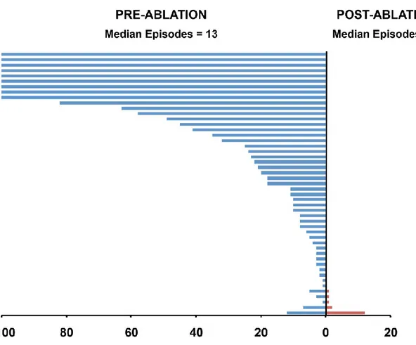
Figure 4. Plot showing the frequency of VT during the year before (blue lines) and after (red
lines) for 49 patients with ICDs before and after ablation. Each line represents an individual patient. Patients are arranged by recurrences during follow-up.
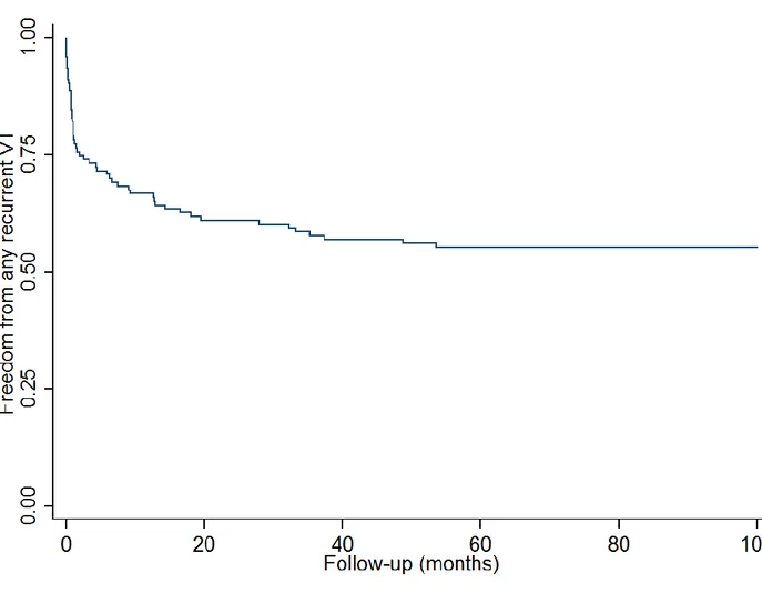
Figure 5. Kaplan-Meier survival curve showing multiple-procedure freedom from any sustained
ventricular arrhythmia in the overall population. Total number of patients followed from last procedure is indicated at the bottom of the figure. Follow-up is truncated at five years.
5.12.2 Use of antiarrhythmic drugs
At the last follow-up, a total of 39 patients were receiving beta-blockers (30) or no drug therapy (9); 21 received either class 1 or 3 AADs sotalol (11 for atrial arrhythmias). Importantly
amiodarone was able to be discontinued after ablation with only 2 patients having amiodarone restarted for a short time prior to transplant for refractory right heart failure. None of the remaining patients required long term amiodarone post ablation.
5.12.3 Other Outcomes
A total of 5 patients had heart (3), heart/liver (1) or heart/kidney (1) transplant at 61.9 ± 61.8 months (median 41.5, range 1 to 154 months) after their last ablation. The indication for heart transplant was refractory heart failure, not refractory ventricular arrhythmias although two of the five patients had documented VT recurrence pre-transplant and were being treated with
amiodarone. Five patients died (ages 51-80 years) during follow up at an average 45.7 ± 54.2 (range 4.9 to 154.3 months). The mode of death was non-arrhythmic in all cases.
5.13 Procedural Complications
A total of 5 (4%) complications and no deaths occurred with the 121 procedures. Two patients had post-procedural deep venous thrombosis with pulmonary embolism documented at imaging studies. These patients received anticoagulant therapy without recurrence over follow-up. After these two thrombotic events, intravenous heparin therapy was routinely used for 48 hours after all pericardial drains were removed. One patient had a late pericardial effusion after an epicardial procedure; this patient did not receive intrapericardial steroids at the end of the procedure. The effusion was not associated with hemodynamic compromise, and was drained percutaneously without consequence. One patient had an RV puncture during percutaneous epicardial access that was directed more anteriorly. Because of continued bleeding after percutaneous drainage in the setting of elevated pulmonary artery pressures, the patient underwent surgical repair with two sutures used to close the small laceration. At the time of surgical repair the patient had concomitant epicardial cryoablation targeting the perivalvular epicardium based on unipolar endocardial
abnormalities and ECG of induced VT. Repeat programmed ventricular stimulation study after the surgical ablation was negative for inducible VT and no recurrent VT was observed after 27 months of follow-up. A final patient developed constrictive pericarditis six months following repeat
epicardial ablation. This patient required multiple passes of the Tuohy needle in order to obtain epicardial access and had a total of 132 radiofrequency lesions applied to epicardial surface in the process of substrate modification. The patient developed post-procedure pericarditis, and ultimately underwent pericardial stripping because of constrictive pericarditis.41 Although this patient did
receive intrapericardial steroids at the end of the procedure, we believe that administration of intrapericardial triamcinolone (2-3mg/kg) is important to minimize the risk of post-procedure pericarditis; of note, this complication has not been observed in any other patient.
5.14 Discussion
The present study describes our institutional experience with radiofrequency catheter ablation of VT in ARVC. It documents the long-term outcomes of ENDO with or without adjuvant EPI
substrate ablation and subsequent use of antiarrhythmic drug therapy in a large cohort of patients with the longest follow-up to date (more than 4 years on average). The main findings are that ENDO with adjuvant EPI substrate modification when indicated (i.e., recurrent VT or persistent inducibility after ENDO-only ablation) provides excellent long-term arrhythmia-free survival, with elimination of VT in the majority of cases and achievement of VT control with infrequent or single isolated recurrent episodes in most of the remaining patients. Importantly, this arrhythmia control was achieved without requiring long-term antiarrhythmic drug therapy treatment with amiodarone. In fact, antiarrhythmic drug therapy with the exception of beta-blockers is uncommonly required in the vast majority of patients unless atrial arrhythmias are also being managed after ablation.
The clinical management of recurrent VT in the setting of ARVC without ablation can be challenging.The efficacy of AADs drug therapy is very limited, with amiodarone being the only drug with a demonstrated benefit in the most recent report from the multicenter North American ARVC registry.30 Given the young age of most ARVC patients, long-term treatment with
amiodarone is an unattractive option due to the substantial and time-dependent risk of end-organ toxicity with this drug.42 In addition, the degree of long-term arrhythmia control with amiodarone is unknown; studies evaluating antiarrhythmic drugs to treat VT in ARVC have limited follow-up.30 Of note, none of the 39 patients who underwent ENDO and adjuvant EPI substrate ablation in our series required long-term amiodarone to achieve VT control making us optimistic that this drug can be avoided in most ARVC patients with VT by successful ablation.
Prior studies evaluating the benefit of radiofrequency catheter ablation of VT in the setting of ARVC have reported only short- and mid-term outcomes in relatively small series of patients frequently combining the results from multiple centers.24, 31, 35-37 The cumulative evidence arising from such studies and now our own large single center experience suggest that an ENDO and EPI substrate-based ablation strategy is important for optimizing management of recurrent VT in patients with ARVC.
We believe that the importance of an aggressive and comprehensive ENDO substrate
modification as part of the overall strategy should be emphasized. Our study supports the notion that ENDO-only ablation may still provide term benefit in selected ARVC cases, as the
long-In our experience, ENDO ablation is particularly important to target the most basal aspect of the typical ARVC perivalvular substrate, which cannot be fully addressed with EPI ablation due to the presence of major coronary vessels and/or fat. Importantly, in this study we reserved EPI ablation only to patients who still had spontaneous or inducible VT after extensive ENDO ablation. Currently, this EPI ablation is typically performed at the same setting. As such, ENDO ablation always represented an important aspect of our procedure and preceded EPI ablation. Therefore, the results of our study do not support a first-line EPI-only ablation approach in patients with ARVC, although adjuvant EPI ablation if often required to achieve long-term VT control. Furthermore, as indicated, 3 out of the 5 total major complications occurring in our study were related to
intrapericardial access and epicardial ablation. This is in line with the findings by Philips et al. in the Johns Hopkins ARVD registry, in which the majority of procedural complications were related to EPI mapping and ablation.36 These data emphasize the importance of performing ENDO ablation first and proceeding to EPI ablation when inducible VT persists to minimize procedural risks.
Given the exceptionally high VT burden in this patient population, with a median of 4 (and up to 14) distinct clinical/inducible VTs per patient, it is important to emphasize the relevance of a comprehensive and extensive substrate based ablation strategy that incorporates ENDO and, if still inducible, EPI ablation in order to achieve the long-term VT control that is reported in the present study. These data, over a more than 10-year experience with EPI VT ablation, have also directed us to be more comprehensive in the extent of substrate ablation particularly whenever epicardial access is obtained. This was done even more aggressively as our clinical experience evolved to try to minimize the need for repeat ablation procedures that were initially commonly observed. It should be emphasized however that our strategy of performing noninvasive programmed stimulation prior to discharge and bringing patients back to the EP lab if inducible VT is found is still advocated. Early repeat ablation to minimize the potential for late adhesion formation that may limit accessibility is advisable.
5.15 Other observations
Remarkably, 2 patients in our study developed a deep venous thrombosis with pulmonary embolism after the procedure. The thromboembolic events may be related to a prothrombotic state associated with the venous stasis due to the underlying significant RV dysfunction. After these events, we have changed our approach including postprocedural therapeutic anticoagulation with heparin in every ARVC patient undergoing catheter ablation for at least 48 hours without further recurrence of deep venous thrombosis and/or venous thromboembolism.
5.16 Study Limitations
This was an observational study summarizing a single-center experience spanning more than 15 years. The choice for the specific ablation approach (i.e., ENDO-only vs. ENDO-EPI) was not randomized and, as expected, the acute ablation endpoints evolved over the multiyear study period. However, given the single center nature of the study, the ablation approaches and protocols adopted