1
UNIVERSITA’ DEGLI STUDI DI CATANIA
DOTTORATO DI RICERCA IN BIOTECHNOLOGIE
XXVIII Ciclo
The role of mTOR signaling in Alzheimer’s disease
______________________TESI DI DOTTORATO
Dottorando:
Dott.ssa Antonella Caccamo
Tutor:
Chiar.ma Prof.ssa Angela Messina
Coordinatore:
Chiar.mo Prof. Vito De Pinto
2 ABSTRACT
The majority (>95%) of Alzheimer’s disease (AD) cases are sporadic and of unknown causes. The single major risk factor for AD is aging and molecular changes that occur in the brain as a function of age may facilitate the development of AD. However, little is known as to how the aging process facilitates the development of AD.
Overwhelming evidence shows that reducing the activity of the mammalian target of rapamycin (mTOR) increases lifespan and health-span in several genetically different species. mTOR is a ubiquitously expressed protein kinase that plays a key role in regulating protein synthesis and cell growth. mTOR also is a negative regulator of autophagy induction. By simultaneously regulating protein synthesis and degradation, mTOR is key in controlling protein homeostasis, a process that is altered in AD and other proteinopathies. Another known function of mTOR signaling is the regulation of synaptic plasticity and function.
Using two widely used animal models of AD, known as Tg2576 and 3xTg-AD mice, we employed multidisciplinary approaches to dissect the role of mTOR signaling in AD. We found that genetic reduction of mTOR signaling reduced amyloid-β deposits and tau pathology and rescued memory deficits in Tg2576 mice. Mechanistically, the reduction in mTOR signaling led to an increase in autophagy induction and restored the hippocampal gene expression signature of the Tg2576 mice to wild type levels. Consistent with these findings, we also found that that genetic reduction of the ribosomal protein S6 kinase 1 (S6K1), a protein kinase directly downstream of mTOR, improved synaptic plasticity and spatial memory deficits, and reduced the accumulation of amyloid-β and tau, in 3xTg-AD mice. Mechanistically, these changes were linked to reduced translation of tau and the beta-site APP cleaving enzyme 1 (BACE-1), a key enzyme in the generation of amyloid-β.
3
Given the overwhelming evidence showing that reducing mTOR signaling increases lifespan and health span, the data presented here have profound clinical implications for aging and AD and provide the molecular basis for how aging may contribute to AD pathology. Our results implicate hyperactive mTOR/S6K1 signaling as a previous unidentified signaling pathway underlying gene-expression dysregulation, synaptic and cognitive deficits in Alzheimer’s disease.
4 SUMMARY 1. Introduction ... 6 1.1. Alzheimer’s disease ... 6 1.1.1. Biology of Aβ... 7 1.1.2. Biology of presenilins ... 8 1.1.3. Biology of tau ... 8
1.1.4. Amyloid cascade hypothesis ... 10
1.1.5. Synaptic dysfunction ... 10
1.1.6. Transgenics mice ... 11
1.1.7. Aβ and tau interaction ... 15
1.2. Mammalian Target of Rapamycin ... 19
1.2.1. mTOR in aging ... 20
1.2.2. mTOR, cognition, and brain aging ... 21
1.3. mTOR and Alzheimer’s disease ... 23
1.3.1. mTOR signaling in AD brains ... 23
1.3.2. Aβ and mTOR ... 24
1.3.3. Tau and mTOR ... 27
1.3.4. Autophagy, mTOR and AD ... 28
2. Aim of the work ... 32
3. Results ... 33
3.1. Genetic reduction of mTOR ameliorates Alzheimer’s disease-like cognitive and pathological deficits by restoring hippocampal gene expression signature ... 33
3.1.1. Removing one copy of the mTOR gene, decreases hippocampal mTOR signaling ... 34
3.1.2. Reducing mTOR signaling rescues cognitive deficits in Tg2576 mice ... 37
3.1.3. Reducing mTOR signaling decreases Aβ levels and burden and increases synaptophysin levels ... 40
3.1.4. Reducing brain mTOR signaling increases autophagy induction ... 43
3.1.5. Reducing mTOR signaling restores the gene expression profile of the Tg2576 mice to CTL levels ... 45
3.1.6. Discussion ... 48
3.2. Reducing ribosomal protein S6 kinase 1 expression improves spatial memory and synaptic plasticity in a mouse model of Alzheimer’s disease ... 51
5
3.2.1. Reducing S6K1 levels prevents synaptic deficits in 3xTg-AD mice ... 52
3.2.2. Reducing S6K1 levels rescues spatial memory deficits in 3xTg-AD mice ... 59
3.2.3. Removing one copy of the S6K1 gene lowers Aβ and tau pathology in 3xTg-AD mice ... 61
3.2.4. Low S6K1 signaling reduces BACE-1 and tau translation ... 65
3.2.5. Discussion ... 69
4. Concluding Remarks ... 73
5. Methods ... 76
5.1. Mice ... 76
5.2. Human tissue ... 76
5.3. Morris water maze ... 77
5.4. Protein extraction ... 78
5.5. Western blot ... 78
5.6. Enzyme-linked immunosorbent assay (ELISA) ... 79
5.7. Immunohistochemistry ... 79
5.8. Microarray ... 80
5.9. Electrophysiology ... 80
5.10. Quantitative real-time polymerase chain reaction ... 81
5.11. Polysome profiling ... 82 5.12. BACE-1 activity ... 82 5.13. S6K1 activity ... 83 5.14. Proteasome activity ... 83 5.15. Antibodies ... 84 5.16. Statistical analyses ... 84 6. References ... 86
6 1. INTRODUCTION
1.1 Alzheimer’s disease
Alzheimer’s disease (AD) is the most common neurodegenerative disorder. It is estimated that 26 million people worldwide are living with AD and by 2050 the number of people with this disorder can reach 100 million (Thies et al., 2013). Currently there are no effective cures for AD and patients perish of comorbidities 3 to 9 years after diagnosis.
More than 95% of AD cases are sporadic and of unknown causes, with aging being the major risk factor for the development of the disease. It is estimated that 33-50% of people older than 85 have AD (Bird, 2008).In the Western Countries, the aging population is the fastest segment of society; thus the number of AD cases is predicted to greatly increase by mid-century (Bird, 2008).
The remaining cases are caused by mutations in one of three genes, presenilin 1 and 2 and amyloid precursor protein (APP; Querfurth and LaFerla, 2010).
One of the first clinical manifestation of AD is deficits in episodic memory (i.e., the ability to recall previous events; Artero et al., 2003; Welsh et al., 1992). As the disease progresses, other cognitive domains are impacted and deficits in executive functions, language, and learning tasks appear evident. Eventually, patients become bedridden and perish due to other comorbidities.
The two hallmark neuropathological lesions that characterize AD are plaques, mainly composed of amyloid-β (A) peptide, and neurofibrillary tangles (NFT), mainly formed by hyperphosphorylated tau (Selkoe, 2001). The events triggering AD pathology and the molecular mechanisms linking aging to AD are not known.
7 1.1.1 Biology of A
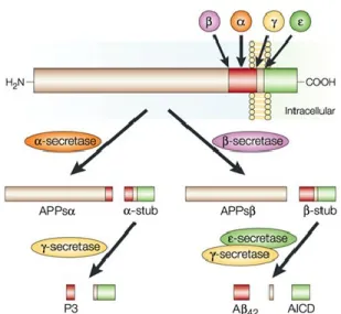
A is derived from a longer precursor molecule, the amyloid precursor protein (APP), which is encoded by a gene located on chromosome 21 (St George-Hyslop et al., 1987). APP can be processed along two different pathways: a non-amyloidogenic pathway, which prevents the generation of A, and an amyloidogenic pathway, which eventually leads to A formation (Selkoe, 2001; Fig. 1). The non-amyloidogenic pathway involves the activity of -secretase, which cleaves APP in the middle of the A sequence, thereby preventing the formation of A. In contrast, the
amyloidogenic pathway involves a cleavage of APP at the β-secretase site by the β-site APP cleaving enzyme (BACE; Vassar et al., 1999), leading to the production of the C99 fragment, which serves as the substrate for another secretase known as -secretase. -secretase cleaves C99 predominantly at two different positions, generating either Aβ40 or Aβ42. The longer species
is more amyloidogenic than the shorter isoforms. Several clinical mutations in the APP gene have
Figure 1. Schematic representation of APP processing. APP is
cleaved by an -secretase, which cuts APP in the middle of the A sequence thus precluding A formation. In the contrary, the sequential activity of - and -secretase enzymes lead to the formation of A. This diagram is from (LaFerla, 2002).
8
been identified and most of them are localized to positions near the , β and -secretase sites (Goate et al., 1991; Mullan et al., 1992) and reviewed by (Haass and Selkoe, 1993). These mutations lead to autosomal-dominant, early-onset familial Alzheimer’s disease (FAD), likely by increasing total Aβ production or by selectively increasing of the longer more amyloidogenic Aβ42
species (Citron et al., 1992; Suzuki et al., 1994).
1.1.2 Biology of presenilins
Two other proteins are directly involved in the pathogenesis of AD: presenilin 1 (PS1) and presenilin 2 (PS2) (Levy-Lahad et al., 1995; Rogaev et al., 1995; Sherrington et al., 1995). These are transmembrane proteins that are part of a high molecular weight complex that is responsible for the -secretase activity (Kimberly et al., 2000; Wolfe et al., 1999). Both PS1 and PS2 are membrane proteins with multiple transmembrane regions that are expressed in the brain and in the periphery (Rogaev et al., 1995; Sherrington et al., 1995). Within neurons, PS2 is mainly expressed in the cell body whereas PS1 is expressed in cell bodies and neuronal processes (Blanchard et al., 1997). Both proteins undergo proteolytic cleavage; PS1 is rapidly cleaved into a ~28 kDa amino terminal and a ~ 19-kDa carboxy-terminal fragment, whereas PS2 is cleaved into two polypeptides of 34 kDa and 20 kDa, respectively. Mutations in the genes encoding these proteins (located on chromosome 14 and 1, respectively) lead to early-onset AD (Rogaev et al., 1995; Sherrington et al., 1995). Indeed, mutations in the PS1 gene, which account for the majority of all early-onset AD cases (Cruts et al., 1996), can cause AD in people as young as 16 years of age. The mechanism by which these mutations lead to AD seems to be linked to A formation (Scheuner et al., 1996).
9
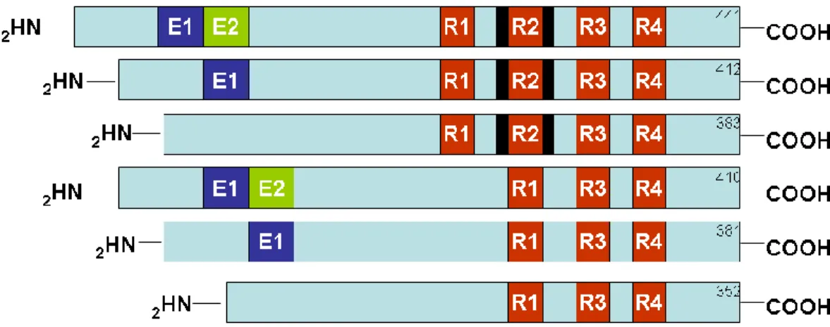
As mentioned above, tau is the main component of NFT. Tau is a microtubule-associated protein that promotes microtubule assembly and stabilization (Drechsel et al., 1992; Lindwall and Cole, 1984; Trinczek et al., 1995). These functions are highly regulated by post-translational modifications including the addition and removal of phosphate groups by the activity of several phosphates and kinases. Tau is highly expressed in neurons although low expression levels are also detected in astrocytes and oligodendrocytes (Binder et al., 1985; LoPresti et al., 1995). Six different isoforms are expressed in the adult brain and all of them are generated from the
alternative splicing of a single gene located on chromosome 17 (Fig. 2). Mutations in the tau gene cause frontotemporal dementia and Parkinsonism linked to chromosome 17 (FTDP-17), a disease characterized by intracellular accumulation of tangles similar to those present in AD brains (Hutton et al., 1998).
Figure 2. Schematic illustration of the human tau gene. Tau is encoded by a single
gene, MAPT, located on chromosome 17. Six isoforms are generated via alternative splicing of exons 2, 3 or 10. Exons 2 and 3 encode repeated regions, whose function is not well understood. The red boxes R1-R4 represent microtubules binding domains. In humans, equal ratio of 3R and 4R is expressed in the adult brain, whereas in mice, the 3R is predominately expressed. This diagram is not drawn to scale and is from (LaFerla and Oddo, 2005).
10 1.1.4 Amyloid cascade hypothesis
As discussed above, the majority of the clinical mutations known to cause AD lead to A mis-metabolism. This genetic evidence was the basis for the formulation of the amyloid cascade hypothesis, which states that A is the trigger of all cases of AD (Hardy and Selkoe, 2002; Hardy and Higgins, 1992; Selkoe, 1991). Based on this hypothesis, all the other pathological events are a consequence of A accumulation, which can be a consequence of mis-metabolism, overproduction, or a reduction in clearance mechanisms. Indeed, steady-state levels of two A-degrading enzymes decrease in the cortex and cerebellum of NonTg mice and humans as a function of age but do not change in the cerebellum. Moreover, the steady-state levels of these enzymes are always higher in the cerebellum (Caccamo et al., 2005). These results clearly suggest that the levels of these enzymes can play an important role in A deposition.
1.1.5 Synaptic dysfunctions
Synaptic loss is another major neuropathological hallmarks of AD and is the best correlate of cognitive deficits (Koffie et al., 2011). Hippocampal synapses begin to degenerate in patients with mild cognitive impairment, where a reduction of about 25% in the presynaptic vesicle protein synaptophysin has been reported (Querfurth and LaFerla, 2010). With the progression of the disease, the loss of synapses becomes severe, especially in brain regions surrounding plaques. These observations suggests that Aβ may be the cause of synaptotoxicity. To this end, Aβ inhibits long-term potentiation (LTP), one of the major cellular mechanisms that
underlies learning and memory, and a measure of synaptic strength and plasticity (Koffie et al., 2011). Consistent with this results, inhibiting the formation of Aβ oligomers rescues LTP induced by treating brain slices with media containing Aβ. In addition to inhibit LTP, Aβ facilitates the induction of long-term depression (LTD) in hippocampal synapses. Impairments in LTP and facilitation of LTD cause synaptic depression and damages in neuronal networks (Palop and
11
Mucke, 2010). The molecular mechanisms underlying the Aβ-mediated synapse dysfunction is very complex and it is an area of active research. For example, Aβ can induce calcium dyshomeostasis, trigger activation of caspases and calcineurin, and modulate the activity of synaptic excitatory receptors and receptor tyrosine kinases, instigating a cascade of molecular events that culminate in synapses loss and eventually impairment of cognitive and memory functions (Koffie et al., 2011).
1.1.6 Transgenic mice
Considering the complex nature of AD pathology, it is very difficult to study the molecular mechanisms underlying this disease in humans. In human post mortem brain tissue, it is difficult to establish the damage due to A and the downstream damage due to degenerative processes occurring in the last years of the patient’s life. Moreover, other complications that can occur during the last years of the patient’s life can complicate the pathological post mortem analysis of these brains. Therefore, several groups have attempted to generate transgenic models of AD, soon after the isolation of the APP gene (Higgins et al., 1993; Mucke et al., 1994; Quon et al., 1991; Sandhu et al., 1991).
APP and PS transgenic models. The first successful model to reproduce A pathology was the PDAPP mice, which overexpress the human APP minigene encoding for APPV717F under the
control of the PDGF promoter (Games et al., 1995). The overexpression levels of the human APP were more than 10-fold over endogenous mouse APP levels, and this probably contributed to their success in reproducing A pathology. These mice develop age-dependent A deposits, which start around 6-9 months of age in the hippocampus, corpus callosum and cerebral cortex. The PDAPP mice also develop neuritic plaques, synaptic loss, astrocytosis and microgliosis (Games et al., 1995). Moreover, they show a behavioral deficit in the radial-maze, Morris
water-12
maze and object-recognition tasks (Chen et al., 2000; Dodart et al., 1999) Another mouse-model (Tg2576) was subsequently developed by overexpressing human APP695 containing the Swedish
double mutation (K670N, M671L) under the control of the PrP (Hsiao et al., 1996). The Tg2576 mice overexpress APP to about 6-fold over endogenous levels, develop all the AD-like pathology observed in the PDAPP mice starting around 9-10 months; they also develop an age-dependent memory deficit tested by the water-maze paradigm (Hsiao et al., 1996; Westerman et al., 2002).
Other groups subsequently developed models showing AD-like pathology by overexpressing the APP gene (Calhoun et al., 1999; Chishti et al., 2001; Davis et al., 2004; Moechars et al., 1999; Mucke et al., 2000; Sturchler-Pierrat et al., 1997). The majority of these animal models develop A pathology, dystrophic neurites and cognitive impairments (Phinney et al., 2003). However, they do not develop NFT nor do they have appreciable cell loss. This is surprising because a single mutation in the APP, PS1 or PS2 genes leads to the development of the full spectrum of AD neuropathology, in humans. Curiously, mice harboring an FAD mutation in PS1 and/or PS2 do not develop plaques (see for example (Chui et al., 1999; Duff et al., 1996; Guo et al., 1999) but show an increase in Aβ42 levels. Double transgenic mice have been generated by crossing
APP and PS1 transgenic mice (see for example (Borchelt et al., 1997; Holcomb et al., 1998)). The common observation in these double transgenic mice is that Aβ deposition is accelerated compared to single APP transgenic mice. Remarkably, despite the increase and early onset in A accumulation in the double transgenic mice, they still do not develop NFT. Several reasons might account for the lack of NFT in these models. For example, it is possible that mice do not live long enough to develop NFT or that the mouse tau gene is somehow protective (Andorfer et al., 2003).
13
the tau gene lead to FTDP-17 (Hutton et al., 1998), a disorder characterized by tangle accumulation in brain regions that are also affected in AD cases. The FTDP-17 patients do not develop any A pathology, suggesting that tau pathology is not likely the factor that triggers AD pathology, but may lead to neurodegeneration downstream of A. Moreover, these cases show that tau pathology can be induced by other factors independent from A.
To better study NFT formation and their involvement in neurodegenerative disease, different groups have generated transgenic models overexpressing human tauP301L, a mutation known to
cause FTDP-17. In 2000, Nature Genetics published the characterization of the first transgenic model harboring the human tauP301 under the control of prion promoter (Lewis et al., 2000). These
mice accumulate NFT in different areas of the brain stem and spinal cord, leading to a profound motor deficit and early death (Lewis et al., 2000). Considering the motor deficit and early mortality that occurs in these mice, their utility for studying neurodegenerative disorders, such as AD or FTDP-17 has been limited. Another group overexpressed the human tauP301L using the Thy1.2
promoter (Gotz et al., 2001a). These mice accumulate NFT in the CA1 region of the hippocampus and in the neocortex. Apoptotic cells were also present in selected areas of the somatosensory cortex (Gotz et al., 2001a). No reports of cognitive impairments in neither of these two transgenic mice are present in the literature.
It is clear that high levels of A in mice do not lead to tau pathology, and likewise high levels of tau pathology do not lead to A pathology. Consequently, to obtain a model with both plaques and tangles, it is necessary to use strategies that are more aggressive. Toward this goal, two major approaches have been used: microinjecting pathological protein into the brains of single transgenic mice (Gotz et al., 2001b) or crossing individual transgenic mice harboring APP and tau mutations (Lewis et al., 2000).
14
Gotz et al. showed that injection of fibrillar A into the somatosensory cortex of transgenic mice overexpressing human tauP301L caused a 5-fold increase in NFT in the amygdala. By injecting a
retrograde tracer in the somatosensory cortex, they showed that the affected neurons in the amygdala project to the injected area. This finding suggests that synaptic damage due to A accumulation can induce NFT in the cell bodies. They also concluded that A can accelerate NFT formation, although no other reports have yet been published that replicate these findings.
Using a different approach, Lewis et al. (2001) arrived at similar conclusions. They crossed two independent transgenic lines, one overexpressing mutant APP and the other overexpressing mutant tau. The double transgenic mice developed A pathology at the same age as the single APP transgenic mice but showed enhanced tau pathology. This led the authors to conclude that either APP or A influences tau pathology in vivo. However, other conclusions might explain their results. For instance, it has been shown that the genetic background of mice can modulate pathological events such as susceptibility to excitotoxic cell death in transgenic mice (Schauwecker and Steward, 1997; Steward et al., 1999). By crossing mice of different genetic backgrounds, Lewis et al. generated a new set of mice where the genetic background was different compared to the parental strain. This might have changed the susceptibility of specific neurons to tau pathology or altered the expression patter of the transgene. Moreover, these mice show a profound motor deficit due to extensive pathology in the brain stem. This deficit and the fact that the mice die prematurely make this model unsuitable for any behavior or immunological studies. Therefore, other animal models with both plaques and tangles in AD-affected brain regions, without the confounding variable of mixing different genetic backgrounds, still need to be derived.
15
3xTg-AD mice. The development of both plaques and tangles is necessary in a model of AD. The 3xTg-AD mice develop age- and region-dependent accumulation of plaques and tangles (Oddo et al., 2003b). These transgenic mice harbor three mutant human genes, APPSwe, TauP301L,
and PSN1M146V. One of the earliest neuropathological manifestation in the 3xTg-AD mice is the
accumulation of intraneuronal Aβ, which is detected in the hippocampus as earlier as four to five months of age (Oddo et al., 2003b), and correlates with the onset of cognitive deficits (Billings et al., 2005). Extracellular plaques appear by 12 months in the entorhinal cortex and become more widespread in the hippocampus and in the cortex by 14 months. Accumulation of tau occur in the hippocampus by 8 months of age and became aggregated and hyperphosphorylated and it is detectable in cortex as well (Oddo et al., 2003b; Oddo et al., 2008). Synaptic dysfunction, LTP deficits and cognitive impairment occur before the accumulation of plaques and tangles. The presence of both lesions is critical to study the molecular interaction between plaques and tangles and to test the efficacy of anti-Alzheimer treatments on both pathologies.
1.1.7 A and tau interaction
One major hypothesis for AD is the amyloid cascade hypothesis (Hardy and Selkoe, 2002; Hardy and Higgins, 1992; Selkoe, 1991), which stipulates that A is the trigger of all cases of AD, and tau pathology is a consequence of the A pathology. In other words, it proposes the existence of a link (direct or indirect) between A and tau, where A is the trigger of a cascade of events that lead to the accumulation of NFT that are then responsible for the cell death observed in AD (Hardy, 2003). Different studies have supported the amyloid cascade hypothesis reaffirming the idea of a direct link between A and tau. However, the molecular mechanisms linking the two pathologies are yet not understood. In the previous section, I referred to some in vivo evidence produced by us and other groups supporting this interaction (Gotz et al., 2001b; Lewis et al., 2001; Oddo et al., 2003a; Oddo et al., 2003b). Here I will analyze some of the in vitro data supporting
16 this interaction.
Several reports have shown that A is toxic to primary rat and human neuron cultures (Busciglio et al., 1993; Pike et al., 1992; Yankner et al., 1989) and some of them have suggested that tau maybe a possible downstream mediator of A toxicity (Alvarez et al., 1999; Busciglio et al., 1995; Takashima et al., 1993). Takashima and colleagues published the first report for a possible role of tau in A-induced pathology. They showed that treatment of primary hippocampal cultures with A causes an increase in the activity of TPK1 (a GSK3β homologous) that correlates with the A-induced neurotoxicity. More interestingly, TPK1 antisense oligonucleotides protect these neurons from A-induced toxicity (Takashima et al., 1993). Considering that A-treated neurons were also positive for Alz50, a conformational specific tau antibody, (Yankner et al., 1989) and that TPK 1 has been involved in the phosphorylation of tau (Ishiguro et al., 1988; Ishiguro et al., 1992), this study implies that A can induce tau pathology. The involvement of TPK 1 / GSK3β in A-induced toxicity has been confirmed by other studies (Hoshi et al., 2003; Hoshi et al., 1996; Imahori and Uchida, 1997).
CDK5 is another key enzyme implicated in tau phosphorylation (Alvarez et al., 2001; Alvarez et al., 1999; Lee et al., 1999; Patrick et al., 1999). The activity of CDK5 is regulated by a small peptide of 35 amino acids (p35), which binds to CDK5 and activates it (Patrick et al., 1999). Proteolytic cleavage of p35 by calpain (a calcium-dependent protease) leads to the formation of a truncated peptide of 25 amino acids (p25), which binds to CDK5 causing its unregulated activation (Patrick et al., 1999). Primary hippocampal neurons treated with A show increased CDK5 activity as well as cell death (Alvarez et al., 1999). When a CDK5 inhibitor or CDK5 antisense oligonucleotides are added to the treated neurons, they partially rescue the A-induced cell death (Alvarez et al., 1999). The mechanism by which A increases the activity of
17
CDK5 is not fully understood. Increases in intracellular Ca2+, which can be caused by A or FAD
mutations (LaFerla, 2002; Leissring et al., 2001; Leissring et al., 1999a; Leissring et al., 1999b; Mattson et al., 1992), has been proposed to be the upstream event leading to increased CDK5 activity. The raise in intracellular Ca2+ concentration activates calpain, which in turn produces
more p25 (Lee et al., 2000; Patrick et al., 1999) eventually leading to higher CDK5 activity and more tau phosphorylation.
Taken together, these studies show that A can enhance the activity of different kinases that phosphorylate tau. It is still possible, however, that tau phosphorylation could be an epiphenomenon, and all these activated kinases are toxic to the neurons by other pathways independent of tau phosphorylation. The direct evidence that tau is actually necessary for A-induced toxicity has been shown using tau knock-out mice. Treatment of hippocampal neurons from tau null mice with fibrillar A did not produce signs of degeneration, whereas neurons from wild-type mice or from mice expressing human tau show profound signs of neurites degeneration within 24 hours of treatment and they die after 96 hours (Rapoport et al., 2002). Moreover, neurons from human tau transgenic mice generated in a tau null background reacquired their susceptibility to A showing the same signs of degeneration as wild-type mice (Rapoport et al., 2002).
It is well established that different factors, including A, can induce apoptosis in both AD and transgenic mice (Gervais et al., 1999; LaFerla et al., 1995; Nakagawa et al., 2000; Rohn et al., 2001; Su et al., 1994; Troy et al., 2000; Zhao et al., 2003). Incubation of primary neurons with fibrillar A induces activation of caspase 3, which can cleave tau removing 20 amino acids from its C-terminal (Gamblin et al., 2003; Rissman et al., 2004). In vitro studies show that the N-terminal portion of this truncated form of tau (delta-au) forms filaments more rapidly in vitro that
18
full-length tau (Gamblin et al., 2003; Rissman et al., 2004). Moreover, when tau is incubated in the presence of small amount of delta-tau, filament formation is greatly increased (Rissman et al., 2004). Delta-tau is also present in NFT that accumulate in AD and transgenic mice (Gamblin et al., 2003; Novak et al., 1993; Rissman et al., 2004) and inversely correlate with cognitive function (Rissman et al., 2004). These data identify a new link between A and tau that is independent of the tau kinases, although it remains to be established if hyperphosphorylated tau is a better substrate for these caspases. If this turns out to be the case, the increase in CDK5 and GSK3β activity, due to A, could facilitate the assembly of tau in NFT.
19 1.2 THE MAMMALIAN TARGET OF RAPAMYCIN
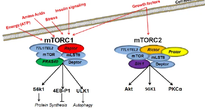
The mammalian target of rapamyicn (mTOR) is a serine/threonine-protein kinase, which is involved in the regulation of both protein synthesis and degradation, longevity, and cytoskeletal formation (Johnson et al., 2013; Wullschleger et al., 2006). mTOR is the key catalytic unit of two separate large multimeric protein complexes, mTOR complex 1 (mTORC1) and 2 (mTORC2; Fig. 3).
Figure 3. The mTORC1 vs. mTORC2 have distinct constituent proteins and regulate different downstream targets. Both mTORC1 and mTORC2 share the catalytic mTOR subunit, the mammalian
lethal with sec-13 protein 8 (mLST8), the Tti1 and Tel2 complex, and the DEP domain containing mTOR-interacting protein (deptor), all depicted in blue. The mTORC1 contains the rapamycin sensitive raptor subunit (depicted in red) and the proline-rich Akt substrate 40 KDa (PRAS40; depicted in green). Downstream effectors ribosomal protein S6 kinase beta-1 (S6K1) and eukaryotic translation initiation factor 4E-binding protein 1 (4EBP1) play a critical role in protein translation by regulating the activation of initiation factors. Inhibition of mTORC1 decreases the phosphorylation of ULK1, which initiates the sequential activation of several autophagy-related proteins, culminating in the formation of AVs. In contrast, the mTORC2 contains the rictor subunit (depicted in orange), the protor component (depicted in yellow), and the mammalian stress-activated map kinase-interacting protein 1 (sin1; depicted in purple). Downstream effectors of mTORC2 are involved in regulating longevity, cellular stress response and cytoskeletal dynamics. The mTORC1 receives signals from growth factors, glucose, inflammatory cell signaling proteins and extracellular amino acids. The mTORC2 receives signals primarily from growth factors.
20
mTORC1 consist of mTOR, raptor, the mammalian lethal with sec-18 protein 8 (mLSTR8), the Dep domain containing mTOR interacting protein (Deptor), the Tti1/Tel2 complex, and the proline-rich Akt substrate 40 KDa (PRAS40) (Wullschleger et al., 2006). mTORC2 consist of mTOR, rictor, mLSTR8, deptor, Tti1/Tel2, and mammalian stress-activated map kinase-interacting protein 1 (Gingras et al., 1999). mTORC1 is regulated by signaling from insulin, growth factors, amino acids, and oxidative stress (Hay and Sonenberg, 2004). It promotes cell growth and proliferation (by facilitating mRNA translation and protein synthesis), lipid biogenesis, regulates mitochondrial metabolism, and modulates autophagy (Kim and Guan, 2015; Perluigi et al., 2015; Wang and Proud, 2006). In contrast, mTORC2 appears to be strictly under control of growth factors and it is thought to function primarily in cytoskeleton assembly and cell size (Frias et al., 2006; Graber et al., 2013). Both mTORC1 and mTORC2 are involved the regulation of longevity (Hay and Sonenberg, 2004; Johnson et al., 2013).
mTORC1 regulates protein translation mainly by controlling the activity of ribosomal protein S6 kinase-1 (S6K1) and eukaryotic initiation factor 4E binding protein 1 (4EBP1), which directly control the activity of several initiation factors (Gingras et al., 1999; Hands et al., 2009; Hay and Sonenberg, 2004; Ruvinsky and Meyuhas, 2006). Malnutrition, stress, insulin, growth factors, and various other signaling pathways that converge on mTOR may differentially activate/inhibit protein synthesis (Gingras et al., 1999; Hay and Sonenberg, 2004).
1.2.1 mTOR in aging
The first evidence of TOR’s involvement in aging came from work conducted in S. cerevisiae. Deletion of the gene encoding the yeast orthologue of S6K1 resulted in a doubling of the chronological lifespan(Fabrizio et al., 2001). Shortly after, inhibition of raptor or S6K1 was shown to extend lifespan in C. elegans (Jia et al., 2004; Vellai et al., 2003). These initial studies have been confirmed and expanded to other species (Johnson et al., 2013; Kapahi et al., 2004). To
21
this end, a landmark report showed that rapamycin, an mTOR inhibitor, fed to genetically heterogeneous mice, increased their lifespan (Harrison et al., 2009). The involvement of mTOR in regulating lifespan in mammals has also been shown using two independent genetic approaches. The first one highlighted that deletion of S6K1, a downstream target of mTOR, extends lifespan and healthspan in both male and female mice by ~9% (Selman et al., 2009). The second report found that mice with two hypomorphic alleles that reduce mTOR expression by 25% compared to wild type levels showed an ~20% increased the median lifespan (Wu et al., 2013). Notably, complete inhibition of TOR signaling during development leads to premature lethality (Montagne et al., 1999; Murakami et al., 2004; Oldham et al., 2000), indicating that TOR signaling is an important and evolutionarily conserved regulator of longevity, which operates within a narrow range in order to maintain homeostasis and health. The role of mTOR in aging has been extensively discussed elsewhere (Johnson et al., 2013; Richardson et al., 2014).
1.2.2 mTOR, cognition, and brain aging
mTOR is highly expressed throughout the brain, primarily in neurons, but it is also found in astrocytes (Li et al., 2015; Meijer et al., 2014). In addition to regulating brain energy levels, mTOR is linked to synaptic plasticity and cognition (Graber et al., 2013; Perluigi et al., 2015; Tang et al., 2002). For example, mTOR activity is necessary for the expression of the late-phase of long-term potentiation (LTP) in the hippocampus, by modulating de novo protein synthesis after LTP induction (Cammalleri et al., 2003; Tang et al., 2002). In addition, mTOR coordinates the timing and location for the synthesis of new proteins (Cammalleri et al., 2003; Tang et al., 2002). A critical role for mTOR in cognition has also been shown by conditionally removing rictor, and therefore mTORC2 activity, from excitatory neurons in limbic and cortical regions after development (Huang et al., 2013). These conditional rictor knockout mice, which still have a fully functional mTORC1, are incapable of establishing LTP and consequently show cognitive deficits
22
(Huang et al., 2013). There deficits appear mediated by alterations in actin-dynamics which are known to regulate the growth of dendritic spines necessary for memory formation (Huang et al., 2013).
While mTOR activity is necessary for normal cognition, mTOR hyperactivity is also detrimental to brain function. The primary evidence comes from clinical cases of tuberous sclerosis (TSC), in which mTOR hyperactivity leads to cognitive deficits (Ehninger et al., 2009). Consistent with these observations, mTOR hyperactivity in mouse models of TSC is linked to synaptic and cognitive deficits (Ehninger et al., 2008). Mechanistically, the cognitive deficits in these mice are mediated by an inability to maintain late-phase LTP, which is regulated by mTOR activity (Cammalleri et al., 2003; Tang et al., 2002). Interestingly, reducing mTOR activity via a two-week administration of rapamycin ameliorates synaptic and cognitive deficits in TSC mice (Ehninger et al., 2008). The link between hyperactive mTOR signaling and cognitive dysfunction has been widely confirmed by others (Costa-Mattioli and Monteggia, 2013; Ricciardi et al., 2011). Currently, there are ongoing clinical trials aimed at determining whether reducing mTOR activity with everolimus and sirolimus (two rapamycin analogs) ameliorates different aspects of TSC symptoms, including brain mTOR hyperactivity and cortical hyperexcitability (ClinicalTrials.gov Identifiers: NCT02451696 and NCT00490789). mTOR is also hyperactive in developmental disorders, such as Down syndrome, Rett syndrome, and fragile X syndrome (Troca-Marin et al., 2012)
mTOR also plays a role in cognitive decline associated with aging. To this end, life-long rapamycin administration ameliorates age-dependent spatial memory deficits in C57Bl/6 mice (Majumder et al., 2012). These rapamycin-mediated improvements were associated with decreased mTOR signaling and brain inflammation, as well as increased hippocampal NMDA signaling (Majumder et al., 2012). Notably, in the same study, mice that were given rapamycin at 15 months of age for three months show no detectable changes in cognitive functions (Majumder et al., 2012). These
23
findings have been confirmed by independent studies, which suggest that in addition to cognition, rapamycin also improves anxiety-related behaviors (Halloran et al., 2012; Kolosova et al., 2013). Further, the age-dependent decrease in autophagy function may lead to increased protein accumulation, which may interfere with normal brain funciton (Hands et al., 2009; Jung et al., 2010; Martinez-Lopez et al., 2015; Rubinsztein et al., 2011). Inhibition of mTOR increases autophagy induciton, which presumably maintains cellular function during aging by counteracting the age-dependent protein accumulation. Given the role of mTOR in several signaling pathways, it is plausible that multiple molecular mechanisms might link the reduction of mTOR signaling to improvements in age-dependent cognitive function. Future studies are necessary to establish why reducing mTOR signaling in older mice has no effects on cognition. It is tempting to speculate that there is a critical period of time before, but not after which, changes in brain function that occur during aging can be mitigated by reducing mTOR. Identifying why some age-dependent changes in brain function are reversible and others are not, may have long-lasting effects on the field’s understanding of age-dependent cognitive deficits.
1.3 mTOR and Alzheimer’s disease
1.3.1 mTOR signaling in AD brains. Disruption of mTOR signaling in the brain affects multiple pathways including glucose metabolism, cell growth and autophagy, all crucial players in age-related cognitive disorders such as Alzheimer disease. Studies done with postmortem human AD brains show that the levels of phospho-mTOR and two of its downstream targets, p70S6K and the eukaryotic translation factor 4E (eIF4E) are increased compared to age-matched control cases, suggesting that mTOR is hyperactive in AD (Oddo, 2012). Immunohistochemical and biochemical study of postmortem AD brain showed that phosphorylated p70S6K levels were significantly higher in AD brains and correlated with Braak's stage and the levels of total and PHF-tau. Interestingly the levels of activated p70S6K are higher in neurons that are known to later develop NFTs (Pei and Hugon, 2008). In addition, phosphorylated eIF4E levels were found to be
24
100-fold higher in AD brains compared to age-matched controls. These study highlighted increase in mTOR could mediate an up-regulation of tau and mTOR activity is elevated in AD brains. In another study from the same group, it has been reported that there is a 3-fold increase in phosphorylated mTOR in the medial temporal cortex of AD cases compared to control cases (Li et al., 2005), which correlates with increased mTOR activity. These studies show that mTOR signaling is augmented in AD brains, and these conclusions are further supported by other studies of human brains (Chang et al., 2002; Onuki et al., 2004; Peel and Bredesen, 2003), and in animal models (Caccamo et al., 2010b; Caccamo et al., 2011b; Khurana et al., 2006; Lin et al., 2013).
1.3.2 Aβ and mTOR. The relationship between Aβ and mTOR has been extensively studied in
vitro and in vivo and a complex picture has emerged. Early in vitro reports showed that exposure
of mouse neuroblastoma cells to 20 μM Aβ42 for 24 hours was sufficient to decrease mTOR signaling (Lafay-Chebassier et al., 2005). However, when applied at lower and more physiological concentrations, Aβ has the opposite effects on mTOR signaling. For example, mTOR is upregulated in Chinese hamster ovary (CHO) cells and mouse neuroblastoma cells (N2A) stably transfected with mutant APP (Caccamo et al., 2010a; Zhou et al., 2008). mTOR hyperactivity was also induced in wild type N2A cells by application of Aβ25-35 (Zhou et al., 2008). In mutant CHO cells, which are known to secrete low concentration of low molecular weight Aβ oligomers(Koo and Squazzo, 1994), the effects on mTOR were prevented by blocking Aβ production(Caccamo et al., 2010a). Consistent with these findings, intrahippocampal injection of naturally secreted Aβ oligomers was sufficient to increase mTOR signaling in the brains of wild type mice (Caccamo et al., 2011a).
25
Work in transgenic mice has also generated conflicting results. To this end, it has been reported that 12-month-old APP/PS1 mice have lower mTOR signaling than age-matched wild type mice (Lafay-Chebassier et al., 2005), which directly contradicts an earlier report showing hyperactive mTOR signaling in 9-month-old APP/PS1 mice (Zhou et al., 2008). In Tg2576 mice, mTOR signaling is down-regulated in young pre-pathological mice. In contrast, in aged Tg2575 mice with established Aβ pathology, mTOR activity is similar to age-matched wild type mice (Ma et al., 2010). We have shown an age- and regional-dependent increase in mTOR signaling in 3xTg-AD mice (Caccamo et al., 2010a; Caccamo et al., 2011a). Notably, genetically or immunologically preventing Aβ accumulation was sufficient to reduce mTOR signaling to wild type levels, indicating that mTOR hyperactivity was due to Aβ accumulation (Caccamo et al., 2011a). The results in the 3xTg-AD mice are in agreement with studies in postmortem human AD brains, which consistently show an upregulation of mTOR signaling (An et al., 2003; Chang et al., 2002; Griffin et al., 2005; Onuki et al., 2004; Pei et al., 2008; Tramutola et al., 2015).
While the mechanisms by which Aβ alters mTOR activity remain elusive, confocal microscopy data showed a direct interaction between intraneuronal Aβ42 and mTOR (Ma et al., 2010).
Furthermore, the Aβ-mediated increase in mTOR activity can be prevented by blocking the phosphorylation of PRAS40, suggesting that the build-up of Aβ may facilitate PRAS40 phosphorylation (Caccamo et al., 2011a). Consistent with this observation, the steady-state levels of phosphorylated PRAS40 were significantly higher in the brains of 3xTg-AD mice (Caccamo et al., 2011a). In summary, a large body of evidence suggests a direct or indirect interaction between Aβ and mTOR; however, this picture is complex as both in vivo and in vitro work have often revealed opposite effects. While it is hard to dissect the causes explaining these divergent effects, strain and age of the mice, as well as different levels of Aβ can have differential effects on mTOR. For example, mTOR hyperactivity in 3xTg-AD mice precedes the formation of Aβ plaques and it is most likely due to high soluble Aβ levels (Caccamo et al., 2010a). In contrast, in APP/PS1 mice
26
mTOR hyperactivity has been reported when the mice have widespread Aβ plaque deposits throughout the brain (Lafay-Chebassier et al., 2005).
Recent evidence suggests that, just as Aβ affects mTOR, mTOR also affects Aβ (Oddo, 2012), indicating that the two proteins interact closely with one another. Elucidating the mechanism(s) of this interaction may reveal previously unknown aspects of AD pathogenesis. The first in vivo evidence indicating that modulation of mTOR signaling had a direct effect on Aβ pathology came from pharmacological studies using 3xTg-AD mice. Specifically, 3xTg-AD and wild type mice were given rapamycin starting at the onset of cognitive deficits, for 10 weeks (Caccamo et al., 2010a). Rapamycin restored the hyperactive mTOR signaling in 3xTg-AD mice to control levels, rescued cognitive deficits, and decreased Aβ and tau pathology (Caccamo et al., 2010a). This study highlighted a crosstalk between Aβ and mTOR as it demonstrated that reducing high levels of mTOR activity reduced Aβ deposition, just as the application of Aβ increase mTOR activity. Consistent with this finding, reducing mTOR signaling by rapamycin or temsirolimus, ameliorated AD-like pathology and cognitive deficits in hAPP(J20) mice or in APP/PS1 mice, respectively (Jiang et al., 2014; Spilman et al., 2010). Further, rapamycin also reduced the formation of Aβ plaques and tangles when administered prior to their formation (Majumder et al., 2011). Conversely, administration of rapamycin to 15-month-old 3xTg-AD mice, with established AD-like neuropathology, had no effect on cognitive deficits or plaque and tangle load (Majumder et al., 2011). The rapamycin-mediated reduction in AD neuropathology was linked to an increase in autophagy induction (Caccamo et al., 2010a; Majumder et al., 2011), which may explain why rapamycin administration to mice with established pathology does not decrease Aβ or tau pathology. To this end, elegant work by the Nixon laboratory has shown that AVs accumulate in human AD brains as well as in a mouse model of AD, suggesting a deficit in their clearance (Boland et al., 2008; Yu et al., 2005). Consistent with this theory, inducing autophagy after the deficit in autophagy flux occurs (most likely following AD-like neuropathology) would simply
27
increase AV formation, which would fail to fuse to lysosomes for content degradation (Nixon and Yang, 2011a; Oddo, 2012). Indeed, compelling evidence suggests that substrate-filled AVs drastically accumulate in AD and animal models (Boland et al., 2008; Majumder et al., 2011; Yu et al., 2005). Thus, increasing autophagy induction (e.g., by rapamycin) may further lead to the accumulation of AVs, which may exacerbate AD pathogenesis as Aβ can be generated in and released from these vesicles (Nixon and Yang, 2011a).
1.3.3 Tau and mTOR. The evidence linking mTOR to tau is less controversial and several laboratories have consistently shown that hyperactive mTOR contributes to tau pathology. In postmortem human AD brains, hyperactive mTOR signaling was found in neurons that were predicted to develop tau pathology (An et al., 2003). Work in animal models has confirmed and expanded on this initial observation. Hyperactive TOR in Drosophila facilitates the development of tau pathology and the associated neurodegeneration (Khurana et al., 2006). Consistent with these observations, blocking TOR signaling rescued tau-induced toxicity, while genetically increasing TOR signaling enhanced tau-induced toxicity in Drosophila (Steinhilb et al., 2007). We have reported that mice with hyperactive mTOR also have increased brain levels of total and phosphorylated tau (Caccamo et al., 2013). Conversely, reducing mTOR has beneficial effects on tau pathology. To this end, reducing mTOR with rapamcyin in a transgenic mouse expressing mutant human tau decreased tau pathology and improved the associated motor deficits (Caccamo et al., 2013). Similar to these observations, chronic treatment with the rapamycin ester CCI-779/Temsirolimus in Tg30 mutant tau mice, decreased mTOR signaling, stimulated autophagy, reduced tau levels and NFT density, which led to an attenution of motor deficits (Frederick et al., 2014).
28
The mechanism underlying these observations is likely multifactorial. For example, hyperactive mTOR signaling decreased autophagy turnover, which is a known degradation pathway for tau (Lee et al., 2013; Wang and Mandelkow, 2012). mTOR can also regulate tau levels by increasing translation of its mRNA. Indeed, direct evidence from primary hippocampal neurons showed that inhibition of mTOR by rapamycin suppresses tau translation, while constitutively active mTOR signaling increased tau translation (Morita and Sobue, 2009). In addition, mTOR can directly regulate tau phosphorylation. To this end, mTOR and S6K1 phosphorylate tau at multiple residues (Pei et al., 2006; Tang et al., 2013; Tang et al., 2015). mTOR also suppresses the activity of the protein phosphatase 2A, an enzyme known to remove phosphate groups from tau (Liu and Gotz, 2013; Wullschleger et al., 2006). One startling implication of these observations is that long-term exposure to hyperactive mTOR might increase tau translation and decrease its degradation/turnover, while concomitantly increasing tau phosphorylation. Collectively, these studies highlight multiple pathways by which mTOR signaling contributes to tau pathology.
1.3.4 Autophagy, mTOR and AD. The autophagic system is a conserved intracellular system designed for the degradation of long-lived proteins and organelles in lysosomes (Cuervo, 2004; Jung et al., 2009; Klionsky and Emr, 2000). Three types of autophagy have been described: macroautophagy, microautophagy, and chaperon-mediated autophagy (CMA). While macro- and microautophagy involve the “in bulk” degradation of regions of the cytosol, CMA is a more selective pathway, and only proteins with a lysosomal targeting sequence are degraded (Cuervo, 2004; Klionsky and Emr, 2000; Majeski and Dice, 2004). Cumulative evidence suggests that an age-dependent decrease in the autophagy/lysosome system may account for the accumulation of abnormal proteins during aging (Cuervo et al., 2005).
Macroautophagy (herein referred to as autophagy) is induced when an isolation membrane is generated surrounding cytosolic components, forming an autophagic vacuole that will eventually
29
fuse with lysosomes for protein/organelle degradation (Cuervo, 2004). Although the molecular mechanisms underlying autophagy induction are not completely understood, an important step in the autophagosome formation is the activation of LC3-I. After its activation, LC3-I is lipidated to form membrane-associated LC3-II, which is incorporated in the growing autophagosome membrane and is often used as a marker of autophagy induction (Kabeya et al., 2000; Tanida et al., 2005). Overall, mTOR negatively regulates autophagy by interfering with its induction (Diaz-Troya et al., 2008).
Several neurodegenerative disorders are characterized by the abnormal accumulation of aggregated proteins and are collectively known as proteinopathies. Based on this premise, it has been suggested that alterations in the cellular quality control system, such autophagy, may be involved in disease pathogenesis (Caccamo et al., 2009; Li et al., 2008; McCray and Taylor, 2008; Nedelsky et al., 2008; Oddo, 2008; Rubinsztein, 2006). Furthermore, the autophagy function decreases with age (the major risk factor for AD and other neurodegenerative disorders), suggesting therefore that the age-dependent decrease in the autophagy function may contribute to the chronic buildup of aggregates in neurons (Cuervo et al., 2005; Martinez-Vicente and Cuervo, 2007). Indeed, genetically reducing autophagy induction leads to profound neurodegeneration and cell loss associated with the accumulation of ubiquitinated inclusions (Hara et al., 2006; Komatsu et al., 2006; Komatsu et al., 2007). Consequently, it has been proposed that inducing autophagy may have beneficial effects in a variety of neurodegenerative disorders (e.g., Berger et al., 2006; Fornai et al., 2008; Ouellet et al., 2009; Pandey et al., 2007; Ravikumar et al., 2004; Rubinsztein, 2006; Shao and Diamond, 2007; Yu et al., 2009).
The role of autophagy in AD is not well understood and contradicting reports have been published. For example, it has been reported that autophagic vacuoles accumulate in AD brains and in APP/PS1 transgenic mice, and this vacuoles maybe a source of Aβ production, suggesting that
30
an increase in autophagy induction may lead to a further accumulation of Aβ (Boland et al., 2008; Lafay-Chebassier et al., 2005; Yu et al., 2005). In contrast, other reports show that autophagy protects neurons from Aβ toxicity (Caccamo et al., 2010b; Hung et al., 2009; Ling and Salvaterra, 2009; Ling et al., 2009; Pickford et al., 2008; Spilman et al., 2010). Along these lines, Wyss-Coray and colleagues showed that beclin-1 levels, a key protein involved in autophagy induction, were decreased in AD patients (Pickford et al., 2008). Furthermore, using complementary genetic approaches, the authors showed that decreasing beclin-1 expression in a transgenic mouse model of AD, decreased autophagy induction and increased Aβ accumulation (Pickford et al., 2008). In contrast, increasing beclin-1 expression in the same mice, increased autophagy induction and reduced intracellular and extracellular Aβ pathology (Pickford et al., 2008), clearly indicating that increasing autophagy induction may be beneficial in AD. More recently, it has been shown that parkin mediates the beclin-dependent autophagic clearance of Aβ (Khandelwal et al., 2011).
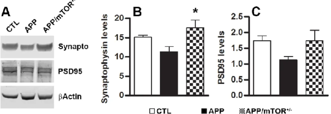
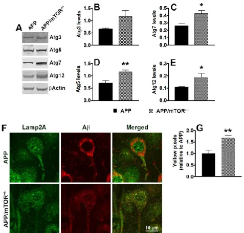
We have shown that pharmacologically reducing mTOR hyperactivity in the brains of the 3xTg-AD mice led to a reduction in soluble Aβ and tau levels (Caccamo et al., 2010b). The effects of rapamycin appear to be mediated by an increase in autophagy induction as we showed that in the brains of rapamycin-treated 3xTg-AD mice there was a significant increase in LC3II and other autophagy related proteins, including Atg5, Atg7 and Atg12 (Caccamo et al., 2010b). Supporting this view, autophagy induction correlated with the decrease in Aβ levels in another mouse model of AD (Spilman et al., 2010). Furthermore, in cell culture experiments, we directly showed that autophagy induction was necessary for the rapamycin-mediated decrease in Aβ levels (Caccamo et al., 2010b). A recent report by Paul Greengard’s group shows that inducing autophagy by small-molecule enhancer of rapamycin in immortalized cell lines and primary neurons led to an 80% reduction in Abeta40 and Abeta42 levels (Tian et al., 2011). These data were further supported by
31
pathology and the associated cognitive decline in a mouse model of AD (Yang et al., 2011). There is an apparent contradiction between the data reported by Nixon and colleagues, who showed that autophagic vacuoles accumulates in AD due to impaired clearance, suggesting that further increase in autophagy may exacerbate the pathology (Yu et al., 2005), and what we and others recently reported, increasing autophagy induction has beneficial effects on AD-like pathology in different animal model of AD (Caccamo et al., 2010b; Pickford et al., 2008; Spilman et al., 2010; Tian et al., 2011). Although the basis of this apparent inconsistency remains to be established, it is tempting to speculate that the relationship between autophagy and Aβ may change with the progression of the disease. At earlier stages of Aβ accumulation, induction of autophagy may facilitate its clearance. As the disease progresses, deficiencies in the clearance of autophagic vacuoles may occur and thus further increasing autophagy may exacerbate the AD phenotype. Indeed, the majority of the studies showing that increasing autophagy induction reduces Aβ and tau accumulation were done in animals with early stages pathology. Thus, when considering the role of mTOR in Aβ and tau pathology and the role of mTOR in autophagy induction, it will be important to determine the effect of increasing autophagy function in mice with established plaques and tangles.
32 2 AIM OF THE WORK
The lack of effective cures or treatments for Alzheimer’s disease (AD) is alarming considering the number of people currently affected by this disorder and the projected increase in incidence and prevalence over the next two decades. Aging is the major risk factor associated with the development of AD; thus, it is plausible that alterations in selective pathways associated with aging may facilitate the development of this insidious disorder (Moll et al., 2014). Overwhelming evidence shows that reducing the activity of the mammalian target of rapamycin (mTOR) increases lifespan and health-span in several genetically different species (Lamming et al., 2013). mTOR is a ubiquitously expressed protein kinase that plays a key role in regulating protein synthesis and cell growth by phosphorylating downstream proteins such as p70 S6 kinase and eukaryotic initiation factor eIF4E binding protein (4E-BP Wullschleger et al., 2006). mTOR also is a negative regulator of autophagy induction (Wullschleger et al., 2006). By simultaneously regulating protein synthesis and degradation, mTOR is key in controlling protein homeostasis, a process that is altered in AD and other proteinopathies (Lee et al., 2013). The Aim of this work is to fully dissect the role of mTOR in the pathogenesis of AD. Specifically, we will test the hypothesis that hyperactive mTOR further contributes to the buildup of Aβ and tau, thereby exacerbating cognitive decline. To test this hypothesis, we employed multidisciplinary approaches in order to identify the mechanistic links between mTOR signaling, Aβ and tau accumulation, and cognitive decline. Overall our results highlight the mTOR/S6K1 pathway as a previously unidentified signaling cascade that plays a key role in AD pathogenesis and offers a potential therapeutic target for this insidious disorder.
33 3 RESULTS
3.1 Genetic reduction of mTOR ameliorates Alzheimer’s disease-like cognitive and pathological deficits by restoring hippocampal gene expression signature
Alzheimer’s disease (AD) is the most common neurodegenerative disorder and the 6th leading
cause of death in the United States. Currently, it is estimated that 26 million people worldwide are living with AD and by 2050 the number of people with this disorder can reach 100 million (Thies et al., 2013). AD is characterized by progressive cognitive deficits associated with the build-up of amyloid-β (Aβ) and neurofibrillary tangles. The majority (>95%) of AD cases are sporadic and of unknown causes, while the remaining cases are caused by mutations in one of three genes, presenilin 1 and 2 and amyloid precursor protein (Querfurth and LaFerla, 2010). The single major risk factor for AD is aging; however, little is known as to how the aging process facilitates the development of AD. Nevertheless, molecular changes that occur in the brain as a function of age may facilitate the development of AD.
Overwhelming evidence shows that reducing the activity of the mammalian target of rapamycin (mTOR) increases lifespan and health-span in several genetically different species (Lamming et al., 2013). mTOR is a ubiquitously expressed protein kinase that plays a key role in regulating protein synthesis and cell growth by phosphorylating downstream proteins such as p70 S6 kinase and eukaryotic initiation factor eIF4E binding protein (4E-BP) (Wullschleger et al., 2006). mTOR also is a negative regulator of autophagy induction(Wullschleger et al., 2006). By simultaneously regulating protein synthesis and degradation, mTOR is key in controlling protein homeostasis, a process that is altered in AD and other proteinopathies (Lee et al., 2013). Another known function of mTOR signaling is the regulation of synaptic plasticity and function (Hoeffer and Klann, 2010;
34
Hoeffer et al., 2008; Santini et al., 2014; Tang et al., 2002). Toward this end, mTOR activity is necessary for memory consolidation (Parsons et al., 2006; Tang et al., 2002); however, mTOR hyperactivity is detrimental and causes cognitive deficits in animals and people (Ehninger, 2013; Ehninger et al., 2008; Puighermanal et al., 2009).
Several laboratories have shown that mTOR signaling is upregulated in AD patients and animal models (An et al., 2003; Caccamo et al., 2010b; Oddo, 2012). Thus, one might expect that lifestyle choices known to increase mTOR signaling, such as a high sugar or high fat diet increase the risk of developing AD. Toward this end, we and others have shown that diabetes increases the risk of developing AD by and mTOR-dependent mechanism (Ma et al., 2013b; Orr et al., 2013). In contrast, some of the beneficial effects of caloric restrictions on brain function might be mediated by reduction in mTOR signaling (Speakman and Mitchell, 2011). To fully dissect the role of mTOR in the pathogenesis of AD, we used a genetic approach and selectively ablated one mTOR gene from the forebrain of an animal model of AD. Our results highlight a previously unidentified signaling pathway as a key player in AD pathogenesis and offer a potential therapeutic target for this insidious disorder.
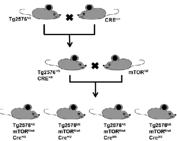
3.1.1 Removing one copy of the mTOR gene, decreases hippocampal mTOR signaling To study the role of mTOR in AD, we genetically and selectively removed one mTOR allele from the brains of the Tg2576 mice, a mouse model of AD (Hsiao et al., 1996). This was accomplished by taking advantage of the Cre-Lox site-specific recombination. Precisely, to obtain brain specific mTOR knockout mice, we first bred hemizygous Tg2576 mice (Tg2576+/0) with homozygous
transgenic mice overexpressing the Cre recombinase enzyme under the control of the neuronal specific CamKII promoter (CRE+/+). As expected ~50% of the offspring had the following
genotype: Tg2576+/0;Cre+/0 (Fig. 4). These mice were bred with homozygous mTOR floxed mice
35
genotypes: (1) Tg2576+/0;mTORfl/wt;Cre+/0; (2) Tg25760/0;mTORfl/wt;Cre+/0; (3)
Tg2576+/0;mTORfl/wt;Cre0/0; (4) Tg25760/0;mTORfl/wt;Cre0/0 (Fig. 4). Notably, all four groups of mice
are littermates, thereby reducing the genetic background effects on the phenotype. Using this breeding strategy, we removed a single copy of the mTOR gene in the forebrain of the Tg2576 mice. For simplicity, herein the mice with the Tg2576+/0;mTORfl/wt;Cre+/0 genotype are referred to
as APP/mTOR+/- mice, the mice with the Tg25760/0;mTORfl/wt;Cre+/0 genotype as mTOR+/- mice,
the mice with the Tg2576+/0;mTORfl/wt;Cre0/0 genotype as APP mice, and the mice with the
Tg25760/0;mTORfl/wt; Cre0/0 genotype as CTL mice.
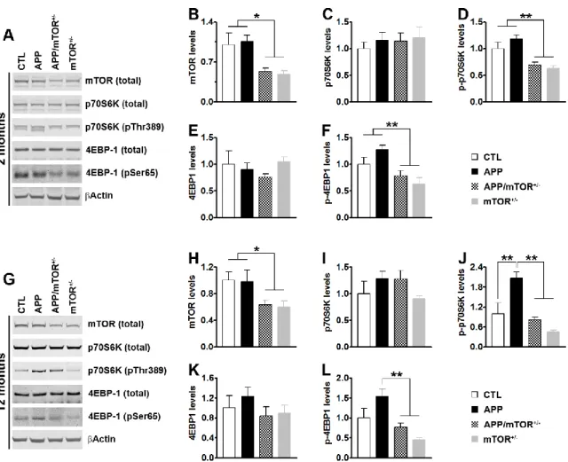
To determine whether removing one copy of the mTOR gene was sufficient to reduce brain mTOR levels and signaling, we measured mTOR levels by Western blots in the hippocampi of 2- and 12-month-old mice (n = 5/genotype/age). We found that in 2-12-month-old mice, mTOR levels were significantly different among the four genotypes (p = 0.01; Fig. 5A-B). Specifically, mTOR levels
Figure 4. Schematic representation of the breeding strategy used to remove one copy of the mTOR gene from the forebrain of the Tg2576 mice. Abbreviations: “+” indicates the presence of a
transgene (APP or CRE). “0” indicates the lack of such transgene. “fl” indicates the presence of a floxed allele. “wt” indicates the presence of a wild type allele.
36
were 47 ± 6% and 52 ± 6% lower in the APP/mTOR+/- and mTOR+/- mice compared to the CTL
mice, respectively. 4EBP-1 and p70S6K are two proteins downstream of mTOR, which directly phosphorylates them at Thr389 and Ser65; the levels of phosphorylated 4EBP-1 and p70S6K are used as an indication of activated mTOR signaling (Guertin and Sabatini, 2007; Hay and Sonenberg, 2004). In the hippocampi of 2-month-old mice, the total levels of p70S6K and 4EBP-1 were similar among the four groups. In contrast, we found that the levels of p70S6K phosphorylated at Thr389 and 4EBP-1 phosphorylated at Ser65 were significantly different among the four genotypes as indicated by one-way ANOVA (p < 0.001 for both; Fig. 5A-F). A
post hoc test with Bonferroni correction showed that the phosphorylation levels of p70S6K and
4EBP-1 were significantly reduced in the APP/mTOR+/- and mTOR+/- mice compared to CTL and
APP mice (Fig. 5D, F). To determine whether the changes in mTOR levels and signaling were maintained in older mice, we repeated these measurements in 12-month-old mice (n = 5/genotype). We found that mTOR levels were 37% and 40% lower in the hippocampi of the APP/mTOR+/- and mTOR+/- mice compared to CTL mice, respectively (Fig. 5G). While these
changes did not affect total p70S6K and 4EBP-1 (Fig. 5 G, I-K), one-way ANOVA indicated that p70S6K levels phosphorylated at Thr389 and 4EBP-1 levels phosphorylated at Ser65 were significantly different among the four groups (p = 0.0003 and 0.0029, respectively). A post hoc test with Bonferroni correction showed that phosphorylated levels of p70S6K were significantly higher in the APP mice compared to CTL mice (p < 0.01). However, the phosphorylated levels of p70S6K in the hippocampi of the APP/mTOR+/- were not statistically different than CTL mice
and were significantly reduced compared to the APP mice (p < 0.01; Fig. 5J). Similarly, the phosphorylated levels of 4EBP-1 were significantly reduced in the APP/mTOR+/- mice compared
37
mTOR gene in the brain is sufficient to reduce mTOR levels and signaling. Notably, this approach restored the elevated levels of phospho-p70S6K of the APP mice to CTL levels.
3.1.2 Reducing mTOR signaling rescues cognitive deficits in Tg2576 mice
Figure 5. Removing one copy of the mTOR gene decreases mTOR levels and signaling. (A)
Representative Western blots of proteins extracted from the hippocampi of 2-month-old mice probed with the indicated antibodies. (B-F) Quantification of the indicated proteins (n = 5 / genotype) showed that mTOR levels and the levels of phosphorylated p70S6K and 4EBP1 were significantly decreased in the hippocampi of mice lacking one copy of the mTOR gene. (G) Representative Western blots of proteins extracted from the hippocampi of 12-month-old mice probed with the indicated antibodies.
(H-L) Quantification of the indicated proteins (n = 5 /genotype) showed that even as the mice age,
mTOR levels and signaling were significantly lower in mice lacking one copy of the mTOR gene. Data were generated by normalizing the levels of the protein of interest to β-actin loading control. Results presented as means ± SEM and analyzed by one-way ANOVA with Bonferroni’s correction. * p < 0.05; ** p < 0.005 .
38
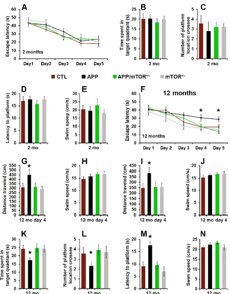
To evaluate the effects of removing one copy of the gene encoding mTOR from the brains of the Tg2576 mice, we tested 2- and 12-month-old mice in the spatial version of the Morris water maze (MWM), using two independent cohorts of mice. Mice received 4 training trials per day for 5 consecutive days to learn the location of a hidden platform using extra maze cues. Their performance was analyzed using a mixed-model, repeated-measures ANOVA, with genotype as the categorically fixed effects, days as the numeric covariate, animals as the random effect, and escape latency as the dependent variable. At two months of age, we found a significant effect for days (p < 0.0001), indicating that the mice learned the task across sessions (Fig. 6A). However, we found a non-significant genotype/day interaction (p > 0.05), indicating that there was no difference in the pace of learning among the four different groups (Fig. 6A). Twenty-four hours after the last training trial, we tested spatial memory by measuring the time mice spent in the target quadrant, the latency to reach the platform location, and the number of platform location crosses over a 60-second probe trial. We found no statistically significant changes in these three measurements among the four different genotypes (Fig. 6B-D). To determine whether the physical performance of the mice may have confounded these results, we measured their swim speed during the probe trials and found that it was not statistically different among the four genotypes (Fig. 6E). This is consistent with data in the literature showing that at this age, the Tg2576 mice are not cognitively impaired (Westerman et al., 2002).
As the Tg2576 mice age, they develop progressive cognitive deficits in several behavioral tests, including the MWM (Westerman et al., 2002). At 12 months of age, we found a significant effect for days (p < 0.0001) and genotype (p = 0.0009; Fig. 6F). The day effect indicates that the mice significantly improve their performance across sessions whereas the genotype effect indicates that one or more genotypes are different from each other. A post hoc test with Bonferroni correction showed that at day 4 and 5, the APP mice performed significantly worse than the other