Università degli Studi di Ferrara
DOTTORATO DI RICERCA IN
Farmacologia e Oncologia Molecolare
CICLO XXVI
COORDINATORE Prof. Antonio Cuneo
A
2A
adenosine receptor over-expression
correlates with motor symptoms in
Parkinson’s disease
Settore Scientifico Disciplinare BIO/14
Dottorando Tutore
Dott.ssa Carmen Corciulo Prof.ssa Katia Varani
SUMMARY
ABSTRACT
Pag. 2
List of abbreviation
Pag. 3
CHAPTER I- INTRODUCTION
Pag. 4
1. Adenosine signaling: the beginning
Pag. 5
2. Adenosine signaling: current knowledge
Pag. 7
3. Future: agonists and/or antagonists of adenosine
receptors in clinical practice
Pag. 18
4. Parkinson’s Disease
Pag. 19
CHAPTER II- AIM OF THE STUDY
Pag. 30
CHAPTER III – METHODS
Pag. 32
CHAPTER IV - RESULTS
Pag. 41
CHAPTER V - DISCUSSION AND CONCLUSIONS
Pag. 60
SUPPLEMENTS
Pag. 65
ABSTRACT
ABSTRACT
Adenosine receptors (ARs) are seven trans-membrane domain G-protein coupled receptors named A1, A2A, A2B and A3. Adenosine, the main agonist, acting on these
receptors exerts a broad spectrum of physiological and pathological functions. Adenosine production increases dramatically when there is a discrepancy between the rates of ATP synthesis and ATP utilization, for example when work load is markedly enhances or when the supply of oxygen and glucose is limiting as in ischemia. Adenosine receptors are expressed in all body tissues with different concentrations and functions. One of the most important role is explain in the regulation of inflammatory function, a field where A2AAR is an important player.
Parkinson’s disease (PD) is a pathology with a complex etiology, involving both genetic and environmental factors. The cardinal signs of PD relate to motor dysfunction, psychiatric and dysautonomic symptoms. PD is characterized by prominent loss of dopaminergic neurons in the substantia nigra pars compacta in relatively early stages of the disease, depletion of striatal dopamine, and the presence of intraneuronal inclusions called Lewy bodies. Current knowledge highlight the important role of inflammation in Parkinson’s disease. The central nervous system was supposed to be an immune privileged site, in which immune cells of the periphery could not enter or rarely entered. Today we know that peripheral immune responses can trigger inflammation and exacerbation of central nervous system degeneration in several neurodegenerative diseases. When cytokines, such as tumor necrosis factor (TNF)-α, are secreted by activated glia in the brain or are present in circulating blood, permeability of the blood brain barrier is increased.
The primary aim of this study was to investigate the expression, affinity and density of adenosine receptors in lymphocytes and neutrophils of PD patients compared to healthy subjects. This study revealed a specific A2AAR alteration correlating with disease
severity: patients with higher A2AAR density and lower affinity were more likely to
exhibit motor complications. An increase in A2AAR density in putamen patients was
found, an alteration that mirrors a similar up-regulation in human peripheral blood cells. Moreover, how expected, we measured high levels of adenosine and TNF-α in plasma of PD patients. Interesting we found out a statistically significant linear correlation among the A2AAR density and TNF-α levels. Elevated levels of TNF-α in PD brains amplify
and sustain the neuroinflammation leading to dopaminergic neurons destruction. Moreover, several studies highlight a close relation between TNF-α release and A2AAR.
To shed some light on the functional adenosine-dopamine interaction, we examined the effects of well-known A2AAR agonists and antagonists on dopamine uptake in the rat
adrenal pheochromocytoma cell line after differentiation into a neuronal phenotype by nerve growth factor. Our results show that A2AAR antagonists decreased dopamine
uptake, and an opposite effect was mediated by A2AAR agonists.
In conclusion our data prove the double importance of A2AAR in Parkinson’s disease: a
LIST OF ABBREVIATION
- ADP: adenosine di-phosphate - AMP: adenosine mono-phosphate - ATP: adenosine tri-phosphate
- CCPA: 2-Chloro-N6-cyclopentyladenosine
- CGS 21680: 4-[[[6-Amino-9-(N-ethyl-β-D-ribofuranuronamidosyl)-9H-purin 2-yl]amino]ethyl]benzenepropanoic acid hydrochloride
- CHO: chinese hamster ovary
- Cl-IB MECA: 2-Chloro-N6-(3-iodobenzyl)-adenosine-5′-N-methyluronamide - CNS: central nervous system
- COX: cyclooxygenase
- CPA: N6-cyclopentyladenosine
- CREB: cAMP response element-binding protein - DA: dopamine
- ENTPD: ectonucleoside triphosphate diphosphohydrolase - HIF: hypoxia inducible factor
- IB-MECA: 1-Deoxy-1-[6-[[(3-iodophenyl)methyl]amino]-9H-purin-9-yl]-N-methyl-β-D-ribofuranuronamide
- IL: interleukine - KO: knock out - LB: Lewy body - LPS: lypopolisaccaride - MAO: monoamino oxidase
- MPTP: 1-metil 4-fenil 1,2,3,6-tetraidro-piridina - NECA: 5'-N-ethylcarboxamido adenosine - NF-kB: nuclear factor kB
- NK cells: natural killer - NOS: nitrous oxide systems - PBS: phosphate buffered saline - PC12: pheochromocytoma cell
- PD81723: 2-Amino-4,5-dimethyl-3-thienyl)-[3-(trifluoromethyl) phenyl] methanone - PGE: prostaglandin
- PKA: protein kinase A - PLC: phospolipase C
- R-PIA: N6-(R-phenylisoprophyl) adenosine
- SCH 58261: 2-(2-Furanyl)-7-(2-phenylethyl)-7H-pyrazolo[4,3-e][1,2,4]triazolo [1,5-c]pyrimidin-5-amine
- Tgf: tumor growth factor - TNF: tumor necrosis factor
- VEGF: vascular endothelium growth factor - WT: wild type
- ZM 241385: 4-(2-[7-Amino-2-(2-furyl)[1,2,4]triazolo[2,3-a][1,3,5]triazin-5-ylamino]ethyl)phenol
CHAPTER I- INTRODUCTION
I
CHAPTER I
INDRODUCTION
1. Adenosine signaling: the beginning
Purines appear to be the most ancient chemical cellular messenger in animals and plants. The purinergic signaling system employs extracellular purines and pyrimidines as signaling molecules both released from living cells. These transmitters act upon target cells through activation of two classes of receptors, the metabotropic P1 receptors to adenosine, and nucleotide receptors of the P2 family, which is subdivided into P2Y metabotropic and P2X ionotropic sub-classes [1].
Adenosine is present ubiquitously and its receptors are widely distributed through the body exerting a broad spectrum of physiological and pathophysiological functions [2]. Data from techniques of molecular biology suggest that P2X ion channel receptors appeared early in evolution while G-protein coupled P1 and P2Y receptors were introduced at the same time or later [1].
The first evidence of adenosine effect date back to eighty years ago. In the University of Cambridge, A.N. Drury and A. Szent-Györgyi, begin their study of a unknown substance extract from bullock’s heart muscle. They chose the heart like study model because “the heart, therefore, is an admirable test object for any substance which may be suppose to influence cell activity”. The injection of this substance into a dog or guinea pig caused bradycardia. After exclusion of vagus nerve involvement and influence of inorganic component used in the protein extraction, they wrote “we are strongly of the opinion, as a result of these observations, than our substance is identical with adenylic acid, and we shall refer in this paper to the substance isolated by us as adenylic acid”. Adenosine prepared from yeast nucleic acid has an identical action to adenylic acid. The lower arterial pressure was due in part to the cardiac slowing and in part to a general arterial dilatation [3].
CHAPTER I- INTRODUCTION
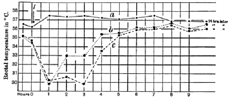
Two years later Bennet and Drury confirmed the previous experiment on the heart and came in light the effect of adenosine on others guinea pig body tissues: uterus constriction, local accumulation of leucocytes, considerable fall of temperature after subcutaneous injection [4].
Fig. 2 Influence of subcutaneous injection of adenosine upon rectal temperature of the guinea-pig. a) Injection of saline, b) and c) injection of 50 mg and 100 mg of adenosine [4].
Severity of trauma, magnitude of the loss of tissue adenine nucleotides and the release of breakdown products correlated with protective effects and vasodilatation, anticoagulatory effect, energy transfer suppose to be due to adenosine nucleotides enter cells. Bennet and Drury studied that a lack of energy owing to hypoxia caused the breakdown of myocardial adenine nucleotides able to cause coronary vasodilatation: increasing oxygen supply to an energy-depleted tissue, subsequently limiting its own formation like a classical tenet of homeostasis and negative feedback [5].
From other study on heart it was understood that methylxanthines acted as adenosine antagonists [6]. From 1960 synthesize of more stable analogues started yielding the adenosine analogue 2-chloroadenosine, R-PIA and NECA [7]. The different effects of these compounds away the idea of a therapeutic benefit but helping to define the subtypes of adenosine receptors (a concept become clear in 1970s).
For many years it was though that the only source of extracellular ATP acting on purinoceptors was damaged or dying cells. It is now recognized that ATP release from healty cells is a physiological mechanism in response to shear stress, stretch or osmotic swelling, hypoxia or stimulation by various agents [8].
The concept of purinergic signaling system was first proposed by Geoffrey Burnstock in 1970, when proposed ATP like a neurotransmitter [9]. In 1978 Burnstock made the important suggestion that there exist a family of receptors called purinergic receptors
that can be subgrouped into two subclasses, P1 and P2 [10]. This classification was based on the selective potency of ATP, ADP, AMP and adenosine, the effectiveness of methylxanthines as antagonists and mediation or not by adenylate cyclase [11]. This hypothesis met considerable resistance because ATP had been established as intracellular energy source and it was thought that such a ubiquitous molecule was unlikely to be involved in selective extracellular signaling [8]. The Purinergic
Hypothesis included the presence of purinoceptors and a few years later Burnstock
proposed a basis for distinguish P1 (adenosine) from P2 (ATP/ADP) receptors and in 1985 distinguished two type of P2 purinoceptor, P2X and P2Y [12]. In 1990s with studies of transduction mechanism and cloning of P2X and P2Y it was proposed that there were two families of P2 receptors, P2X ionotropic ligand-gated ion channel receptors and P2Y metabotropic G-protein-coupled receptors.
A classification into A1 and A2 adenosine receptor was proposed in 1979 [13] along
with the A2A and A2B nomenclature for the two cloned receptors showing considerable
sequence homology and similar signal transduction mechanism, distinguished by pharmacological criteria [14].
2. Adenosine signaling: current knowledge
ADENOSINE
Adenosine is the main agonist at the P1 receptor class and in addition the adenosine metabolite inosine can activate at least some of the receptors [15]. The concentration of adenosine in the extracellular compartment is the consequence of many biological processes, including extracellular adenosine production from intracellular sources, transport, and metabolism to inosine or AMP. ATP can be release from vescicles with a “kiss and run” mechanism (vescicles open and close transiently), from the lysosome by exocytosis or by an uncontrolled leakage from necrotic cells and inflammatory or vascular endothelial cell release.
Adenosine can increase dramatically from the basal level, estimate to be in the range of 30-200 nM. Very minor changes in steady state ATP levels in the cell will translate into major changes in the intracellular adenosine concentrations (100,000 times higher). The adenosine production from hydrolysis of adenosine nucleotides, from ATP or ADP
CHAPTER I- INTRODUCTION
ADP to AMP by ENTPD1 or CD39 followed by AMP hydrolysis to adenosine by ecto-5’-nucleotidase (NT5E or CD53) [16]. Adenosine generated intracellulary is transported into the extracellular space mainly via specific bi-directional transporters through facilitated diffusion. In some tissues there are nucleoside transporter protein capable of maintaining high adenosine concentration gradient: ENT1 and ENT2, CTN1 and CTN2 [17,18].
Adenosine is formed intracellulary whenever there is a discrepancy between the rates of ATP synthesis and ATP utilization, for example when work load is markedly enhanced or when the supply of oxygen and glucose is limiting as in ischemia. In ischemic areas of after a massive tissue trauma leading to cell death by necrosis can increase to perhaps 30 μM [19].
Fig.3 Adenosine synthesis, metabolism and transport in physiological condition [7].
The malfunction of adenosine metabolism is the cause of some serious pathologies. In human, absence of adenosine deaminase induces immunodeficiency (SCID-ADA). Patients with this pathology show skeletal [20], lung, liver [21] and neural abnormalities [22]. The absence of ADA induces increase of deoxyadenosine converted in deoxyadenosinetriphosphatase (dATP) by adenosine kinase. dATP causes lymphocytoxicity in thymus through induction of apoptosis in developing thymocytes [23].
ADENOSINE RECEPTORS
Forty years ago was postulated the existence of adenosine receptors, an hypothesis based on the competitive antagonist of adenosine activity by methylxanthines. Twenty years later four receptors were cloned from several mammalian species and identified as members of G protein-coupled family. These receptors show an high sequence homology among different species [24].
Fig.4 Tree graph showing adenosine receptors gene homology among different species [24].
A
1adenosine receptor
A1 adenosine receptor (A1AR) is the most conserved receptor subtype among species.
The gene, ADORA1, have chromosomal localization 1q32.1. It is expressed throughout the body with the highest levels found in the brain (cortex, hippocampus and cerebellum) especially at excitatory nerve endings, but also in heart, kidney, testis and adipose tissue. The A1AR has been shown to couple with Gi-1, Gi-2, Gi-3 and G0 but not
with Gs or Gz proteins. Responses to A1ARs activation are blocked by pertussis toxin
which is compatible with an involvement of the Gi/G0 family of G-proteins [14]. The
activation of A1R induces inhibition of adenylate cyclase, activates potassium channels,
block transiently calcium and IP3 levels by activating PLC. A model for this receptor
was proposed after studies based on sequence analysis and computer-assisted molecular modeling [25].
CHAPTER I- INTRODUCTION
Fig.5 Model of A1AR protein based on sequence analysis and computer-assisted molecular
modeling [14].
Chemical modification of histidine residues in the receptors transmembrane domain, one in helix VI and one in helix VII, strongly affects ligand binding. Glutamate residues in the second extracellular loop as being important for ligand recognition. From extensive mutagenesis analysis, consisting of single aminoacid replacement in human, canine and bovine species, other aminoacids are resulted very important in ligand recognition [24].
Biological functions of A1AR were studied in mice lacking of ADORA1 gene. This
animals showed a reduction of fertility, lifespan [26] and an increase of seizures risk [27]. A1AR in afferent arterioles mediate vasoconstriction and inhibition of renin
secretion and tubular A1AR appear to modify tubular NaCl absorption [28]. In brain
A1AR are down-regulated by hypoxia in glioma cells and up-regulates in human
temporal lobe in epilepsy. A deeply hypothermic and hypometabolic state was pharmacologically induced in a nonhibernating rat by A1AR agonist CHA [29]. In
murine astrocytes A1AR indirectly reduce the LPS-mediated HIF-1α accumulation, a
master regulator of oxygen homeostasis [30]. It is known that astrocytes regulate the reduction of depressive symptoms after sleep deprivation, an effect that seems to be mediate by A1AR: pharmacological activation of this receptor mimicked the effect of
sleep deprivation on depression phenotypes [31]. It has been already demonstrated that IL-6 can increase the survival of retinal ganglion cells in culture and this trophic effect is mediated by adenosine receptor A1AR activation [32].
Activation of A1AR in the central and peripheral nervous systems produces an
antinociceptive effect. The local application of CCPA in a pain model induced by a
tibial nerve injury inhibited thermal hyperalgesia, but was less effective against mechanical allodynia [33]. The analgesic effect during inflammation could be ascribes at the activation of opioid receptors [34].
Activation of the A1AR in adipocytes causes inhibition of lipolysis also in pathological
conditions like insulin resistance, diabetes and dyslipidemia [35].
From a study that compare WT and A1AR KO mice results that after administration of
an adenosine analogue, insulin induced lipogenesis in A1AR (+/+), but not in A1R (-/-)
adipocytes. Body weight was similar in young A1AR (+/+) and A1AR (-/-) mice, but old
male A1AR (-/-) mice were heavier than wild type controls. Plasma levels of free fatty
acids, glycerol and triglycerides were significantly lower in A1AR (+/+) than in A1AR
(-/-) [36]. After 8-16 h of CPA injection increase of 2-to 10-fold serum level of leptin, the appetite hormone [37].
A
2Aadenosine receptor
A2A adenosine receptor (A2AAR) gene is localized on 22q11.23 chromosome, have
multiple exons, which encode alternative transcripts, whose expression is driven by at least four independent promoters. The regulation of these promoters is highly responsive to alterations in the extracellular environment and particular sensitive to changes in the concentrations of exogenous and endogenous factors involved in inflammation [38]. This is a receptor coupled to Gs protein, and if over-expressed can interact with other G-proteins like Golf in striatum [39].
CHAPTER I- INTRODUCTION
Stimulation of A2AAR activates PKA that phosphorylates CREB on Ser 133. CREB
activation competed with NFkB-p65 for CBP mediating the anti-inflammatory effect [41]. A2AAR have independent G-protein action by C-terminus tail (120 aminoacids
highly conserved among species) interaction with other proteins. One of this protein is F-actin, cross linking protein that mediates the internalization of A2AARs after
stimulation contributing to receptor desensitization [42].
High levels of A2AAR are found in the striatum, immune cells of the spleen, thymus,
leukocytes and blood plates, but also, any less, in heart, lung and blood vessels [7], where is involved in many physiological and pathological process. A2AAR in the brain
interacts with several neurotransmitters to regulate motor activity, psychiatric behaviors, sleep-wake cycle and neuronal cell death. In peripheral tissue A2AAR have a crucial role
in the modulation of inflammation, myocardial oxygen consumption, coronary blood flow, angiogenesis and control of cancer pathogenesis [7]. In immune system adenosine binding A2AAR controls lymphocyte trafficking in response to tissue injury or infection
mediating extravasations of lymphocytes through blood vessels.
It was demonstrated that activation of A2AAR significantly suppresses the deposition of
collagen types I and III, reduces the infiltration of CD4+ T lymphocytes, and attenuates the expression of TGF-β1 and ROCK1, in a mouse model of renal interstitial fibrosis, a pathological process of chronic kidney diseases leading to renal function deterioration [43].
Adenosine regulates the function of the innate and adaptive immune systems mainly through A2AAR activation on monocytes/macrophages, dendritic cells, mast cells,
neutrophils, endothelial cells, eosinophils, epithelial cells, as well as lymphocytes, NK cells, and NKT cells [38].
In the heart A2AAR engagement protects against ischemia-reperfusion injury following
an acute ischemic episode and inducing profound oxidative/nitrosative stress, capillary pluging, edema and reduction in coronary vascular flow [44]. In the lung inosine acting on A2AARs can regulate ovalbumin induced allergic inflammation modulating
inflammatory processes and cytokines release [45].
In lung, deficiency of A2AAR increases activation of proinflammatory transcription
factor Nf-KB and augmented expression of inducible NO synthase [46]. Moreover adenosine by this receptor reduces bowel inflammation [47].
In the liver A2AAR is expressed on Kuppfer cells, hepatocytes and hepatic stellate cells.
injury [48] and ischemia reperfusion injury [49]. In the kidney A2AAR is expressed in
microvasculature [50] and his activation reduces injury following ischemia-reperfusion in rats [51].
Methotrexate, a therapeutic drug for rheumatoid arthritis, has immunosuppressive and anti-inflammatory effects mediated by increasing of adenosine levels, effect less effective in A2AAR KO mice [52]. In the joint A2AAR exert his anti-inflammatory
action in leukocytes, chondrocytes [53] and synovial cells [38]. A2AARs regulate also
the intrinsic circadian clock in immune cells playing an important role in circadian rhythms in rheumatoid arthritis [54]. It was investigated A2AAR density and
functionality in RA progression by using a longitudinal study in patients afflicted by rheumatoid arthritis before and after methotrexate, anti-TNFα agents or rituximab treatments: in lymphocytes obtained from RA patients, the A2AAR up-regulation was
gradually reduced in function of the treatment time. Moreover in adjuvant-induced arthritis model in rats it was showed the efficacy of the A2AAR agonist, CGS 21680, in
comparison with standard therapies [55].
Within brain, A2AAR expression levels are highly concentrated in dorsal and ventral
striatum (on striatopallidal) as well as in olfactory tubercle. It is also recognized that A2AARs are expressed at substantially lower levels outside striatum in brain regions
including hippocampus and cortex [56].
A2AAR is involved in sleep induction and it was demonstrate that caffeine induces
wakefulness in A1AR but not in A2AAR KO mice, demonstrating that this effect is
mediated by antagonism on A2AAR [57,58]. A genetic variant of the A2AAR gene in
humans is associated with individual sensitivity to effect of caffeine on sleep [59]. KO mice for A2AAR show a reduced explorative activity, higher anxiety, slow response
to acute pain stimuli and male are much more aggressive than WT mice [60].
A2AAR inactivation in the brain has been associated with protection against brain
damage after ischemia, excitotoxicity, traumatic injury and neurodegeneration in Parkinson and Alzheimer disease. Adult mice exposed to A2AAR antagonists including
caffeine during pregnancy and lactation displayed loss of hippocampal GABA neurons and some cognitive deficits demonstrating the important role of A2AAR on the neural
development [61].
Interestingly, while pharmacological blockade of A2AAR by antagonists induces motor
stimulation, adult gb-A2AAR KO mice consistently exhibit reduced spontaneous activity
CHAPTER I- INTRODUCTION
acute/short-term blockade of A2AARs by selective antagonists compared to complete
absence and/or long-term depletion of the receptor in these knockout models [63]. The involvement of A2AARs in neurodegenerative disease, where the inflammatory
component is considerable, was deeply investigated by our research group. Data from a study on Huntington’s disease patients indicate the existence of an aberrant A2AAR
phenotype in the peripheral blood cells of subjects carrying the HD mutation [64] and the alteration of A2AAR in lymphocytes reflects the presence of the mutant protein [65].
In R6/2 HD transgenic mouse model, it was seen a transient increase in A2AAR density
and A2AAR cAMP production at early presymptomatic ages and a decreasing A2AAR
mRNA, a discrepancy that suggests a compensatory mechanism [66]. A positive correlation was found between A2AAR density in lymphocytes of patients afflicted of
neurodegenerative diseases and gravity and progression of the pathology, like as Amyotrophic and Lateral Sclerosis, Multiple Sclerosis and Parkinson’s disease [67-69] indicating a possible protective effect of this receptor subtype.
A2AAR play a major role in coronary vasodilatatory properties of adenosine, and it is
expressed in coronary endothelial cells as well as in coronary smooth muscle cells. Along with vascular cells, A2AAR is expressed in additional cell types in the
myocardium [70]. Many studies show that optimal A2AAR agonist-mediated protection
occurs with low doses infused for at least 1 hour. Higher concentrations are associated with significant hypotension and reflex tachycardia and could also induce coronary steal [71,72].
A
2Badenosine receptor
A2B adenosine receptor (A2BAR) is codificated by ADORA2B gene with chromosomic
localization 17p12-p11.2 and contains a single intron that interrupt the coding region [73].
A2BARs are expressed on various cells of hematopoietic origin: neutrophils [74],
lymphocytes [75] and even platelets [76].
A2BAR is rarely achieved under physiological conditions because requires micromolar
adenosine concentrations. Nevertheless Adora2B-KO mice have very strong phenotypes, show low-grade inflammation [77] and vascular leakiness in several organs [78]. During conditions in which adenosine levels are elevated, such as hypoxia, ischaemia or inflammation, A2BAR is involved in tissue adaptation to hypoxia,
identified a strong dependence of A2BAR by HIF-1α: binding of HIF to a hypoxia
responsive element on the promoter resulting in increased of A2BAR mRNA and protein
[79].
Inflammatory mediators associated with inflammation have been demonstrated to increase A2BAR gene transcription, like bacterial lipopolysaccharide, TNF-α and IL-1β
[80]. Other inflammatory mediator including PGE2, IL-6 and IL-4 are not associated with an increase in transcription but rather with stabilization of A2BAR mRNA during
inflammation.
A2BAR couples to Gs to activate adenyl cyclase and increase intracellular cAMP levels
[81] and can couple to the Gq family of G-proteins to activate PLC and increase intracellular calcium [82]. It appears the G protein coupling of the receptor is cell type dependent and coupling to Gs results in an anti-inflammatory response while coupling to Gq could potentially results in a pro-inflammatory response [80].
Activation of the A2BAR has been shown to promote bone cell differentiation, control
glucose homeostasis [83,84] and regulate hyperlipidaemia and atherosclerosis [85]. Stimulation of A2BAR protects against trauma-hemorrhagic shock-induced lung injury
[86].
A2BAR promote tumor cell growth by VEGF in Lewis lung carcinoma A2BKO mice:
these mice exhibited significantly attenuated tumor growth and longer survival time compared to WT controls [87].
Pharmacologic activation of A2BAR results in proinflammatory effects relevant to the
progression of asthma but genetic removal of A2BARs leads to exaggerate responses in
models of acute inflammation. A2BAR gene ablation in ovalbumin-induced chronic
pulmonary inflammation attenuates allergen-induced chronic pulmonary inflammation [88].
In a mouse model of COPD it was demonstrated that the blockade of ADORA2B is able to attenuate the development of a pulmonary hypertension phenotype that correlates with reduced levels of hyaluronan deposition in the vessels and down regulation of genes involved in the synthesis of hyaluronan [89].
Conversely, A2BARs were proposed to play an inhibitory role in degranulation of mouse
bone marrow derived mast cells (BMMCs), based on the finding that A2BKO mice show
an exaggerated antigen-induced mast cell degranulation [90]. Studies in HMC-1 cells showed that only A2BAR, but not A2AARs or A3ARs stimulate secretion of angiogenic
CHAPTER I- INTRODUCTION
A2BARs expression has been detected in vascular endothelium and smooth muscle cells
where it has been implicated in the regulation of vascular tone through receptor-mediated vaso-dilatory effects counteracting A1AR-mediated vasoconstriction [95].
Due to their large surface area, mucosal organs are particularly prone to hypoxia and ischemia associated inflammation then adenosine via the A2BARs dampens mucosal
inflammation and tissue injury during intestinal ischemia or colitis [96], and A2BAR KO
mice show a reduction in induced colonic inflammation compared with their wild-type counterparts [97].
A
3adenosine receptor
The human A3 adenosine receptor (A3AR) was cloned in 1993 from a striatal cDNA
library. There is a considerable variation in the pharmacology and distribution, and hence function, of A3ARs among species. The protein, of 318 amino acids, exhibits
almost 30% difference at the amino acid level between human and rat [7].
KO of the A3AR in mice was surprising, resulting in marked phenotypes even at
loca-tions where the receptors are very sparse.
The phenotype of mice that lack the A3AR shows a marked diurnal variation in activity,
heart rate and reduction of body temperature [98].
A3AR represents a good therapeutic target for tumoral pathologies because is
overexpressed in cancer and in inflammatory cells, while low expression is found in normal cells. An overexpression was found in different neoplastic cells including leukemia, lymphoma, astrocytoma, melanoma, mesothelioma and pineal tumor cells [99].
Interesting study were made about the increase expression of A3AR in tumor tissues
derived from patients with colon, breast, small cell lung, pancreatic and hepatocellular carcinomas and melanoma in comparison with adjacent normal cells [100]. In breast cancer tissue A3AR mRNA increase of 1.27-fold respect to normal tissues [101], and in
patients with colorectal adenocarcinomas A3AR expression was higher in cancer tissue
compared with normal mucosa from the same individuals, moreover the expression of these receptors in peripheral blood cells was approximately 3-fold higher compared with healthy subjects [102].
The pharmacological stimulation of A3ARs with agonist IB-MECA resulted in a
inhibition of androgen-independent PC-3 prostate human carcinoma cells by deregulation of Wnt and NF-kappa B signaling pathways [104].
A3AR activation with the specific agonist CF101 inhibits the development of cancer
growth in a murine model of colon carcinoma via modulation of GSK-3 β and NF-kB [105].
An A3AR over expression was described in human and experimental animal model of
inflammatory disease. A3ARs reduce the LPS-mediated HIF-1α accumulation in murine
astrocytes, resulting in a downregulation of genes involved in inflammation and hypoxic injury [30]. In a murine model of ischemia-reperfusion activation of A3AR by
Cl-IB-MECA attenuates lung dysfunction, inflammation, and neutrophil activation and chemotaxis [106]. In some case disrupting neutrophils function could adversely affect innate immune responses conferring to A3AR a pro-inflammatory role [107]. In a mice
model of acute pancreatitis, administration of IB-MECA attenuate the histological parameters of inflammation [108]. In patients with rheumatoid arthritis, lymphocytes show an increase of A3ARs in comparison with healthy controls and activation of these
receptors inhibit NF-kB pathway and diminished inflammatory cytokines such as TNF-α, IL-1β and IL-6. Moreover A3AR density inversely correlated with clinical
parameters, DAS28 and DAS, suggesting a direct role of the endogenous activation of these receptors in the control of RA joint inflammation [109]. In an experimental model of autoimmune uveitis, CF101 inhibiting pro-inflammatoy cytokines production and apoptosis of inflammatory cells, improved uveitis clinical score [110].
A3AR could be considered a biological marker because levels of A3AR in PBMCs
mirror the receptor expression levels in tumor or inflammatory tissue: infact, it is known that pro-inflammatory cytokines, especially TNF-α, binding their receptors, initiate downstream signaling that result in an upregulation of transcription factors, like NF-kB, that bind A3AR promoter region [99]. In vitro and in vivo model of hepatocellular
carcinoma, CF102 treatment decreases the expression level of NF-kB, and TNF-α and prevented apoptosis in the liver with a decreased expression levels of pro-apoptotic proteins Bax and Bad. In addition, CF102-induced apoptosis of Hep-3B cells via de-regulation of the PI3K-NF-κB signaling pathway, resulting in up-de-regulation of pro-apoptotic proteins [111]. Normally, adenosine modulates colonic cholinergic motility via activation of A3ARs in the myenteric plexus. A3AR-mediated tonic inhibitory
control by adenosine was impaired in inflamed bowel, despite increased density of functioning and pharmacologically recruitable A3ARs [112].
CHAPTER I- INTRODUCTION
3. FUTURE:
AGONISTS
AND/OR
ANTAGONISTS
OF
ADENOSINE RECEPTORS IN CLINICAL PRACTICE
Due to the involment of adenosine in numerous pathological processes (Fig. 7), the importance of its therapeutically targeting is clear. In the beginning, because the complexity of their signaling, adenosine agonists or antagonists cannot be delivered in a manner that is clinically effective and safe [7]. Actually adenosine molecule named Adenocard was approved by Food and Drug Administration (FDA) in 1989 for the treatment of paroxysmal supraventricular tachycardia restoring normal sinus rhythm in patients. Adenoscan was approved by FDA in 1995, indicated as an adjunct to thallium-201 myocardial perfusion scintigraphy in patients unable to exercise adequately for its vasodilatory effect (FDA web site).
In 2008 Regadenoson (Lexiscan, Astellas Pharma), an A2AAR agonist is approved by
FDA for myocardial perfusion imaging in patients with suspected coronary disease.
4.
5.
6.
7.
8.
9.
10.
11.
12.
13.
4. PARKINSON DISEASE
Parkinson’s disease (PD) is a pathology with a complex etiology, involving both genetic and environmental factors.
Currently its definition is not so different from the first description made by James Parkinson:
“Involuntary tremulous motion, with lessened muscular power, in parts not in action and even
when supported; with a propensity to bend the trunk forwards, and to pass from a walking to a
running pace: the senses and intellects being uninjured” (J.Parkinson, 1817).
The cardinal signs of PD relate to motor dysfunction and include resting tremor, bradykinesia, rigidity and postural reflex impairment. Other manifestations include psychiatric symptoms such as anxiety and depression and dysautonomic symptoms such as hypotension and constipation, paresthesias, cramps, olfactory dysfunction, seborrheic dermatitis and increase of cognitive deficit with the disease progression.
The main pathological characteristic associated with the motor deficits of PD is degeneration of the dopaminergic neurons of the pars compacta of the substantia nigra (SNpc) leading to loss of dopamine in the striatum. The loss of these neurons, which normally contains neuromelanin, produces the classic SNpc depigmentation [114]. Symptoms do not develop until about 50–60% of the nigral neurons are lost and 80– 85% of the dopamine content of the striatum is depleted.
The combination of asymmetry of symptoms and signs, the presence of a resting tremor with the classic 4-to-6-Hz frequency, and a good response to levodopa best differentiate PD from parkinsonism due to other causes.
PD is characterized by the progressive death of selected but heterogeneous populations of neurons, including the neuromelanin-laden dopaminergic neurons of SNpc, selected dopaminergic brain-stem nuclei (both catecholaminergic and serotoninergic), the cholinergic nucleus basalis of Meynert, hypothalamic neurons, and small cortical neurons (particularly in the cingulate gyrus and entorhinal cortex), as well as the olfactory bulb, sympathetic ganglia, and parasympathetic neurons in the gut [115].
CHAPTER I- INTRODUCTION
The Lewy Body (LB) is an eosinophilic hyaline inclusion consistently observed in selectively vulnerable neuronal populations. LBs in the brain stem and basal forebrain are usually more than 15 μm in diameter, with a spherical, dense hyaline core, a clear halo.
The LB contains a variety of other constituents, elements of LB filaments, proteins that represent a cellular response to LB formation, enzymes such as phosphatases and kinases, and other cytosolic proteins that probably become trapped in LB during their formation [116]. Elevation of iron levels detected in the pars compacta of the substantia nigra in patients with PD is believed to be an important factor in causing oxidative stress [117]. The metabolism of endogenous dopamine may also produce a number of toxic bioproducts that could contribute to the heightened state of oxidative stress in PD [118].
PD is a complex network disorder in which abnormal activity in groups of neurons in the basal ganglia strongly affect the excitability, oscillatory activity, synchrony and sensory responses of areas of the cerebral cortex that are involved in the planning and execution of movements.
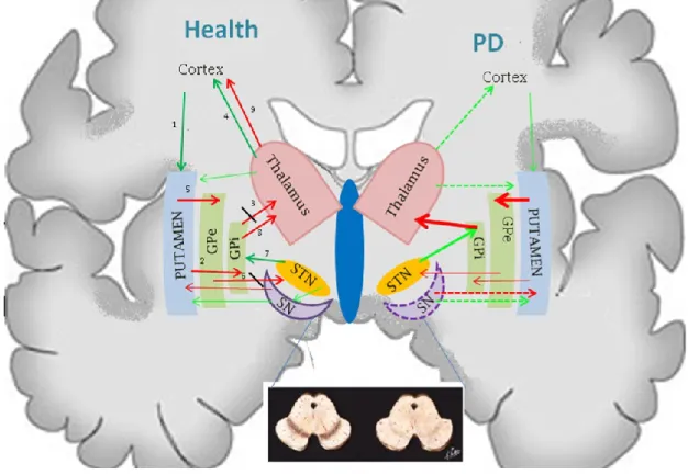
In the basal ganglia function the stimulation of the direct and indirect striatal pathways provokes motor activation and motor inhibition. In PD the equilibrium of the basal ganglia circuits is last due to the depletion in striatal dopamine caused by degeneration of SN dopaminergic neurons [119]. Direct pathway is a monosynaptic inhibitory pathway that is mediated by GABA, whereas the indirect pathway is a polysynaptic net excitatory pathway that includes the Globus pallidum (GP) and STN. Input to the striatal neurons comes from cortex and thalamus. Increased activity of the direct pathway results in increased movement via an inhibitory effect on thalamocortical projection neurons, whereas increased activity of the indirect pathway results in the opposite effect. Dopamine receptors in the motor circuit can be either excitatory (D1) or
inhibitory (D2). The release of striatal dopamine decrease GPi and SNr activity via
stimulation of the direct pathway (D1 receptors in the SNc) and inhibition of the indirect
pathway (D2 receptors). Inhibition of dopamine release has the opposite effect [120].
DA deficiency causes overactivity of the indirect pathway, resulting in an excessive glutamatergic stimulation drive to the internal segment of the GP and pars reticulata of the SN and reduced activity of the inhibitory GABAergic direct pathway, further disinhibiting the activity of the internal segment of the GP and of the SNr. Because these structures use the inhibitory neurotransmitter GABA, the increased output of the
basal ganglia leads to excessive inhibition and, effectively, to a shutdown of the thalamic and brainstem nuclei that receive their outflow (Fig.8). The excessive thalamic inhibition leads to suppression of the cortical motor system, possibly resulting in akinesia, rigidity, and tremor, whereas the inhibitory descending projection to brain-stem locomotor areas may contribute to abnormalities of gait and posture [115].
Fig. 8 - Basal ganglia neurotransmission in health and Parkinson’s Disease. Numbers (from 1 to 9) indicate the neuro-stimulation/inhibition motor control pathway (red arrow: inhibition of neurotransmission; green arrow: stimulation of neurotransmission; dotted arrow: impairment of the neurotransmission).
CHAPTER I- INTRODUCTION
EPIDEMIOLOGICAL INFORMATIONS
PD is a very common disease with a decline in incidence in older age groups which may be the result of difficulties in assigning a PD diagnosis in patients with extensive co-morbidities.
Incidence rates for PD in studies that reported results for all age groups ranged between 1.5 and 22 per 100,000 person-years; studies restricted to older populations (above 55 or 65 years) reported overall incidence rates between 410 and 529 per 100,000 person-years. Variations across ethnic groups occur maybe because different environmental exposures and different susceptibility genes in own genetic pattern. In Europe incidence rates is between 9 and 22 per 100,000 person-years, with a rate of 410 and 529 per 100,000 person-years in studies based on populations above 55 or 65 years. In North American studies incidence rates ranged between 11 and 13 per 100,000 person-years, with an incidence rate of 224 per 100,000 person-years in individuals 65 years or above. In Asia incidence rates is between 1.5 and 17 per 100,000 person-years.
The incidence of PD seems to be higher in men than in women whit a ratio of 1.49 and this suggested possible protective effect of estrogens, higher frequency of intensity of occupational toxin exposure and recessive susceptibility genes on the X chromosome. Gender difference decreased with increasing age, as men had higher mortality rates than women.
Overall prevalence ranged between 31 and 970 per 100,000. In US and Europe PD prevalence among people 65 years or older it was estimated at 950 per 100,000. Another study estimated the number of individuals above age 50 with PD in the world at between 4.1 and 4.6 million in 2005. By year 2030 the number was projected to more than double to between 8.7 and 9.3 million [121].
RISK FACTORS
As with many neurodegenerative diseases, age appears to be a clear risk factor for PD: both men and women have an increasing risk of PD with increasing age.
Men have a higher incidence rate of PD than women and one interpretation is that there are potentially protective effects of estrogen because there is some evidence that women with PD were more likely to have undergone a hysterectomy or oophorectomy than those who did not, develop PD. Testosterone could have harmful effects on the development of PD, could be a recessive susceptibility gene on chromosome X, or male lifestyle could predispose to this pathology [122].
Genetic mutations in the α-Syn gene [123] (SNCA) are rare and include point mutations and whole-locus multiplications. Duplications are detected in approximately 1% to 2% of the PD families, and these patients often display a classical PD phenotype; triplications and point mutations are exceedingly rare, found in a few families of Greek, German and Spanish origin and show more severe phenotypes [124,125].
Mutations in leucine-rich repeat kinase 2 (LRRK2) are the most common known cause of autosomal dominant PD. LRRK2 mutations explain approximately 10% of patients and the most common mutation is Gly2019Ser. This mutation is frequent in some populations from South Europe, but very common among Arab patients from North Africa and among Ashkenazi Jewish patients [126].
The most recently described gene involved in autosomal dominant PD is VPS35 within affected members of a Swiss kindred with late-onset, autosomal dominant PD.
Mutation in PRKN, PINK1, and DJ-1 genes cause autosomal-recessive forms of PD [127,128]. Mutations in ATP13A2 (PARK9), PLA2G6 (PARK14), and FBXO7 (PARK15), cause more-rare forms of recessive parkinsonism, usually with very early onset (<30 years) and atypical clinical features [129].
PD is associated with rural living or farming and numerous epidemiological studies have suggested that pesticide or herbicides exposure is closely related to the development of PD, with a risk increased of 1.8-fold (pesticide 1.6-fold and to herbicides 1.4-fold) [130]. No association was observed with fungicides, rodenticides, organochlorines, and organophosphates but 2-fold increase in risk for exposure to paraquat [131].
MPTP, a meperidine analogue occasionally used accidentally by heroin addicts, is a potent neurotoxin with selective effects on nigral dopaminergic neurons. MPTP toxicity is due to the inhibition of complex I (NADH–ubiquinone oxidoreductase) of the mitochondrial electron-transport chain, leading to energy failure and cell death.
INFLAMMATORY COMPONENT IN PD
The central nervous system was supposed to be an “immune privileged” site, in which immune cells of the periphery could not enter or rarely entered, and thus the two systems had little to no interaction.
Today we know that peripheral immune responses can trigger inflammation and exacerbation of CNS degeneration in several neurodegenerative diseases, increasing
CHAPTER I- INTRODUCTION
neuronal vulnerability to other neurodegenerative insults, such as those caused by aging, oxidative stress, environmental toxins or genetic predisposition (fig.9) [132].
Fig.9- Peripheral inflammatory reactions can also initiate neurodegenerative changes that are often associated with the preclinical phase of many neurodegenerative disease [132].
The cause of the neuronal loss in PD is poorly understood but neuroinflammatory mechanism might contribute to the cascade of events leading to neuronal degeneration. This mechanism comprise microglial activation, astrogliosis, and lymphocytic infiltration. Inflammation in PD is not merely a consequence of neuronal degeneration but may be involved in its progression by producing deleterious proinflammatory molecules [133]. Data from post-mortem studies revealed the presence of activated microglia cells within the substantia nigra of PD patients [134]. The density of astrocytes in SNpc is lower in post mortem brain patients than in healthy individuals, this implies few surrounding cells witch detoxify oxygen free radicals [135]. In addition some peripheral cells can enter the brain parenchyma during the neurodegenerative process because changes in blood brain barrier function might occur in the brain of patients with PD, in fact was found an increase density of endothelial cells and change in brain capillaries in the SN of patients was found, maybe like a consequence of reactive species released by activate microglia [136]. In striatum of post mortem brain
of patients it was found an increase of TNF-α, TGF-β1, IL-1β, IL-6 and IL-2. Furthermore dopaminergic neurons express receptors for these cytokines suggesting that they are sensitive to them which in this way can exert toxic effects [133]. Proinflammatory cytokines can indirectly induce a toxic effect leading expression of the inducible form of NOS or COX- 2 [137]. In body fluids of PD patients there is an increase of active T-cells, CD4+ bright CD8+ dull lymphocytes usually increase after a viral infection, a condition that might contribute to the pathogenesis of the disease [138]. Other studies, supporting the inflammatory role in PD progression, show that men with high levels of IL-6 in blood have increased risk to develop PD. However the risk of this disorder was lower in people who regularly took non-steroidal anti-inflammatory drugs [139].
A recent study provides evidence that enteric inflammation, occurring in PD patients, reinforce the role of peripheral inflammation in the initiation and/or the progression of the disease [140].
LRRK2 is a gene related to Parkinson’s disease susceptibility and also to Crohn’s disease and numerous scientist support the hypothesis that the detrimental effects mediated by Parkinson’s disease LRRK2 mutations may initiate in the periphery and extend to the central nervous system as a consequence of increased levels of pro-inflammatory factors permeable to the blood brain barrier [141].
MANAGEMENT OF PD
The management of PD includes symptomatic and regenerative treatments. Because of the link to dopamine depletion, treatment of PD has centered on various dopamine replacement strategies including dopamine precursor, dopamine agonist and agents that inhibit the metabolism of dopamine.
MAO-B inhibitors were developed as a means to increase synaptic dopamine levels and half-life. Two MAO-B inhibitors, selegilin and rasagiline, are currently licensed in Europe and North America for the symptomatic improvement of early Parkinson’s disease. A third MAO-B inhibitor (safinamide) is currently under development in phase III clinical trials as adjuvant therapy to either a dopamine agonist or levodopa [142].
Levodopa/Carbidopa, respectively, Levodopa/Benserazide are the most effective treatment for PD. Levodopa induces complications such as dyskinesias motor fluctuations: “wearing-off ” and “on–off ” phenomena.
CHAPTER I- INTRODUCTION
Motor fluctuations occur in 50 percent of patients after 5 years of levodopa therapy and the proportion affected increases to 70 percent among those treated for more than 15 years. Wearing off can be defined as a perception of loss of mobility or dexterity, usually taking place gradually over a period of minutes (up to an hour) and usually having a close temporal relation to the timing of antiparkinsonian medications. On–off effects are unpredictable and generally sudden occurrences (lasting seconds to minutes) of shifts between on and off periods that are not apparently related to the timing of antiparkinsonian medication. These off periods last minutes to hours and do not include transient episodes of “freezing” (also referred to as “motor blocks”; the initiation or continuation of a motor act such as walking is arrested for few seconds) or stress-induced tremor, which are both components of the underlying disease and occur even in the absence of treatment. Once fluctuations and dyskinesia emerge, the pharmacodynamic response changes, resulting in a narrowing of the “therapeutic window” and a specific levodopa threshold is needed for a sufficient clinical response [143].
Diagnoses
McGhee and colleagues summarized the main characteristics of a good biomarker for PD: his change have to follow the neurodegeneration, have to show an association with the clinical phenotype, have a direct association with the disease progression and have not be influenced by symptomatic treatment, be sensitive reflecting small changes in disease progression, quick and cheap to measure, safe and tolerable for the patients, suitable for measurement across different centers. They analyzed numerous scientific work on serum/plasma/blood biomarkers, brain SPECT, brain PET, CSF, brain MRI, concluding that they found insufficient evidence to recommend the use of any biomarker for disease progression in PD [144].
Diagnoses in PD is based on medical history of patients, evaluation of signs and symptoms, neurological and physical examination. Imaging test includes single proton emission computed tomography (SPECT), sonography, positron emission tomography (PET), functional magnetic resonance imaging (fMRI) [145]. In 2011 the FDA approved a specialized imaging technique called DaTscan that allows doctors to capture detailed pictures of the dopamine system in brain. Unfortunately, because there is no definitive test for PD, and because these disease symptoms are similar to those of other neurological conditions, the misdiagnosis rate remains significant. PET and SPECT
imaging use a number of radiotracers for in vivo assessment of normal and abnormal brain function. Cerebral blood flow and glucose can be mapped with radiolabeled water or glucose. SPECT imaging is less expensive and more available than PET that has a superior spatial resolution and higher sensitivity. fMRI is available, don’t require the injection of a radiotracer [146].
The Unified Parkinson's Disease Rating Scale (UPDRS) is a scale that was developed as an effort to incorporate elements from existing scales to provide a comprehensive means to monitor PD, related disability and impairment. The scale has four components, largely derived from preexisting scales: part I-Mentation, Behaviour and Mood; part II-Activities of daily living; Part III-Motor; Part IV-complications. UPDRS is often accompanied with Hoehn and Yahr scale [147]. Hoehn and Yahr scale analyzed the motor decline progression, deterioration in quality of life, neuroimaging studies of dopaminergic loss and it is focused on issues of unilateral versus bilateral disease [148]. The “mini mental state examination” (MMSE) is useful for the evaluation of the cognitive deficit and it is named “mini” because excludes questions concerning mood and abnormal mental experience [149]. Usually for the PD diagnosis are used also the Geriatric Depression Scale (GDS) and the Frontal Assessment Battery: the first test represent a reliable and valid self-rating depression screening scale for elderly populations [150] and the second is a bedside cognitive and behavioral battery to assess frontal lobe functions [151].
Evidences of A2AAR useful in Parkinson’s disease management
In recent years much research was focused on identifying non dopaminegic compounds to be used as adjuvants to L-DOPA therapy. Among several new classes of drugs, A2AAR antagonists have emerged as the most promising candidates [119]. From a 30
years study emerged that incidence of PD decline consistently with increased amount of coffee intake, from 10.4 per 10000 person-years in men who drank no coffee to 1.9 per 10000 person years in men who drank at least 28 oz/day: caffeine, an adenosine receptors antagonist, intake is associated with a significantly lower incidence of PD [152].
Patients with PD have altered adenosine A2AAR expression in the basal ganglia,
suggesting a pathogenic role for these receptor. A2AAR mRNA levels (+129%) and
CHAPTER I- INTRODUCTION
patients compared to controls. This increase was also significant compared with dyskinetic PD patient (+60% and +24% for mRNA and [3H]-SCH58261 specific binding respectively). These data suggest that A2AAR increase is associated with the
development of dyskinesias [153].
The deletion of the A2AAR in mice protects against dopaminergic neuron degeneration
induced by a mutant human α-synuclein transgene. The A2AAR KO completely prevents
loss of dopamine and dopaminergic neurons suggesting that A2AARs appear required
for neurotoxicity in a mutant α-synuclein model of PD [154].
The A2AAR is largely coexpressed with D2DR in the striatum where modulates
dopaminergic activity and antagonizes D2DR mediates neurochemical effects by direct
A2AAR-D2DR interaction. The role of this interaction was analyzed by using mice
genetically deficient in A2AARs or D2DRs or both. In D2 KO and wild-type mice the
A2AAR agonist reduced spontaneous and amphetamine-induced locomotion, instead in
the same mice A2AAR antagonist produce motor stimulation [155].
Neurochemical studies have shown that activation of A2AARs reduces the binding
affinity of D2 agonist to their receptors [156].
By immunofluorescence experiments was demonstrated the co-localization of A2AAR
and D2DR in cell membranes of SH-SY5Y human neuroblastoma cell stably transfected
with human D2DR and in cultured striatal cells (figure 10). Furthermore long term
exposure to A2AAR and D2DR agonists in D2DR cotransfected neuroblastoma cells
resulted in coaggregation, cointernalization and codesensitization of A2AAR and D2DR
[157].
Fig.10- Double immunofluorescence staining in SH-SY5Y neuroblastoma cells stably transfected with the human D2DR. a) A2AAR immunoreactivity (green); b) D2DR
immunoreactivity (red); c) superimposition of images revealing the colocalization of A2AAR and D2DR (yellow) [157].
A2AAR and D2DR have reciprocal antagonistic interactions that regulate GABAergic
neurons. A2AAR activation in the indirect pathway opposes the action of D2DR
activation by means of an intramembrane interaction, whereas stimulation of D2DRs
GABA release whereas D2DR stimulation inhibits this process. Inhibition of A2AAR and
D2DR produces opposite effects [120].
The antiparkinsonian effect of A2AAR antagonist was well studied in different PD
animal models. In 6-OHDA lesioned rats rendered dyskynetic by prior treatment with levodopa, A2AAR antagonist produce and additive reduction in motor disability with
levodopa [158]. In MPTP treated common marmosets, oral administration of istradefylline increased locomotor activity [159]. In levodopa primed MPTP treated cynomologous monkeys, istradefylline significantly improved locomotor function, comparable to that observed with levodopa (50 mg) but with little or no dyskinesia [160].
In human a potent and selective A2AAR antagonist, preladenant, was tested on 253
patients of 15 countries. This study revealed that 5 and 10 mg of preladenant twice daily (fig. 11) might be clinically useful to reduce off time in patients with PD and motor fluctuation [161].
Fig.11- Preladenant administration: mean change in daily off and on time from baseline to week 12. *p=0·024 versus placebo. †p=0·0489 versus placebo. ‡p=0·0486 versus placebo. §p=0·019 versus placebo[161].
CHAPTER II- AIM OF THE STUDY
CHAPTER II
AIM OF THE STUDY
AIM OF THE STUDY
PD is a neurodegenerative disorder with a strong inflammatory component. Increasing evidences suggest a direct involvement of adenosine in PD and its key role in the regulation of neurotransmission. For example epidemiological studies suggest that higher coffee and caffeine (unspecific antagonist for adenosine receptors) assumption, is associated with a significantly lower incidence of PD [162]. A2A is the main adenosine
receptor involved in this disorder. In the brain is localized prevalently in the striatum and it is co-expressed with D2 dopamine receptors: here exerts an antagonist effect on
D2DR and if up-regulated induces motor impairment.
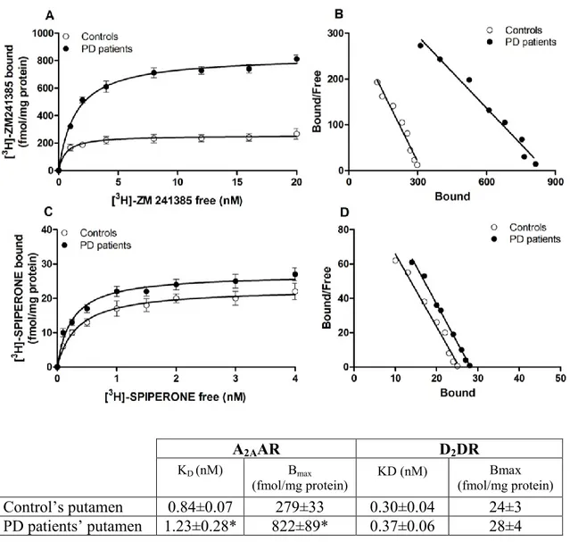
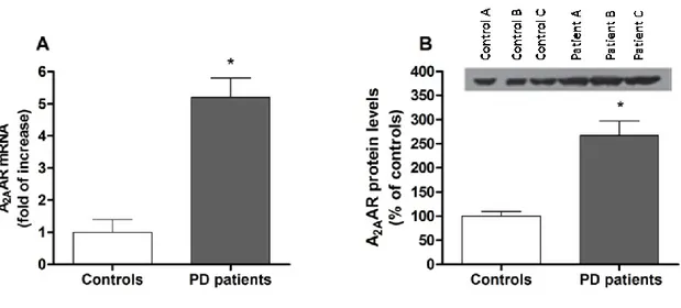
The aim of this study was to investigate the presence and a possible density and functional alteration of A2AARs and D2DRs in post mortem putamen of PD patients
compared to control subjects. Furthermore from analysis on blood of patients we found high levels of TNF-α in plasma, then we were interested to understand the involvement of inflammation, in particular of this cytokine, in the neuronal up-regulation of A2AAR.
For this purpose we analyzed the effect of TNF-α on A2AAR density and cAMP
production in PC12 cell line before and after treatment with NGF to induce a neuronal phenotype in these cells. Moreover we are interested to understand if the involvement of A2AAR in PD is also associated to a modification in dopamine reuptake. For this
purpose we measured the dopamine quantity after stimulation and/or inhibition of A2AAR with specific agonist and antagonist.
The other goal of our study is the identification of a possible correlation between A2AAR density in the blood and clinical parameters of PD patients to understand if
peripheral A2AAR alteration could mirror the brain damage and then use it like a
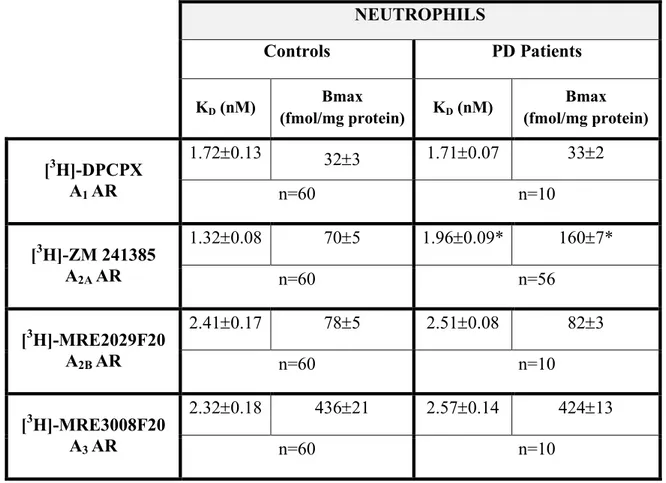
biomarker for PD. To this end it was evaluated the affinity and density of A1-, A2A, A2B-
and A3ARs in lymphocyte and neutrophil membranes from PD patients and healthy
control subjects. These data were correlated with score values of different scales estimating motor and cognitive impairment of PD patients.
CHAPTER III-METHODS
CHAPTER III
METHODS
METHODS
1. Subjects
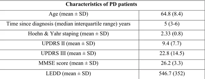
Brains of postmortem PD patients and healthy controls were derived from Netherlands Brain Bank of the Department of the Netherlands Institute for Neuroscience, Amsterdam, Netherlands. Human brain samples from 7 PD patients (3 men and 4 women) were obtained as well as from 7 controls (3 men and 4 women) who died with no neurological disorders. All autopsies were carried out within 12 hours of death, and the brain fragments were immediately frozen at -80°C until use in the experimental assays (Table 1M).
We collected peripheral blood samples of 129 PD patients (82 men and 47 women) attending the Neurological Clinic of the University of Ferrara, Italy. PD diagnosis was based on established diagnostic criteria. All patients were followed up by the same neurologist. Clinical data were obtained from medical records of patients and direct interviews, throughout a structured questionnaire administered to patients and their relatives. Demographic data, family and clinical history, and age at onset of initial symptoms were recorded. Pharmacological data regarding antiparkinsonian drugs and other concurrent treatments were also registered. The levodopa equivalent daily dose (LEDD) was computed according to standard conversion factors. The severity of PD symptoms and signs was assessed using the Unified Parkinson’s Disease Rating Scale (UPDRS) and the Hoehn and Yahr’s staging. The mini–mental state examination (MMSE), the Geriatric Depression Scale (GDS), and the Frontal Assessment Battery (FAB) were also administered. The blood samples were obtained from PD patients on the same day of the clinical examination and interview. Clinical features of included PD subjects are summarized in Table 2M.
123 healthy control subjects (79 men and 44 women) were volunteers from Ferrara University Hospital Blood Bank and were matched for similar age to the cohort of PD patients. The study was approved by the local Ethics Committee of the University Hospital of Ferrara, and informed consent was obtained from each participant in accordance with the principles outlined in the Declaration of Helsinki.
CHAPTER III-METHODS
NBB autopsy code Diagnosis Sex
Age at death (years) Delay to autopsy (hours:min) Brain tissue pH 96-032 96/052 191 J non-demented control F 60 08:25 6.60 97-043 S97/133 100 W1 non-demented control M 68 10:10 7.08 93-110 *93/215 119 F non-demented control M 73 08:45 6.11 04-049 S04/158 351 B non-demented control F 77 08:20 6.48 04-021 S04/057 351 B non-demented control M 80 06:30 6.43 98-056 S98/123 100 W1 non-demented control F 83 05:15 7.30 96-078 S96/238 191 J non-demented control F 87 08:00 6.91
03-078 S03/239 300 PUT3 PD with dementia F 61 04:30 6.30
94-026 S94/069 136 K2 PD M 68 06:00 6.70
05-080 S05/298 300 PUT3 PD M 73 06:35 6.28
94-092 S94/245 104 G PD F 77 09:40 6.86
93-007 S93/014 104 G PD M 79 05:25 6.15
94-021 S94/062 136 K2 PD F 83 05:00 6.58
05-045 S05/160 300 PUT3 PD with dementia F 87 05:25 6.44
Table 1M- Characteristics of postmortem healthy controls and PD patients.
Characteristics of PD patients
Age (mean ± SD) 64.8 (8.4)
Time since diagnosis (median interquartile range) years 5 (3-6)
Hoehn & Yahr staping (mean ± SD) 2.33 (0.8)
UPDRS II (mean ± SD) 9.4 (7.7)
UPDRS III (mean ± SD) 22.8 (14.5)
MMSE score (mean ± SD) 26.2 (3.3)
LEDD (mean ± SD) 546.7 (352)
Table 2M- Demographic and clinical characteristics of included PD patients involved in the blood examinations.
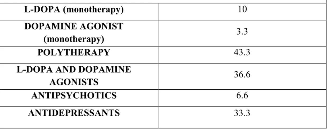
Table 3M- Percentage of PD patients involved in our study that assumed specific drugs.
2. Preparation of human putamen membranes
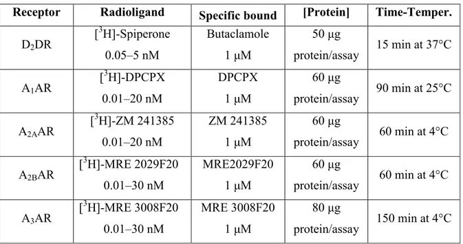
Human putamen of PD patients and controls was homogenized in 50 mM Tris-HCl buffer, pH 7.4, with a Polytron (Kinematica Inc., Bohemia, NY, USA) and centrifuged for 20 min at 48,000 g. To study A2AARs, the membrane pellet was suspended in 50
mM Tris-HCl buffer, pH 7.4, containing 10 mM MgCl2 and was incubated with 2 IU/ml
adenosine deaminase for 30 min at 37°C. Similar aliquots of membranes were suspended in 50 mM Tris-HCl buffer, pH 7.4, with the aim of investigating D2DRs.
3. Preparation of blood peripheral cells or membranes
Lymphocytes or neutrophils were isolated and prepared from the peripheral blood of PD patients and control subjects. The blood was supplemented with 6% (by weight) Dextran T500 solution (Sigma, St. Louis, MO, USA). Cells were pelletted by centrifugation for 5 min at 250 g, suspended in Krebs-Ringer phosphate buffer, and layered onto 10 ml of Fycoll-Hypaque (GE Healthcare, Little Chalfont, UK). After centrifugation, lymphocytes or neutrophils were diluted at 1 x 10⁶ cells/sample and used immediately in cAMP experiments. To obtain membrane suspensions, cell fractions were centrifuged in hypotonic buffer at 20,000 g for 10 min. The pellet was resuspended in Tris-HCl 50 mM buffer, pH 7.4, containing 2 UI/ml adenosine deaminase (Sigma), incubated for 30 min at 37°C, and used for radioligand binding assays. The proteins concentration was determined by a BCA protein assay kit (Pierce, Rockford, IL, USA) with bovine albumin as the reference standard.
Patients drug assumption (% of patients)
L-DOPA (monotherapy) 10
DOPAMINE AGONIST
(monotherapy) 3.3
POLYTHERAPY 43.3
L-DOPA AND DOPAMINE
AGONISTS 36.6
ANTIPSYCHOTICS 6.6
CHAPTER III-METHODS
4. PC12 cell culture
PC12 cells were maintained in DMEM medium supplemented with 5% FCS, 10% horse serum, 2 mM glutamine, 100 μg/ml penicillin, and 100 μg/ml streptomycin in a humidified atmosphere (5% CO2) at 37°C. Cells were subcultured 3x/wk at a density of
5x10⁵/ml. Differentiation was achieved by treatment with 50 ng/ml NGF (Sigma) for 1 week.
Cells were treated with TNF-α (100 ng/ml; Sigma) for 24 h and then harvested for radioligand binding experiments to examine A2AARs and D2DRs. PC12 cells were also
treated with a potent A2AAR agonist, CGS 21680 (1 μM; Sigma) and with a selective
A2AAR antagonist, SCH 58261 (1 μM; Tocris, Bristol, UK) for 24 h and then used in
A2AAR and D2DR saturation binding experiments.
5. Real-time RT-PCR experiments
Total cytoplasmic RNA was extracted from human putamen fractions and human lymphocytes by the acid guanidinium thiocyanate phenol method. Quantitative real-time RT-PCR assay of A1-, A2A-, A2B-, and A3R mRNAs was carried out using a gene
specific fluorescently labeled TaqMan MGB probe (minor groove binder) in an ABI Prism 7700 Sequence Detection System (Applied Biosystems, Foster City, CA, USA). For the real-time RT-PCR of A1-, A2A-, A2B-, and A3ARs, the Assay-on-Demand Gene
Expression Products NM 000674, NM000675, NM 000676, and NM 000677 (Applied Biosystems) were used, respectively. For the real-time RT-PCR of the reference gene, the endogenous control human β-actin kit was used, and the probe was fluorescently labeled with VIC (Applied Biosystems).
6. Western blotting analysis
Human putamen fractions and human lymphocytes were washed with ice-cold phosphate buffered saline (PBS) containing 1 mM sodium orthovanadate, 104 mM
4-(2-aminoethyl)-benzenesulfonyl fluoride, 0.08 mM aprotinin, 2 mM leupeptin, 4 mM bestatin, 1.5 mM pepstatin A, and 1.4 mM E-64. Then cells or tissues were lysed in Triton lysis buffer, and the protein concentration was determined using a BCA protein assay kit (Pierce, Rockford, IL, USA). Aliquots of total protein sample (50 μg) were analyzed using antibodies specific for human A2AARs (Alpha Diagnostic, San Antonio,
TX, USA; 1 μg/ml dilution). Filters were washed and incubated for 1 h at room temperature with peroxidase-conjugated secondary antibodies (1: 2000 dilution).
![Fig. 7- Possible disease targets for selective adenosine receptor ligands [113].](https://thumb-eu.123doks.com/thumbv2/123dokorg/4723290.45759/19.892.140.782.525.1038/fig-possible-disease-targets-selective-adenosine-receptor-ligands.webp)