Research Article
Conjugation of LasR Quorum-Sensing Inhibitors with
Ciprofloxacin Decreases the Antibiotic Tolerance of
P. aeruginosa Clinical Strains
Daria Bortolotti ,
1Claudio Trapella,
2Alessandra Bragonzi,
3Paolo Marchetti,
2Vinicio Zanirato,
2Andrea Alogna,
1,2Valentina Gentili,
1,2Carlo Cervellati ,
4Giuseppe Valacchi ,
5,6Mariangela Sicolo,
1Giulia Turrin,
2Anna Fantinati,
2Dario Di Luca,
1and Roberta Rizzo
1,71Department of Medical Sciences, Section of Microbiology and Medical Genetics, University of Ferrara, Via Luigi Borsari, 46-44121 Ferrara, Italy
2Department of Chemical and Pharmaceutical Sciences, University of Ferrara, Via Fossato di Mortara, 17, 44121 Ferrara, Italy 3Division of Immunology, Transplantation and Infectious Diseases, IRCCS San Raffaele Scientific Institute, Milano, Italy 4Department of Biomedical and Specialist Surgical Sciences, Section of Medical Biochemistry, Molecular Biology and Genetics,
University of Ferrara, Via Luigi Borsari, 46-44121 Ferrara, Italy
5Department of Life Sciences and Biotechnology, Section of Biology and Evolution, University of Ferrara, Via Luigi Borsari, 46-44121 Ferrara, Italy
6NC State University, Plants for Human Health Institute, Animal Science Deptment, NC Research Campus, Kannapolis, NC, USA 7LTTA-Electron Microscopy Center, University of Ferrara, Ferrara, Italy
Correspondence should be addressed to Daria Bortolotti; [email protected]
Received 9 November 2018; Revised 15 January 2019; Accepted 10 February 2019; Published 11 March 2019 Academic Editor: Ajaya Kumar Singh
Copyright © 2019 Daria Bortolotti et al. This is an open access article distributed under the Creative Commons Attribution License, which permits unrestricted use, distribution, and reproduction in any medium, provided the original work is properly cited.
Pseudomonas aeruginosa is a Gram-negative bacterium that commonly infects subjects with weakened immune system causing
deadly infections above all at pulmonary level. During infection, P. aeruginosa produces a well-organized bacterial structure, called biofilm, activating the quorum-sensing (QS) signaling, a mechanism of gene regulation. In this work, we synthesized already known QS inhibitors (QSi) designed on the scaffold of the N-(3-oxododecanoyl) homoserine lactone (3O-C12-HSL) QS molecule and conjugated them with ciprofloxacin to inhibit P. aeruginosa biofilm formation and increase the antibiotic susceptibility of clinical strains. We identified, for the first time, a QSi conjugated with ciprofloxacin (ET37), that is able to reduce the formation of biofilm and the onset of tolerant clones in P. aeruginosa clinical strains. This compound could have a wide application in clinical setting. The possibility to affect biofilm formation in chronically infected patients, such as patients affected by cystic fibrosis, and to reduce the onset of ciprofloxacin resistance would improve patient healing and allow to decrease antibiotic drug dosage.
1. Introduction
Microbial infections can result in complications, such as bacteremia, kidney failure, and toxic shock syndrome. For this, the identification of the systems to counteract bacterial infection is one of the challenges of modern medicine. The absence of novel molecules that control complications due to
bacterial infections suggests the defective comprehension of the mechanisms used by bacteria to control host immune response and resist treatment. During host infection, several bacteria organize a bacterial population, and Pseudomonas
aeruginosa is one of the most commonly studied. P. aeru-ginosa is a Gram-negative bacterium that especially infects
subjects with weakened immune system causing deadly
Volume 2019, Article ID 8143739, 13 pages https://doi.org/10.1155/2019/8143739
infections above all at pulmonary level. In fact, cystic fibrosis (CF) or HIV patients exhibit increased susceptibility to P.
aeruginosa lung infections. Since P. aeruginosa is a
ubiq-uitous bacterium, exposure to this pathogen in the hospital setting results to be frequent, making it one of the most problematic nosocomial infections.
The antibiotics used to treat P. aeruginosa infection include ciprofloxacin, tobramycin, ceftazidime, gentamicin, and imipenem. P. aeruginosa bacteria present a high degree of resistance to these antibiotics. Interestingly, the response to ciprofloxacin is very effective at the beginning of the treatment, but high-level resistance is rapidly acquired by P.
aeruginosa, making the treatment ineffective in the 30% of
strains obtained from clinical isolates [1].
The mechanisms that could increase P. aeruginosa an-tibiotic susceptibility still remain unclear [2–4].
P. aeruginosa is characterized by a low permeability of its
cell wall that increases its resistance to antibiotics because of a lower drug uptake or higher efflux pumps expression that cause decreased intracellular drug concentrations [5]. Al-terations in QRDR of the target sites DNA gyrase (gyrA) and
topoisomerase IV, encoded by parC and parE subunits, are
considered the main reason of bacterial resistance to qui-nolones [6, 7].
The acquisition of resistance to ciprofloxacin is a mul-tistage process in P. aeruginosa: in stage I, the exposure to the drug kills the susceptible cells; in stage II, a small population survives antibiotic exposure without increasing the re-sistance and maintaining a slow growth; in stage III, a the population is reconstituted by a slow-growing population with an increased drug resistance [8]. The appearance of a drug-resistant population limits the possibility to use pro-longed therapies with existing or newly developed antibiotic drugs. We suggest the possible use of the inhibitor of quorum-sensing (QS). One of the main defense mechanisms adopted by P. aeruginosa is represented by biofilm forma-tion, which allows the bacteria to avoid both host immune system and antibiotics effects [9, 10]. QS is a mechanism of gene regulation sensitive to population density that enables host colonization contrasting the immune surveillance through biofilm formation and the expression of virulence factors [10] via the production of self-generated extracellular signal molecules [11–13]. P. aeruginosa is characterized by two QS systems, Las and Rhl, based on acylhomoserine lactone [14] molecules [15]. The las system is controlled by the transcriptional activator LasR and the autoinducer synthase enzyme LasI, which directs the synthesis of N-(3-oxododecanoyl) homoserine lactone (3O-C12-HSL). Simi-larly, the Rhl system is regulated by the transcriptional activator RhlR and the RhlI AI synthase that synthesizes N-butyryl homoserine lactone (C4-HSL). 3O-C12-HSL binds LasR and activates the LasR/3O-C12-HSL complex; the multimerization will promote the transcription of RhlR, RhlI, LasI genes, and other virulence genes that are con-nected to the regulon [16–18]. In a similar way, RhlR/C4-HSL complex dimerizes and activates the expression of its own regulon and RhlI [19]. LasR/3O-C12-HSL regulates the quinolone signal by inducing expression of PqsR, as well as PQS [20]. PQS, in turn, increases the transcription of RhlI
and the production of C4-HSL [21]. Interestingly, PqsR expression is inhibited by RhlR/C4-HSL [21], suggesting that the ratio between 3O-C12-HSL and C4-HSL concentrations controls the Pqs signaling system.
Since QS molecules are important during infection, the interference on QS signaling represents a potential strategy to contrast bacterial virulence, decreasing antibiotics dosage and facilitating the natural bacterial clearance by host im-mune response. Brackman et al. have shown that QS in-hibitors (QSi) increase the susceptibility of bacterial biofilms to different types of antibiotics [22]. We report the results of our work on the effect of different QSi compounds, designed on the scaffold of QS inducer 3O-C12-HSL, conjugated with ciprofloxacin. These results will set up the initial steps de-veloping new strategies that may subvert the ciprofloxacin resistance of P. aeruginosa.
2. Materials and Methods
2.1. Chemical Synthesis. The compounds tested in this work
are antagonists of LasR and are designed on 3O-C12-HSL scaffold [23]. Previous evaluations of these derivatives showed that S absolute configuration at the 3-position of the homoserine lactone was important for the activity of the compounds. On the contrary, the R derivative was not ef-fective. For this reason, we synthetized all the compounds in the S absolute configuration. The chemical synthesis was performed as described in Geske et al. [23]. The compounds tested in this study derived from libraries of AHL mimics designed to be capable of intercepting the LasR and RhlR QS system which are specific for P. aeruginosa. The LasR an-tagonists interact with the N-terminal ligand-binding do-main of LasR, blocking the binding site for QS molecules [23].
2.2. Samples. Ten sequential P. aeruginosa isolates from four
patients with CF were chosen from the strains collection of the CF clinic in Hannover. We selected two strains from patients SG (SG1 and SG58, both LasR wild type (wt)), three strains from patients AA (AA2 and AA12 LasR wt and AA11 LasR mutant), three strains from patient TR (TR2 LasR wt and TR1 and TR66 LasR mutants), and two strains from patient KK (KK1 and KK72, both LasR mutants). These strains were characterized in previous work [24]. Patients were checked after the diagnosis of CF, and meanwhile, the respiratory specimens were sampled. The “early” isolates of
P. aeruginosa strains were collected from the first positive
cultures, whereas late isolates were collected 7 to 16 years after colonization or prior to death or lung transplantation. CF isolates behaviour was compared with the laboratory strain PAO1 for their phenotypic diversity [25, 26].
2.3. Phenotypic Analysis for LasR Mutants. Colony surface
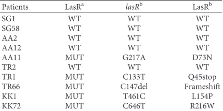
iridescence and metallic sheen were evaluated and consid-ered as a phenotypic characteristic of LasR mutants [25, 26]. The DNA sequence confirmed the presence of LasR mu-tations (Table 1).
2.4. Biofilm Formation Inhibition and Cell Growth Evaluation. P.aeruginosa PAO1 or clinical strains were grown in a static
culture in M9 medium for liquid culture. Flat-bottomed polystyrene 96-well plates were used for biofilm formation experiments, and optical density measurements were per-formed in a plate reader at 600 nm. In order to screen compounds for QS inhibition, a microplate-based assay was used. Briefly, P. aeruginosa strains were seeded and grown in
M9 for 24 hours at 37°C. The optical density (OD600) at
inoculation was 0.04. The compounds were dissolved in DMSO added to planktonic cell cultures. Cell growth was evaluated by reading absorbance at 600 nm, while biofilm formation was analysed as previously described [27]. Briefly, after 24 hours of treatment, the planktonic cells were gently removed, and the wells were washed three times with PBS 1x.
The plate was let at 60°C for 1 hour in order to dry the
biofilm. The biofilm mass was measured by staining with crystal violet 1% w/v and then resuspended in 200 µl of 33% glacial acetic acid and read at 570 nm. 3O-C12-HSL and DMSO were used as positive and negative controls, re-spectively. Results are reported as mean ± SD and are ob-tained from three independent experiments.
2.5. Syto9 Assay. P.aeruginosa was cultured in 8-chamber
slides and treated and left for 24 hours at 37°C. Planktonic
cells were removed, and each chamber was washed with PBS 1x. 200 µl of Syto9 working solution prepared by diluting Syto9 stock solution (5 mM in DMSO, Invitrogen) 1 : 1200 was added and kept in dark for 30 minutes at RT. The chambers were then washed two times with PBS 1x, and the biofilm was visualized by fluorescent microscopy (ZOE Fluorescent Cell Imager, Biorad). Pictures of the biofilm were compared to the untreated control.
2.6. Minimal Inhibitory Concentration. The MIC for each
strain was determined in triplicate using M.I.C. Evaluator (Oxoid, Basingstoke, Hants, UK) containing ciprofloxacin in final concentrations of 32, 16, 8, 4, 2, 1, 0.5, 0.25, 0.12, 0.06, 0.03, 0.015, 0.008, 0.004, 0.002, and 0 μg/ml. The M.I.C. was evaluated after 3 passages in antibiotic-free media [28]. The inoculum was Mueller-Hinton agar
in-oculated with a standard inoculum (105 to 106CFU/ml)
according to the European Committee on Antimicrobial Susceptibility Testing guidelines [29].
2.7. Time-Kill Studies. Ciprofloxacin and ET37 time-kill
studies were performed at concentrations equal to the theoretical plasma peak (4 μg/mL), then at concentrations
equal to 1/2, 2, 4, 8, X MIC50of the antibiotic used. Bacteria
were cultured for 24 h at 37°C on Mueller-Hinton agar
(MHB, BioM´erieux, France) and used to prepare the
ex-ponential growth phase at standard inoculum (106CFU/mL)
in Mueller-Hinton broth. The inoculum of 106CFU/ml was
supplemented with ciprofloxacin or ET37 at the different
concentrations and cultured for 24 h at 37°C. 100 ml of
culture supernatants were collected at 2, 4, 6, and 24 h and plated on agar for colony counts.
2.8. Pyocyanin and Elastase Assay. P. aeruginosa strains were
cultured overnight at 37°C with shaking. Cultures were back
diluted 1 : 1,000 into fresh medium and grown for 18 h. The cells were pelleted by centrifugation, and the culture supernatants were filtered through 0.22 μm filters. The
production of pyocyanin was reported as A380/A600 ratio
[30]. The production of LasB elastase was assessed through the measurement of elastase activity using elastin-Congo red
and reported as A495/A600ratio [31].
2.9. GyrA and ParC Amplification and DNA Sequencing.
QRDR amplification of gyrA and parC from resistant and intermediate isolates was carried out using specific primers: GyrA-1 (5′-GTGTGCTTTATGCCATGAG-3′) and GyrA-2 (5′-GGTTTCCTTTTCCAGGTC-3′) for the amplification of 287 bp of the fluoroquinolone resistance-determining region of the gyrA gene and ParC-1 (5′-CATCGTCTACGC-CATGAG-3′) and ParC-2 (5′-AGCAGCACCTCGGAA-TAG-3′) were used to amplify 267 bp of the fluoroquinolone resistance-determining region of ParC as previously re-ported [14]. Amplified products were then separated using 1.5% agarose gels, and PCR products were sequenced, and the sequence of each of the sample was compared with P.
aeruginosa PAO1 sequence. The sequences were multiple
aligned by ClustalW2 (http://www.ebi.ac.uk/Tools/msa/ clustalw) in order to detect mutations. Nucleotide se-quences were translated by Expasy Bioinformatics Resource Portal (http://web.expasy.org/translate/) then compared with P. aeruginosa PAO1 protein sequence using ClustalW2 to find changes in amino acid sequences.
2.10. Disk Diffusion Ciprofloxacin Susceptibility Testing.
The antibiotic susceptibility of bacterial isolates was de-termined using the disk diffusion method standardized
according to the EUCAST (http://www.eucast.org/
ast_of_bacteria/disk_diffusion_methodology/), with
in-terpretation based on the EUCAST Clinical Breakpoint Tables v. 9.0, valid from 2019 to 01-01, the zone diameter breakpoint >26 for susceptible strains (S), and <26 for re-sistant strains (R). The bacterial strains were inoculated onto Mueller-Hinton agar. Antibiotic disks were placed on the
Table 1: Mutations in LasR in P. aeruginosa isolates. Patients LasRa lasRb LasRb
SG1 WT WT WT
SG58 WT WT WT
AA2 WT WT WT
AA12 WT WT WT
AA11 MUT G217A D73N
TR2 WT WT WT
TR1 MUT C133T Q45stop
TR66 MUT C147del Frameshift
KK1 MUT T461C L154P
KK72 MUT C646T R216W
aLasR status evaluated by phenotypic analysis.bNumbering is based on the
surface, and this was followed by incubation at 35 ± 1°C in air
for 18 ± 2 h ours. Inhibition zone diameter values were read by a pair of calipers, in millimeters to one decimal place. Ciprofloxacin disks (5 µg) were obtained from Oxoid Ltd (Oxoid AB, Hampshire, UK).
2.11. Determination of Minimum Biofilm Inhibitory Con-centration (MBIC). The MBICs of ciprofloxacin and ET37
were determined in PAO1 strain, as previously reported [32]. The experiments were done in 96-well polystyrene microtiter plates with round bottoms. An overnight culture with a turbidity equivalent to that of a 0.5 McFarland standard, obtained with TSB, was aliquoted into the wells of
microtiter plates. The plates were incubated for 24 h at 37°C.
The wells were washed three times with phosphate-buffered saline (PBS) to remove unattached bacteria and dried in an inverted position. Volumes of 100 µL of appropriate two-fold dilutions of the ciprofloxacin or ET37 in Mueller-Hinton broth were transferred into the dried wells with established biofilms. The microtiter plates were incubated
for 18–20 hours at 37°C, and minimum biofilm inhibitory
concentration (MBIC) was determined, which corresponds to the lowest concentration of antibiotic which inhibits growth of biofilm cells as indicated by absence of visible growth in the wells. A positive control and a negative control were included in all experiments. The experiment was re-peated three times.
2.12. Determination of Minimum Duration for Killing 99% of the Population (MDK99). MDK99was determined by mea-suring the time to kill 99% of the population [33]. MDK99 was tested in P. aeruginosa clinical strains with concen-tration 1x the MIC of ciprofloxacin or 1x the MIC ET37 in P.
aeruginosa wild-type and LasR mutant clinical strains.
3. Results
3.1. Effect of LasR Antagonists on Biofilm Formation in P. aeruginosa PAO1 Laboratory Strain. We selected 4
com-pounds reported to be active antagonists of LasR of P.
aeruginosa and designed on 3-O-C12-HLS scaffold
(Fig-ure 1, # 1, 2, 3, 4) [23]. The synthesis was performed as described in Geske et al. and references cited therein.
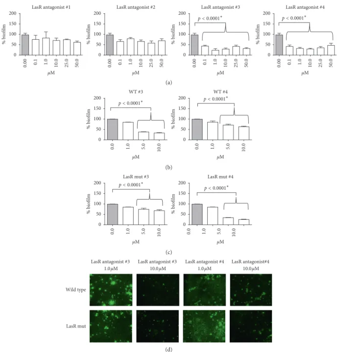
We evaluated the effect of the selected 4 LasR antagonists on cell growth and biofilm formation of P. aeruginosa PAO1 laboratory strain. We treated PAO1 immediately after seeding the planktonic bacteria with LasR antagonists at different concentrations (0.1, 1.0, 10.0, 25.0, and 50.0 µM) for 24 hrs. We evaluated cell growth by reading the optical density at 600 nm, and we observed no effect on this pa-rameter (data not shown). Considering biofilm formation, the treatment with number 3 and number 4 compounds reduced the formation of biofilm of more than 50% (Figure 2(a)) (p < 0.0001; Studentʼs t-test). In particular, in the concentration range of 1.0–10.0 µM, we observed the highest effect on biofilm inhibition for both number 3 and number 4 compounds. On the contrary, compounds number 1 and 2 failed to significantly inhibit biofilm
formation, also increasing the time of exposure to 48–72 hrs (data not shown). On the basis of these results, we selected number 3 and number 4 compounds and the range of concentrations 1.0–10.0 µM for further investigations on clinical isolates.
3.1.1. Effect of Number 3 and Number 4 Compounds on Biofilm Formation in Clinical Strains. First, we evaluate the
efficacy of QSi in acting as LasR antagonists in P. aeruginosa clinical strains. We selected two subgroups of P. aeruginosa clinical strains: (i) wild type for the expression of 3O-C12-HSL receptor (LasR); (ii) P. aeruginosa LasR mutant. LasR mutant clinical strains were phenotypically evidenced as producing iridescent and metallic colonies [34, 35]. We performed our analysis on 10 different isolates from four CF individuals (SG, AA, TR, and KK) wild type or mutant for LasR. In order to evaluate LasR functionality, we treated wild-type (N � 5) and LasR mutant (N � 5) clinical strains immediately after seeding the planktonic bacteria with LasR agonist (1.0, 5.0, 10.0, 25.0, and 50.0 µM) for 24 hours. We analysed cell growth by reading the optical density at 600 nm, and we observed no effect on this parameter (data not shown). Concerning biofilm formation in wild-type strains, we observed biofilm inhibition of 60% with the compound number 3 at the concentrations of 5.0–10.0 µM (Figure 2(b)) and compound number 4 reduced biofilm formation of the 30–40% (Figure 2(b)). We observed also a reduction in biofilm formation in LasR mutant clinical strains treated with the same two compounds (number 3 and
4) at the concentrations of 5.0–10.0 µM, reporting a decrease
of the 30% and 60–70%, when treated with LasR antagonist number 3 and 4, respectively, (Figure 2(c)). To confirm the results obtained by crystal violet staining, we performed the Syto9 assay, based on fluorescence staining. We choose to use Syto9 staining to confirm our data and to overcome the variability of the results that could arise by crystal violet staining of P. aeruginosa biofilm [36]. Similarly, Syto9 assay showed a clear decrease of biofilm formation after the ad-dition of the compounds number 3 and 4 in both wild-type and mutant strains (Figure 2(d)), with the highest effect observed at the concentration of 10.0 µM.
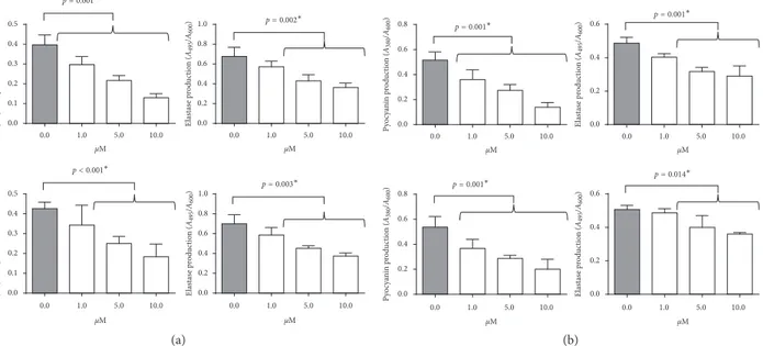
Since QS inhibition results in a reduced expression of the virulence factors pyocyanin and elastase and in an alteration of biofilm morphology/phenotype [37], the effect of com-pound numbers 3 and 4 was tested also on the expression of these genes. We observed that both compound numbers 3 and 4 were able to reduce the production of pyocyanin and elastase in wild-type and LasR mutant strains (Figure 3).
3.1.2. ET37 Effect on PAO1 Strain Susceptibility to Ciprofloxacin. Ciprofloxacin belongs to the group of
fluo-roquinolones, active against bacterial infections. It has demonstrated a high efficacy towards P. aeruginosa infection and experienced in clinical practice. Since both compounds
3 and 4 resulted efficient in decreasing biofilm formation, we
selected both of them to associate with ciprofloxacin, on the basis of its chemical structure. The chimeric derivatives
O N H O Br O
Chemical formula: C12H12BrNO3
Exact mass: 297.000 Molecular weight: 298.1326 (a) N H O O OH Chemical formula: C18H27NO3 Exact mass: 305.1991 Molecular weight: 305.4119 (b) N H O Chemical formula: C15H29NO Exact mass: 239.2249 Molecular weight: 239.3969 (c) N H O O O Chemical formula: C11H19NO3 Exact mass: 213.1365 Molecular weight: 213.2735 (d)
Figure1: Molecular structure of P. aeruginosa QS antagonists (#1, 2, 3, 4) designed on 3-O-C12-HSL scaffold. (a) LasR antagonist #1. (b) LasR antagonist #2. (c) LasR antagonist #3. (d) LasR antagonist #4.
0 50 100 150 200 μM % biofilm LasR antagonist #1 0.00 0.1 1.0 10.0 25.0 50.0 0 50 100 150 200 μM % biofilm LasR antagonist #2 0.00 0.1 1.0 10.0 25.0 50.0 0 50 100 150 200 μM % biofilm LasR antagonist #3 p< 0.0001∗ 0.00 0.1 1.0 10.0 25.0 50.0 0.00 0.1 1.0 10.0 25.0 50.0 0 50 100 150 200 μM % biofilm p< 0.0001∗ LasR antagonist #4 (a) 0 50 100 150 200 μM % biofilm p< 0.0001∗ WT #3 0.0 1.0 5.0 10.0 0 50 100 150 200 μM % biofilm p<0.0001WT #4∗ 0.0 1.0 5.0 10.0 (b) 0 50 100 150 200 μM % biofilm p< 0.0001LasR mut #3∗ 0.0 1.0 5.0 10.0 0.0 1.0 5.0 10.0 0 50 100 150 200 μM % biofilm p< 0.0001∗ LasR mut #4 (c) Wild type LasR antagonist #3
1.0μM LasR antagonist #3 10.0μM LasR antagonist #41.0μM LasR antagonist#4 10.0μM
LasR mut
(d)
Figure2: Evaluation of biofilm mass after LasR antagonists (#1, 2, 3, 4) treatment on (a) P. aeruginosa PAO1 strain and after treatment with number 3 and number 4 compounds on (b) wild-type and (c) LasR mutant clinical strains, using crystal violet assay. (d) Evaluation of biofilm mass after treatment with number 3 and number 4 compounds, using Syto9 assay in LasR wild-type (upper line) and mutant clinical (lower line) strains.∗Studentʼs t-test.
between compounds 3 and 4 and ciprofloxacin are depicted in Figure 4.
The molecules ET37 and ET39 were synthesized as depicted in Figure 5; the decanedioic acid monoethylester 5 was condensed to the amine moiety of the piperazine ring present in the commercially available ciprofloxacin antibiotics to obtain compound 6 that was subjected to 2N NaOH sa-ponification to achieve acid derivative 7 [38]. Finally, com-pound 7 was condensed to the cyclopentylamine and L-homoserine lactone using WSC/HOBt as activating agents to obtain, respectively, compound ET37 and compound ET39 that were fully characterized by NMR and exact mass spectrometry.
ET37 and ET39 were preliminarily tested on PAO1 and
P. aeruginosa clinical strains. ET37 maintained its ability to
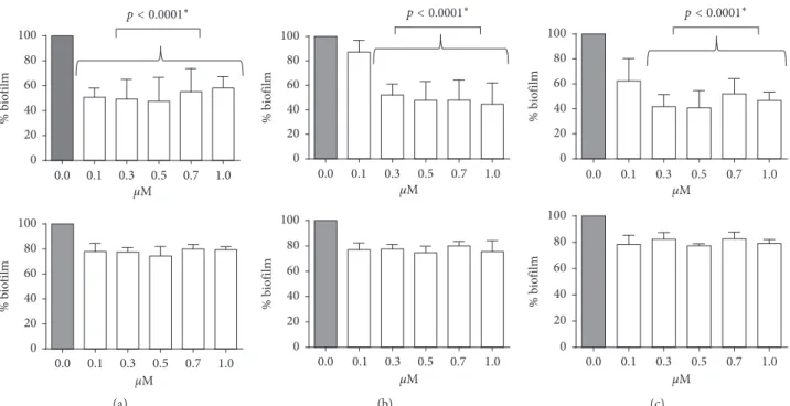
decrease biofilm formation in PAO1 (Figure 6(a)), wild-type (Figure 6(b)) and LasR mutant clinical strains (Figure 6(c)). On the contrary, ET39 lost the efficacy of compound number 4 (Figure 6). Similarly, pyocyanin and elastin were reduced by
ET37 treatment in both wild-type (Figure 7(a)) and LasR
mutant clinical strains (Figure 7(b)). Starting with PAO1 strain, we evaluated the minimal inhibitory ciprofloxacin concen-tration [28] using the Etest to establish a baseline. The MIC50
was within the expected ranges, with the PAO1 MIC50 at
0.5 μg/ml ciprofloxacin, corroborating previous research [39]
μM Py oc ya ni n p ro du ct io n ( A380 /A600 ) 0.0 1.0 5.0 10.0 0.0 0.1 0.2 0.3 0.4 0.5 p= 0.001∗ μM El ast as e p ro duc tio n ( A495 /A600 ) 0.0 1.0 5.0 10.0 0.0 0.2 0.4 0.6 0.8 1.0 p= 0.002∗ μM Py oc ya ni n p ro du ct io n ( A380 /A600 ) 0.0 1.0 5.0 10.0 0.0 0.1 0.2 0.3 0.4 0.5 p< 0.001∗ μM El as ta se pro du ct ion (A495 /A600 ) 0.0 1.0 5.0 10.0 0.0 0.2 0.4 0.6 0.8 1.0 p= 0.003∗ (a) Py oc ya ni n p ro du ct io n ( A380 /A600 ) 0.0 0.2 0.4 0.6 0.8 p= 0.001∗ El as ta se p ro du ct io n ( A495 /A600 ) 0.0 0.2 0.4 0.6 p= 0.001∗ Py oc ya ni n p ro du ct io n ( A380 /A600 ) 0.0 0.2 0.4 0.6 0.8 p= 0.001∗ El as ta se p ro duc tio n ( A495 /A600 ) 0.0 0.2 0.4 0.6 p= 0.014∗ μM 0.0 1.0 5.0 10.0 μM 0.0 1.0 5.0 10.0 μM 0.0 1.0 5.0 10.0 μM 0.0 1.0 5.0 10.0 (b)
Figure3: Evaluation of pyocyanin and elastin production after compound number 3 (upper panels) or compound number 4 (lower panels) treatment of (a) wild-type and (b) LasR mutant clinical strains.∗Studentʼs t-test.
N F N N O COOH O O H N (a) N F N N O COOH O O O O HN (b)
O O O O O O O O O O O O O O ET39 ET37 O O O O b a c d O O O O HO O O OH F F F F N N N N N N N N N N 8 8 8 8 N N N F N HN 5 6 7 OH OH OH OH OH H N H N
Figure5: Schematic synthesis of ET37 and ET39 compounds. Conditions: (a) WSC/HOBt, DMF, 0°C, o.n., Y 88%. (b) NaOH 2N, EtOH,
r.t. 4 h, Y quant. (c) WSC/HOBt, cyclopentylamine, DMF, 0°C, 12 h, Y 71%. (d) WSC/HOBt, L-homoserine, NMO, DMF, 12°h, Y 63%.
p< 0.0001∗ 0 20 40 60 80 100 % b io film 0.1 0.3 0.5 0.7 1.0 0.0 μM 0 20 40 60 80 100 % b io film 0.1 0.3 0.5 0.7 1.0 0.0 μM (a) p< 0.0001∗ 0 20 40 60 80 100 % b io film 0.1 0.3 0.5 0.7 1.0 0.0 μM 0 20 40 60 80 100 % b io film 0.1 0.3 0.5 0.7 1.0 0.0 μM (b) p< 0.0001∗ 0 20 40 60 80 100 % b io film 0 20 40 60 80 100 % b io film 0.1 0.3 0.5 0.7 1.0 0.0 μM 0.1 0.3 0.5 0.7 1.0 0.0 μM (c)
Figure6: Evaluation of biofilm mass after ET37 (upper panel) or ET39 (lower panel) treatment on (a) P. aeruginosa PAO1, (b) wild-type, and (c) LasR mutant clinical strains, using crystal violet assay.∗Studentʼs t-test.
and at 0.2 μg/ml ET37. The MIC90 and modal MIC were ≥32 μg/ml.
We evaluate the population dynamics in response to ciprofloxacin unconjugated and conjugated to compound number 3 (ET37). The exposure was at concentrations ranging from 0.5 to 8x the MIC in PAO1 strain (Figure 8).
After the treatment with ciprofloxacin, a large part of the bacteria was killed by the drug (Figure 8(a)), as the expected response to an antibiotic treatment with an efficacy of 24 h, as illustrated in Figure 5(a). At lower exposure levels, there was a surviving subpopulation of “drug-tolerant” cells, that survived after ciprofloxacin treatment (Figure 8(a), red
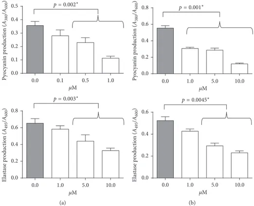
μM Elastase production ( A495 /A600 ) 0.0 0.2 0.4 0.6 0.8 p= 0.003∗ 0.0 1.0 5.0 10.0 μM Pyocyanin production ( A380 /A600 ) 0.0 0.1 0.2 0.3 0.4 0.5 p= 0.002∗ 0.0 0.1 0.5 1.0 (a) Pyocyanin production ( A380 /A600 ) 0.0 0.2 0.4 0.6 0.8 p=0.001∗ Elastase production ( A495 /A600 ) 0.0 0.2 0.4 0.6 p= 0.0045∗ μM 0.0 1.0 5.0 10.0 μM 0.0 1.0 5.0 10.0 (b)
Figure7: Evaluation of pyocyanin (upper panels) and elastin (lower panels) production after compound ET37 treatment of (a) wild-type and (b) LasR mutant clinical strains.∗Studentʼs t-test.
1/2x MIC (0.25 μg/ml) 1x MIC (0.5 μg/ml) 2x MIC (1 μg/ml) 4x MIC (2 μg/ml) 8x MIC (4 μg/ml) No ciprofloxacin added 0 2 4 6 8 10 Log CFU/ ml 10 20 30 40 50 0 Exposure time (a) 1/2x MIC (0.1 μg/ml) 1x MIC (0.2 μg/ml) 2x MIC (0.4 μg/ml) 10 20 30 40 50 0 Exposure time 0 2 4 6 8 10 Log CFU/ ml 4x MIC (0.8 μg/ml) 8x MIC (1.6 μg/ml) No ET37 added (b)
Figure8: Time-kill studies with PAO1 with concentration ranging from 0.5 to 8x the MIC of (a) ciprofloxacin or (b) ET37 treatment. Red arrow: “drug-tolerant” cells in ciprofloxacin-treated PAO1; blue arrow: “drug-tolerant” cells in ET37-treated PAO1.
arrow). The “drug-tolerant” cell population was not present in ET37 treated PAO1 cultures (Figure 8(b), blue arrow).
Previous data showed that ciprofloxacin reduced viru-lence factors and biofilm formation decreasing the pro-duction of QS signal molecules in P. aeruginosa [40]. We tested the minimal biofilm inhibitory concentration (MBIC) of both ciprofloxacin and ET37 on PAO1. Results
dem-onstrated that ET37 MBIC was similar to MIC50(0.22 μg/
ml) while ciprofloxacin MICB was eight times higher
(4.0 μg/ml) than MIC50(0.5 μg/ml).
3.1.3. ET37 Effect on P. aeruginosa Clinical Strains Suscep-tibility to Ciprofloxacin. The efficacy of ciprofloxacin was
then tested also on P. aeruginosa clinical strains. The
resulting MIC50are reported in Table 2.
Three strains were resistant to ciprofloxacin (TR2, TR1, and KK72), three where intermediate (AA2, TR66, and KK1), and four were susceptible (SG1, SG58, AA12, and AA11). We evaluated the genetic background at the basis of ciprofloxacin resistance. QRDRs regions of GyrA and ParC, implicated in P. aeruginosa resistance to quinolone [6, 7], were amplified from resistant isolates and were sequenced for mutations involved in ciprofloxacin resistance. One strain resistant to ciprofloxacin (TR1) had a mutation in GyrA (Thr83⟶Ile) and a mutation in ParC (Ser87⟶ Leu or Trp). Two strains resistant to ciprofloxacin (TR2 and KK72) and three intermediate strains (AA2, TR66, and KK1) had a mutation in GyrA (Thr83⟶Ile).
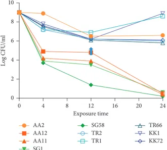
When we evaluated the time-kill studies with the ET37 concentration of 4 μg/ml, equal to the theoretical plasma peak [41], we observed 6-log decrease at 24 h for strain SG1, SG58, AA12, and AA11 (Figure 9), which are the most susceptible according to their MICs (Table 2). Interestingly, a bactericidal activity was observed with 3.0-log inoculum reduction at 12 h for strains AA2 and TR66, that were categorized as intermediate and TR2 and KK72, that were considered resistant to ciprofloxacin (Table 2) (Figure 9). A bacterial growth reoccurred at 24 h for strains TR1 and KK1 (Figure 9), the most resistant strains according to their MICs (Table 2).
3.1.4. Time-Kill Studies on P. aeruginosa Clinical Strains.
Time-kill studies were performed in parallel, on P.
aerugi-nosa clinical strains, with 1/2, 2, 4, 8, X MIC50, for both ciprofloxacin and ET37 (Table 2). Following 24 h treatment with ciprofloxacin, there was a surviving subpopulation of “drug-tolerant” cells only in low-dose ciprofloxacin-treated
P. aeruginosa clinical strains (Figure 10(a)), while both
wild-type and LasR mutant clinical strains did not change over 48 h treatment with ET37 (Figure 10(b)).
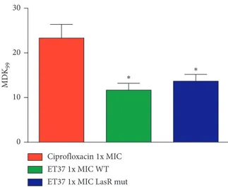
The clinical strains were then tested for their tolerance to ciprofloxacin or ET37 treatment. The measured MDK99 decreased in the ET37-treated strains in comparison with the ciprofloxacin-treated strains (Figure 11). Both P.
aeru-ginosa wild-type and LasR mutant clinical strains showed a
MDK99of 12 hours after treatment with ET37 in
compar-ison with the MDK99 of 24 hours observed in the same
strains without ET37 treatment (p< 0.1).
4. Discussion
In this research, for the first time, we analysed the effect of selected LasR antagonists, on P. aeruginosa clinical strains, and we identified two compounds designed on 3O-C12-HSL scaffold, number 3 and number 4, which exhibit an in-hibitory effect on both LasR wild-type and mutant strains. These LasR antogonists interact with the N-terminal ligand-binding domain of LasR, blocking the ligand-binding site for QS molecules.
Since clinical P. aeruginosa strains are commonly LasR mutants, it is fundamental to obtain QSi with a demon-strated functionality also in LasR mutant clinical strains. Both number 3 and 4 compounds are able to counteract biofilm formation also in LasR mutant strains, possibly due to the shorter aliphatic chain that results in a lower steric hindrance that could facilitate the cross interaction with RhlR [17, 18]. In fact, it has been reported that LasR an-tagonist could be effective also on other QS receptors, as
Table2: MIC (μg/ml) of the P. aeruginosa clinical strains. Clinical
strain statusLasR MICcipro50 Diameter inhibitoryzone (mm)∗ Categorization
SG1 WT 0.12 32 S SG58 WT 0.25 33 S AA2 WT 1.0 27 I AA12 WT 0.008 36 S AA11 MUT 0.5 34 S TR2 WT 2.0 10 R TR1 MUT 2.0 13 R TR66 MUT 1.0 27 I KK1 MUT 1.0 27 I KK72 MUT 2.0 11 R
∗According to the EUCAST Clinical Breakpoint Tables v. 9.0, valid from
2019 to 01-01, the zone diameter breakpoint>26 for susceptible strains (S)
and<26 for resistant strains (R).
Exposure time Lo g CFU/ml 0 4 8 12 16 20 24 0 2 4 6 8 10 AA2 AA12 AA11 SG1 SG58 TR2 TR1 TR66 KK1 KK72
Figure9: Time-kill studies with P. aeruginosa clinical strains with a concentration of ET37 equal to the theoretical plasma peak (4 μg/ml). Blue arrow: “drug-tolerant” cells in ET37 treated P.
RhlR [17, 19]. We speculate that, in the presence of LasR mutations, LasR antagonists numbers 3 and 4 could interact with other QS receptors that are still functional in LasR mutant strains. It has been described that some virulence factors, such as pyocyanin, are still produced in LasR mu-tants, confirming that QS system is still functional. This could be explained by the ability of P. aeruginosa to cir-cumvent QS deficiency using rhl and Pqs systems [20]. Similarly, 3O-C12-HSL is known to bind not only its re-ceptor LasR, but also other QS rere-ceptor, as RhlR and PqsR. This is in line with the results of Kalaiarasan and coauthors reporting the ability of two anti-QS compounds to decrease biofilm production in two P. aeruginosa clinical isolates, affecting both lasR and rhlR transcription [42].
The identification of new mechanisms to block the ap-pearance of drug resistance in P. aeruginosa is important
because it is characterized by a quick adaptation to resist to new drug compounds [1], causing chronic lung infection in individuals with CF [43] or chronic obstructive pulmonary disease (COPD) [44], and the 10% of hospital-acquired infections.
The antibiotics used to treat P. aeruginosa infection include ciprofloxacin, tobramycin, ceftazidime, gentamicin, and imipenem. P. aeruginosa bacteria present a high degree of resistance to these antibiotics. Interestingly, the response to ciprofloxacin is very effective at the beginning of the treatment, but high-level resistance is rapidly acquired by P.
aeruginosa, making the treatment ineffective in the 30% of
strains obtained from clinical isolates [1]. The mechanisms that could increase P. aeruginosa antibiotic susceptibility still remain unclear [2–4] and few studies tried to develop ther-apeutic strategies to decrease the insurgence of resistances.
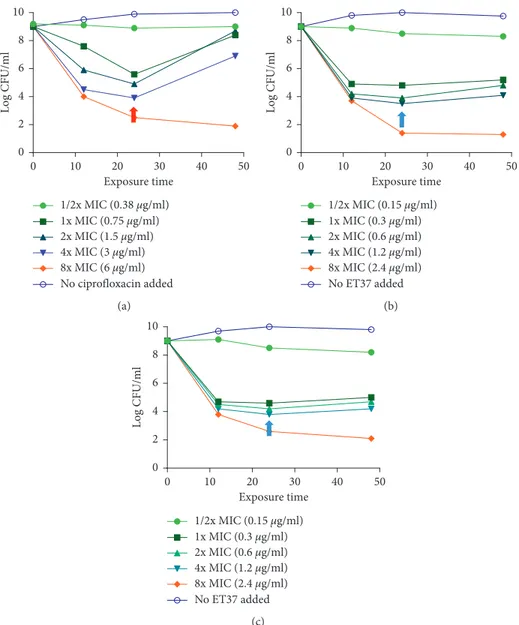
Exposure time Log CFU /ml 0 10 20 30 40 50 0 2 4 6 8 10 1/2x MIC (0.38 μg/ml) 1x MIC (0.75 μg/ml) 2x MIC (1.5 μg/ml) 4x MIC (3 μg/ml) 8x MIC (6 μg/ml) No ciprofloxacin added (a) Exposure time Log CF U/ml 0 10 20 30 40 50 0 2 4 6 8 10 1/2x MIC (0.15 μg/ml) 1x MIC (0.3 μg/ml) 2x MIC (0.6 μg/ml) 4x MIC (1.2 μg/ml) 8x MIC (2.4 μg/ml) No ET37 added (b) Exposure time Log CFU/ ml 0 10 20 30 40 50 0 2 4 6 8 10 1/2x MIC (0.15 μg/ml) 1x MIC (0.3 μg/ml) 2x MIC (0.6 μg/ml) 4x MIC (1.2 μg/ml) 8x MIC (2.4 μg/ml) No ET37 added (c)
Figure10: (a) Time-kill studies with P. aeruginosa clinical strains with concentration ranging from 0.5 to 8x the MIC of ciprofloxacin. (b) Time-kill studies with P. aeruginosa wild-type clinical strains with concentration ranging from 0.5 to 8x the MIC of ET37 treatment. (c) Time-kill studies with P. aeruginosa LasR mutant clinical strains with concentration ranging from 0.5 to 8x the MIC of ET37 treatment. Red arrow: “drug-tolerant” cells in ciprofloxacin-treated P. aeruginosa clinical strains; blue arrows: “drug-tolerant” cells in ET37-treated P.
The onset of resistant mutants seems to be connected with the use of antibiotic concentrations that can select mutants, that
ranges between 0.5 and 8 μg ml−1 [45], in line with the
concentrations used in our work. As drug concentrations increased, we observed the selection of particular tolerant clones (“drug-tolerant”). The use of ET37 reduced the for-mation of biofilm, the expression of virulence genes (e.g., pyocyanin and elastin), and the onset of tolerant clones. This was obtained also in those strains that presented a genetic mutation in GyrA and ParC, that are now known to play an integral role in quinolone resistance in P. aeruginosa [6, 7]. From the time-kill curves with ciprofloxacin and ET37, it seems that the pharmacodynamics of ciprofloxacin changes from concentration-/dose-dependent killing, as reported in Figure 8(a), to more time-dependent killing, as we observed similar time-killing curves at different ET37 concentrations (1x, 2x, and 4x MICs), as reported in Figure 8(b). This is also evident in the time-kill curves of the clinical isolates presented in Figure 10. This might suggest that the mechanisms involved in ciprofloxacin tolerance is quorum-sensing related. In fact, when the clinical strains were tested for their tolerance to ciprofloxacin or ET37 treatment, we observed a decrease in
MDK99after ET37 treatment in comparison with the
cipro-floxacin (Figure 11). Both P. aeruginosa wild-type and LasR
mutant clinical strains showed an MDK99 of 12 hours after
treatment with ET37 in comparison with the MDK99 of
24 hours observed in the same strains without ET37 treatment. The reduced incidence of tolerant bacteria after ET37 treat-ment could be associated with a modified gene expression [34]. In particular, ciprofloxacin treatment induces the production of hydroxyl radicals that might cause oxidation-mediated cell death [46]. The onset of drug-tolerant bacteria might be as-sociated with the increase in the phosphorylation of two proteins, PA0265 (succinate-semialdehyde dehydrogenase (SSADH), encoded by GabD) and PA3570
(methylmalonate-semialdehyde dehydrogenase (MMSADH), encoded by MmsA) [8], that seems to induce the production of NADH as a reaction product. NADH is a reducing agent, induced in response to environmental stress [35] to prevent oxidative damage. Therefore, the upregulation of NADH by the phosphorylation of these two proteins might buffer oxidative stress induced by ciprofloxacin and facilitate the onset of tolerant bacteria. ET37 compound could facilitate the accu-mulation of ciprofloxacin within the cell, increasing the
in-tracellular superoxide anion (O2−) concentration causing
more oxidative stress that cannot be buffered by SSADH/ MMSADH, than in P. aeruginosa strains treated with cipro-floxacin alone. The benefit of ET37 in comparison with ciprofloxacin is its ability to reduce the formation of biofilm and consequently diminishing the physical barrier to cipro-floxacin penetration into bacterial cells. In fact, the MBIC of
ET37 was similar to MIC50(0.22 μg/ml), while ciprofloxacin
MBIC was eight times higher (4.0 μg/ml) than MIC50(0.5 μg/
ml). Moreover, the inhibition of QS could also affect the production of rhamnolipid. The production of rhamnolipid in
P. aeruginosa is controlled by the transcriptional regulator
RhlR of the QS system [47]. Even if the role of rhamnolipids in bacterial physiology is not clear, they seem to take part to the assimilation of insoluble substrates [36], to antimicrobial activities [48], to hemolytic activity [49], to the solubilization of the quinolone signal, and to the swarming motility [50]. Moreover, rhamnolipids seem to reduce the activation of host innate immunity, facilitating P. aeruginosa survival and col-onization on compromised epithelia. The decrease in rham-nolipids production by QSi could facilitate the elimination of the infection by the host’s immune system.
5. Conclusions
On the basis of our results, we reported, for the first time, the reduction of biofilm formation and ciprofloxacin tolerance in
P. aeruginosa clinical strains treated with ET37 compound.
This molecule, generated by the conjugation of a QSi and ciprofloxacin, could have a wide application in clinical setting. The possibility to affect biofilm formation in chronically infected patients, such as CF and COPD patients, and to reduce the onset of ciprofloxacin tolerance would improve patient healing and allow to decrease antibiotic drug dosage.
Data Availability
The data used to support the findings of this study are available from the corresponding author upon request.
Disclosure
An earlier version of this study was presented as a poster in “Congresso Nazionale della Societ`a Italiana di Micro-biologia, 2018”.
Conflicts of Interest
The authors declare that there are no conflicts of interest regarding the publication of this paper.
0 10 20 30 M DK 99 Ciprofloxacin 1x MIC ET37 1x MIC WT ET37 1x MIC LasR mut
∗ ∗
Figure11: MDK99was determined by measuring the time to kill
99% of the population, in P. aeruginosa clinical strains with concentration 1x the MIC of ciprofloxacin (red histogram), P.
aeruginosa wild-type clinical strains (green histogram, WT) and
LasR mutant clinical strains (blue histogram, LasR mut) with 1x the MIC ET37.∗pvalue< 0.01, obtained by Studentʼs t-test. Data are
Authors’ Contributions
Daria Bortolotti and Claudio Trapella equally contributed to the work.
Acknowledgments
We thank Iva Pivanti for technical support and Linda M. Sartor for language revision of the manuscript. This work was supported by University of Ferrara FAR 2017 and 2018.
Supplementary Materials
The experimental part of the synthesis of the chemical compounds and NMR data are reported in the Supple-mentary Material file. (SuppleSupple-mentary Materials)
References
[1] G. Manno, M. Cruciani, L. Romano, S. Scapolan, M. Mentasti, and R. L. Lorini, “Antimicrobial use and Pseudomonas
aer-uginosa susceptibility profile in a cystic fibrosis centre,” In-ternational Journal of Antimicrobial Agents, vol. 25, no. 3,
pp. 193–197, 2005.
[2] D. C. Minicucci, “Mechanisms of action and resistance of older and newer fluoroquinolones,” Clinical Infectious
Dis-eases, vol. 31, no. 2, pp. 24–28, 2000.
[3] A. Kureishi, J. M. Diver, B. Beckthold, T. Schollaardt, and L. E. Bryan, “Cloning and nucleotide sequence of
Pseudo-monas aeruginosa DNA gyrase GyrA gene from strain PAO1
and quinolone-resistant clinical isolates,” Antimicrobial
Agents and Chemotherapy, vol. 38, no. 9, pp. 1944–1952, 1994.
[4] C. Walsh, “Molecular mechanisms that confer antibacterial drug resistance,” Nature, vol. 406, no. 6797, pp. 775–781, 2000.
[5] R. E. W. Hancock and D. P. Speert, “Antibiotic resistance in
Pseudomonas aeruginosa: mechanisms and impact on
treat-ment,” Drug Resistance Updates, vol. 3, no. 4, pp. 247–255, 2000.
[6] H. Y. Chenia, B. Pillay, and D. Pillay, “Analysis of the mechanisms of fluoroquinolone resistance in urinary tract pathogens,” Journal of Antimicrobial Chemotherapy, vol. 58, no. 6, pp. 1274–1278, 2006.
[7] L. S. Redgrave, S. B. Sutton, M. A. Webber, and L. J. V. Piddock, “Fluoroquinolone resistance: mechanisms, impact on bacteria, and role in evolutionary success,” Trends
in Microbiology, vol. 22, no. 8, pp. 438–445, 2014.
[8] H.-C. Su, K. Ramkissoon, J. Doolittle et al., “The development of ciprofloxacin resistance in Pseudomonas aeruginosa in-volves multiple response stages and multiple proteins,”
An-timicrobial Agents and Chemotherapy, vol. 54, no. 11,
pp. 4626–4635, 2010.
[9] M. J. Richards, J. R. Edwards, D. H. Culver, and R. P. Gaynes, “Nosocomial infections in medical intensive care units in the United States,” Critical Care Medicine, vol. 27, no. 5, pp. 887–892, 1999.
[10] T. R. De Kievit, R. Gillis, S. Marx, C. Brown, and B. H. Iglewski, “Quorum-sensing genes in Pseudomonas
aeruginosa biofilms: their role and expression patterns,” Applied and Environmental Microbiology, vol. 67, no. 4,
pp. 1865–1873, 2001.
[11] I. Joint, J. Allan Downie, and P. Williams, “Bacterial con-versations: talking, listening and eavesdropping. An
introduction,” Philosophical Transactions of the Royal Society
B: Biological Sciences, vol. 362, no. 1483, pp. 1115–1117, 2007.
[12] P. Williams, “Quorum sensing, communication and cross-kingdom signalling in the bacterial world,” Microbiology, vol. 153, no. 12, pp. 3923–3938, 2007.
[13] S. B. von Bodman, J. M. Willey, and S. P. Diggle, “Cell-cell communication in bacteria: united we stand,” Journal of
Bacteriology, vol. 190, no. 13, pp. 4377–4391, 2008.
[14] N. Gorgani, S. Ahlbrand, A. Patterson, and N. Pourmand, “Detection of point mutations associated with antibiotic re-sistance in Pseudomonas aeruginosa,” International Journal of
Antimicrobial Agents, vol. 34, no. 5, pp. 414–418, 2009.
[15] J. Lee and L. Zhang, “The hierarchy quorum sensing network in Pseudomonas aeruginosa,” Protein & Cell, vol. 6, no. 1, pp. 26–41, 2015.
[16] P. Kiratisin, K. D. Tucker, and L. Passador, “LasR, a tran-scriptional activator of Pseudomonas aeruginosa virulence genes, functions as a multimer,” Journal of Bacteriology, vol. 184, no. 17, pp. 4912–4919, 2002.
[17] A. Latifi, M. Foglino, K. Tanaka, P. Williams, and A. Lazdunski, “A hierarchical quorum-sensing cascade in
Pseudomonas aeruginosa links the transcriptional activators
LasR and RhlR (VsmR) to expression of the stationary-phase sigma factor RpoS,” Molecular Microbiology, vol. 21, no. 6, pp. 1137–1146, 1996.
[18] E. C. Pesci, J. P. Pearson, P. C. Seed, and B. H. Iglewski, “Regulation of las and Rhl quorum sensing in Pseudomonas
aeruginosa,” Journal of Bacteriology, vol. 179, no. 10,
pp. 3127–3132, 1997.
[19] I. Ventre, F. Ledgham, V. Prima, A. Lazdunski, M. Foglino, and J. N. Sturgis, “Dimerization of the quorum sensing regulator RhlR: development of a method using EGFP fluo-rescence anisotropy,” Molecular Microbiology, vol. 48, no. 1, pp. 187–198, 2003.
[20] G. Xiao, J. He, and L. G. Rahme, “Mutation analysis of the
Pseudomonas aeruginosa mvfR and pqsABCDE gene
pro-moters demonstrates complex quorum-sensing circuitry,”
Microbiology, vol. 152, no. 6, pp. 1679–1686, 2006.
[21] S. L. McKnight, B. H. Iglewski, and E. C. Pesci, “The Pseu-domonas quinolone signal regulates Rhl quorum sensing in
Pseudomonas aeruginosa,” Journal of Bacteriology, vol. 182,
no. 10, pp. 2702–2708, 2000.
[22] G. Brackman, P. Cos, L. Maes, H. J. Nelis, and T. Coenye, “Quorum sensing inhibitors increase the susceptibility of bacterial biofilms to antibiotics in vitro and in vivo,”
Anti-microbial Agents and Chemotherapy, vol. 55, no. 6,
pp. 2655–2661, 2011.
[23] G. D. Geske, J. C. O’Neill, and H. E. Blackwell, “Expanding dialogues: from natural autoinducers to non-natural ana-logues that modulate quorum sensing in Gram-negative bacteria,” Chemical Society Reviews, vol. 37, no. 7, pp. 1432– 1447, 2008.
[24] A. Bragonzi, M. Paroni, A. Nonis et al., “Pseudomonas aer-uginosa microevolution during cystic fibrosis lung infection establishes clones with adapted virulence,” American Journal
of Respiratory and Critical Care Medicine, vol. 180, no. 2,
pp. 138–145, 2009.
[25] D. A. D’Argenio, M. Wu, L. R. Hoffman et al., “Growth phenotypes of Pseudomonas aeruginosa LasR mutants adapted to the airways of cystic fibrosis patients,” Molecular
Microbiology, vol. 64, no. 2, pp. 512–533, 2007.
[26] L. R. Hoffman, H. D. Kulasekara, J. Emerson et al.,
fibrosis lung disease progression,” Journal of Cystic Fibrosis, vol. 8, no. 1, pp. 66–70, 2009.
[27] S. Chugani, B. S. Kim, S. Phattarasukol et al., “Strain-dependent diversity in the Pseudomonas aeruginosa
quorum-sensing regulon,” Proceedings of the National
Academy of Sciences, vol. 109, no. 41, pp. 2823–2831, 2012.
[28] D. M. Michaelson, “APOE ε4: the most prevalent yet understudied risk factor for Alzheimer’s disease,” Alzheimer’s
& Dementia, vol. 10, no. 6, pp. 861–868, 2014.
[29] EUCAST, “Clinical breakpoints,” May 2018, http://www. eucast.org/clinical_breakpoints/.
[30] T. Rasamiravaka, O. M. Vandeputte, L. Pottier et al., “Pseudomonas aeruginosa biofilm formation and persistence, along with the production of quorum sensing-dependent virulence factors, are disrupted by a triterpenoid coumarate ester isolated from dalbergia trichocarpa, a tropical legume,”
PLoS One, vol. 10, no. 7, Article ID e0132791, 2015.
[31] U. Muh, M. Schuster, R. Heim, A. Singh, E. R. Olson, and E. P. Greenberg, “Novel Pseudomonas aeruginosa quorum-sensing inhibitors identified in an ultra-high-throughput screen,” Antimicrobial Agents and Chemotherapy, vol. 50, no. 11, pp. 3674–3679, 2006.
[32] L. Cernohorsk´a and M. Votava, “Antibiotic synergy against biofilm-forming Pseudomonas aeruginosa,” Folia Microbiol
(Praha), vol. 53, pp. 57–60, 2008.
[33] A. Brauner, N. Shoresh, O. Fridman, and N. Q. Balaban, “An experimental framework for quantifying bacterial tolerance,”
Biophysical Journal, vol. 112, no. 12, pp. 2664–2671, 2017.
[34] M. D. Brazas and R. E. W. Hancock, “Ciprofloxacin induction of a susceptibility determinant in Pseudomonas aeruginosa,”
Antimicrobial Agents and Chemotherapy, vol. 49, no. 8,
pp. 3222–3227, 2005.
[35] R. A. Proctor and A. von Humboldt, “Bacterial energetics and antimicrobial resistance,” Drug Resistance Updates, vol. 1, no. 4, pp. 227–235, 1998.
[36] R. A. Al-Tahhan, T. R. Sandrin, A. A. Bodour, and R. M. Maier, “Rhamnolipid-induced removal of lipopoly-saccharide from Pseudomonas aeruginosa: effect on cell surface properties and interaction with hydrophobic sub-strates,” Applied and Environmental Microbiology, vol. 66, no. 8, pp. 3262–3268, 2000.
[37] K. M. Smith, Y. Bu, and H. Suga, “Library screening for synthetic agonists and antagonists of a Pseudomonas
aeru-ginosa autoinducer,” Chemistry & Biology, vol. 10, no. 6,
pp. 563–571, 2003.
[38] S.-J. Zhang and W.-X. Hu, “Synthesis, antiproliferation, and docking studies of N-phenyl-lipoamide and 8-mercapto-N-phenyloctanamide derivatives: effects of C6 position thiol moiety,” Medicinal Chemistry Research, vol. 21, no. 10, pp. 3312–3320, 2012.
[39] J. M. Andrews, “Determination of minimum inhibitory concentrations,” Journal of Antimicrobial Chemotherapy, vol. 48, no. 1, pp. 5–16, 2001.
[40] P. Gupta, S. Chhibber, and K. Harjai, “Subinhibitory con-centration of ciprofloxacin targets quorum sensing system of
Pseudomonas aeruginosa causing inhibition of biofilm
for-mation & reduction of virulence,” Indian Journal of Medical
Research, vol. 143, no. 5, pp. 643–651, 2016.
[41] A. Grillon, F. Schramm, M. Kleinberg, and F. Jehl, “Com-parative activity of ciprofloxacin, levofloxacin and moxi-floxacin against Klebsiella pneumoniae, Pseudomonas aeruginosa and stenotrophomonas maltophilia assessed by
minimum inhibitory concentrations and time-kill studies,”
PLoS One, vol. 11, no. 6, Article ID e0156690, 2016.
[42] E. Kalaiarasan, K. Thirumalaswamy, B. N. Harish, V. Gnanasambandam, V. K. Sali, and J. John, “Inhibition of quorum sensing-controlled biofilm formation in
Pseudomo-nas aeruginosa by quorum-sensing inhibitors,” Microbial Pathogenesis, vol. 111, pp. 99–107, 2017.
[43] J. A. Bosso, “Use of ciprofloxacin in cystic fibrosis patients,”
American Journal of Medicine, vol. 87, no. 5, pp. 123–127,
1989.
[44] D. Lieberman and D. Lieberman, “Pseudomonal infections in patients with COPD,” American Journal of Respiratory
Medicine, vol. 2, no. 6, pp. 459–468, 2003.
[45] G. T. Hansen, X. Zhao, K. Drlica, and J. M. Blondeau, “Mutant prevention concentration for ciprofloxacin and levofloxacin with Pseudomonas aeruginosa,” International Journal of
An-timicrobial Agents, vol. 27, no. 2, pp. 120–124, 2006.
[46] M. A. Kohanski, D. J. Dwyer, B. Hayete, C. A. Lawrence, and J. J. Collins, “A common mechanism of cellular death induced by bactericidal antibiotics,” Cell, vol. 130, no. 5, pp. 797–810, 2007.
[47] V. Jensen, D. Lons, C. Zaoui et al., “RhlR expression in
Pseudomonas aeruginosa is modulated by the Pseudomonas quinolone signal via PhoB-dependent and independent
pathways,” Journal of Bacteriology, vol. 188, no. 24, pp. 8601–8606, 2006.
[48] X. L. Wang, L. Y. Gong, S. K. Liang, X. R. Han, C. J. Zhu, and Y. B. Li, “Algicidal activity of rhamnolipid biosurfactants produced by Pseudomonas aeruginosa,” Harmful Algae, vol. 4, no. 2, pp. 433–443, 2005.
[49] K. Fujita, T. Akino, and H. Yoshioka, “Characteristics of heat-stable extracellular hemolysin from Pseudomonas
aerugi-nosa,” Infection and Immunity, vol. 56, pp. 1385–1387, 1988.
[50] E. Deziel, F. Lepine, S. Milot, and R. Villemur, “RhlA is re-quired for the production of a novel biosurfactant promoting swarming motility in Pseudomonas aeruginosa: 3-(3-hydroxyalkanoyloxy) alkanoic acids (HAAs), the precursors of rhamnolipids,” Microbiology, vol. 149, no. 8, pp. 2005–2013, 2003.
Hindawi www.hindawi.com International Journal of Volume 2018
Zoology
Hindawi www.hindawi.com Volume 2018 Anatomy Research InternationalPeptides
Hindawi www.hindawi.com Volume 2018 Hindawi www.hindawi.com Volume 2018 Journal of Parasitology ResearchGenomics
International Journal of Hindawi www.hindawi.com Volume 2018 Hindawi Publishing Corporationhttp://www.hindawi.com Volume 2013 Hindawi www.hindawi.com
The Scientific
World Journal
Volume 2018 Hindawi www.hindawi.com Volume 2018Bioinformatics
Advances inMarine Biology
Journal ofHindawi www.hindawi.com Volume 2018 Hindawi www.hindawi.com Volume 2018