Selection of our books indexed in the Book Citation Index in Web of Science™ Core Collection (BKCI)
Interested in publishing with us?
Contact [email protected]
Numbers displayed above are based on latest data collected. For more information visit www.intechopen.com Open access books available
Countries delivered to Contributors from top 500 universities
International authors and editors
Our authors are among the
most cited scientists
Downloads
We are IntechOpen,
the world’s leading publisher of
Open Access books
Built by scientists, for scientists
12.2%
117,000
130M
TOP 1%
154
Vitamin C Against Cancer
Domenico Mastrangelo, Lauretta Massai,
Giuseppe Fioritoni and Francesco Lo Coco
Additional information is available at the end of the chapter http://dx.doi.org/10.5772/intechopen.68746
Abstract
The selective anticancer properties of vitamin C are known since at least four decades. However, only recently in vitro studies have shown that vitamin C, in high enough con-centrations, can efficiently and selectively kill a number of different human tumor cell lines, and these data have been confirmed in experimental animal tumor models. The first human clinical trials revealed that high doses of vitamin C administered by intrave-nous injection are not only very well tolerated but also substantially improve the quality of life of patients with clinically advanced cancer. However, the clinical evidence of the effectiveness of vitamin C in fighting off cancer is still controversial. The present chapter outlines the importance of vitamin C for a number of physiological functions, within the human body, and shows that there is a solid rationale for its use in the routine treatment of cancer, either alone or in combination with conventional treatment.
Keywords: vitamin C, sodium ascorbate, cancer, oxidative stress, free radicals, high dose intravenous ascorbate
1. Historical background
The anticancer effects of vitamin C (ascorbic acid) are known since 1969, when Benade et al. published a paper showing that the sodium salt of this nutrient (sodium ascorbate) is highly toxic or lethal to Ehrlich ascites carcinoma cells in vitro [1].
A few years before this discovery, the American biochemist, Irwin Stone, had already pub-lished some interesting reports on the genetic origin of scurvy, the lethal disease produced by severe deficiency of vitamin C. He had also coined the term “hypoascorbemia,” to define the
© 2017 The Author(s). Licensee InTech. This chapter is distributed under the terms of the Creative Commons Attribution License (http://creativecommons.org/licenses/by/3.0), which permits unrestricted use, distribution, and reproduction in any medium, provided the original work is properly cited.
inability of humans and a few other species to synthesize vitamin C because of the lack of the enzyme L-gulonolactone oxidase (GLO), due to an “inborn error of carbohydrate metabolism.” After decades of research in this field, Stone became convinced that given the lack of GLO, and the low amount of vitamin C introduced with food, man easily undergoes a condition of “chronic subclinical scurvy (CSS),” and CSS is our most widespread disease. The long-term biochemical outcome of CSS, according to the scientist, sets the stage for the development of the serious medical problems of later life, including, among others, cardiovascular diseases (CVDs), collagen diseases, and cancer. Clinical tests reveal that mega levels of vitamin C are useful in the prevention and treatment of cancer and other diseases. Moreover, to correct CSS at least 10 g of vitamin C per day depending upon the incident stresses is required. Under stress, the daily requirement of vitamin C may be up to 200–300 g/day.
Stone noted that in the past decades, “micro” daily levels of vitamin C had wiped out acute frank scurvy, but did not prevent the epidemic incidence of CSS, the more insidious and more dangerous, relatively asymptomatic form of scurvy. The full correction of CSS is, therefore, the first step in any preventive medicine procedure. Even though the recommended dietary allowances (RDA) [2] prescribe daily amounts of vitamin C in the order of a few milligrams, these, according to Stone, will only prevent the appearance of the terminal symptoms of scurvy, but will not do much else.
On this ground, Stone concluded that “vitamin C” is not a real “vitamin,” and proposed the use of the term “ascorbate,” to better define this missing human liver metabolite [3, 4].
Therefore, according to the scientist, cancer, as well as almost any other known human dis-eases, depends on both the inability of humans to synthesize vitamin C and the insufficient amount of the nutrient normally assumed with food, leading to a deficiency (“hypoascorbemia”), which, in the long term, transforms into CSS, thereby predisposing to all kind of diseases. The evidence that among the mammals producing their own vitamin C, an unstressed 70 kg goat is capable of producing 13 g of this liver metabolite, [5] and much more under stress had convinced Stone that vitamin C RDAs were largely underestimated. Therefore, he proposed the use of mega doses or doses ranging from 300 to several thousand times, the amount sug-gested by the RDAs of the nutrient to treat and prevent different diseases including cancer [6]. However, although formally proposed by Stone, the therapeutic use of mega doses of vitamin C was not really a novelty.
In 1949, Frederick Klenner had reported the successful treatment of 60 cases of bulbar polio-myelitis, with high doses of vitamin C, administered by mouth and, simultaneously, by intramuscular and intravenous injection, continuously, for 72–96 hours, until the complete remission of the symptoms [7]. Klenner proposed his anti-Polio, vitamin C–based treatment after reading a series of studies published by Jungeblut, between 1935 and 1937 [8], but he also treated with success, a number of other viral diseases, by using the same high dose vitamin C protocol.
The plea for a substantial revision of the dosage of vitamin C used in clinics had already come from the Nobel Prize Albert Szent-Györgyi, the discoverer of vitamin C, who, in the
introduction to the Stone’s book, “The healing factor,” wrote, “The medical profession itself took
a very narrow and wrong view. Lack of ascorbic acid caused scurvy, so if there was no scurvy there was no lack of ascorbic acid. Nothing could be clearer than this. The only trouble was that scurvy is not a first symptom of lack but a final collapse, a premortal syndrome, and there is a very wide gap between scurvy and full health … But nobody knows what full health is! … Full health, in my opinion, is the condition in which we feel best and show the greatest resistance to disease. This leads us into statistics, which demand organization. However, there is another, more individual difficulty. If you do not have sufficient vitamins and get a cold, and as a sequence pneumonia, your diagnosis will not be “lack of ascorbic acid” but “pneumonia.” So you are waylaid immediately” [3].
Therefore, Szent-Györgyi had already warned the medical establishment about the need to radically review vitamin C RDAs (in the order of milligrams) that he considered sufficient to prevent scurvy, but largely insufficient to grant a condition of “full health.”
The twofold Nobel Laureate, Linus Pauling later formalized this concept, in an article pub-lished in 1974 [9]. In this article, Pauling, after illustrating in detail the arguments in favor of the use of mega doses of vitamin C to treat a number of different diseases, suggests that the RDA of 45 mg/day, be renamed minimum dietary allowance, and the recommended dietary intake be introduced, ranging from 250 to 4000 mg/day for adults.
In the same year, Cameron and Campbell published an article concerning the treatment of 50 patients with advanced cancer, with 10 g of vitamin C administered by vein for the first few days, and then by mouth, for the rest of their lives. The results of this study indicated that high doses of vitamin C are useful as a routine supportive measure to reinforce standard treatment of earlier and more favorable cases [10].
Two years later, Cameron and Campbell demonstrated that the use of the protocol proposed by Cameron and Pauling significantly prolonged the survival and improved the quality of life of terminal cancer patients [11], but their work raised a number of criticisms, especially focused on the randomization procedure. To respond to the critics, the authors decided to undertake a second investigation, but this new study further confirmed that patients on mega doses of vitamin C lived, on average, 251 days longer than the untreated controls [12]. The same authors [13] and a group of Japanese clinicians [14] later confirmed the results formerly obtained by Cameron and Pauling.
In an attempt to either duplicate or refute the results reported by Cameron and Pauling, the Mayo Clinic initiated another investigation, which seemed to disprove the efficacy of the mega doses of vitamin C against cancer [15]. However, according to Pauling, the inclusion criteria used by the Mayo Clinic scientists were not conformed to the ones he had used. In fact, the Mayo Clinic study included patients previously treated with chemotherapy that compro-mises the immune response, while a functioning immune system is, according to Pauling, a fundamental prerequisite for an effective anticancer action of mega doses of vitamin C. Therefore, based on this and other criticisms, the Mayo Clinic group undertook a second clini-cal investigation that substantially confirmed the results of the first one [16], and this study represented, for the scientific community, the definitive evidence of the inefficacy of mega doses of vitamin C against cancer.
As an undoubted evidence of the biases affecting this study, it will be worth mentioning that the second clinical trial performed at Mayo included only patients affected by colorectal cancer (CRC), that are clearly not representative of the entire complex and variegated range of cancer types affecting humans. However, to remain confined to just CRC as a paradigm of cancer, it will be worth mentioning the recent reports showing that vitamin C in high doses kills BRAF and KRAS mutants of CRC, which are resistant to the standard chemo-therapeutic regimens, thus, substantially disproving the results of the second Mayo Clinic investigation [17–19].
2. Mechanistic explanation of the anticancer properties of vitamin C
Vitamin C is an essential nutrient with a number of beneficial functions, for the organism, since it
• helps the metabolism of tyrosine, folic acid, and tryptophan; • increases the elimination of cholesterol;
• contributes to the synthesis of catecholamines; • helps the body to absorb and breakdown histamine; • enhances the absorption of nonheme iron;
• promotes the synthesis of collagen (its most widely known physiological function);
• neutralizes free radicals (it is a reducing agent, “scavenger” of free radicals, and a founder among the natural antioxidants);
• protects the DNA from damage due to free radicals and mutagens; • reduces the risk of premature death;
• fights off widespread environmental pollutants;
• prevents the development of nitrosamines, and much more.
Vitamin C is ubiquitous, but humans, guinea pigs, some primates, a particular type of fruit-eating bat, the majority of fishes and birds do not produce it, and therefore they depend on diet for the assumption and use of this fundamental nutrient [20].
Regarding the anticancer properties of vitamin C, different authors have proposed various mechanistic explanations; among others:
2.1. The prooxidant pathway
As mentioned in the previous section, the hypothesis that vitamin C in high concentrations, administered by intravenous infusion, acts as a prooxidant, rather than antioxidant, leading to the formation of H2O2, with consequent oxidative damage to cancer cells, was formerly
proposed in 1969 [1]. According to this hypothesis, vitamin C kills cancer cells through the intracellular generation of toxic hydrogen peroxide (H2O2) produced upon its oxidation by the cells themselves.
Although relevant for clinical cancer treatment, this discovery remained “hidden” for more than four decades, and the theory of the prooxidant activity of vitamin C in high concentra-tion was proposed again 36 years later, though with no menconcentra-tion of the original work done in 1969 [21, 22].
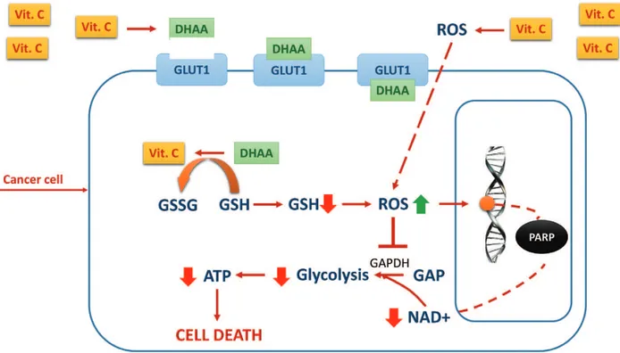
The theory was further investigated, very recently, by Yun et al. [17–19] who defined the chain of events leading vitamin C to behave as a prodrug of H2O2 thereby killing selectively cancer cells, both in vitro and in vivo. Briefly, vitamin C in high doses administered by intra-venous injection exerts its selective cytotoxic effect on cancer cells, because, after parenteral administration, it behaves as a peroxide delivery system for the generation of sustainable ascorbate radical and H2O2 in the extracellular space, with consequent oxidative damage to cancer cells (Figure 1). The selectivity of the cytotoxic effect of parenteral vitamin C depends on the fact that cancer cells, compared to their normal counterpart, show a reduced level of antioxidant enzymes such as catalase, superoxide dismutase, and glutathione peroxi-dase. The reduced level of antioxidant enzymes leads to cellular damage by accumulation of H2O2, with consequent intracellular redox imbalance and oxidative damage to different cellular structures.
Figure 1. Prooxidant effect of vitamin C (see text). Legend: Vit. C = vitamin C, ATP = adenosin triphosphate, DHAA =
dehydroascorbic acid, GSH = glutathione, GSSG = glutathione disulfide, GAP = glyceraldehyde 3-phosphate, GAPDH = glyceraldehyde 3-phosphate dehydrogenase, GLUT = glucose transporter, NAD = nicotinamide adenine dinucleotide, ROS = reactive oxygen species, PARP = poly ADP-ribose polymerase.
Yun et al. [17–19] have recently showed that the death of KRAS and BRAF cell mutants of CRC is imputable to the oxidized form of vitamin C: dehydroascorbic acid (DHAA). DHAA competes with glucose, for intracellular uptake by glucose transporters (GLUTs), mainly one and four subtype receptors. Interestingly, both KRAS and BRAF activating mutations are responsible of the upregulation of GLUT1 expression in different types of cancer, including CRC, although the upregulation of GLUT1 expression is not always associated with increased sensitivity of tumor cell lines to the cytotoxic effects of DHAA.
Investigation into the metabolic makeup of KRAS and BRAF CRC-derived cell lines shows that there is an accumulation of glycolytic intermediates upstream glyceraldehyde-3-phos-phate-dehydrogenase (GAPDH) and a contemporary depletion of the metabolites down-stream GAPDH, indicating an inhibition or severe reduction of its enzymatic activity, which appears to be the key of the cytotoxic effect of DHAA.
In summary, the data reported by Yun et al. on the effect of DHAA on CRC cell lines indicate that in glycolysis-addicted KRAS and BRAF mutated cell lines, high amounts of DHAA enter the cancer cells, thanks to the overexpressed GLUT-1 receptors. DHAA is then reduced again to vitamin C inside the cells. The reduction of DHAA to vitamin C scavenges glutathione (GSH), thus inducing redox imbalance and oxidative stress. Oxidative stress, in turn, leads to inactivation of GAPDH, inhibition of glycolysis, and energetic crisis, which leads to cancer cell death.
According to this mechanistic explanation, vitamin C, functioning as a prooxidant, would induce an increase in the intracellular reactive oxygen species (ROS), which leads to increased DNA damage, with consequent activation of poly ADP-ribose polymerase (PARP), an enzyme necessary to repair damaged DNA. PARP activation would in turn consume NAD+, with NAD+ depletion and consequent ADP depletion, leading to energetic crisis and death of can-cer cells [23].
The theory according to which vitamin C in high doses would act as a prodrug of H2O2, beyond being criticized by different authors, seems somewhat controversial and overlooks a few important aspects as follows:
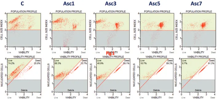
• Although reported in 2005 by Chen et al. [21, 22], it is not new, since, as we have seen, it had been already proposed by Benade et al. in 1969 [1]. Interestingly, while according to Benade, H2O2 forms inside the cell, starting from vitamin C, Chen et al. conclude that H2O2 is formed outside the cell, staring from DHAA. Our experience with high concentrations of vitamin C to treat, in vitro, retinoblastoma (Y79) (Figures 2 and 3) [24], uveal melanoma (C918, OCM1) [25], human promyelocytic leukemia (HL60) [26], and different human my-eloid leukemia (HL60, U937, K562, NB4, NB4-R1, and NB4/As) cell lines [27] indicates that H2O2 forms inside the cells, rather than outside. In fact, in our experiments, the cytotoxic effect of vitamin C on cancer cells in culture persists for hours/days after the removal of vitamin C from the culture medium.
• H2O2 is a metabolite normally produced by the cells of the body and usually overproduced by cancer cells. Therefore, H2O2 itself could be an optimal substitute for vitamin C, as an an-ticancer compound. In this regard, it could be useful remark that Reginald Holman, in 1957, published a paper in “Nature” showing that rat implanted with Walker 256 adenocarcinoma
Figure 2. Flow cytometric analysis of Y79 human retinoblastoma cell line viability, after treatment with increasing
concentrations of vitamin C (Asc) in vitro. C = control sample, Asc1 = vitamin C 1 mM, Asc3 = vitamin C 3 mM, Asc5 = vitamin C 5 mM, Asc7 = vitamin C 7 mM. Starting from a viability of about 74% (control sample), the percentage of viable cells after 1 hour of treatment with vitamin C and 18–24 hours of incubation are about 66% at 1 mM, 41% at 3 mM, 11% at 5 mM, and 9% at 7 mM of vitamin C.
Figure 3. Microphotographs of Y79 human retinoblastoma cell lines: A: hematoxylin/eosin staining of cultured Y79.
Interestingly, cells in culture tend to form the typical Flexner-Wintersteiner «rosettes» (black arrows) commonly seen in pathology specimends (original magnification: 200×). B: May-Grunwald Giemsa staining showing morphological details: large cells with loose chromatin and highly basophilic cytoplasm (original magnification: 400×). C: Contrast phase microphotographs of Y79 human retinoblastoma cell lines in culture (control sample): cells tend to form clusters in culture (original magnification: 200×). D: Contrast phase microphotograph of Y79 human retinoblastoma cell lines after treatment with vitamin C 7 mM. At a concentration of 7 mM, vitamin C destroys the vast majority of cells in culture. A few residual cells appear swollen and necrotic (original magnification: 200×). E and F: Hoechst/PI staining of Y79 human retinoblastoma cell line in the culture. With Hoechst/PI, live cells not distinguishable in the black/withe caption. In the control sample (E), there is a clear prevalence of cells stained by the Hoechst dye (alive) while in the sample treated with 7 mM of vitamin C (F), the prevalent color is given by PI (dead cells)(original magnification: 200×).
and treated by simply replacing their drinking water with 0.45% hydrogen peroxide showed a rate of cure of 50–60% [28]. The time for complete disappearance of the tumor varied from 15 to 50 days depending on the tumor size at the beginning of treatment. Holman’s work was based on the assumption (later confirmed by studies on vitamin C) that malignant cells are deficient in catalase, and as such unable to detoxify high fluxes of H2O2. As a conse-quence, an in vivo measurement of catalase activity in tumors would represent a useful di-agnostic tool to predict which cancers will respond to pharmacological doses of vitamin C therapy (or H2O2) [29].
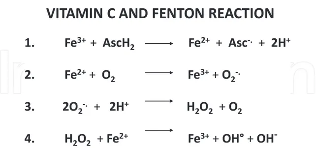
• According to the prooxidant theory, vitamin C in high concentrations induces the produc-tion of H2O2 through a Fenton-like reaction (Figure 4). This reaction is the oxidation of organic substrates by iron and hydrogen peroxide. Trivalent iron (Fe3+) is fundamental
for the reaction, but, since Fenton-like reactions are usually controlled, in vivo, because of iron sequestration by metal-binding proteins, the prooxidant effect of vitamin C, in vivo, is considered scarcely significant by different authors [30, 31], and other mechanisms should be hypothesized.
• Cancer cells produce high amounts of H2O2, and high levels of this metabolite have been associated with key features in cancer, such as DNA alterations, cell proliferation, apop-tosis resistance, metastatic spread, angiogenesis, and hypoxia-inducible factor 1 (HIF-1) activation. On the contrary, decreasing the cellular levels of H2O2 may reverse the ma-lignant phenotype. Therefore, H2O2 can be either proapoptotic or antiapoptotic, either carcinogenic or anticarcinogenic, depending on its concentration and localization within the cell [32, 33];
Figure 4. Fenton reaction mediated by vitamin C. (1) Vitamin C (ascorbic acid, AscH2) reduces ferric ions (Fe3+) to
ferrous ions (Fe2+). (2) Ferrous ion reacts with oxygen to produce superoxide. (3) Dismutation of superoxide leads to hydrogen peroxide. (4) Hydrogen peroxide reacts with ferrous ions to form hydroxyl radicals. Legend: Asc = ascorbic acid, vitamin C, Fe = iron, O = oxygen, H2O2 = hydrogen peroxide, OH = hydroxyl radical.
• According to the “Fenton chemistry,” invoked to explain the selective cytotoxic effect of vita-min C against cancer cells, trivalent iron (Fe3+) is necessary for the formation of H
2O2, starting
from vitamin C. However, some literature data seem to demonstrate that the exact oppo-site is true. In particular, Mojic et al. using two prostate cancer cell lines (LNCaP and PC-3) showed that iron at physiological concentrations in the cell culture medium and human plas-ma abrogates the anticancer/cytotoxic effects of vitamin C. According to these authors, iron at physiological concentrations promotes both production and decomposition of H2O2, the latter being mediated by a Fenton reaction, which prevents the accumulation of H2O2, thus abolishing the cytotoxic effect of vitamin C. Therefore, as the authors conclude, the in vitro in-vestigations on the anticancer properties of vitamin C may have been overestimated because all suffered the bias of a low amount of Fe3+ in the culture medium, if compared to normal
plasma and body fluids. To repeat in vivo the results obtained in vitro, the authors suggest that the simultaneous administration of vitamin C and chelating agents remove iron [34]. • Vitamin C (ascorbate) readily undergoes pH-dependent autoxidation producing
hydro-gen peroxide, and catalytic metals accelerate the oxidation process. This means that cata-lytic iron is not strictly necessary for the production of H2O2, starting from vitamin C, and therefore, the Fenton reaction may not be essential for this purpose. The autoxidation, i.e., oxidation in the absence of catalytic metals, occurs via the ascorbate dianion (Asc2−). In
par-ticular, that at pH 7.0, 99.9% of ascorbate (vitamin C) is in the form of monoanion (AscH−).
Asc2− increases by a factor 10, with one unit increase in the pH. Therefore, while the
pro-duction of H2O2 may be scarcely relevant in the absence of catalytic iron (as in the “Fenton chemistry”), it may become considerable when the concentration of ascorbate is in the or-der of the millimoles, as in the case of the use of vitamin C as an anticancer compound. To give an example, an aqueous solution containing 20 mM of vitamin C in the form of sodium ascorbate (the sodium salt of the ascorbic acid) will contain 1 μM of Asc2− which, in turn,
will result in a flux of H2O2 on the order of 10 nM/s in a typical cell culture experiment [35]. • In most laboratory settings, autoxidation of vitamin C is due to adventitious catalytic met-als, as part of the buffers used or contaminating of lab equipment. It is not by chance that the methods underpinning the “Fenton Paranoia” are in vitro methods using either isolated tissue cultures or d samples exposed to the air. Indeed, as reported by some authors: “…
unless extreme care is taken, every time vitamin C is added to blood samples (and urine samples) outside the body there are a whole host of oxidative products and markers produced …” [36].
• Both the antioxidant and prooxidant activities of vitamin C in high doses may not necessar-ily be mutually exclusive. Studies on chelation therapy have shown that 5 g of the sodium salt of vitamin C added to the ethylenediaminetetraacetic acid (EDTA) chelation cocktail results in acute oxidative stress, but this effect is transitory, and after multiple sessions of EDTA-based chelation treatment, a prolonged, protective, antioxidant effect of the treat-ment becomes evident [37]. These data confirm the evidence reported by Mojic et al.[34] regarding the inhibitory effect of iron on the prooxidant activity of vitamin C, and also the idea, formerly illustrated by Klenner, according to which vitamin C in high concentration may act as a “flash oxidizer” [38].
In summary, increasing the concentration of vitamin C with mega doses of the nutrient injected in vein may lead to a substantial increase in the spontaneous generation of H2O2, with toxic consequences to cancer cells, according to the chain of biochemical reactions more recently described by Yun et al., and briefly summarized herein. This does not mean that the administration of vitamin C in high doses by intravenous injection abrogates its anti-oxidant properties, but most probably that both proanti-oxidant and antianti-oxidant effects coexist. It rather implies that both antioxidant and prooxidant properties are simultaneously present in the molecule, the latter resulting more pronounced when the concentration of the nutrient reaches values in the order of millimoles (20 mM).
2.2. The antioxidant pathway
“Collagen” is a collective name to designate group of fibrous proteins that occur in verte-brates as the chief constituent of connective tissue fibrils and in bones. Many of the symptoms of scurvy (the syndrome of acute deficiency of vitamin C) depend on the defective produc-tion of collagen. Since the beginning of the history of this nutrient, scientists know that vita-min C is essential for the synthesis of collagen. Indeed, vitavita-min C is a cofactor of collagen prolyl-4-hydroxylase (C-P4H), the enzyme responsible for the formation of hydroxyproline, the essential component of collagen. Under conditions of vitamin C deficiency, C-P4H loses its activity, and the organism does not form collagen properly, with consequent connective tissue deterioration, as it happens in scurvy (Figure 4).
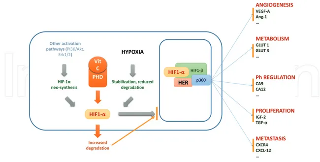
Prolyl-hydroxylases are an entire family of enzymes, also known as 2-oxoglutarate-depen-dent dioxygenases (2-OGDDs) with a wide range of biological functions [39], and members of this family include HIF-hydroxylases [40]. These vitamin C–dependent enzymes are of extreme importance in tumor biology since hypoxia and induction of HIFs are a hallmark of many tumors [41].
HIF is a heterodimeric transcription factor discovered in 1991 [42]. In normal oxygen pressure conditions (“normoxia”), the HIF-1α unit is downregulated by vitamin C–dependent hydrox-ylases, while in hypoxic conditions, such as those so frequently found in solid tumors, there is a repression of the hydroxylation of HIF-1α, with consequent increase of HIF-dependent gene transcription, neoangiogenesis, and tumor growth and progression. The important consequence of the central role of vitamin C in the synthesis of HIF-1α hydroxylases is that low levels of vitamin C promote tumor growth and progression (as already hypothesized by Stone, Szent-Györgyi, and Pauling), by reducing HIF-1α hydroxylation, thereby stabilizing HIF-1α [43], and high levels of HIF render cancer cells more sensitive to vitamin C–induced toxicity [44].
Recently, Kuiper et al. have confirmed the above data by finding an inverse relationship between intratumor levels of vitamin C and HIF activity in both endometrial cancer and CRC [45–47].
For a better understanding of the centrality of the relationship between hypoxia and HIF in tumor biology, we must consider that cancer hypoxia (a very common feature in cancer) is associated with HIF activity that mediates angiogenesis, epithelial-mesenchymal transition
(EMT), stem cell maintenance, invasion, metastasis, and resistance to radiation therapy and chemotherapy [48]. Therefore, attempts to downregulate HIF synthesis and activity may rep-resent a step forward in the search of an effective and selective anticancer drug [49, 50]. Is vitamin C such a molecule?
The current evidence shows that vitamin C has a close relationship with the function of HIF, and therefore, being a natural compound, it is the best-suited, natural molecule for the pur-poses of inhibiting cancer growth through HIF-mediated mechanisms.
Tian et al. note that the overexpression of HIF greatly enhances vitamin C–induced toxicity on cancer cells, since HIF increases the intracellular uptake of oxidized vitamin C through its transcriptional target GLUT1, synergizing with the uptake of its reduced form through sodium-dependent vitamin C transporters (SVCTs) [44].
Other authors, working with human leukemia cell lines, showed that vitamin C inhibits the growth of human leukemic cells not only through the generation of H2O2 but also and more importantly through the downregulation of HIF-1α transcription [51].
Further important insights into the role of HIF have come from studies of three tumori-genic models in vivo, showing that the antitumoritumori-genic effects of antioxidants such as N-acetylcysteine (NAC) and vitamin C are not due to their ability to squelch DNA damage and genomic instability mediated by ROS but due to their capacity to downregulate HIF levels [52].
These results are of extreme interest for the following reasons:
• Whether we consider its prooxidant activity, leading to cancer cell damage through the generation of H2O2 or its antioxidant (more typical) activity, leading to the enzymatic breakdown, and nonenzymatic downregulation of the HIF, vitamin C in high doses always shows the characteristics of a simple, natural, and effective anticancer molecule;
• Antioxidants (all of them!) have anticancer effects;
• Vitamin C and other antioxidants may have a role as adjuvant therapy to prevent progres-sion or recurrence of HIF-dependent tumors;
• Vitamin C may show anticancer properties even when administered by mouth, with caution about the dose, which must be sufficiently high.
Recent investigations have shown that ascorbate therapy has a significant effect on the expres-sion of several genes relevant to the development or inhibition of cancer. In particular, the reduced expression of tumor-promoting genes, such as HIF, and the increased expression of tumor-suppressor genes, such as p53, support the notion that vitamin C can act as a potential agent for the suppression of tumor development [53].
2.3. The epigenetic pathway
As we have seen, vitamin C has a number of beneficial biological functions, many of which are related to its action as an electron donor for adjusting the redox state of iron-containing
enzymes. Recent studies have implicated Fe2+-dependent oxidative modification activities in
normal tissue homeostasis and experimentally induced reprogramming. The loss of these activities is associated with epigenetic defects and compromised cell differentiation or devel-opmental potential (Figure 5).
One of the most important properties of Fe2+-2OG-dependent dioxygenases is their
suscep-tibility to environmental factors (as we have seen in the case of HIF-hydroxylases). Vitamin C is necessary to maintain the function of these enzymes, and different oncometabolites can inhibit their activity.
Recent studies show that vitamin C may enhance reprogramming of pluripotent stem cells, and the available data suggest a strong link between vitamin C, dioxygenase func-tion, and stem cell differentiafunc-tion, that is of great relevance for human disease [54]. In fact, vitamin C (in the form of sodium ascorbate), as a member of Fe2+- and 2OG-dependent
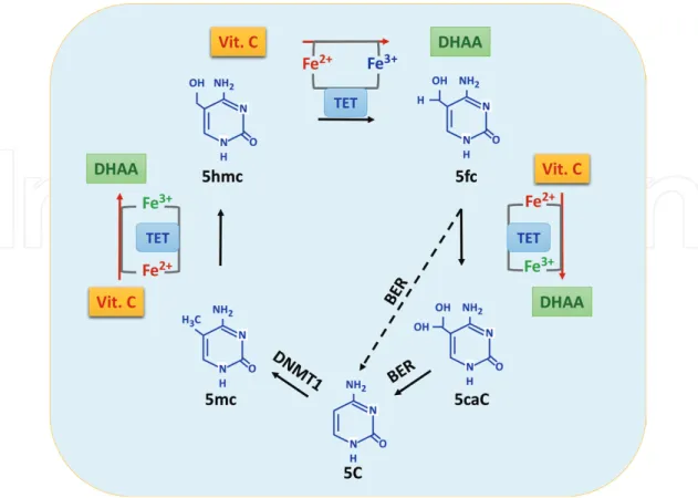
dioxygenases, plays a critical role in the demethylation of DNA and histone, as a cofactor for a group of enzymes termed methylcytosine dioxygenase ten-eleven translocation (TET, including TET1, TET2, and TET3) and some Jumonji-C (JmjC) domain-containing histone demethylases [55, 56].
(Note 1: TET1 catalyzes the conversion of the modified DNA base 5-methylcytosine (5-mC) to 5-hydroxymethylcytosine (5-hmC). TET1 produces 5-hmC by oxidation of 5-mC in an iron and alpha-ketoglutarate-dependent manner [8]. The conversion of 5-mC to 5-hmC has been proposed as the initial step of active DNA demethylation in mammals.)
(Note 2: TETs belong to the Fe2+- and 2OG-dependent dioxygenase superfamily.)
Figure 5. Hypoxia stabilizes HIF-1α as the rate of prolyl-hydroxylases (PHD)-mediated hydroxylation of a subunit is
limited. HIF-1α accumulates in the cytoplasm and passes into the nucleus, where it binds to HIF1-β. Cofactor p300 allows the binding to hypoxia response elements (HREs) of target genes involved in angiogenesis, metabolism, pH regulation, proliferation, and metastasis. Vitamin C, as a cofactor of PHD, determines increased degradation of HIF1-α and, as a consequence, inhibits the expression of genes activated by HIF1-α.
Given its role as an essential cofactor of enzymes involved in epigenetic gene regulation/ expression, vitamin C can be involved in embryonic development, postnatal development, aging, cancer, and other diseases [57].
In summary, the epigenetic gene regulation functions of vitamin C encompass:
• The regulation of DNA demethylation as an essential cofactor for TET dioxygenases; • The regulation of histone demethylation as an essential cofactor for JmjC
domain-contain-ing histone demethylases;
• The role of interface between the genome and environment;
• The critical role in maintaining the epigenome, especially at early embryonic stages;
• The contribution to different disease in the case of deficiency, which causes reduction of the catalytic activity of TET dioxygenases and JmjC domain-containing histone demethylases [58]. According to some authors, vitamin C causes widespread, consistent, and remarkably specific DNA demethylation of 1847 genes in human embryonic stem cells (hESCs), including impor-tant stem cell genes, with a clear bias toward demethylation at CpG island boundaries [59]. Other epigenetically relevant effects of vitamin C include:
• The development of dopamine neuron differentiation in fetal midbrain [60], the induction of pluripotent state in mouse embryonic stem cells [61, 62];
• The enhancement of the demethylating activity of 5-azacytidine, and induction of cytotox-icity [63, 64];
• The inhibition of the malignant phenotype on melanoma cells in vitro, by partially reestab-lishing the global content of 5-hydroxymethylcytosine (5-hmC) and the consequent altera-tion in the transcriptome [65];
• The upregulation of several microRNA (miRNA) involved in tumor suppression and drug resistance, the most prominent of which correlates with increased overall survival of breast cancer or nasopharyngeal carcinoma [66];
• The inhibition of the proliferation, migration, and epithelial-mesenchymal-transition (EMT) of lens epithelial cells by destabilizing HIF-1α [67].
2.4. The immunologic pathway
Vitamin C concentrations in the plasma and leukocytes rapidly decline during infections and stress, and vitamin C supplementation improves a number of different immunologic func-tions, including, among others: antimicrobial and natural killer (NK) activities, lymphocyte proliferation, chemotaxis, and delayed-type hypersensitivity. Furthermore, by maintaining the cellular redox integrity, vitamin C protects the cells of the immune system against ROS [68, 69]. Immunocompetent cells such as lymphocytes, neutrophils, and monocytes have vita-min C levels 10-–100-fold higher than the plasma, and accumulate it against a concentration gradient (Figure 6) [70].
A decrease in the intracellular content of vitamin C may result in locally increased apoptosis of immune cells and immunosuppression [71].
Vitamin C is essential for immunoglobulin synthesis [72] and active phagocytosis [73] enhances the production of interferon [74] and suppresses the production of interleukin-18 (IL-18), a key regulator in malignant skin tumors, including melanomas, squamous cells carcinomas, and a number of other tumors [75].
The concept that the immune system can help fighting cancer has deep roots. In 1909, the German scientist Paul Ehrlich proposed that the incidence of cancer would be much higher, were it not for the action of our immune system in recognizing and eliminating tumor cells [76]. Half a century later, two scientists, Lewis Thomas and Frank Macfarlane Burnet, took Paul Ehrlich’s original idea a step further and proposed the model of “immune surveillance,” according to which the cells of the immune system actively patrol the body looking for cancerous cells and eliminate them as they arise. This idea became a grounding principle of the new field of cancer immunology that took shape beginning in the 1950s [77].
More recently, the relationship between cancer and the host immune system has become clear with the introduction of the concept of “immune editing,” a three-phase process leading can-cer cells to escape the control of the immune system, with consequent cancan-cer progression [78].
Figure 6. As a cofactor for TET dioxygenases, vitamin C participates in the conversion of 5-mC to 5-hmC, and further
5fC and 5caC, thus modulating DNA demethylation. Legend to figure: 5C = cytosine, 5-mC = 5-methylcytosine, 5-hmC = 5-hydroxymethylcytosine, 5fC = 5-formylcytosine, 5caC = 5-carboxylcytosine, BER = base excision repair, DNMT1 = DNA methyltransferase-1, DHAA = dehydroascorbic acid, Vit. C = vitamin C.
Therefore, since no doubts exist, nowadays, about the role of the immune system in controlling cancer development, progression and spread, natural substances such as vitamin C, whose action spreads over a wide range of effectors of both the innate and adaptive immunity, repre-sent a new promising therapeutic tool against cancer.
Much of the “booster” action of vitamin C on the immune system depends on its anti-oxidant nature. Indeed, vitamin C downregulates ROS-dependent expression of pro-inflammatory interleukin genes, via inhibition of transcription of NF-κB (nuclear factor kappa-light chain-enhancer of activated B cells), which, in turn, regulates the expression of pro-inflammatory cytokines, such as IL-1 and tumor necrosis factor-alpha (TNFα) [79]. Recent evidence shows that vitamin C enhances antioxidant defenses of T-cells [80] and increases T-cell responsiveness to antigens, thus suggesting that it has a definite role in regulating immune function [81].
Confirm of the role of vitamin C in regulating the immune system function come from stud-ies showing that increased vitamin C concentrations inhibit antigen-induced, withdrawal-induced, steroid-withdrawal-induced, and spontaneous T-cell apoptosis [82], fas-induced apoptosis of monocytes [83], and increased cytotoxic activity of natural killer cells in humans [84]. All these data indicate that it can modulate the immune system by inhibiting T-cell apoptosis signaling pathways [85]. Similar results apply to monocyte-derived dendritic cells (DCs) [86]. As mentioned in the “historical background,” more than four decades ago, Pauling attrib-uted to vitamin C the role of a “booster” of the immune system. This was his main argu-ment against the negative results reported by the Mayo Clinic scientists, since they should not have included, in their clinical trials, cancer patients previously treated with chemotherapy, because chemotherapy destroys immunocompetent cells and severely impairs the individual immune response to cancer.
The Mayo Clinic scientists apparently did not understand this argument and, in reply to the legendary scientist wrote “… Any contention that previous chemotherapy prevented our patients
from achieving the extraordinary survival increase claimed by Drs. Cameron and Pauling must be con-sidered highly speculative at best. Our patients were entered into the study only when they were well past any acute immune-suppressive effects of previous therapy” [87].
The evidence, today, indicates that a healthy immune system is necessary to control the malignant disease and that immune suppression associated with cancer and cancer chemo-therapy contributes to its progression [88]. Therefore, in agreement with Pauling and the cur-rent evidence, it is likely to assume that since cancer chemotherapy produces long-lasting deleterious effects on the immune system [89, 90], patients previously treated with cytotoxic chemotherapy should not expect positive outcomes from the treatment with vitamin C as a “booster” of the immune system.
2.5. The collagen pathway
William McCormick first proposed, about 6 decades ago, the hypothesis that cancer could be due to a defect in the metabolism of collagen [91]. Given the fundamental role of vitamin C, in
the synthesis of collagen [92], the scientist concluded that a deficient intake of vitamin C (as in full blown or chronic subclinical scurvy) could determine cancer by disrupting the synthesis of collagen, thus creating a local permissive environment for cancer to grow and spread to other organs and tissues (Figure 7) [93].
As we have seen in the previous sections, vitamin C is required for collagen synthesis by act-ing as a cofactor for nonheme iron α-ketoglutarate-dependent dioxygenases such as prolyl-4-hydroxylase. It stimulates all types of collagen synthesis by donating electrons required for hydroxylation reactions of proline and lysine in procollagen, normally performed by specific hydroxylating enzymes.
In recent years, the role of basement membrane (BM) in the dynamic regulation of cell behav-ior and cell-signaling pathways has clearly emerged. The BM defines the tumor microen-vironment and provides significant host-derived regulatory signals during progression of tumor growth and metastasis. The major component of basement membrane is type IV colla-gen, and recent studies indicate that in cancer progression there is a disruption of the normal assembly and organization of the basement membrane [94].
These studies support the notion that the loss of collagen type IV chains may provide a per-missive microenvironment for cancer invasiveness, as hypothesized by McCormick, more than 5 decades ago [95].
Metastatic spread is a crucial event in the evolution of cancer, being responsible of the major-ity of cancer-related deaths. The steps leading to metastatic spread of cancer encompass the detachment of cancer cells from the primary tumor, the disruption of the BM, the invasion of the surrounding stroma, cancer cell entry into and transport through the vascular or lymphatic
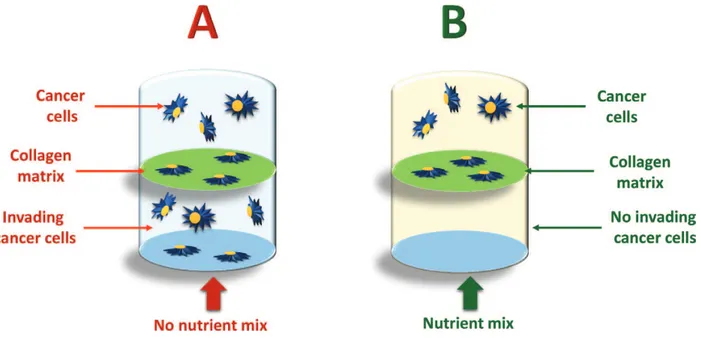
Figure 7. The “nutrient mix” used is a mixture of vitamin C, L-lysine, L-proline, and epigallocatechin gallate (EGCG).
Cancer cells can destroy enzymatically the collagen matrix (A) between the two chambers of the vial, and spread over the second chamber. When the nutrient mix is added (B), cancer cells are no longer able to destroy the collagen matrix and therefore they remain confined to the superior chamber (see Refs. [99, 100]).
system to distal sites (liver, lungs, brain, etc.), and extravasation, tumor cell proliferation and angiogenesis at distal sites [96].
A critical event in tumor cell invasion is the degradation of the extracellular matrix (ECM), a complex network of extracellular macromolecules such as collagen, proteoglycans, fibronec-tin, laminin, and other glycoproteins that act as a barrier to the spread of cancer cells to distal sites by restricting tumor growth and invasion.
Matrix metalloproteinases (MMPs) are calcium-dependent endopeptidases, which require coordination of a zinc ion to mediate catalysis. As implied by their name, MMPs operate on a variety of substrates belonging to the ECM [97], and owing essentially to their vast diversity, MMPs participate in nearly every biological process, involving the remodeling of the ECM, from implantation of an embryo into the uterine wall to tissue necrosis.
A major structural protein for ECM and basement membrane is type IV collagen. Therefore, type IV collagenases MMP-2 (72-kd gelatinase A) and MMP-9 (92-kd gelatinase B), usually overexpressed in malignancy, are the focus of research in this field.
All types of cancer cells form tumors and spread in the body by degrading the ECM by means of various matrix MMPs. The activity of these enzymes correlates with the aggressiveness of tumor growth and invasiveness of the cancer.
In 1992, Pauling and Rath hypothesized that natural compounds such as lysine and vitamin C could inhibit ECM proteolysis and, as such, had the potential to modulate cancer growth and spread [98]. These nutrients would exert their anticancer activity by both inhibiting MMPs and strengthening the connective tissue surrounding the tumor.
Several lines of evidence support an indispensable role for vitamin C in maintaining good-quality collagen. Vitamin C assists the posttranslational modification of collagen by reducing iron in the participating enzymes, lysyl-hydroxylase, and prolyl-hydroxylase. Experimental
in vitro data show incubation of cancer cells with a nutrient mixture containing vitamin C,
L-lysine, L-proline, and epigallocatechin gallate (EGCG), they are no longer able to invade the collagen matrix [99, 100] and spread at distant sites (Figure 7).
Although the mechanisms through which the nutrient mixture used in these experiments inhibits MMPs deserves further investigation, it is quite clear that the role of vitamin C is to stabilize collagen, and contribute to tumor cell toxicity, through one or more of the mechanisms illustrated in the previous sections.
Experiments on Gulo-knockout (GULO-KO) mice challenged with murine B16FO cancer cells show that vitamin C–supplemented mice developed smaller tumors with more collagen encapsulation and fibrous capsule inter digitation. On the contrary, Gulo-KO mice deprived of ascorbate hosted large tumors with poorly defined borders showed more necrosis and mitosis, [101], thus reinforcing the notion that vitamin C plays a central role in the prevention and control of tumor growth, progression, and metastatic spread.
To further confirm their data, the author showed that vitamin C-supplemented GULO-KO mice injected with B16FO melanoma cells demonstrated a significant reduction (by 71%, p = 0.005) in tumor metastasis compared to GULO-KO mice on the control diet [102].
2.6. The antitoxic/chemopreventive pathway
“Ascorbic acid is a potent detoxicant which counteracts and neutralizes the harmful effects of many
poi-sons in the body. It will combat various inorganic poipoi-sons, such as mercury and arsenic, and it neutral-izes the bad reactions of many organic poisons, drugs, and bacterial and animal toxins. Ascorbic acid detoxifies carbon monoxide, sulphur dioxide, and carcinogens, so it is the only immediate protection we have against the bad effects of air pollution and smoking” [3].
This sentence defines yet another way through which vitamin C can prevent and fight off can-cer, i.e., by neutralizing chemical carcinogens. Whether this effect depends on its antioxidant, anti-inflammatory effect, or yet other mechanisms, it is clear that vitamin C is an essential factor in cancer chemoprevention, and this cancer-preventive capacity is more likely associated with its protective effect against oxidative stress mediated by ROS [103].
Free radicals and other ROS are molecular species capable of independent existence that con-tains an unpaired electron in an atomic orbital. Free radicals derive either from normal meta-bolic processes or from external sources, such as exposure to X-rays, ozone, cigarette smoking, air pollutants, and industrial chemicals.
An antioxidant is a molecule stable enough to donate an electron to a rampaging free radical and neutralize it, thus reducing its capacity to damage. Antioxidants delay or inhibit cellular damage mainly through their free radical scavenging property.
Given this peculiarity, vitamin C, as noted by Stone, can combat the toxic effects of different organic and inorganic poisons, thus preventing and fighting cancer due to environmental pollutants. Recently, the US Department of Agriculture and the National Cancer Institute recommend the consumption of a minimum of five servings of fruit and vegetables to prevent cancer, with vitamin C being able to reduce the risk of stomach, mouth, pharynx, esophagus, lung, pancreas, and cervical cancers [104]. Furthermore, both epidemiologic and observational studies based on food intake provide evidence for a strong, protective role of vitamin C against cancer [105, 106]. Different studies show that the vitamin C is capable of preventing nitrosation and interfere with experimentally induced carcinogenesis [107, 108].
Vitamin C administered together with diethylnitrosamine (NDEA) shows an inhibitory effect on the experimental esophageal carcinogenesis in Wistar rats [109]. It also protects against the toxic effects of a number of pesticides/insecticides, including malathion [110], imidacloprid [111], endosulfan [112], dimethoate [113], fenvalerate [114], and many others.
More importantly, vitamin C reduces the toxic effects of different anticancer agents, includ-ing cisplatin [115, 116], cyclophasphamide [117], selenium-cisplatin conjugates [118] radiation [119], arsenic [120], doxorubicin [121], alkylating agents [122], and many others.
2.7. The “adjuvant” anticancer effect
As shown in the previous section, vitamin C protects normal cells from the oxidative, geno-toxic effects of chemotherapeutic agents, but this does not imply that it counteracts the cytotoxic effects of cancer chemotherapy and radiotherapy.
Regarding the clinical use of high doses of vitamin C in combination with standard antican-cer chemotherapy, for a long time, and even today, detractors of vitamin C (that are still a multitude, within the scientific community!) warn against the risk that antioxidants (such as vitamin C) may enhance cancer cell proliferation.
The role of micronutrients with antioxidant properties (including vitamin C) as a useful adjunct to conventional chemotherapy and /or radiotherapy has been controversial, essen-tially because they could protect cancer cells from the deleterious effects of free radicals generated by the therapy, thereby preventing cancer cell death.
After an exhaustive review of the literature, encompassing more than 44 scientific articles on the effectiveness of vitamin C alone, or with other vitamins, with chemotherapy, the authors concluded that: “… antioxidants [including vitamin C] do not protect cancer cells against free radical
and growth-inhibitory effects of standard therapy. On the contrary, they enhance its growth-inhibitory effects on tumour cells, but protect normal cells against its adverse effects” [123]. This literature
review suggests that the use of vitamin C alone with chemotherapy results in increased sur-vival, enhancement of chemotherapy, inhibition of tumor growth, decrease in the overall toxicity, modulation of genotoxicity linked to chemotherapy, distinct potentiating effect of chemotherapy, improved quality of life, and a whole series of other positive effects on the outcome of treated patients.
Recent evidence suggests that vitamin C can efficiently
• aid low-dose methotrexate (MTX) in inducing cell death in Hep3B cells [124], • synergize arsenic trioxide in acute promyelocytic leukemia [26],
• improve chemosensitivity of ovarian cancer, reducing, at the same time, the toxicity of chemotherapy [125],
• sensitize tumor cells toward cytostatic drugs [126], and
• improve the quality of life of patients undergoing chemo/radiotherapy [127, 128].
In a position paper published in the Journal of the American College of Nutrition in 2001, the authors highlight that “… none of the published data on the effect of antioxidants in com-bination with radiation or chemotherapeutic agents on tumour cells supports this hypoth-esis” [129]. As the authors observe, normal and tumor cells differ in their responses to antioxidants; low-dose and high-dose antioxidants differ in their effect on tumor cells, some actions of antioxidants on tumor cells are unrelated to scavenging of free radicals, and anti-oxidants have profound effects on the regulation of gene expression in tumor cells.
3. What to do next
As we have seen, the expectations generated from in vitro and animal studies on vitamin C still wait for the confirmation of clinical studies. The discrepancy between in vitro and in vivo results is due to several factors presented in the next.
3.1. The confusion about the dose
Among the many mystifying (and sometimes pseudoscientific) data regarding the anticancer effects of vitamin C, the most blatant is surely the one concerning the dose used. While it is clear that therm “high” designates doses of vitamin C (generally administered by intra-venous injection) leading to plasma concentrations in the order of millimoles (from one to several), current “institutional” clinical trials pass off as “high” doses of vitamin C of 1000 mg (1 g)! This is the case of a number of clinical trials, among which we could mention:
• A phase II trial of arsenic trioxide and ascorbic acid with temozolomide in patients with metastatic melanoma with or without central nervous system metastases [130],
• A clinical experience on the combination of arsenic trioxide and ascorbic acid in patients with refractory metastatic colorectal carcinoma [131],
• A phase I study on combination of decitabine, arsenic trioxide, and ascorbic acid for the treatment of myelodysplastic syndrome and acute myeloid leukemia [132], and many oth-ers. Clinical trials designed as the ones reported above are of no value in verifying the role of high doses of vitamin C in the treatment of cancer, since 1000 mg of the nutrient, even if administered in vein, is not a high (“pharmacologic”) dose.
3.2. The level of tissue oxygenation
If we assume that one of the main mechanisms through which vitamin C in pharmacologic doses is toxic to cancer cells is the production of H2O2, then oxygen becomes a fundamental part of the cytotoxic activity of vitamin C against cancer. Solid tumors often contain areas subjected to acute or chronic hypoxia. Although severe or prolonged hypoxia is deleterious, adaptation to a hypoxic microenvironment, allows cancer cells to survive and proliferate in a hostile milieu. More importantly, since cell culture experiments are usually performed in an oxygen-rich environment, while solid tumors usually show a very low content of oxygen, this difference in oxygen content may explain the different outcome in vitamin C cancer cell killing in vitro, compared to what happens in vivo [133]. Overcoming cancer hypoxia may therefore represent one of the main ways to improve the anticancer activity of high doses of vitamin C in clinical settings, as commonly realized with either hyperbaric oxygen (HBO) or ozonated autohemotherapy.
3.3. The pharmaceutical preparation
Sodium ascorbate, rather than ascorbic acid, may be the preferred preparation for intravenous injection. Ascorbic acid produces a very acidic solution, when dissolved in water or saline solution, and, as such, unsuitable for intravenous injection. Therefore, in order to obtain a neutral solution, it is necessary to buffer it with either sodium bicarbonate or sodium hydrox-ide. However, adding a buffer may represent a major problem, in terms of stability of the solution, and therefore the sodium salt of vitamin C, which produces a pH of around 7.0, is clearly preferable.
3.4. The administration schedule
According to some author, a constant “flow” of vitamin C in the blood works as if the body would produce the nutrient on its own (the “dynamic flow” hypothesis) [134]. The slow, con-stant infusion of vitamin C is the best option to maintain a stable plasma level of the nutrient, by intravenous injection [135] even though this approach is not common in the treatment of cancer patients with intravenous high doses of the nutrient. In fact, the great majority of the cancer clinical trials performed so far with intravenous vitamin C use the infusion of vitamin C on alternate days, withdrawing the treatment during the weekend. With this treatment modality, systemic conditioning (the accelerated metabolism or disposal of ascorbic acid) may occur after prolonged supplementation of high doses of vitamin C. Thus, if vitamin C supplementation were to cease abruptly, the accelerated disposal of the nutrient may create a deficiency state (“rebound scurvy”), and this may represent a serious inconvenience, when treating cancer patients. This is why, since Klenner’s experience with multiple administration routes, it may turn out to be useful to combine both intravenous and oral administration of large doses of vitamin C [7].
3.5. The glucose-ascorbate antagonism (GAA)
John Ely first proposed the glucose-ascorbate antagonism (GAA) theory in the 1970s [136]. According to this theory, the chemical structure of vitamin C and glucose is very similar and therefore they compete for the same transport system to enter the cells. As a consequence, both vitamin C and its oxidized form, DHAA, transported into different cell types (includ-ing adipocytes, erythrocytes, granulosa cells, neutrophils, osteoblasts, and smooth muscle cells), are inhibited by high blood glucose. Although in vivo studies are missing, investiga-tions on diabetic patients have confirmed the theory. Therefore, given the inverse relainvestiga-tionship between glucose and vitamin C blood levels, maintaining blood glucose levels within the normal range may greatly enhance the anticancer effect of vitamin C.
4. The efficacy of vitamin C in high doses against cancer: the facts
As a necessary premise to an evaluation of the anticancer properties of vitamin C, a realistic look at the state of the art on cancer chemotherapy can be helpful. The “war on cancer,” offi-cially declared by President Richard Nixon, with the National Cancer Act, in 1971, has been largely considered a failure, by the experts [137, 138], because of the following:
• The major improvements in survival rates mainly concern cancers of children and young adults, which account for 1.3% of all known cancers, and this has a little impact on the overall picture. Therefore, in most cases and for most forms of cancer, the war (the “war” metaphor) has been lost [139, 140].
• Targeted therapies (the “magic bullet” metaphor) are not curative because cancer usually adapts itself, becoming resistant to every new “weapon” used [141].
• Since the overwhelming majority of cancer is due to environmental, particularly lifestyle, factors, prevention, rather than cure, should be the foremost aim [142].
• The industry continues to be developed and the institutional organisms approve new can-cer drugs, based on marginal improvements in survival at an unsustainably high financial cost [143].
• Furthermore, cancer chemotherapy has an inherent toxicity, which, in many instances, encompasses, among others, nausea, vomiting, mucositis, hair loss, bone marrow toxic-ity, cardiac, neurologic, and renal toxictoxic-ity, and, in the long term, sterility and secondary malignancy.
• Finally, both early and recent reports demonstrate that cancer chemotherapy can be either ineffective/useless [144, 145] or definitely harmful [146, 147].
Compared to the current chemotherapeutic agents, vitamin C in high (“pharmacologic”) concentration has the following advantages:
• It is a natural compound that is usually produced by the vast majority of plants and animals, but not (no longer!) by humans. As any other natural product, it is neither pat-entable nor commercially exploitable.
• Both tissues and plasma of cancer/leukemia patients show reduced levels of this nutrient and therefore the routine administration of adequate amounts of vitamin C to these pa-tients is not only warranted, but highly desirable.
• High concentrations of vitamin C within tumor cells are associated with extended disease-free survival, while low concentrations are associated with aggressive tumor phenotype.
• It has no relevant side effects, with the exception of a slight diarrhea, a “guiding symptom” that indicates that the body “saturation” with the nutrient (“bowel tolerance”). To control the mild diarrhea that follows the body saturation threshold, it is sufficient to reduce or fractionate the doses, but in clinical practice, it is useful to maximize the effects of vitamin C, when assumed by mouth. Given the almost total absence of side effects, undue and sometimes laughable attempts to warn against its use have come, from time to time, from detractors of vitamin C, dealing with the possibility that several grams per day could lead to oxalate stone formation. In this regard, it will suffice to mention the European Food Safety Authority (EFSA) report n. EFSA-Q-2003-018, which, on this matter, clearly affirms: “No significant relationships were found in an analysis of data from 5214 men and 5785 women
between serum vitamin C concentrations and the prevalence of kidney stones” [148]. Moreover, the
hyperoxaluria associated with the use of high-dose vitamin C is primarily due to a labora-tory artifact, resulting from the conversion of vitamin C to oxalate ex vivo (i.e., after it has left the body, while it is in the collection bottle) [149]. The only contraindication to the treat-ment with high doses of intravenous vitamin C is the deficiency of glucose-6-phosphate dehydrogenase (G6-P-D), a rare genetic disorder in which a number of different drugs are usually contraindicated.
Beyond all these advantages, vitamin C in high doses is clearly cytotoxic for a large number of human tumor cell lines. At plasma concentrations achieved by intravenous administration, vitamin C induces death in 75% of 48 cancer cell lines tested in vitro [150], but has no toxic effect on human peripheral white blood cells, fibroblasts, or epithelial cells. This represents the realization of the dream of the “magic bullet,” even though the “scientific community” seems to continue to ignore it!
Regarding the anticancer efficacy of vitamin C in vivo, although the results of the first clinical trials have been rather disappointing, an unbiased analysis of the data currently available reveals an excellent safety profile, a clear-cut improvement of the quality of life [127], and a potentially important antitumor activity even though further, well-designed, controlled studies are strongly required.
5. Still looking for a “rationale”: is too much of it good for nothing?
Almost 50 years after the discovery of the anticancer properties of vitamin C, scientists are still looking for a rationale for the use of this nutrient in the treatment of cancer. However, as we have seen, this rationale not only exists, but it is also evidence-based, well-founded, com-plex, and variegated, given the many extraordinary benefits of vitamin C for human health. Therefore, faced with such an overwhelming evidence in favor of the efficacy of vitamin C against cancer, the question may become “why mega doses of vitamin C have not yet entered the routine clinical treatment of cancer?”
As sad as it may appear, the many advantages of vitamin C as an anticancer agent represent likewise limitations to its use in clinical practice. In fact, vitamin C is a natural compound, and this implies that no pharmaceutical company can effectively exploit it for commercial pur-poses. Drug companies must patent the molecular structure of the active ingredient of their products in order to make a profit. Natural substances, such as vitamin C, cannot undergo any patent submission procedure, because they exist in nature.
Another important issue may be the price: compared to the high costs of cancer drugs, some of which may reach the 30,000 USD for a single dose [151], vitamin C with its price ranging from 20 to 40 USD per kilogram (depending on the country) represents a real outsider within such an expensive market. No pharmaceutical company would ever invest in the clinical development of such an inexpensive product!
Going deeper into this apparent “lack of interest” for vitamin C as an anticancer compound, we can find the outstanding issue of its selectivity of action (the “magic bullet” principle); an aspect that still fascinates the clinical oncologists. As we have seen, contrary to the chemo-therapeutic agents, vitamin C kills cancer cells by exploiting a substantial metabolic difference between them and their normal counterpart; a property virtually unknown to the vast majority of the chemotherapeutic agents of common use in clinical practice!
The ethical implications of the above considerations are clear, and we will not discuss them herein. However, there are aspects, in this incomprehensible “indifference” to vitamin C as an anticancer molecule, which go far beyond plausibility and common sense.
One is surely the evidence that vitamin C deficiency is common in patients with advanced cancer, and, at the same time, patients with low plasma concentrations of vitamin C almost invariably show shorter survival, if compared to those with normal/higher concentrations [152]. Should not this evidence alone compel the clinicians to use vitamin C supplementation in cancer patients on a routine basis? A vitamin C deficiency, as we have seen in the first sec-tion of this chapter, is most probably in play in the genesis and development of cancer, and traces of this deficiency often remain, unless patients use supplements on their own, in blood and tissues of affected individuals. Is not this data alone sufficient to warrant the routine administration of vitamin C to cancer patients?
The other, not less relevant, aspect is the safety of vitamin C. The LD50 for a mouse (who normally produces its own vitamin C), is more than 3.3 g/kg of body weight, but most prob-ably, even more than that, for mammals not producing their own vitamin, such as humans. This has clearly emerged from clinical studies on intravenous injection of mega doses of the nutrient, which have also showed a definite and unequivocal improvement in the quality of life of the treated patients.
The last aspect, regarding the importance of vitamin C in the treatment of cancer, is the demonstration of the capacity of this nutrient to reduce the side effects and improve the anticancer activity of conventional chemotherapeutic agents, when combined with them [125].
Do we really need more information to introduce high doses of vitamin C in the routine treat-ment of cancer?
Acknowledgements
This chapter has been realized, in part, thanks to the financial support of the “Pescara Cell Factory Foundation,” and the “Associazione Italiana dei Genitori dei bambini affetti da Retinoblastoma” (AIGR).
Author details
Domenico Mastrangelo1*, Lauretta Massai1, Giuseppe Fioritoni2 and Francesco Lo Coco3
*Address all correspondence to: [email protected]
1 Department of Medical, Surgical and Neurological Sciences, University of Siena, Polo Scientifico San Miniato, Siena, Italy
2 Pescara Cell Factory Foundation Onlus, Corso Vittorio Emanuele, Pescara, Italy
References
[1] Benade L, Howard T, Burk D. Synergistic killing of Ehrlich ascites carcinoma cells by ascorbate and 3-Amino-1, 2, 4-triazole. Oncology. 1969;23:33-43
[2] Panel on Dietary Antioxidants and Related Compounds; Subcommittee on Upper Reference Levels of Nutrients; Subcommittee on Interpretation and Uses of DRIs; Standing Committee on the Scientific Evaluation of Dietary Reference Intakes; Food and Nutrition Board; Institute of Medicine. Dietary Reference Intakes for Vitamin C, Vitamin E, Selenium, and Carotenoids. 2000; 529 pages, ISBNs: Paperback: 978-0-309-06935-9; Hardcover: 978-0-309-06949-6. DOI:https://doi.org/10.17226/9810
[3] Stone I. The Healing Factor. “Vitamin C” Against Disease. New York: Grosset and Dunlap Inc.; 1972
[4] Stone I. Eight decades of scurvy. The case history of a misleading dietary hypothesis. Orthomolecular Psychiatry. 1979;8:58-62
[5] Chatterjee IB. Evolution and the biosynthesis of ascorbic acid. Science. 1971;182:1271-1272 [6] Stone I. The genetics of scurvy and the cancer problem. Journal of Orthomolecular
Psychiatry. 1976;5:183-190
[7] Klenner F. The treatment of poliomyelitis and other virus diseases with vitamin C. Southern Medicine and Surgery. 1949;111:209-214
[8] Jungeblut CW. Further observations on vitamin C therapy in experimental poliomyeli-tis. Journal of Experimental Medicine. 1937;66:459-477
[9] Pauling L. Are recommended daily allowances for vitamin C adequate? Proceedings of the National Academy of Sciences of the United States. 1974;71:4442-4446
[10] Cameron E, Campbell A. The orthomolecular treatment of cancer II: Clinical trial of high dose ascorbic acid supplement in advanced human cancer. Chemico-Biological Interactions. 1974;9:285-315
[11] Cameron E, Pauling L. Supplemental ascorbate in the supportive treatment of cancer: Prolongation of survival times in terminal human cancer. Proceedings of the National Academy of Sciences of the United States. 1976;73:3685-3689
[12] Cameron E, Pauling L. Supplemental ascorbate in the supportive treatment of cancer: Reevaluation of prolongation of survival times in terminal human cancer. Proceedings of the National Academy of Sciences of the United States. 1978;75:4538-4542
[13] Cameron E, Pauling L. Cancer and Vitamin C. New York: W. W. Norton & Co.; 1979. [Updated and Expanded Cancer and Vitamin C, Cameron E, Pauling L, Camino Books, Inc., Phila., PA 19102; 1993]