UNIVERSITY
OF
CATANIA,
SCHOOL
OF
MEDICINE
P
H
D
P
ROGRAM IN
P
RECLINICAL AND
C
LINICAL
P
HARMACOLOGY
XXIII Cycle Beatrice Nardone
H
ISTOPATHOLOGIC ANDI
MMUNOHISTOCHEMICALC
HARACTERIZATION OFR
ASH TOH
UMANE
PIDERMALG
ROWTHF
ACTORR
ECEPTOR1
(HER1)
ANDHER1/2
I
NHIBITORS INC
ANCERP
ATIENTSPhD Thesis
Coordinator:
Prof. Renato Bernardini Tutor:
Prof. Giuseppe Micali Co-Tutor:
Prof. Dennis P. West
Academic Year 2009/2010
Beatrice
Nardone
Digitally signed by Beatrice Nardone
DN: cn=Beatrice Nardone, o, ou, email=b-nardone@northwestern. edu, c=US
TABLE OF CONTENTS
1. INTRODUCTION...2
1.1. HERTARGETED AGENTS... 3
1.2. EFFECTS OF HERINHIBITORS ON SKIN... 7
1.3. PAPULOPUSTULAR RASH DUE TO HER INHIBITORS... 9
2. EXPERIMENTAL DESIGN...13
3. MATERIALS AND METHODS ...14
4. RESULTS ...20
5. DISCUSSION AND CONCLUSIONS ...28
6. REFERENCES...35
ACKNOWLEDGEMENTS ...43
APPENDIX A ...44
IMMUNOHISTOCHEMICAL DATA FROM ACISII... 44
APPENDIX B ...74
2
1. INTRODUCTION
Human epidermal receptor (HER) also known as EGFR, is a 170-kd transmembrane glycoprotein that belongs to the ErbB family of receptor tyrosine kinases. (1, 2). It consists of an extracellular ligand-binding domain, a hydrophobic transmembrane domain, and an intracellular domain that possesses tyrosine kinase activity. It is activated by EGF-like ligands, including EGF, transforming growth factor alpha, amphiregulin, heparin-binding EGF-like growth factor,
betacellulin, and epiregulin. Overexpression of HER1, and alterations
in its signaling networks, has detrimental effects on normal cell
growth, resulting in tumorigenesis (2). HER normally plays an
important role in the control of cell growth and differentiation (1). Human epidermal receptor inhibitors (HERi) have shown effectiveness against a wide variety of solid tumors. In particular, they are used in malignancies overexpressing HER1 and 2, including head and neck, breast, lung, pancreatic, and colorectal cancers (3, 4).
The rationale for the use of HER1i in anticancer therapy is their attenuation effect in HER signaling pathways that lead cell differentiation, proliferation, migration, angiogenesis, and apoptosis (5, 6).
3
1.1. HERTARGETED AGENTS
Two main classes of HER targeted agents for cancer therapy have been developed to date: extracellular and cytoplasmic. In particular, monoclonal antibodies (mAb) block the extracellular domain of the receptor, thereby preventing ligand-dependent activation and downstream signaling, while small molecule, Tirosyn Kinase inhibitors (TKI) that are orally administered, are low molecular weight compounds directed against the intracellular tyrosine kinase domain blocking the intracytoplasmic ATP-biding site on the receptor, thereby preventing downstream signal transduction (7).
Two anti-HER1 monoclonal antibodies, cetuximab and panitumumab, have been approved by the U.S. Food and Drug Administration (FDA) and European Medicines Agency (EMEA) to date. Cetuximab, a chimeric human-murine IgG1 monoclonal antibody that binds to the extracellular domain of HER, is approved for second-line treatment of metastatic colorectal carcinoma as well as for advanced squamous cell carcinoma of the head and neck (8). Panitumumab is a fully human IgG2 monoclonal antibody approved for treatment of metastatic colorectal carcinoma refractory to standard chemotherapy (9). A
4
number of other HER-binding monoclonal antibodies, including zalutumumab, nimotuzumab and matuzumab, are currently in clinical development.
Of the small molecule HER1 tyrosine kinase inhibitors, erlotinib is indicated as second-line therapy for advanced non-small cell lung cancer (NSCLC) and in combination with gemcitabine for advanced pancreatic cancer (10). Gefitinib was approved in 2003 for the treatment of locally advanced or metastatic NSCLC refractory to both platinum-based and docetaxel chemotherapies; however, in 2005 the FDA labeling was modified to only allow continued treatment of those patients who are benefiting or have benefited from gefitinib therapy (11).
Lapatinib ditosylate is an oral dual kinase inhibitor targeting both the HER1 and HER2 receptors. Increased expression and activation of HER1 and HER2 in breast cancer are associated with a high risk for recurrence after primary treatment and consequently a poor clinical outcome (12). HER1 is reportedly overexpressed in up to 30% of breast tumors and HER2 is reportedly overexpressed in up to 25% of the 1.5 million new breast cancers that are diagnosed annually worldwide (13). Lapatinib reversibly binds to the intracellular cytoplasmic ATP-binding site of the tyrosine kinase domain and
5
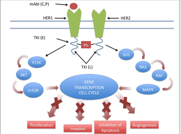
blocks receptor phosphorylation and activation, thereby blocking downstream signaling pathways, namely, simultaneous activation of extracellular signal–related kinase 1/2 and phosphatidylinositol 3 kinase/Akt (14) (Fig. 1) .
6
Figure 1. Mechanism of action of inhibitors of HER1 and HER 2.
mAbI= monoclonal antibody inhibitor; TKI=tyrosine kinase inhibitor; Ph= phosphorylation site; HER=human epidermal receptor; L= lapatinib; E= erlotinib; C=cetuximab; P= panitumumab. Monoclonal antibodies bind the extracellular domain, preventing ligand binding and subsequent activation of the receptor. Moreover, receptor internalization and degradation occurs after antibody binding. TKIs bind the intracytoplasmic domain of the receptors, preventing docking of the phosphate group, thereby abrogating activation and downstream signaling.
7
1.2. EFFECTS OF HERINHIBITORS ON SKIN
In skin, HER1 is primarily expressed in basal and suprabasl keratinocytes, sebocytes, and the outer root sheath of the hair follicle (15). Activation of HER1 by its ligands, epidermal growth factor (EGF), transforming growth factor-α (TGFα), amphiregulin, and heparin-binding EGF (HBEGF), has been shown to regulate keratinocyte proliferation, differentiation, migration and survival (16). HER-driven proliferation results in downstream activation of PI3K (phosphatidylinositol 3-kinase) -Akt and MAPK (mitogen activated protein kinase) pathways regulating keratinocyte survival, proliferation, and differentiation.
Blockade of HER1 in skin induces apoptosis in normal keratinocytes, which increases five-fold between therapy days 4 and 12, and which correlates with median time to rash onset in patients (17). Increased chemokine expression after HER1 blockade has been shown to be regulated by Extracellular Regulated Kinase 1 and 2 (ERK1/2), resulting in enhanced skin inflammation (18). HER1 inhibition induces early differentiation by upregulating the expression of terminal differentiation markers, such as KRT1 (keratin1) and KRT10 (19). In addition, increased STAT3 staining in the basal layer
8
of the epidermis (20) occurs, indicative of premature differentiation. Decreased expression of cytoskeletal proteins, f-actin-binding protein vinculin and the actin-binding protein ACTN1 (actinin-α1), resulting in decreased motility (21) ensues, along with increased attachment via cadherin-associated protein CTNND2 (catenin-δ2) and DSG2 (desmoglein 2) (22).
9
1.3. PAPULOPUSTULAR RASH DUE TO HER INHIBITORS
Although HERi are usually well-tolerated compared to standard cancer therapies such as chemotherapy and radiation, they may be associated with significant side effects. Cutaneous reactions are the most common adverse effects, and are associated with all HER inhibitors. Consequently, dermatologic toxicities are considered to be a class-specific side effects that are likely due to HER inhibition. Cutaneous and mucous membrane toxicities most commonly associated with HERi include papulopustular rash, paronychia, hair changes, dry skin, hypersensitivity reacton and mucositis (23).
Other common side effects include diarrhea, nausea, vomiting and asthenia.
The most common clinical toxicity associated with the use of HERi is a papulopustular (sometimes referred to as acneiform) rash that develops in up to 90% of patients (24-26).
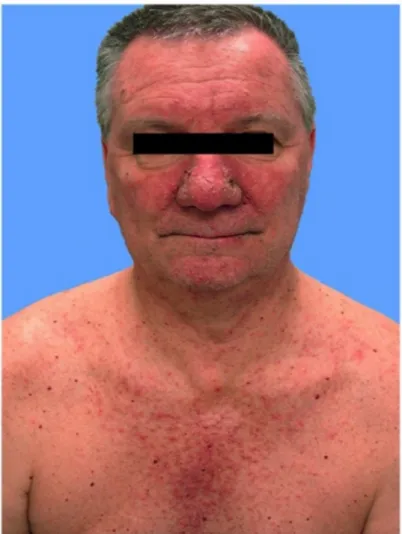
This rash is clinical characterized by erythematous papulopustules in 45-100% of patients and commonly affects the face and upper body (back and chest) (Fig.2) while scalp, extremities, abdomen and buttocks are affected less frequently (27).
10
Figure 2. Clinical presentation of papulopustular rash due to HER inhibitors.
HER-induced skin rash typically occurs within 8-10 days with a peak at 2 weeks of treatment. The most commonly involved sites are face, upper chest and back. Permanent scarring generally does not occur; however, significant scarring and postinflammatory hyperpigmentation following resolution of severe rash have been described (23, 28). Papulopustular eruptions seem to be
dose-11
dependent (29).
Histologic analysis of HERi-induced skin rash is characterized by a suppurative folliculitis. An early finding may consist of T lymphocyte infiltration surrounding the follicular infindibulum. Consequently, a predominantly neutrophilic infiltrate is seen that may also include lymphocytes and histiocytes, that surround follicles. Severe eruptions may reveal destruction of follicles.
In contrast to the findings in acne vulgaris, sebaceous glands are not affected in HERi-induced papulopustular rash. Thinning of the stratum corneum and a loss of the normal basketweave appearance is observed. Enlarged follicles may contain keratin plugs (28, 30-32). The mechanism by which HER1i are associated with cutaneous toxicity is unknown, but the alteration of physiologic HER1-mediated signaling processes in the epidermis and hair follicle seems to play a key role. It has been shown that HERi leads to a decrease in expression of Ki-67 within the epidermis, thereby reflecting inhibition of keratinocyte proliferation, as well as leading to induction of p27, a cyclin-dependent kinase inhibitor that arrests the growth of keratinocytes (30, 33).
Papulopustular rash due to HER1i has a negative impact as it relates to quality of life (34), cost (35), secondary infections (36) and
12
ability to mantain antineoplastic therapy without interruption (37), all of which may also affect clinical outcome.
13
2. EXPERIMENTAL DESIGN
Clinically, HER1/2 inhibition by lapatinib results in a lower incidence of rash (41%) (38), when compared to the HER1 inhibitors erlotinib, cetuximab and panitumumab 75-90% (24-26). Based on rash severity, patients on HER1/2i are less likely to require dose modifications when compared to HER1i.
The mechanisms responsible for this differing skin toxicity profile remain unknown, as preclinical data show a similar inhibitory profile for these agents.
This study investigated histological and immunohistochemical characteristics that may explain some of the clinical differences in skin toxicity between HER1 inhibitors (HER1i) and the HER1/2 inhibitors (HER1/2i).
14
3. MATERIALS AND METHODS
PATIENTSUpon IRB approval, existing medical records including archived skin biopsy specimens, dermatopathology reports of patients with rash attributed to treatment with lapatinib, cetuximab, panitumumab or erlotinib were analyzed. All patients were seen between January 2006 and December 2007. A total of 8 samples per patient/inhibitor were collected, with a total of 32 patient specimens analyzed for this study. Anatomic site of skin biopsy specimens was variable and based on the location of rash, with a majority located on the upper trunk.
IMMUNOHISTOCHEMISTRY
For each subject, immunohistochemical (IHC) studies were performed on 5 µ m sections of formalin-fixed paraffin-embedded tissue by using an Envision kit (Dako, Carpinteria, CA), a peroxidase-conjugated polymer detection system and diaminobenzidine (DAB) as chromagen on a Dako autostainer. An automated cellular imaging system (ACIS II; ChromaVision Medical Systems, Inc, San Juan Capistrano, CA) was used to quantify the staining of each molecular marker. The ACIS II software also calculates the average percentage
15
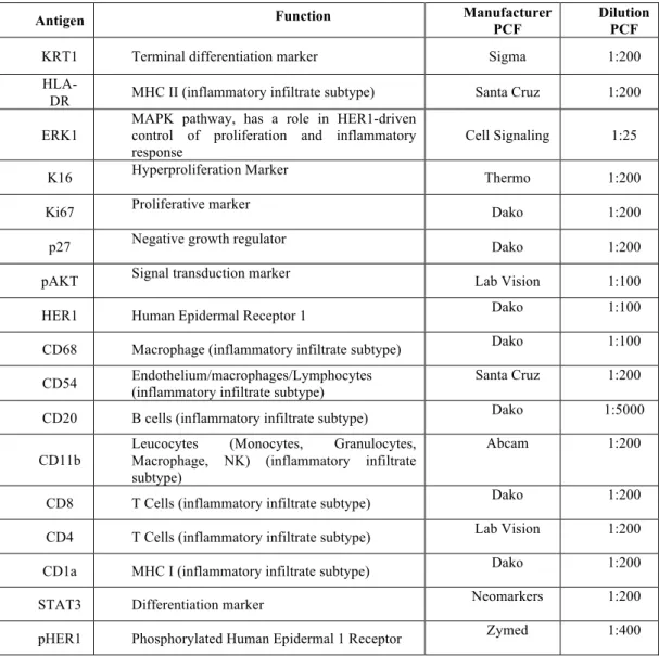
and intensity of stained cells. Positive staining was calculated by applying two thresholds with one recognizing blue background (hematoxylin stained) on cells and another recognizing brown (DAB) positive cells. The percentage of positivity was the area detected by the brown threshold divided by the sum of the area detected by the brown and blue thresholds. The intensity was calculated by masking out all areas not selected by the brown threshold and calculating the integrated optical density of brown within the remaining area. This value was divided by the area in pixels of the brown mask to calculate an average intensity of a selected area (39). Seventeen IHC biomarkers were used for each case and included Keratin 1 (KRT1), HLA-DR, Extracellular Regulated Kinase 1 (ERK1), K16, Ki67, p27, pAKT, HER1, pHER1, CD68, CD54, CD20, CD11b, CD4, CD8, CD1a, and STAT3 (Table 1).
16
Antigen Function Manufacturer PCF Dilution PCF
KRT1 Terminal differentiation marker Sigma 1:200
HLA-DR MHC II (inflammatory infiltrate subtype) Santa Cruz 1:200 ERK1
MAPK pathway, has a role in HER1-driven control of proliferation and inflammatory
response Cell Signaling 1:25 K16 Hyperproliferation Marker Thermo 1:200 Ki67 Proliferative marker Dako 1:200 p27 Negative growth regulator Dako 1:200 pAKT Signal transduction marker Lab Vision 1:100 HER1 Human Epidermal Receptor 1 Dako 1:100 CD68 Macrophage (inflammatory infiltrate subtype) Dako 1:100 CD54 Endothelium/macrophages/Lymphocytes (inflammatory infiltrate subtype) Santa Cruz 1:200 CD20 B cells (inflammatory infiltrate subtype) Dako 1:5000 CD11b
Leucocytes (Monocytes, Granulocytes, Macrophage, NK) (inflammatory infiltrate subtype)
Abcam 1:200
CD8 T Cells (inflammatory infiltrate subtype) Dako 1:200 CD4 T Cells (inflammatory infiltrate subtype) Lab Vision 1:200 CD1a MHC I (inflammatory infiltrate subtype) Dako 1:200 STAT3 Differentiation marker Neomarkers 1:200 pHER1 Phosphorylated Human Epidermal 1 Receptor Zymed 1:400
Table 1. Immunohistochemical markers analyzed. KRT1=Keratin 1; ERK=, Extracellular Regulated Kinase 1; HER1=Human Epidermal Receptor 1; pHER1=Phosphorylated Human Epidermal Receptor 1.
17
HISTOPATHOLOGY
Each biopsy was a 4-mm archived skin specimen obtained for research purposes with Northwestern University IRB approval. Following formalin fixation and paraffin embedding, staining with hematoxylin and eosin was performed and specimens were submitted to two dermatopathologists for independent blinded assessment of the epidermis, dermis, follicle and inflammatory infiltrate (P.G. and J.G). Biopsies were separately evaluated for the presence of epidermal, dermal, follicular, eccrine gland, and sebaceous gland alterations. Specifically, the epidermis was evaluated for presence of ulceration, parakeratosis, acanthosis, epidermal atrophy, dysmaturation, dyskeratosis, and infiltrates of neutrophils, monocytes, or eosinophils. The dermis was evaluated for the presence of neutrophilic, monocytic, or eosinophilic infiltrates. The follicle was evaluated for bacterial colonies/concretions, neutrophilic pustules, dysmorphic features, dyskeratosis, and neutrophilic, monocytic, and eosinophilic follicular infiltrates. Finally, eccrine and sebaceous glands were evaluated for inflammatory infiltrates; eccrine glands were also evaluated for necrosis and dyskeratosis. Histologic features were rated as 0 (absent) or 1 (present), with the exception of infiltrate, rated from 0 (absent) to 3 (most prominent). If a specific structure such as a follicle or eccrine
18
gland was not present in the specimen, the case was not included in the statistical analysis.
STATISTICAL ANALYSIS
Seventeen IHC biomarkers and 23 dermatopathology features were statistically analyzed for the 8 specimens in each of 4 drug treatment groups. The biomarkers were continuous and analyzed using analysis of variance methods. For each specimen positive staining calculations were determined. A two-factor nested repeated measures analysis of variance was used, with drug as the between subject factor, with subject nested within drug and skin layer (epidermis or dermis) as the within subject factor. A drug by skin layer interaction term is included in this analysis to determine whether the pattern of differences in means across drugs differed by skin layer. In addition, p-values for main effects of drug and of skin layer are reported. Within each layer, a one-way analysis of variance (ANOVA) was performed to determine differences across drugs. A p-value comparing the dual HER1/2i (lapatinib) with all other single HER1i combined was also reported. Pairwise comparisons were done across drugs using independent sample t-tests without correction for multiple comparisons. In all these analyses, separate variances were estimated
19
for each drug-skin layer combination. Histopathologic features were compared across groups using Fisher’s exact test. P-values less than 0.05 were considered statistically significant.
20
4. RESULTS
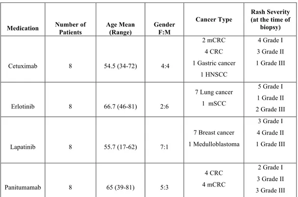
PATIENTSA total of 32 specimens were analyzed, 8 for each HERi. Complete demographic data for these patients is shown in Table 2. For patients on erlotinib, rash severity was grade 1 (n=5, 63%), grade 2 (n=1, 13%), grade 3 (n=2, 25%); for cetuximab, severity was grade 1 (n=4, 50%), grade 2 (n=3, 37.5%), grade 3 (n=1, 12.5%); for panitumumab, grade 1 (n=2, 25%), grade 2 (n=3, 37.5%), and grade 3 (n=3, 37.5%); and for lapatinib, severity was grade 1 (n=3, 37.5%), grade 2 (n=4, 50%), and grade 3 (n=1, 12.5%).
Medication Number of Patients Age Mean (Range) Gender F:M
Cancer Type
Rash Severity (at the time of
biopsy) Cetuximab 8 54.5 (34-72) 4:4 2 mCRC 4 CRC 1 Gastric cancer 1 HNSCC 4 Grade I 3 Grade II 1 Grade III Erlotinib 8 66.7 (46-81) 2:6 7 Lung cancer 1 mSCC 5 Grade I 1 Grade II 2 Grade III Lapatinib 8 55.7 (17-62) 7:1 7 Breast cancer 1 Medulloblastoma 3 Grade I 4 Grade II 1 Grade III Panitumamab 8 65 (39-81) 5:3 4 CRC 4 mCRC 2 Grade I 3 Grade II 3 Grade III
Table 2. Patient characteristics (N=32). mCRC=metastatic colorectal cancer; HNSCC= Head and neck squamous cell carcinoma; mSCC= metastatic squamous cell carcinoma.
21
DUAL HER1/2 INHIBITION IS ASSOCIATED WITH INCREASED PAKT,
DECREASED K16 AND P27 IN SKIN
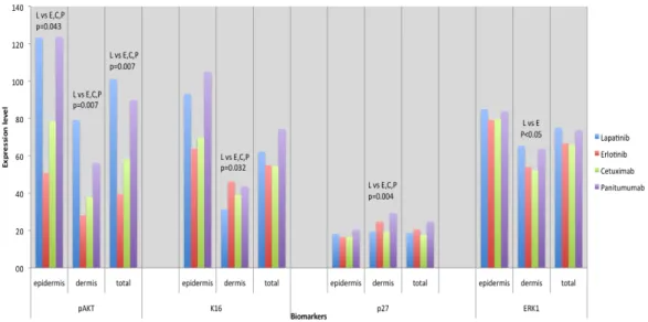
Significant IHC results are summarized in Figure 3. When comparing HER1/2i to the 3 HER1i, IHC analysis revealed a significantly increased expression of pAKT in both epidermis (p=0.043) and dermis (p=0.007) (Fig. 4), and a decreased expression of K16 (p=0.032) and p27 (p=0.04) in the dermis. Also, HER1 expression was significantly lower in the epidermis (p=0.03) and significantly higher in the dermis (p=0.0002).
Figure 3. Most notable immunohistochemical results.
Immunohistochemical analysis highlighting significant differences in epidermis and dermis between different agents. L= lapatinib; E= erlotinib; C=cetuximab; P=panitumumab; pAkt= phospho Akt; K16=keratin 16; ERK1= Extracellular Regulated Kinase 1
22
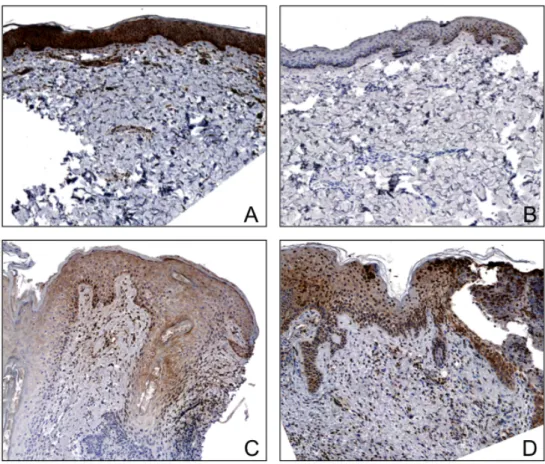
Figure 4. Immunohistochemistry of representative specimens for each HER inhibitor for pAKT.
A) Specimen from a patient on lapatinib showing increased pAKT expression in the epidermis as compared to cetuximab (B), erlotinib (C), and panitumumab (D), (X40 magnification).
ERK1 EXPRESSION IN THE DERMIS IS HIGHER FOR LAPATINIB COMPARED TO ERLOTINIB
When comparing low molecular weight HER inhibitors, a significantly higher expression of ERK1 in the dermis (p=0.028) was detected for lapatinib compared to erlotinib.
23
VARIATIONS IN MARKER EXPRESSION BETWEEN HER INHIBITORS
Lower expression of pHER1 (epidermis), CD68 (dermis), CD54 (in both layers), and CD4 (dermis) was observed for samples from patients on cetuximab when compared to panitumumab (p<0.05). A higher expression of CD8 was found in the dermis for panitumumab when compared to erlotinib and a higher expression of CD1a (epidermis) was found for panitumumab and lapatinib compared to cetuximab (p<0.05). Decreased expression of Ki67 was noted for cetuximab compared to lapatinib (epidermis) (p<0.05). STAT3 expression was decreased for cetuximab when compared to panitumumab in the epidermis and for cetuximab compared to all three other drugs in the dermis (p<0.05).
ATROPHY, DYSKERATOSIS AND DYSMATURATION IN EPIDERMIS ARE LESS PROMINENT WITH HER1/2I
To determine differences in cutaneous architecture, archived histopathologic specimens from 8 patients on each HERi were analyzed. Detailed histopathologic analyses are shown in Table 4 with representative histologic sections in Figure 5.
Total Number of Cases
Histopathologic Finding Cetuximab Erlotinib Lapatinib Panitumumab 1pvalue 2pvalue
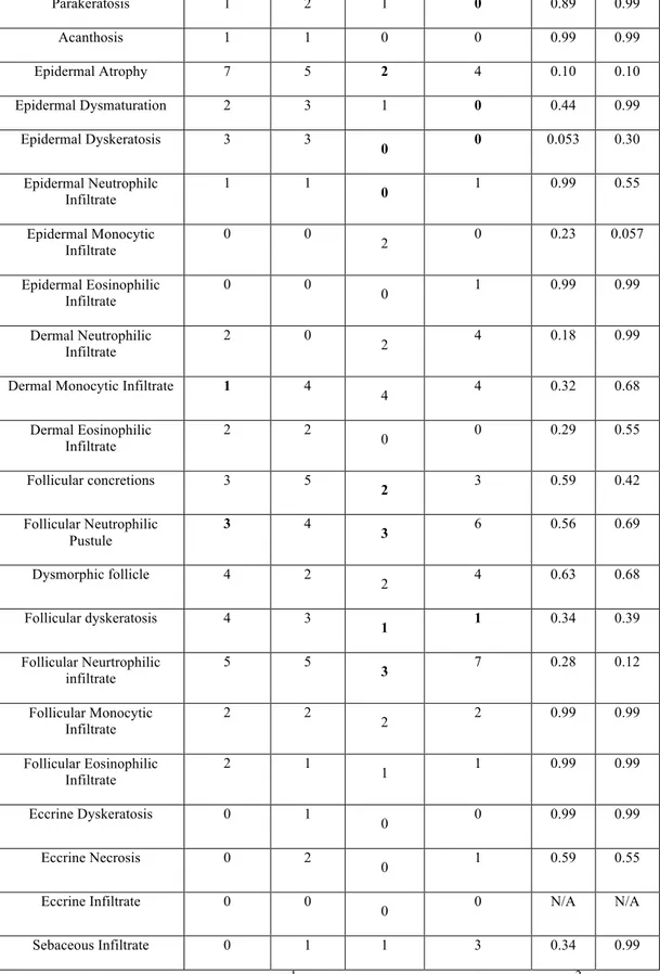
24 Parakeratosis 1 2 1 0 0.89 0.99 Acanthosis 1 1 0 0 0.99 0.99 Epidermal Atrophy 7 5 2 4 0.10 0.10 Epidermal Dysmaturation 2 3 1 0 0.44 0.99 Epidermal Dyskeratosis 3 3 0 0 0.053 0.30 Epidermal Neutrophilc Infiltrate 1 1 0 1 0.99 0.55 Epidermal Monocytic Infiltrate 0 0 2 0 0.23 0.057 Epidermal Eosinophilic Infiltrate 0 0 0 1 0.99 0.99 Dermal Neutrophilic Infiltrate 2 0 2 4 0.18 0.99 Dermal Monocytic Infiltrate 1 4
4 4 0.32 0.68 Dermal Eosinophilic Infiltrate 2 2 0 0 0.29 0.55 Follicular concretions 3 5 2 3 0.59 0.42 Follicular Neutrophilic Pustule 3 4 3 6 0.56 0.69 Dysmorphic follicle 4 2 2 4 0.63 0.68 Follicular dyskeratosis 4 3 1 1 0.34 0.39 Follicular Neurtrophilic infiltrate 5 5 3 7 0.28 0.12 Follicular Monocytic Infiltrate 2 2 2 2 0.99 0.99 Follicular Eosinophilic Infiltrate 2 1 1 1 0.99 0.99 Eccrine Dyskeratosis 0 1 0 0 0.99 0.99 Eccrine Necrosis 0 2 0 1 0.59 0.55 Eccrine Infiltrate 0 0 0 0 N/A N/A Sebaceous Infiltrate 0 1 1 3 0.34 0.99
Table 3. Histopathologic results. 1 p-value among the four drugs; 2 p-value between lapatinib compared to all 3 drugs. Bolded values are representative of the drug with the lowest number of specimens with that particular histologic finding.
25
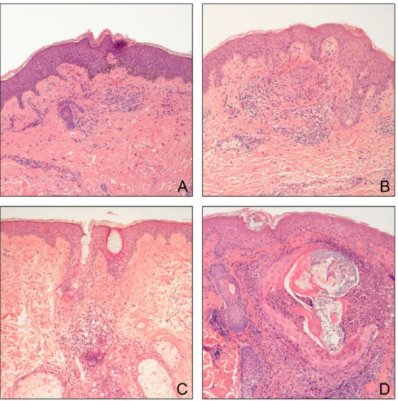
Figure 5. Histologic findings of representative specimens for each HER inhibitor. A) Skin from a patient on lapatinib exhibiting a mild periadnexal and interstitial infiltrate consisting of mononuclear cells and neutrophils. B) Specimen from a patient on cetuximab illustrating a more intense periadnexal neutrophilic infiltrate with mild follicular dyskeratosis. C) Specimen from a patient on erlotinib showing an intense primarily neutrophilic perifollicular infiltrate with follicular dyskeratosis and mild epidermal atrophy. D) Specimen from a patient on panitumumab demonstrating a prominent follicular neutrophilic pustule with follicular dysmorphic features and dyskeratosis with overlying mild atrophy (X10 magnification).
26
The most frequent finding in the epidermis was atrophy, seen in 7 of 8 (87.5%) patients on cetuximab, 5 of 8 (62.5%) patients on erlotinib, 4 of 8 (50%) patients on panitumumab, and 2 of 8 (25%) patients on lapatinib. A comparison of the extent of atrophy between lapatinib compared to cetuximab, erlotinib, and panitumumab combined, trended towards statistical significance (p=0.10). Dyskeratosis and dysmaturation were more frequently observed with cetuximab and erlotinib (3 of 8 for both). Both of these findings were rare or absent with lapatinib (dyskeratosis = 0 of 8 patients; dysmaturation = 1 of 8 patients) and panitumumab (0 of 8 for both). Lapatinib was the only drug that was found to have monocytes within the epidermis (2 of 8, p=0.057). Epidermal infiltrates were otherwise not frequent amongst all four drugs (0-2 of 8 patients).
Dermal presence of neutrophilic, monocytic, and eosinophilic infiltrates were more frequently seen with panitumumab (4 of 8), compared to cetuximab (2 of 8 patients) and erlotinib (2 of 8 patients). Presence of dermal monocytes were variable for all four drugs (1 of 8 for cetuximab, and 4 of 8 for erlotinib, lapatinib, and panitumumab).
Follicles were not found in the biopsies of 2 patients on cetuximab, and in 1 patient each on erlotinib, lapatinib, and
27
panitumumab. Bacterial concretions were more frequently seen with erlotinib (5 of 7). The presence of a neutrophilic pustule was noted for all drugs, but in highest frequency with panitumumab (6 of 7). Follicular dyskeratosis was seen in 4 of 6 patients on cetuximab and 3 of 7 patients on erlotinib, as opposed to 1 of 7 for both lapatinib and panitumumab.
FOLLICULAR NEUTROPHILIC INFILTRATES ARE MORE FREQUENT DURING HER1I THERAPY
Neutrophilic infiltrate within the follicle was less frequent for patients on lapatinib (3 of 7) as compared to cetuximab (5 of 6), erlotinib (5 of 7), and panitumumab (7 of 7), with a trend toward statistical significance (p=0.12). Follicular monocytic (2 for each drug) and eosinophilic infiltrates (2 of 6 patients for cetuximab and 1 of 7 patients all others) were similar amongst the four drugs.
Overall, alterations in eccrine glands were unusual, with erlotinib patients more likely to have changes (1 of 8 having dyskeratosis and 2 of 8 with necrosis). Similarly, sebaceous glands were evaluated for presence of an inflammatory infiltrate. Sebaceous gland infiltrates were most commonly found with panitumumab, where 3 of 5 specimens had infiltration present.
28
5. DISCUSSION AND CONCLUSIONS
Skin rash is the most frequently reported toxicity to HER1 inhibitors and, although not life threatening, often results in discontinuation and/or interruption of anticancer therapy, which may also affect clinical outcome. At present, the exact mechanism of this rash is not clearly understood. Altered keratinocyte growth and differentiation and enhanced inflammation induced by HER inhibition seems to play a critical role in the development of rash, especially in hair follicle epithelium (40). There is also some evidence that these agents may also alter the immune system (41). More recently, a preclinical model demonstrated the role of Tumor Necrosis Factor-α (TNF-α) and interleukin-1 (IL-1) in the development of HER1i-associated skin rash and suggested a possible therapeutic role for anti-TNF agents (32). Several studies have aimed to identify histologic and immunohistochemical features of skin in patients undergoing therapy with HER1 inhibitors. In our study, we describe histologic and immunohistochemical differences in skin from patients treated with HER1 and HER1/2 inhibitors, consistent with clinical data of decreased rash severity after dual inhibition.
29
sizes in each of the four treatment groups. Even with these small sample sizes, significant differences were found between the epidermis and dermis in skin of patients treated with lapatinib, when compared to other HER inhibitors. A larger sample size would likely lead to more differences in biomarkers and histopathological features and is currently being planned as part of a prospective study.
Use of the HER1/2 inhibitor lapatinib results in decreased HER1 phosphorylation and activation by reversibly binding to the cytoplasmic ATP-binding site, preventing subsequent downstream signaling of ERK-1/2 and PI3K/Akt (12). Clinical data suggest that the HER1/2i lapatinib is associated with a lower incidence of rash compared to single HER1 inhibitors (24-26, 38). Consistent with these clinical data, our findings show a more intact immunohistochemical and histological pattern in the skin of patients treated with the HER1/2 inhibitor lapatinib. HER1 kinase activity is an important signal for pAKT/PI3K pathway activation (42), and pAKT has a key role in cell survival, with increasing activity during keratinocyte differentiation and stratification (43) The protective role of pAKT in skin treated with HER1 inhibitors has been shown in patients treated with erlotinib, in which greater pAKT expression at baseline correlated with decreased rash severity (44).
30
We also found decreased dermal expression of the proliferation marker K16 and the negative growth regulator p27 (33) in HER1/2i treated patients, compared to those on HER1i therapy. In the skin of cetuximab treated patients, p27 was upregulated in epidermis (30), suggesting growth inhibition of basal keratinocytes. Lower p27 in HER1/2i treated patients suggests decreased inhibition of proliferation in skin, consistent with lower skin toxicity. Increased ERK1 expression for lapatinib compared to erlotinib, suggests greater pathway activity, which may account for lower inflammmation of rash. Previous studies have demonstrated that suppression of the HER1/ERK signaling pathway enhances skin inflammation by increasing chemokine expression in keratinocytes (18, 45).
Histologic findings in HER1i-induced rash include a mixed inflammatory infiltrate, suppurative folliculitis with follicular rupture, and epidermal dyskeratosis (30). Perieccrine inflammation and dyskeratosis have also been reported. All of these findings have also been observed in our analyzed samples. These findings have also been demonstrated in mice treated with an anti-EGFR monoclonal antibody, where follicular plugging with increased sebaceous gland size and a neutrophilic follicular infiltrate are observed (32). Enlargement of sebaceous units was not detected in our patients,
31
which could potentially relate to timing of biopsies. The time from onset of rash to biopsy was not uniform in the current study. Early enlargement of sebaceous glands, may be explained by a mouse study in which enlargement was noted to occur prior to the appearance of inflammatory infiltrates (32).
There was a lower incidence of epidermal atrophy in HER1/2i-treated patients. In addition, no patients on the HER1/2i lapatinib showed evidence of epidermal dyskeratosis, in contrast to 3 of 8 patients on the HER1i cetuximab and erlotinib. Similarly, follicular dyskeratosis was seen in 1 of 7 patients receiving the HER1/2i compared to 4 of 6 patients on cetuximab and 3 of 7 patients on erlotinib. In summary, a more benign histopathologic pattern was observed for patients on the HER1/2i lapatinib compared to the single inhibitors of HER1.
This phenomenon could be explained by the decreased expression of p27 for the HER1/2i as compared to the single HER1 inhibitors, which has been associated with impaired cell growth and differentiation in the follicle. This suggests improved keratinocyte survival, cell differentiation, and normalization of keratinization may possibly relate to an increase in pAKT activity as demonstrated in our current IHC data. Increased activity in the pAKT pathway may also
32
lead to decreased dysmaturation and dyskeratosis in the epidermis with HER1/2 inhibition. Moreover, patients on cetuximab had lower levels of Stat3 as compared to the other drugs; as a differentiation marker this may lead to greater alterations in both epidermis and follicle and thus lead to such findings.
Inflammatory infiltrates for patients on lapatinib were predominantly composed of monocytes. Overall, there were few differences between all HERi when was analyzed the inflammatory infiltrate. However, fewer patients on lapatinib (3 of 7) had a neutrophilic follicular infiltrate as compared to cetuximab (5 of 6), erlotinib (5 of 7), and panitumumab (7 of 7), with a trend towards statistical significance (p=0.12). This reduction in follicular inflammation with lapatinib is consistent with a lower incidence of rash (38). Similar numbers of monocytes and eosinophils within the follicle are identified for all four HERi. Moreover, dermal monocytic infiltrates are less prominent for patients on cetuximab, which is consistent with lower levels of CD68, CD54, and CD4 seen in these patients.
Randomized clinical trials show a benefit of prophylactic management of HER1 inhibitor rash with minocycline (46) or a skin treatment regimen consisting of doxycycline, topical hydrocortisone,
33
moisturizers, and sunscreen (47). Both of these published studies were conducted in patients receiving the anti-HER monoclonal antibodies cetuximab and panitumumab, respectively. There are no controlled studies on the management of rash to small molecule anti-HER agents erlotinib and lapatinib. However, our observations described here in showing a lower inhibition of the pAKT pathway and a decreased expression of K16 and p27 in the dermis, suggest that the design of trials against lapatinib-induced rash would require prophylactic interventions at lower doses or frequency, or conceivably that anti-rash interventions could be instituted in a reactive fashion, since the alterations at the cellular level appear to be of less significance, concordant with clinical observations showing that the lapatinib rash is of less frequency and severity as compared to erlotinib, cetuximab, or panitumumab. All of which would necessitate confirmation in a separate study with the aforementioned HER inhibitory agents.
Taken as a whole, there are fewer histologic and immunohistochemical alterations in the skin of patients treated with an inhibitor of HER1/2 compared to single HER1 inhibitors. The finding of greater pAKT expression, decreased p27 and epidermal atrophy underscore cellular differences in skin toxicity induced by HER1/2i vs HER1i. These findings also suggest that interventions to
34
identify risk factors, prevent, and treat rash due to HER1 and HER 1/2 inhibitors should be tailored to the causative agent.
35
6. REFERENCES
1. Reiter JL, Maihle NJ. A 1.8 kb alternative transcript from the human epidermal growth factor receptor gene encodes a truncated form of the receptor. Nucleic Acids Res 1996;24:4050-6.
2. Alroy I, Yarden Y. The ErbB signaling network in
embryogenesis and oncogenesis: signal diversification through combinatorial ligand-receptor interactions. FEBS Lett 1997;410:83-6.
3. Hu JC, Sadeghi P, Pinter-Brown LC, et al. Cutaneous side
effects of epidermal growth factor receptor inhibitors: clinical presentation, pathogenesis, and management. J Am Acad Dermatol 2007;56:317-26.
4. Heidary N, Naik H, Burgin S. Chemotherapeutic agents and the
skin: An update. J Am Acad Dermatol 2008;58:545-70.
5. Klapper LN, Kirschbaum MH, Sela M, et al. Biochemical and
clinical implications of the ErbB/HER signaling network of growth factor receptors. Adv Cancer Res 2000;77:25-79.
6. Ciardiello F, Caputo R, Bianco R, et al. Inhibition of growth factor production and angiogenesis in human cancer cells by ZD1839 (Iressa), a selective epidermal growth factor receptor tyrosine kinase inhibitor. Clin Cancer Res 2001;7:1459-65.
36
7. Mendelsohn J, Baselga J. Status of epidermal growth factor
receptor antagonists in the biology and treatment of cancer. J Clin Oncol 2003;21:2787-99.
8. Food and Drug Administration. [cited; Available from:
http://www.fda.gov/NewsEvents/Newsroom/PressAnnouncements/20
04/ucm108244.htm. Accessed on Dec 2, 2010.
9. Food and Drug Administration. [cited; Available from:
http://www.fda.gov/NewsEvents/Newsroom/PressAnnouncements/20
06/ucm108745.htm. Accessed on Dec 2, 2010.
10. Food and Drug Administration. [cited; Available from:
http://www.fda.gov/NewsEvents/Newsroom/PressAnnouncements/20
04/ucm108378.htm. Accessed on Dec 2, 2010.
11. Food and Drug Administration. [cited; Available from:
http://www.fda.gov/NewsEvents/Newsroom/PressAnnouncements/20
04/ucm108383.htm. Accessed on Dec 2, 2010.
12. Moy B, Goss PE. Lapatinib: current status and future directions in breast cancer. Oncologist 2006;11:1047-57.
13. Chu I, Blackwell K, Chen S, et al. The dual ErbB1/ErbB2 inhibitor, lapatinib (GW572016), cooperates with tamoxifen to inhibit both cell proliferation- and estrogen-dependent gene expression in antiestrogen-resistant breast cancer. Cancer Res 2005;65:18-25.
37
14. Rusnak DW, Lackey K, Affleck K, et al. The effects of the novel, reversible epidermal growth factor receptor/ErbB-2 tyrosine kinase inhibitor, GW2016, on the growth of human normal and tumor-derived cell lines in vitro and in vivo. Mol Cancer Ther 2001;1:85-94. 15. Nanney LB, Stoscheck CM, King LE, et al. Immunolocalization of epidermal growth factor receptors in normal developing human skin. J Invest Dermatol 1990;94:742-8.
16. Jost M, Kari C, Rodeck U. The EGF receptor - an essential regulator of multiple epidermal functions. Eur J Dermatol 2000;10:505-10.
17. Rodeck U, Jost M, Kari C, et al. EGF-R dependent regulation of keratinocyte survival. J Cell Sci 1997;110:113-21.
18. Pastore S, Mascia F, Mariotti F, et al. ERK1/2 regulates epidermal chemokine expression and skin inflammation. J Immunol 2005;174:5047-56.
19. Peus D, Hamacher L, Pittelkow MR. EGF-receptor tyrosine kinase inhibition induces keratinocyte growth arrest and terminal differentiation. J Invest Dermatol 1997;109:751-6.
20. Hauser PJ, Agrawal D, Hackney J, et al. STAT3 activation accompanies keratinocyte differentiation. Cell Growth Differ 1998;9:847-55.
38
21. Woodworth CD, Michael E, Marker D, et al. Inhibition of the epidermal growth factor receptor increases expression of genes that stimulate inflammation, apoptosis, and cell attachment. Mol Cancer Ther 2005;4:650-8.
22. Lorch JH, Klessner J, Park JK, et al. Epidermal growth factor receptor inhibition promotes desmosome assembly and strengthens intercellular adhesion in squamous cell carcinoma cells. J Biol Chem 2004;279:37191-200.
23. Lynch TJ, Kim ES, Eaby B, et al. Epidermal growth factor receptor inhibitor-associated cutaneous toxicities: an evolving paradigm in clinical management. Oncologist 2007;12:610-21.
24. Cunningham D, Humblet Y, Siena S, et al. Cetuximab monotherapy and cetuximab plus irinotecan in irinotecan-refractory metastatic colorectal cancer. N Engl J Med 2004;351:337-45.
25. Shepherd FA, Rodrigues Pereira J, Ciuleanu T, et al. Erlotinib in previously treated non-small-cell lung cancer. N Engl J Med 2005;353:123-32.
26. Van Cutsem E, Peeters M, Siena S, et al. Open-label phase III trial of panitumumab plus best supportive care compared with best supportive care alone in patients with chemotherapy-refractory metastatic colorectal cancer. J Clin Oncol 2007;25:1658-64.
39
27. Agero AL, Dusza SW, Benvenuto-Andrade C, et al. Dermatologic side effects associated with the epidermal growth factor receptor inhibitors. J Am Acad Dermatol 2006;55:657-70.
28. Sipples R. Common side effects of anti-EGFR therapy: acneform rash. Semin Oncol Nurs 2006;22:28-34.
29. Perez-Soler R, Delord JP, Halpern A, et al. HER1/EGFR inhibitor-associated rash: future directions for management and investigation outcomes from the HER1/EGFR inhibitor rash management forum. Oncologist 2005;10:345-56.
30. Busam KJ, Capodieci P, Motzer R, et al. Cutaneous side-effects in cancer patients treated with the antiepidermal growth factor receptor antibody C225. Br J Dermatol 2001;144:1169-76.
31. Lacouture ME. Mechanisms of cutaneous toxicities to EGFR inhibitors. Nat Rev Cancer 2006;6:803-12.
32. Surguladze D, Deevi D, Claros N, et al. Tumor necrosis factor-alpha and interleukin-1 antagonists alleviate inflammatory skin changes associated with epidermal growth factor receptor antibody therapy in mice. Cancer Res 2009;69:5643-7.
33. Busse D, Doughty RS, Ramsey TT, et al. Reversible G(1) arrest induced by inhibition of the epidermal growth factor receptor tyrosine
40
kinase requires up-regulation of p27(KIP1) independent of MAPK activity. J Biol Chem 2000;275:6987-95.
34. Joshi SS, Ortiz S, Witherspoon JN, et al. Effects of epidermal growth factor receptor inhibitor-induced dermatologic toxicities on quality of life. Cancer 2010;116:3916-23.
35. Abraham T, Rademaker A, Ortiz S, et al. Economic impact associated with the management of dermatologic adverse drug reactions (dADRs) induced by EGFR inhibitors (EGFRIs) in lung cancer. J Clin Oncol 2008;26.
36. Eilers RE, Gandhi M, Patel JD, et al. Dermatologic infections in cancer patients treated with epidermal growth factor receptor inhibitor therapy. J Natl Cancer Inst 2010;102:47-53.
37. Boone SL, Rademaker A, Liu D, et al. Impact and management of skin toxicity associated with anti-epidermal growth factor receptor therapy: survey results. Oncology 2007;72:152-9.
38. Lacouture ME, Laabs SM, Koehler M, et al. Analysis of dermatologic events in patients with cancer treated with lapatinib. Breast Cancer Res Treat 2009;114:485-93.
39. Jiang Z, Wu CL, Woda BA, et al. Alpha-methylacyl-CoA racemase: a multi-institutional study of a new prostate cancer marker. Histopathology 2004;45:218-25.
41
40. Giovannini M, Gregorc V, Belli C, et al. Clinical Significance of Skin Toxicity due to EGFR-Targeted Therapies. J Oncol 2009;2009:849051.
41. Hammond-Thelin LA. Cutaneous reactions related to systemic immunomodulators and targeted therapeutics. Dermatol Clin 2008;26:121-59, ix.
42. Calautti E, Li J, Saoncella S, et al. Phosphoinositide 3-kinase signaling to Akt promotes keratinocyte differentiation versus death. J Biol Chem 2005;280:32856-65.
43. Thrash BR, Menges CW, Pierce RH, et al. AKT1 provides an essential survival signal required for differentiation and stratification of primary human keratinocytes. J Biol Chem 2006;281:12155-62. 44. Tan AR, Steinberg SM, Parr AL, et al. Markers in the epidermal growth factor receptor pathway and skin toxicity during erlotinib treatment. Ann Oncol 2008;19:185-90.
45. Pastore S, Mascia F, Mariani V, et al. The epidermal growth factor receptor system in skin repair and inflammation. J Invest Dermatol 2008;128:1365-74.
46. Scope A, Agero AL, Dusza SW, et al. Randomized double-blind trial of prophylactic oral minocycline and topical tazarotene for
42
cetuximab-associated acne-like eruption. J Clin Oncol 2007;25:5390-6.
47. Lacouture ME, Mitchell EP, Piperdi B, et al. Skin toxicity evaluation protocol with panitumumab (STEPP), a phase II, open-label, randomized trial evaluating the impact of a pre-Emptive Skin treatment regimen on skin toxicities and quality of life in patients with metastatic colorectal cancer. J Clin Oncol 2010;28:1351-7.
43
ACKNOWLEDGEMENTS
I would like to express my sincere thanks to my advisor Prof. Dennis West, for his invaluable support, guidance and patience throughout this project and these 2 years in the US.
I also wish to thank Prof. Mario Lacouture, for his continuous assistance, advice, support and valuable contribution to this project and the sharing of his expertise and scientific knowledge.
I am grateful to Prof. Amy Paller, Chair of the Department of Dermatology, Northwestern University for her support and the opportunity to conduct research in a very conducive environment. I would like to thank Prof. Mary Martini, Director Pigmented Lesion Clinic, for her continuous personal and scientific support throughout my time in Chicago.
Special thanks to all Department of Dermatology, Northwestern University and Clinical staff, and especially the Clinical Trials Unit staff, for their wonderful assistance during the 2 years.
I would also like to express my gratitude to Prof. Giuseppe Micali, my mentor, for his invaluable contribution throughout my scientific growth and for his continuous encouragement in realizing my PhD research experience.
I am also very grateful to Prof. Renato Bernardini, for his continuous and valuable support in making this experience possible.
Part of this thesis has been accepted for publication:
Nardone B, Nicholson K, Newman M, Guitart J, Gerami P, Talarico N, Yang XJ, Rademaker A, West DP, Lacouture ME*. Histopathologic and immunohistochemical characterization of rash to human epidermal growth factor receptor 1 (HER1) and HER1/2 inhibitors in cancer patients. Clin Cancer Res 2010; Sep 1;16(17):4452-60.
*Special thanks go to all of the authors for their valuable contribution to this project.
44
APPENDIX A
IMMUNOHISTOCHEMICAL DATA FROM ACISII(CHROMAVISION
MEDICAL SYSTEMS,INC.)
KRT1
Dearm Path Number Area Intensity Percentage (%)
DP07-2010 Epidermis 150,00 64,22 Dermis 111,75 5,80 DP07-3486 Epidermis 174,75 82,46 Dermis 112,50 3,82 DP07-2495 Epidermis 168,25 70,56 Dermis 101,50 3,53 DP07-1561 Epidermis 178,25 82,88 Dermis 102,50 2,85 DP07-5876 Epidermis 142,25 64,74 Dermis 114,50 4,07 DP07-5878 Epidermis 165,75 87,12 Dermis 109,25 3,70 DP08-541 Epidermis 168,00 95,20 Dermis 123,50 6,98 DP08-2678 Epidermis 146,50 86,38 Dermis 105,25 2,86 DP07 10082-A Epidermis 160,00 76,04 Dermis 109,25 3,60 DP07 17053 Epidermis 147,00 87,73 Dermis 100,00 2,56 DP07 17250 Epidermis 179,50 82,87 Dermis 113,00 2,98 DP06-2974 Epidermis 153,50 76,63 Dermis 98,75 5,28 DP07 19209 Epidermis 136,75 80,79 Dermis 103,25 3,88 DP07-6221 Epidermis 203,25 92,96
45 Dermis 103,50 5,48 DP07-8218 Epidermis 126,50 87,99 Dermis 97,25 3,44 DP2619-06 Epidermis 133,00 82,72 Dermis 100,50 3,30 DP845-07 Epidermis 171,75 90,49 Dermis 101,25 4,83 DP07-7169 Epidermis 159,75 87,53 Dermis 106,50 1,86 DP07-8617 Epidermis 131,50 79,55 Dermis 108,00 3,44 DP254-07 Epidermis 140,75 81,01 Dermis 110,00 4,87 DP07-6294 Epidermis 155,75 69,39 Dermis 116,75 4,53 DP07-8786 Epidermis 167,75 74,54 Dermis 107,25 2,78 DP06-11523 Epidermis 172,00 83,57 Dermis 103,50 6,97 DP07-6295 Epidermis 127,25 89,53 Dermis 94,75 2,30 DP07-1826 Epidermis 147,25 85,68 Dermis 92,50 3,27 DP07-5446 Epidermis 157,50 95,58 Dermis 114,25 6,83 DP07-4746 Epidermis 122,25 79,15 Dermis 99,50 3,54 DP08 15669 Epidermis 157,25 95,74 Dermis 97,50 4,90 DP07 18707 Epidermis 134,25 88,83 Dermis 94,75 4,92 DP07-1157 Epidermis 137,25 84,06 Dermis 101,00 4,29 DP07-1905 Epidermis 135,50 60,86 Dermis 105,75 6,21 DP06-2953 Epidermis 152,75 74,93
46
Dermis 114,75 4,37
HLA-DR
Derm Path Number Area Intensity Percentage (%)
DP07-2010 Epidemis 166,50 6,23 Dermis 166,00 24,95 DP07-3786 Epidemis 167,00 7,51 Dermis 160,50 13,02 DP07-2495 Epidemis 163,00 8,17 Dermis 160,50 13,71 DP07-1561 Epidemis 160,50 7,67 Dermis 159,00 18,55 DP07-5876 Epidemis 156,00 6,75 Dermis 162,00 13,92 DP07-5878 Epidemis 165,00 7,60 Dermis 159,00 12,57 DP08-541 Epidemis 159,00 15,24 Dermis 152,50 11,33 DP08-2678 Epidemis 165,00 9,85 Dermis 171,50 37,03 DP07 10082-A Epidemis 165,50 11,15 Dermis 167,00 18,84 DP07 17053 Epidemis 160,50 7,61 Dermis 149,00 7,55 DP07-17250 Epidemis 172,00 10,33 Dermis 158,50 10,40 DP06 2974 Epidemis 134,00 17,15 Dermis 133,00 16,41 DP07 19209 Epidemis 137,00 6,10 Dermis 133,00 14,77 DP07-6221 Epidemis 140,00 13,91 Dermis 123,00 16,90 DP07-8218 Epidemis 138,50 8,50 Dermis 136,00 36,75 DP2619-06 Epidemis 146,50 13,17 Dermis 150,50 48,61
47 DP854-07 Epidemis 138,50 28,51 Dermis 134,50 51,31 DP07-7619 Epidemis 129,50 21,29 Dermis 128,00 20,54 DP07-8617 Epidemis 138,50 29,82 Dermis 133,50 46,86 DP254-07 Epidemis 143,00 13,53 Dermis 149,00 64,56 DP07-6294 Epidemis 128,50 14,88 Dermis 128,50 16,92 DP07-8786 Epidemis 143,00 18,28 Dermis 135,00 25,72 DP07-6295 Epidemis 154,50 2,23 Dermis 158,50 9,44 DP06-11523 Epidemis 128,00 16,31 Dermis 122,50 23,10 DP07-1826 Epidemis 163,50 5,24 Dermis 154,00 5,80 DP07-5446 Epidemis 170,50 10,26 Dermis 177,00 19,06 DP07-4746 Epidemis 166,00 11,66 Dermis 174,00 19,08 DP08 15667 Epidemis 157,50 1,69 Dermis 148,00 6,06 DP07 18707 Epidemis 169,00 4,54 Dermis 156,00 7,04 DP07-1157 Epidemis 157,00 3,51 Dermis 153,00 7,09 DP07-1925 Epidemis 133,50 1,01 Dermis 139,00 6,54 ERK1
Derm Path Number Area Indensity Percentage (%)
DP07-2495 Epidermis 111,67 72,58 Dermis 119,40 47,16 DP07-1561 Epidermis 133,00 84,04
48 Dermis 137,20 56,13 DP07 17250 Epidermis 130,33 90,26 Dermis 129,40 48,92 DP07 17053 Epidermis 128,00 41,68 Dermis 130,00 47,78 DP07 10082-A Epidermis 115,67 94,05 Dermis 138,80 54,00 DP07-8786 Epidermis 121,00 61,65 Dermis 118,40 38,68 DP07-2010 Epidermis 136,33 97,27 Dermis 144,60 53,27 DP07-5876 Epidermis 113,00 38,83 Dermis 114,00 41,06 DP07-5878 Epidermis 121,33 91,09 Dermis 127,20 56,98 DP07-6294 Epidermis 127,00 94,33 Dermis 131,60 51,45 DP08-541 Epidermis 148,67 85,98 Dermis 135,80 47,76 DP08-2678 Epidermis 111,67 54,24 Dermis 125,80 50,92 DP07-3486 Epidermis 119,00 92,31 Dermis 143,60 59,70 DP07-6221 Epidermis 99,67 36,45 Dermis 103,00 38,90 DP07-8218 Epidermis 111,67 75,04 Dermis 123,40 50,28 DP07 19209 Epidermis 108,33 58,81 Dermis 132,40 52,66 DP06-2953 Epidermis 145,33 88,16 Dermis 139,60 50,28 DP06-2974 Epidermis 134,33 73,70 Dermis 125,60 46,66 DP06-11523 Epidermis 146,00 42,35 Dermis 120,20 20,50 DP254-07 Epidermis 108,00 88,97
49 Dermis 122,80 51,01 DP845-07 Epidermis 120,00 69,73 Dermis 120,40 51,62 DP07-6295 Epidermis 132,00 34,77 Dermis 133,40 49,93 DP07-7169 Epidermis 113,67 65,57 Dermis 121,80 49,72 DP07-8617 Epidermis 112,67 91,47 Dermis 127,40 49,15 DP07-1826 Epidermis 98,33 27,95 Dermis 110,60 28,31 DP07-5446 Epidermis 161,00 39,49 Dermis 136,80 42,94 DP07-4746 Epidermis 126,00 72,15 Dermis 130,80 46,69 DP08 15669 Epidermis 122,67 79,07 Dermis 129,60 38,02 DP07-1905 Epidermis 114,33 25,28 Dermis 117,00 34,58 DP07-1157 Epidermis 100,67 50,57 Dermis 124,40 28,48 DP07-18707 Epidermis 118,33 45,03 Dermis 113,80 48,07 K16
Derm Path Number Area of Interest Intensity
Percentage (%) DP06-2953 Epidermis 174,00 88,17 Dermis 132,00 13,22 DP06-2974 Epidermis 114,50 65,78 Dermis 118,00 43,77 DP2619-06 Epidermis 117,00 15,80 Dermis 120,00 44,43 DP07-1561 Epidermis 147,50 88,73 Dermis 116,00 38,96 DP07-2010 Epidermis 163,00 87,87
50 Dermis 138,50 26,62 DP07-2495 Epidermis 120,00 69,28 Dermis 127,00 35,55 DP07-3486 Epidermis 152,50 93,27 Dermis 124,50 23,11 DP07-5876 Epidermis 132,50 32,94 Dermis 120,50 19,80 DP07-5878 Epidermis 156,50 51,97 Dermis 145,50 19,28 DP07-6221 Epidermis 123,50 30,74 Dermis 127,00 24,98 DP07-6294 Epidermis 127,50 74,02 Dermis 119,00 27,21 DP07-7169 Epidermis 115,50 60,75 Dermis 113,50 24,40 DP07-8218 Epidermis 117,50 66,21 Dermis 110,00 34,11 DP07-8617 Epidermis 114,50 44,35 Dermis 114,50 41,97 DP07-8786 Epidermis 106,50 31,62 Dermis 109,00 19,42 DP07 10082-A Epidermis 126,00 77,53 Dermis 138,00 18,71 DP07-17053 Epidermis 131,50 58,85 Dermis 130,00 15,04 DP07 17250 Epidermis 124,50 72,94 Dermis 130,00 15,78 DP07 19209 Epidermis 120,00 56,88 Dermis 122,00 28,31 DP254-07 Epidermis 117,50 23,31 Dermis 122,00 43,02 DP845-07 Epidermis 103,50 23,11 Dermis 100,00 13,10 DP08-541 Epidermis 143,50 79,14 Dermis 137,00 20,84 DP08-2678 Epidermis 149,00 82,64
51 Dermis 140,00 27,08 DP06-11523 Epidermis 128,50 86,60 Dermis 120,50 64,95 DP07-1157 Epidermis 131,00 91,98 Dermis 112,00 59,14 DP07-1826 Epidermis 123,00 85,11 Dermis 117,50 52,24 DP07-1905 Epidermis 111,50 24,02 Dermis 121,00 33,22 DP07-4746 Epidermis 135,00 88,96 Dermis 101,00 61,93 DP075446 Epidermis 122,00 30,94 Dermis 110,00 44,50 DP07-6295 Epidermis 141,50 89,51 Dermis 114,00 45,91 DP07-18707 Epidermis 111,50 28,20 Dermis 112,00 36,72 DP08-15669 Epidermis 121,50 94,45 Dermis 124,50 57,08 p27
Derm Path Number Area Intensity Percentage (%)
DP07-2010 Epidermis 166,40 20,31 Dermis 144,50 29,59 DP07-3486 Epidermis 154,60 7,83 Dermis 144,25 31,69 DP07-2495 Epidermis 155,40 11,09 Dermis 157,50 30,89 DP07-1561 Epidermis 154,80 8,89 Dermis 126,75 8,99 DP07-5876 Epidermis 138,20 5,24 Dermis 124,25 12,96 DP07-5878 Epidermis 144,00 5,50 Dermis 138,25 17,95 DP08-541 Epidermis 141,20 29,51 Dermis 131,25 12,86
52 DP08-2678 Epidermis 143,00 7,06 Dermis 128,75 15,38 DP07 10082-A Epidermis 135,20 14,27 Dermis 122,00 15,60 DP07 17053 Epidermis 152,00 6,60 Dermis 141,75 18,01 DP07 17250 Epidermis 147,20 22,76 Dermis 131,25 12,28 DP06-2974 Epidermis 135,40 10,71 Dermis 117,50 14,35 DP07-6221 Epidermis 133,00 10,87 Dermis 125,75 17,83 DP07-7169 Epidermis 110,60 8,96 Dermis 122,75 19,36 DP07-8218 Epidermis 135,60 7,05 Dermis 127,75 15,37 DP07 19209 Epidermis 130,80 7,04 Dermis 125,00 15,54 DP845-07 Epidermis 140,80 29,09 Dermis 128,50 25,17 DP07-6294 Epidermis 136,60 6,42 Dermis 121,75 13,99 DP07-8786 Epidermis 147,20 10,61 Dermis 124,25 16,98 DP06-11523 Epidermis 150,40 23,95 Dermis 122,25 20,99 DP07-6295 Epidermis 127,00 13,84 Dermis 115,00 19,88 DP07-8617 Epidermis 140,80 6,70 Dermis 125,75 19,44 DP07-1826 Epidermis 125,40 22,62 Dermis 111,50 19,91 DP254-07 Epidermis 130,60 11,83 Dermis 121,25 20,01 DP07-5446 Epidermis 145,80 23,01 Dermis 123,75 17,49
53 DP07-4746 Epidermis 129,20 10,93 Dermis 120,75 13,69 DP07 15669 Epidermis 125,60 8,62 Dermis 122,75 31,01 DP2619-06 Epidermis 135,00 6,13 Dermis 122,00 11,37 DP07-1905 Epidermis 145,20 9,08 Dermis 131,75 17,63 DP07-1157 Epidermis 145,60 11,97 Dermis 129,00 11,17 DP07-18707 Epidermis 136,80 14,47 Dermis 127,25 11,59 pAKT
Derm Path Number Area Intensity Percentage(%)
DP07-2010 Epidermis 145,40 94,09 Dermis 115,50 21,94 DP07-2495 Epidermis 166,80 97,75 Dermis 117,50 14,36 DP07-3486 Epidermis 154,00 95,74 Dermis 139,50 62,99 DP07-1561 Epidermis 135,80 96,69 Dermis 131,50 59,65 DP07-5876 Epidermis 127,60 52,84 Dermis 142,50 69,46 DP07-5878 Epidermis 161,60 99,01 Dermis 149,00 80,81 DP08-541 Epidermis 174,00 96,43 Dermis 154,00 58,12 DP08-2678 Epidermis 133,20 92,76 Dermis 143,50 72,31 DP08 10082-A Epidermis 150,00 94,87 Dermis 139,50 37,22 DP07-17053 Epidermis 155,20 81,65 Dermis 151,50 29,05 DP07 17250 Epidermis 167,20 97,19
54 Dermis 132,00 41,07 DP06-2794 Epidermis 146,40 61,92 Dermis 137,50 14,15 DP07 19209 Epidermis 125,40 32,76 Dermis 124,50 14,71 DP07-6221 Epidermis 131,80 24,47 Dermis 137,00 33,25 DP07-8218 Epidermis 141,60 33,01 Dermis 114,00 5,09 DP845-07 Epidermis 128,60 69,24 Dermis 119,50 45,01 DP07-7169 Epidermis 133,60 39,04 Dermis 117,00 17,22 DP07-8617 Epidermis 133,00 17,18 Dermis 120,00 11,85 DP254-07 Epidermis 112,00 2,81 Dermis 99,00 2,15 DP07-6294 Epidermis 152,20 86,86 Dermis 129,33 26,12 DP07-8786 Epidermis 142,00 33,10 Dermis 134,00 11,72 DP06-11523 Epidermis 153,00 73,08 Dermis 133,00 58,10 DP07-6295 Epidermis 135,20 89,88 Dermis 134,67 71,41 DP07-1826 Epidermis 122,40 66,22 Dermis 129,00 45,75 DP07-5446 Epidermis 148,00 87,19 Dermis 137,33 66,14 DP07-4746 Epidermis 120,80 60,87 Dermis 125,67 37,42 DP08 15669 Epidermis 125,20 58,26 Dermis 127,67 39,88 DP07-1905 Epidermis 104,00 0,61 Dermis 116,00 8,46 DP07-1157 Epidermis 99,20 75,79
55
Dermis 116,33 46,76 DP07-18707 Epidermis 123,20 81,76 Dermis 101,00 48,73
HER1
Derm Path Number Area Intensity Percentage(%)
DP07-2010 Epidermis 130,00 63,56 Dermis 125,50 20,79 DP07-3486 Epidermis 111,00 22,16 Dermis 121,60 68,32 DP07-2495 Epidermis 123,00 26,40 Dermis 112,80 51,19 DP07-1561 Epidermis 117,00 16,96 Dermis 111,40 88,89 DP07-5876 Epidermis 100,50 39,31 Dermis 144,80 88,87 DP07-5878 Epidermis 117,00 18,37 Dermis 137,20 76,12 DP08-541 Epidermis 124,00 18,89 Dermis 135,80 84,70 DP08-2678 Epidermis 145,50 26,59 Dermis 124,80 71,37 DP07 10082-A Epidermis 117,50 24,66 Dermis 127,00 87,04 DP07 17053 Epidermis 113,00 15,38 Dermis 129,20 81,49 DP07 17250 Epidermis 109,50 17,84 Dermis 132,20 87,85 DP06-2953 Epidermis 117,00 13,08 Dermis 161,80 88,30 DP06-2974 Epidermis 102,50 22,17 Dermis 119,20 73,34 DP07 19209 Epidermis 116,50 15,25 Dermis 130,00 80,24 DP07-8218 Epidermis 112,00 18,61 Dermis 125,00 15,99
56 DP845-07 Epidermis 111,40 72,24 Dermis 99,00 9,82 DP07-7169 Epidermis 109,80 76,35 Dermis 97,50 13,10 DP07-8617 Epidermis 153,60 87,12 Dermis 115,50 18,57 DP254-07 Epidermis 166,40 91,09 Dermis 127,50 16,74 DP07-6294 Epidermis 156,00 85,33 Dermis 110,50 39,65 DP07-8786 Epidermis 151,20 91,24 Dermis 119,50 34,43 DP06-11523 Epidermis 162,40 96,92 Dermis 108,00 39,43 DP06-11523 Epidermis 142,20 64,30 Dermis 122,00 16,40 DP07-6295 Epidermis 144,60 81,41 Dermis 111,50 22,08 DP07-5446 Epidermis 145,20 40,62 Dermis 132,50 23,53 DP07-1826 Epidermis 189,40 88,38 Dermis 140,50 28,73 DP08-15669 Epidermis 106,40 36,97 Dermis 126,50 24,34 DP07-4746 Epidermis 190,40 91,26 Dermis 123,50 15,68 DP07 17053 Epidermis 125,20 40,21 Dermis 135,50 17,42 DP07-1905 Epidermis 137,20 68,27 Dermis 116,00 12,85 DP07-1157 Epidermis 121,20 11,60 Dermis 129,50 26,25 DP07-18707 Epidermis 95,20 14,05 Dermis 138,00 16,72 CD68
57
Derm Path Number Area Intensity Percentage (%)
DP07-2010 Epidermis 125,50 1,67 Dermis 141,40 16,63 DP07 17250 Epidermis 114,00 0,63 Dermis 141,60 12,38 DP07 17053 Epidermis 103,00 1,00 Dermis 125,40 12,25 DP07 10082-A Epidermis 133,50 2,06 Dermis 130,20 11,83 DP08-2678 Epidermis 110,00 0,65 Dermis 124,80 9,54 DP08-541 Epidermis 110,00 4,98 Dermis 118,80 8,67 DP06-2974 Epidermis 108,50 1,41 Dermis 126,20 11,07 DP07-1561 Epidermis 124,00 0,07 Dermis 120,00 14,49 DP07-2495 Epidermis 118,50 0,35 Dermis 137,60 16,42 DP07-3486 Epidermis 115,00 1,47 Dermis 127,60 16,33 DP07-5876 Epidermis 101,50 0,35 Dermis 129,00 13,46 DP07-6294 Epidermis 129,00 1,44 Dermis 130,20 12,21 DP07-8786 Epidermis 101,00 0,15 Dermis 128,20 8,82 DP254-07 Epidermis 73,00 0,04 Dermis 94,00 4,62 DP07-8617 Epidermis 99,50 0,41 Dermis 127,20 12,73 DP854-07 Epidermis 125,50 1,53 Dermis 134,80 23,36 DP07-7169 Epidermis 122,50 2,02 Dermis 132,40 8,10 DP2619-06 Epidermis 105,50 0,28
58 Dermis 133,00 16,23 DP07-8218 Epidermis 128,50 0,21 Dermis 116,40 9,83 DP07-6221 Epidermis 101,00 0,13 Dermis 131,40 10,31 DP07-6295 Epidermis 103,00 4,95 Dermis 135,00 25,58 DP07-1826 Epidermis 122,00 1,58 Dermis 113,60 11,45 DP07-5446 Epidermis 148,50 13,86 Dermis 135,80 21,06 DP07-4746 Epidermis 116,00 3,84 Dermis 141,40 19,01 DP08 15669 Epidermis 110,50 0,44 Dermis 135,80 26,79 DP07 19209 Epidermis 103,50 0,31 Dermis 129,20 5,31 DP06-11523 Epidermis 119,00 2,91 Dermis 123,00 9,45 DP06 2953 Epidermis 0,00 0,00 Dermis 128,80 13,52 DP07-1905 Epidermis 105,00 0,87 Dermis 122,60 7,98 DP07-1157 Epidermis 120,50 1,45 Dermis 127,20 14,17 DP07-18707 Epidermis 118,00 0,74 Dermis 122,00 9,67 CD54
Derm Path Number Area of Interest Intensity
Percentage (%) DP06 2953 Epidermis 147,50 18,71 Dermis 140,00 19,19 DP06-2974 Epidermis 124,50 19,02 Dermis 144,00 36,48
59 DP06-11523 Epidermis 118,50 9,87 Dermis 134,00 30,61 DP2619-06 Epidermis 141,00 18,04 Dermis 166,00 74,23 DP07-1157 Epidermis 120,50 48,82 Dermis 159,50 54,79 DP07-1561 Epidermis 138,50 23,03 Dermis 152,00 72,96 DP07-1826 Epidermis 117,00 35,53 Dermis 149,00 49,93 DP07-1905 Epidermis 118,50 8,62 Dermis 136,50 35,65 DP07-2010 Epidermis 151,50 55,02 Dermis 157,50 32,86 DP07-2495 Epidermis 146,50 28,18 Dermis 164,00 66,04 DP07-3486 Epidermis 150,00 28,84 Dermis 167,00 53,81 DP07-4746 Epidermis 156,00 86,26 Dermis 166,50 92,26 DP07-5446 Epidermis 158,00 42,43 Dermis 171,00 66,79 DP07-5876 Epidermis 124,00 23,36 Dermis 153,50 65,28 DP07-5878 Epidermis 134,00 28,78 Dermis 145,00 25,25 DP07-6221 Epidermis 130,50 21,64 Dermis 144,50 25,26 DP07-6294 Epidermis 112,50 16,61 Dermis 135,50 20,01 DP07-6295 Epidermis 125,00 35,33 Dermis 147,50 58,29 DP07-7169 Epidermis 168,00 54,04 Dermis 146,00 24,97 DP07-8218 Epidermis 111,00 11,00 Dermis 133,00 59,54
60 DP07-8617 Epidermis 127,50 10,79 Dermis 158,00 57,18 DP08-8786 Epidermis 133,00 17,74 Dermis 150,50 35,54 DP07 10082-A Epidermis 150,50 64,76 Dermis 161,50 49,85 DP07 17053 Epidermis 130,50 11,82 Dermis 153,50 28,98 DP07 17250 Epidermis 131,50 35,27 Dermis 160,50 46,94 DP07 18707 Epidermis 139,50 38,28 Dermis 142,00 48,50 DP07 19209 Epidermis 116,00 14,12 Dermis 141,50 29,84 DP254-07 Epidermis 137,00 24,82 Dermis 147,50 47,18 DP854-07 Epidermis 123,00 32,60 Dermis 153,50 77,36 DP08-541 Epidermis 125,00 45,40 Dermis 138,50 36,33 DP08-2678 Epidermis 145,50 26,72 Dermis 157,00 59,48 DP08 15669 Epidermis 115,50 41,42 Dermis 144,00 54,44 CD20
Derm Path Number Area Intensity Percentage (%)
DP06 11523 Epidermis 119,00 4,65 Dermis 114,20 5,90 DP07-8786 Epidermis 122,00 0,18 Dermis 116,40 1,91 DP07-6294 Epidermis 139,00 1,08 Dermis 117,60 4,25 DP254-07 Epidermis 122,00 0,19 Dermis 126,00 5,92 DP07-8619 Epidermis 123,33 0,16
61 Dermis 123,60 1,59 DP06 2953 Epidermis 0,00 0,00 Dermis 124,20 6,48 DP06 2974 Epidermis 123,33 1,37 Dermis 116,20 3,12 DP2619-06 Epidermis 116,67 0,21 Dermis 117,00 2,52 DP07-1561 Epidermis 100,33 0,51 Dermis 118,20 2,13 DP07-1826 Epidermis 111,00 0,59 Dermis 126,20 2,02 DP07-2010 Epidermis 114,00 0,94 Dermis 148,40 27,28 DP07-2495 Epidermis 114,00 1,02 Dermis 120,20 3,07 DP07-3486 Epidermis 129,00 0,94 Dermis 114,20 2,98 DP07-4746 Epidermis 103,33 2,65 Dermis 116,00 3,16 DP07-5446 Epidermis 143,00 18,60 Dermis 120,40 6,28 DP07-5876 Epidermis 89,67 0,20 Dermis 125,60 7,79 DP07-5878 Epidermis 101,33 0,16 Dermis 112,60 2,09 DP07-6221 Epidermis 115,00 0,58 Dermis 125,60 3,40 DP07-6295 Epidermis 96,67 0,81 Dermis 106,00 1,33 DP07-7169 Epidermis 128,67 1,00 Dermis 125,80 4,37 DP07-8128 Epidermis 108,67 0,17 Dermis 112,60 2,64 DP07-8617 Epidermis 129,33 0,40 Dermis 136,00 3,07 DP07 10082A Epidermis 127,33 1,60
62 Dermis 114,00 4,23 DP07 17053 Epidermis 132,00 1,04 Dermis 124,20 5,37 DP07 17250 Epidermis 131,00 0,75 Dermis 123,80 4,95 DP07 19209 Epidermis 101,33 0,25 Dermis 116,80 2,96 DP854-07 Epidermis 104,00 0,44 Dermis 131,60 5,28 DP08-541 Epidermis 112,67 5,00 Dermis 118,60 3,20 DP08-2678 Epidermis 114,67 0,24 Dermis 121,00 3,10 DP08 15669 Epidermis 101,33 0,38 Dermis 108,40 2,68 DP07-1905 Epidermis 131,33 1,00 Dermis 109,80 2,79 DP07-1157 Epidermis 98,33 0,48 Dermis 123,00 2,57 DP07-18707 Epidermis 116,67 1,94 Dermis 130,40 5,58 CD11b
Derm Path Number Area of Interest Intensity
Percentage (%) DP06-2953 Epidermis 140,00 3,73 Dermis 150,00 11,06 DP06-2974 Epidermis 106,00 0,68 Dermis 116,00 2,79 DP2619-06 Epidermis 118,50 0,29 Dermis 114,50 2,26 DP07-1561 Epidermis 126,00 1,05 Dermis 134,50 3,37 DP07-2010 Epidermis 130,00 2,05 Dermis 127,50 2,94
63 DP07-2495 Epidermis 141,50 0,92 Dermis 126,50 3,03 DP07-3486 Epidermis 126,50 1,20 Dermis 133,50 4,44 DP07-5876 Epidermis 132,50 0,66 Dermis 139,50 2,82 DP07-5878 Epidermis 159,00 2,53 Dermis 138,00 3,28 DP07-6221 Epidermis 152,50 13,07 Dermis 119,00 2,01 DP07-6294 Epidermis 120,50 0,90 Dermis 120,00 2,34 DP07-7169 Epidermis 140,00 0,68 Dermis 139,50 0,95 DP07-8218 Epidermis 130,50 0,19 Dermis 110,00 1,51 DP07-8617 Epidermis 124,00 0,70 Dermis 135,50 9,27 DP07-8786 Epidermis 102,50 0,33 Dermis 121,50 2,20 DP07 10082-A Epidermis 106,50 0,65 Dermis 135,00 4,16 DP07-17053 Epidermis 116,50 0,97 Dermis 119,50 1,85 DP07 17250 Epidermis 122,00 0,81 Dermis 112,50 1,84 DP07 19209 Epidermis 111,50 0,38 Dermis 118,50 2,08 DP254-07 Epidermis 136,00 0,55 Dermis 120,00 3,87 DP845-07 Epidermis 124,00 1,10 Dermis 140,50 4,29 DP08-541 Epidermis 135,50 3,86 Dermis 134,00 3,36 DP08-2678 Epidermis 137,50 1,62 Dermis 126,00 3,38