We have read with great interest the excellent review on cardioimmunology by Filip K. Swirski & Matthias Nahrendorf (Cardioimmunology: the immune system in cardiac homeostasis and disease.
Nat. Rev. Immunol. 18, 733–744 (2018))1. The authors extensively discussed the role of immune cells in normal and diseased heart, specifically in myocardial infarction, myocarditis and endocarditis, heart failure and rhythm disorders1. Regarding rhythm disorders, they speculated that the immune
immuno-cardiac electrophysiology. In fact, accumulating data indicate that the immune system can promote cardiac arrhythmias by means of autoantibodies and/or inflammatory cytokines that directly affect the function of specific ion channels on the surface of cardiomyocytes2–4.
Several autoantibodies have been described that target cardiac Ca2+, K+ or Na+
channels and that have arrhythmogenic effects in the absence of evident histological changes in the heart; these are known as autoimmune cardiac channelopathies2,4. Indeed, it is well recognized that anti- Sjögren’s- syndrome-related antigen A (anti- SSA) antibodies (also known as anti- Ro/SSA antibodies) can cross react with the pore region of both L- type Ca2+ channels
(CaV1.2 and CaV1.3) and T- type Ca2+
channels (CaV3.1 and CaV3.2). By inhibiting
the related Ca2+ currents, these antibodies
promote conduction disturbances, such as sinus bradycardia and atrioventricular (AV) block5,6(Fig. 1a). Similar clinical consequences were also shown for autoantibodies
recognizing the extracellular loop of domain system could contribute to arrhythmias
through two mechanisms — a crosstalk between immune cells and fibroblasts and/or myocytes, leading to insulating fibrosis, or a direct participation of leukocytes (macrophages) in the electrical regulation of conducting cells, by interacting through connexin 43 (CX43)-containing gap junctions1.
However, the authors substantially disregarded a third important mechanism of arrhythmias in this new field of
Cardioimmunology of arrhythmias:
the role of autoimmune and
inflammatory cardiac channelopathies
Pietro Enea Lazzerini , Franco Laghi- Pasini, Mohamed Boutjdir and Pier Leopoldo Capecchi
volume 19 | january 2019 | 63
C o r r e s p o n d e n C e
nature reviews | Immunology
Ca2+ Anti-SSA antibody Anti-SSA antibody Anti-SSA antibody NaV1.5-targeting autoantibody Ca2+ CaV1.2 or CaV1.3 CaV3.1 or CaV3.2 CaV1.2 NaV1.5 Extracellular Intracellular Intracellular Intracellular
Sinus bradycardia AV block
SA node Aorta atrium ventricle AV node Atrial fibrillation – – Na+– – – CX40 CX43 Cations Cations TNF TNF
and IL-1 TNFand IL-6 TNF IL-6
a SA node and AV node cells cAtrial myocytes
Autoantibodies Inflammatorycytokines
K+ K+ K+ KV 4.1-targeting autoantibody KV 7.1-targeting autoantibody KV11.1 LQTS SQTS LQTS KV1.4 KV7.1 – – + K+ K+ K+ KV11.1 KV4.2, KV4.3 or KV1.4 KV7.1 Extra-cellular Intra-cellular Extra-cellular Intra-cellular – – – Ca2+ +
b Ventricular myocytes d Ventricular myocytes
Intercellular space
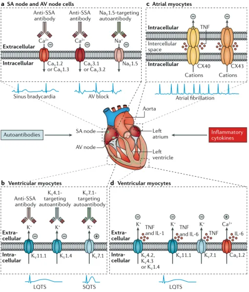
Fig. 1 | Autoimmune and inflammatory cardiac channelopathies and arrhythmias: molecular basis. Autoantibodies and inflammatory cytokines
can modulate the function of cardiac ion chan nels and promote arrhythmias. a | Bradyarrhyth
mias and conduction dis turbances can be induced by antiSjögren’s syndromerelated antigen A (antiSSA) antibodies (also known as anti Ro/SSA antibodies), which target the L type and/or Ttype Ca2+ channels and inhibit the related currents, or
by autoantibodies targeting the NaV1.5 Na+ chan
nel, which inhibit the Na+ current, in cells of the
sinoatrial (SA) node and atrioventricular (AV) node. b | Long QT syndrome (LQTS) can be
induced by antiSSA antibodies, which target the KV11.1 K+ channel (also known as hERG) and
inhibit the rapidly activating repolarizing compo nent of the delayed rectifier K+ current, or by
autoantibodies targeting the KV1.4 K+ channel,
which might inhibit the transient outward K+ cur
rent in ventricular myocytes. Short QT syndrome (SQTS) can be induced by autoantibodies target ing the KV7.1 K+ channel, which increase the slowly
activating repolarizing component of the delayed rectifier K+ current in ventricular myocytes. c | Atrial fibrillation can be induced by tumour
necrosis factor (TNF), which impairs the expression and/or distribution of connexin 40 (CX40) and CX43 and inhibits the function of gap junctions in atrial myocytes. d | LQTS can be induced by TNF,
which targets KV4.2, KV4.3, KV11.1 and KV7.1 K+
channels and inhibits the respective currents; by IL-1, which inhibits the transient outward K+
current; or by IL6, which targets the L type Ca2+channel (Ca
V1.2) and increases the L type Ca2+
current, in ventricular myocytes. For simplicity, IL6 and TNF receptors are not shown in panel d.
I S5–S6 of the NaV1.5 Na+ channel7. These
antibodies, which can be detected in patients with idiopathic AV block, inhibit Na+
currents and induce conduction disturbances in experimental models7.
Other autoantibodies that target ion channels can affect the action potential duration (APD) of ventricular myocytes, leading to long- QT syndrome (LQTS) or short- QT syndrome (SQTS) and associated malignant arrhythmias2. LQTS can be induced by anti- SSA antibodies, which inhibit the rapidly activating repolarizing K+ current
by targeting the extracellular pore loop of the KV11.1 K+ channel (also known as hERG)8,9,
as well as by autoantibodies targeting KV1.4
K+ channels, possibly through blockade of the
transient outward K+ current10. Conversely, agonist- like autoantibodies targeting KV7.1 K+
channels that enhance the slowly activating repolarizing K+ current were associated with
SQTS11(Fig. 1b).
Moreover, inflammatory cytokines — in particular, tumour necrosis factor (TNF), IL-1 and IL-6 — can be arrhythmogenic by directly affecting the function of cardiac ion channels; these are known as inflammatory cardiac channelopathies4. Specifically, it has been shown that TNF induces dysfunction of gap junctions in atrial myocytes through impaired expression and/or distribution of CX40 and CX43 and that these changes promote atrial fibrillation by favouring a slow and heterogeneous conduction in the atria12(Fig. 1c). In addition, cytokines can favour the development of LQTS by decreasing specific cardiac K+ currents
and/or increasing L- type Ca2+ currents3,4. TNF inhibits transient outward, rapidly activating repolarizing and slowly activating
3. Lazzerini, P. E., Capecchi, P. L. & Laghi- Pasini, F. Systemic inflammation and arrhythmic risk: lessons from rheumatoid arthritis. Eur. Heart J. 38, 1717–1727 (2017).
4. Lazzerini, P. E. et al. Emerging arrhythmic risk of autoimmune and inflammatory cardiac channelopathies. J. Am. Heart Assoc. 7, e010595 (2018).
5. Xiao, G. Q., Hu, K. & Boutjdir, M. Direct inhibition of expressed cardiac L- and T-type calcium channels by IgG from mothers whose children have congenital heart block. Circulation 103, 1599–1604 (2001).
6. Karnabi, E. et al. Congenital heart block: identification of autoantibody binding site on the extracellular loop (domain I, S5-S6) of α1D L- type Ca channel. J. Autoimmun. 34, 80–86 (2010).
7. Korkmaz, S. et al. Provocation of an autoimmune response to cardiac voltage- gated sodium channel NaV1.5 induces cardiac conduction defects in rats. J. Am. Coll. Cardiol. 62, 340–349 (2013). 8. Yue, Y. et al. Pathogenesis of the novel
autoimmune-associated long- QT syndrome. Circulation 132, 230–240 (2015). 9. Lazzerini, P. E. et al. Arrhythmogenicity of
anti- Ro/SSA antibodies in patients with torsades de pointes. Circ. Arrhythm. Electrophysiol. 9, e003419 (2016).
10. Suzuki, S. et al. Cardiac involvements in myasthenia gravis associated with anti-KV1.4 antibodies. Eur. J. Neurol. 21, 223–230 (2014).
11. Li, J. et al. Anti- KCNQ1 K+ channel autoantibodies increase IKs current and are associated with QT interval shortening in dilated cardiomyopathy. Cardiovasc. Res. 98, 496–503 (2013). 12. Sawaya, S. E. et al. Downregulation of
connexin40 and increased prevalence of atrial arrhythmias in transgenic mice with cardiac- restricted overexpression of tumor necrosis factor. Am. J. Physiol. Heart Circ. Physiol. 292, H1561–H1567 (2007).
13. Wang, J. et al. Impairment of HERG K+ channel function by tumor necrosis factor- alpha: role of reactive oxygen species as a mediator. J. Biol. Chem.
279, 13289–13292 (2004).
14. Monnerat, G. et al. Macrophage- dependent IL-1β production induces cardiac arrhythmias in diabetic mice. Nat. Commun. 7, 13344 (2016).
15. Hagiwara, Y. et al. SHP2-mediated signaling cascade through gp130 is essential for LIF- dependent I CaL, [Ca2+]i transient, and APD increase in cardiomyocytes. J. Mol. Cell. Cardiol. 43, 710–716 (2007). 16. Aromolaran, A. S. et al. Interleukin-6 inhibition of
hERG underlies risk for acquired long QT in cardiac and systemic inflammation. PLoS One. 13(12), e0208321 (2018).
Competing interests
The authors declare no competing interests. repolarizing K+ currents as a result of the
downregulation of channel expression and/or alterations in channel- gating kinetics, which are also associated with prolongation of the APD and/or QT interval4,13. Similar effects are mediated by IL-1, which reduces the transient outward K+ current14, and by IL-6, which enhances the L- type Ca2+ current
through CaV1.2 phosphorylation15 and
inhibits the rapidly activating repolarizing K+ current through a pathway involving the
IL-6 receptor and Janus kinase16(Fig. 1d). In terms of translational medicine, emphasizing the role of autoimmune and inflammatory cardiac channelopathies in arrhythmogenesis may lead to innovative anti- arrhythmic therapies based on the targeted modulation of the immune– inflammatory system, such as cytokine- targeting monoclonal antibodies or short decoy peptides that divert autoantibodies from their binding sites on ion channels.
There is a reply to this letter by Swirski, F. K. & Nahrendorf, M. Nat. Rev. Immunol. https://
doi.org/10.1038/s41577-018-0099-y (2018).
Pietro Enea Lazzerini 1*, Franco Laghi- Pasini1,4,
Mohamed Boutjdir2,3,4 and Pier Leopoldo Capecchi1,4 1Department of Medical Sciences, Surgery and
Neurosciences, University of Siena, Siena, Italy.
2VA New York Harbor Healthcare System, SUNY
Downstate Medical Center, New York, NY, USA.
3NYU School of Medicine, New York, NY, USA. 4These authors contributed equally: Franco Laghi-
Pasini, Mohamed Boutjdir, Pier Leopoldo Capecchi. *e- mail: [email protected] https://doi.org/10.1038/s41577-018-0098-z 1. Swirski, F. K. & Nahrendorf, M. Cardioimmunology:
the immune system in cardiac homeostasis and disease. Nat. Rev. Immunol. 18, 733–744 (2018). 2. Lazzerini, P. E. et al. Autoimmune channelopathies as
a novel mechanism in cardiac arrhythmias. Nat. Rev. Cardiol. 14, 521–535 (2017).
64 | january 2019 | volume 19 www.nature.com/nri