Background: The evidence on the use of dexamethasone im-
plants in the treatment of Behçet’s disease (BD)-related uveitis is limited to a few cases.
objectives: To evaluate the efficacy of dexamethasone im-
plants on ocular functional, morphological, and clinical para- meters in BD patients with severe refractory uveitis.
methods: Five eyes from five BD patients were enrolled. A single
intravitreal dexamethasone injection was applied to each eye. Best corrected visual acuity (BCVA), central macular thickness (CMT) assessed with optical coherence tomography, retinal vasculitis assessed by fluorescein angiography, vitreous haze score (Nussenblatt scale), intraocular pressure (IOP), and lens status (LOCS III, Lens Opacities Classification System III) were recorded at baseline and at 1, 3, and 6 month follow-up visits.
Results: At baseline, all eyes showed marked macular edema
and 4/5 had concomitant active retinal vasculitis. Mean BCVA was increased from baseline at each control visit with a mean improvement of 0.26 ± 0.18 lines at 6 months follow-up. Mean CMT decreased from baseline at each control visit with a mean improvement at 6 months follow-up of 198.80 ± 80.08 μm. At the end of the study, none of the eyes showed macular edema and the mean CMT was 276.80 ± 24.94 μm. Retinal vasculitis resolved in all eyes. One eye experienced an IOP spike during treatment that resolved spontaneously, and one eye developed a clinically significant lens opacity at 6 months follow-up.
Conclusions: Treatment with a dexamethasone implant in
BD-uveitis and inflammatory macular edema was safe and effective as an additional treatment combined with systemic immunomodulatory drugs.
IMAJ 2017; 19: 415–419
uveitis, macular edema, Behçet’s disease, intravitreal treatment, dexamethasone implant
intravitreal Dexamethasone implant as an Adjunct
Weapon for severe and Refractory uveitis in
Behçet’s Disease
Claudia Fabiani MD PhD1, Giacomo Emmi MD PhD2, Giuseppe Lopalco MD4, Lorenzo Vannozzi MD PhD3, Daniela Bacherini MD3, Silvana Guerriero MD PhD5, Rossella Franceschini MD6, Bruno Frediani MD7, Florenzo Iannone MD PhD4, Gian Marco Tosi MD6, Donato Rigante MD8* and Luca Cantarini MD7*
1Department of Ophthalmology, Humanitas Clinical and Research Center, Rozzano, Milan, Italy
2Department of Experimental and Clinical Medicine and 3Department of Surgery and Translational Medicine, Eye Clinic, University of Florence, Florence, Italy 4Department of Emergency and Organ Transplantation, Rheumatology Unit and 5Department of Ophthalmology and Otolaryngology, University of Bari, Bari, Italy 6Ophthalmology and Neurosurgery Department and 7Research Center of Systemic Autoinflammatory Diseases and Behçet's Disease Clinic, Department of Medical Sciences,
Surgery and Neurosciences, University of Siena, Siena, Italy
8Institute of Pediatrics, Fondazione Policlinico Universitario A. Gemelli, Università Cattolica Sacro Cuore, Rome, Italy
ABsTRAcT:
KEY woRds:
*The last two authors contributed equally to this study
B
ipolar aphthosis and severe intraocular inflammation lead-ing to sight-threatenlead-ing sequelae are the main features of Behçet’s disease (BD) [1]. BD-related posterior uveitis and panu-veitis are the most significant causes of morbidity ranging from 50 to 70% of cases, and blindness is reported with a frequency rate of about 25% [2]. According to the European League against Rheumatism (EULAR) recommendations, any patient with BD-related inflammatory eye disease affecting the posterior segment should be treated with azathioprine and systemic glu-cocorticoids [3]. In case of severe eye disease, defined as > 2 lines of drop in visual acuity on a 10/10 scale and/or retinal disease (retinal vasculitis or macular involvement), it is recommended that either cyclosporine A or infliximab be used in combination with azathioprine and glucocorticoids. Alternatively interferon-alpha with or without glucocorticoids might be used instead [3].Over the past years, increasing evidence has shown that the intravitreal delivery of glucocorticoids may be useful to induce the remission of intraocular inflammation in refractory BD-related ocular involvement or in the most severe cases. A few studies have explored the efficacy and safety of intravitreal injection of triamcinolone acetonide and fluocinolone aceton-ide implant in BD (Retisert
®
, Bausch & Lomb, Rochester, NY, USA) [4]. Most authors have also suggested the need to closely monitor intraocular pressure (IOP) and lens clarity since a con-sistent percentage of treated eyes may develop glaucoma and/or cataract. However, the bio-erodible intravitreal dexamethasone implant, now licensed for the treatment of adult and pediatric noninfectious intermediate and posterior uveitis in the United States and Europe, has shown a better safety profile compared to other drugs. Moreover the clinical efficacy of a dexametha-sone implant in the treatment of non-infectious posterior uve-itis has been assessed in several randomized controlled clinical trials [5-7]. Nevertheless, with regard to BD-related uveitis, theevidence on the use of a dexamethasone implant is limited to a few cases [8].
In the present study we aimed at evaluating the efficacy of a dexamethasone implant on ocular functional, morphological, and clinical parameters in a small case series of BD patients with severe refractory and/or long-standing uveitis.
PATIEnTs And mEThods
We performed a retrospective review of the medical records of patients treated with a dexamethasone implant (Ozurdex
®
; Allergan, Inc, Irvine, CA, USA) for persistent macular edema secondary to BD-related uveitis in three uveitis referral centers in Italy (Universities of Siena, Florence, and Bari). Diagnosis of BD was based on the International Study Group Criteria (ISGC) [9] and/or International Criteria for BD (ICBD) [10]. Patients were considered to have persistent inflammatory mac-ular edema if they had central macmac-ular thickness (CMT) greater than 300 microns and fluid in the macula (cysts or subretinal fluid), as assessed with optical coherence tomography (OCT) and fluorescein angiography (FA), for longer than 3 months before a dexamethasone implant treatment.Demographic and clinical characteristics of patients are shown in the Table 1. The following ophthalmic clinical parameters were recorded at all-time points (baseline, 1 month, 3 month, 6 month follow-up): best corrected visual acuity (BCVA), CMT measured by OCT, presence of macular edema
and retinal vasculitis assessed with FA, vitreous haze (VH) grading according to Nussenblatt scale, and IOP values [11]. Lens status and cataract grading according to LOCS III clas-sification system was recorded at baseline and at 6 months [12].
Only patients who had been followed for at least 6 months were included in the study. The implant was injected by a stan-dard procedure under topical anesthesia. After the injection, a topical antibiotic was applied 4 times a day for 7 days. All patients gave written informed consent for the dexamethasone implantation. Data were statistically described by mean values and standard deviation for continuous variables and by ranges for discrete variables.
REsulTs
Five eyes of five BD patients were included in the study. All patients fulfilled both the ISGC and ICBD diagnostic criteria for BD. Three patients were male and two patients were female, with age ranging from 38 to 52 years. Four eyes were phakic and one eye was pseudophakic. At baseline OCT and FA revealed the presence of cystoid macular edema (CME) in all eyes, and 4/5 eyes showed concomitant fluorangiographic evidence of active retinal vasculitis. None of the patients had activation of uveitis in the fellow eye during the follow-up period. Table 2 shows previous and concomitant treatments with disease modi-fying anti-rheumatic drugs (DMARDs), biologic agents, gluco-corticoids and colchicine. Only one eye (patient II) experienced
Table 1. Demographic and clinical characteristics of patients
Patient gender Age (years) Age at disease onset (years) hlA B51 (Y/n) disease duration
(years) characteristics of eye involvement
mucosal involvement
(Y/n) skin (Y/n) cns (Y/n) snP (Y/n) Fever (Y/n) gut (Y/n) vascular (Y/n) Joint (Y/n) Isgc (Y/n) IcBd (Y/n)
1 F 52 49 Y 3 Bilateral panuveitis Y Y N N N N N Y Y Y
2 M 38 18 Y 20 Panuveitis Y Y Y N Y N Y Y Y Y
3 M 46 42 N 4 Bilateral panuveitis Y Y N N N N N N Y Y
4 M 45 28 N 17 Bilateral posterior uveitis Y N N N Y Y N Y Y Y
5 F 43 35 Y 8 Posterior uveitis Y Y N N Y Y Y N Y Y
Y = yes, N = no, F = female, M = male, CNS = central nervous system, SNP = peripheral nervous system, ISGC = International Study Group Criteria, ICBD = International Criteria for Behçet’s Disease
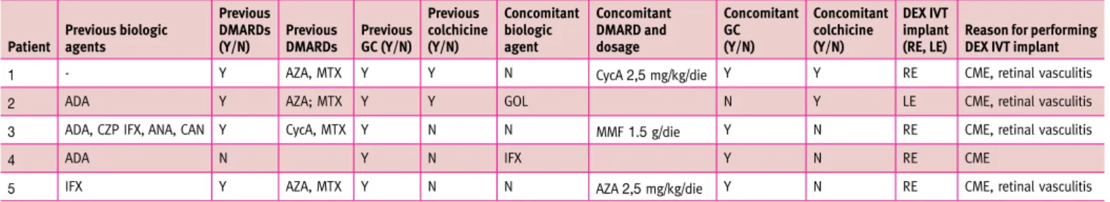
Table 2. Previous and concomitant treatments of the patients enrolled
Patient Previous biologic agents
Previous dmARds
(Y/n) Previous dmARds Previous gc (Y/n) Previous colchicine (Y/n) concomitant biologic agent concomitant dmARd and dosage concomitant gc (Y/n) concomitant colchicine (Y/n) dEX IvT implant
(RE, lE) Reason for performing dEX IvT implant
1 - Y AZA, MTX Y Y N CycA 2,5 mg/kg/die Y Y RE CME, retinal vasculitis
2 ADA Y AZA; MTX Y Y GOL N Y LE CME, retinal vasculitis
3 ADA, CZP IFX, ANA, CAN Y CycA, MTX Y N N MMF 1.5 g/die Y N RE CME, retinal vasculitis
4 ADA N Y N IFX Y N RE CME
5 IFX Y AZA, MTX Y N N AZA 2,5 mg/kg/die Y N RE CME, retinal vasculitis
Y = yes, N = no, DMARDs = disease modifying antirheumatic drugs, GC = glucocorticoids, DEX = dexamethasone, IVT = intravitreal, RE = right eye, LE = left eye, CME = cystoid macular edema, ADA = adalimumab, CZP = certolizumab pegol, IFX = infliximab, GOL = golimumab, ANA = anakinra, CAN = canakinumab, MTX = methotrexate, AZA = azathioprine, CycA = cyclosporine A
the eyes showed signs of macular edema with a mean CMT of 276.80 ± 24.94. At baseline retinal vasculitis was diagnosed in 4/5 eyes, while it was not observed in all eyes at 3 and 6 month follow-up visits. One eye experienced an IOP spike (> 20 mmHg) during treatment that resolved spontaneously, and only one eye developed a clinically significant lens opacity at 6-month follow-up. No additional side effects that could be related to the dexamethasone implant injection were observed. In one eye (patient IV) vitreoretinal surgery was performed along with cataract extraction before the dexamethasone implant was given, because of the occurrence of hemovitreous.
dIscussIon
In BD ocular involvement the cumulative structural damage and the vision loss result from recurrent episodes of inflamma-tion [13]. The goal of treatment should not only be to suppress inflammation when it occurs, but also to prevent severe recur-rent attacks of intraocular inflammation and to attain complete remission of inflammation in the longer term. In our patients, a single intravitreal injection of a dexamethasone implant was safe and effective in the treatment of BD posterior uveitis or panu-veitis, refractory to systemic glucocorticoids and/or DMARDs or biologic agents. Indeed, at the 6-month follow-up, treatment with dexamethasone implant was highly effective in resolving CME and retinal vasculitis in all cases. These findings occurred in parallel with the improvement of visual function (BCVA), which was also recorded in all eyes. Our observations are con-sistent with the results of the pivotal clinical trials and real-world data on the use of dexamethasone implants in the treatment of non-infectious uveitis. Indeed, in most studies, a high efficacy has been observed in terms of improvement of all ocular func-tional and morphological parameters [5, 14-18]. Our results are also in line with a recently published retrospective multicenter study on BD refractory uveitis [8]. Coşkun et al. [8] investigated the results of a single dexamethasone implant in the treatment of 17 eyes of 12 patients with refractory Behçet posterior uveitis at 1, 3, 6, and 12 months follow-up. BCVA, CMT, vitreous haze a mild recurrence of vitreous haze at the 6 month follow-up
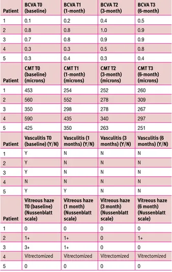
visit, while the remaining eyes did not show the recurrence of uveitis during the entire follow-up. Table 3 illustrates the main functional and morphological parameters collected at baseline and at each follow-up visit in the affected eyes (BCVA, CMT, vasculitis and vitreous haze). Table 4 summarizes the lens status at baseline and at the end of the study, and IOP values at each time-point.
The mean BCVA, CMT, and vitreous haze score improved over the follow-up period. The mean BCVA increased from baseline (0.44 ± 0.29) at each control visit with a mean improve-ment at the 1 month visit of 0.06 ± 0.05, at the 3 month visit of 0.18 ± 0.10, and at the 6 month visit of 0.26 ± 0.18. The mean CMT decreased from baseline (475.60 ± 98.81) at each control visit with a mean improvement at the 1 month visit of 97.80 ± 77.77, at the 3 month visit of 193.40 ± 81.90, and at the 6 month follow-up of 198.80 ± 80.08. At the end of treatment none of
Table 3. BCVA, CMT, vasculitis and vitreous haze at baseline and at
each follow-up visit of the five BD-patients enrolled
Patient BcvA T0 (baseline) BcvA T1 (1-month) BcvA T2 (3-month) BcvA T3 (6-month)
1 0.1 0.2 0.4 0.5 2 0.8 0.8 1.0 0.9 3 0.7 0.8 0.9 0.9 4 0.3 0.3 0.5 0.8 5 0.3 0.4 0.3 0.4 Patient cmT T0 (baseline) (microns) cmT T1 (1-month) (microns) cmT T2 (3-month) (microns) cmT T3 (6-month) (microns) 1 453 254 252 260 2 560 552 278 309 3 350 298 278 267 4 590 435 340 297 5 425 350 263 251
Patient vasculitis T0 (baseline) (Y/n) vasculitis (1 months) (Y/n) vasculitis (3 months) (Y/n) vasculitis (6 months) (Y/n)
1 Y N N N 2 Y N N N 3 Y N N N 4 N N N N 5 Y Y N N Patient vitreous haze T0 (baseline) (nussenblatt scale) vitreous haze (1 month) (nussenblatt scale) vitreous haze (3 month) (nussenblatt scale) vitreous haze (6 month) (nussenblatt scale) 1 0 0 0 0 2 1+ 1+ 0 1+ 3 3+ 1+ 0 0
4 Vitrectomized Vitrectomized Vitrectomized Vitrectomized
5 0 0 0 0
Y = yes, N = no, BCVA = best corrected visual acuity, CMT = central macular thickness
Table 4. The lens status at baseline and at the end of the study and the IOP at
baseline and at each follow-up visit following the intravitreal dexamethasone implantation
Patient
lens status before implant (locs III)
lens status after implant (6 months) (locs III) IoP T0 (baseline) (mmhg) IoP T1 (1 month) (mmhg) IoP T2 (3 months) (mmhg) IoP T4 (6 months) (mmhg) 1 0 0 13 13 16 13 2 0 0 13 14 19 17 3 NCP1 NCP4 14 16 16 15 4 Pseudophakic Pseudophakic 12 15 14 14 5 0 0 16 16 16 16
IOP = intra ocular pressure, T = time, NCP = nuclear cortical and posterior/subcapsular, LOCS III = Lens Opacities Classification System III
Additional benefits are noted when ocular inflammation is unilateral or asymmetric, when local therapy may prevent the need to increase the dose of systemic administered medica-tions. Individual patients’ characteristics should guide the treatment with dexamethasone implant in the daily clinical practice.
correspondence
Dr. C. Fabiani
Dept. of Ophthalmology, Humanitas Clinical and Research Center, Rozzano, Milan 53100, Italy
phone: (+39) 34084-84873 Fax: (+39) 06481-9648 email: [email protected]
References
1. Hatemi G, Seyahi E, Fresko I, Talarico R, Hamuryudan V. Behçet’s syndrome: a critical digest of the 2013-2014 literature. Clin Exp Rheumatol 2014; 32: S112-22. 2. Tugal-Tutkun I, Onal S, Altan-Yaycioglu R, Huseyin Altunbas H, Urgancioglu
M. Uveitis in Behçet disease: an analysis of 880 patients. Am J Ophthalmol 2004; 138: 373-80.
3. Hatemi G, Silman A, Bang D, et al. EULAR recommendations for the management of Behçet disease. Ann Rheum Dis 2008; 67: 1656-62.
4. Fabiani C, Alió JL. Local (topical and intraocular) therapy for ocular Adamantiades-Behçet›s disease. Curr Opin Ophthalmol 2015; 26: 546-52. 5. Lowder C, Belfort R, Lightman S, et al. Ozurdex HURON Study Group.
Dexamethasone amethasone intravitreal implant for noninfectious intermediate or posterior uveitis. Arch Ophthalmol 2011; 129: 545-53.
6. Lightman S, Belfort R Jr, Naik RK, et al. Vision-related functioning outcomes of dexamethasone intravitreal implant in noninfectious intermediate or posterior uveitis. Invest Ophthalmol Vis Sci 2013; 54: 4864-70.
7. Williams GA, Haller JA, Kuppermann BD, et al. Dexamethasone DDS Phase II Study Group. Dexamethasone posterior-segment drug delivery system in the treatment of macular edema resulting from uveitis or Irvine-Gass syndrome.
Am J Ophthalmol 2009; 147: 1048-54.
8. Coşkun E, Celemler P, Kimyon G, et al. Intravitreal dexamethasone implant for treatment of refractory Behçet posterior uveitis: one-year follow-up results.
Ocul Immunol Inflamm 2015; 23: 437-43.
9. Criteria for diagnosis of Behçet’s disease. International Study Group for Behçet’s Disease. Lancet 1990; 335: 1078-80.
10. International Team for the Revision of the International Criteria for Behçet’s Disease (ITR-ICBD). The International Criteria for Behçet’s Disease (ICBD): a collaborative study of 27 countries on the sensitivity and specificity of the new criteria. J Eur Acad Dermatol Venereol 2014; 28: 338-47.
11. Nussenblatt RB, Palestine AG, Chan CC, Roberge F. Standardization of vitreal inflammatory activity in intermediate and posterior uveitis. Ophthalmology 1985; 92: 467-71
12. Davison JA, Chylack LT. Clinical application of the lens opacities classification system III in the performance of phacoemulsification. J Cataract Refract Surg 2003; 29: 138-45.
13. Ozyazgan Y, Ucar D, Hatemi G, Yazici Y. Ocular involvement of Behçet’s syndrome: a comprehensive review. Clin Rev Allergy Immunol 2015; 49: 298-306. 14. Zarranz-Ventura J, Carreño E, Johnston RL, et al. Multicenter study of
intravitreal dexamethasone implant in noninfectious uveitis: indications, outcomes, and reinjection frequency. Am J Ophthalmol 2014; 158: 1136-45. 15. Tomkins-Netzer O, Taylor SRJ, Bar A, et al. Treatment with repeat dexa-
methasone implants results in long-term disease control in eyes with noninfectious uveitis. Ophthalmology 2014; 121: 1649-4.
16. Cao JH, Mulvahill M, Zhang Li, Joondeph BC, Dacey MS. Dexamethasone intravitreal implant in the treatment of persistent uveitic macular edema in the absence of active inflammation. Ophthalmology 2014; 121:1871-6.
17. Khurana RN, Porco TC. Efficacy and safety of dexamethasone intravitreal implant for persistent uveitic cystoid macular edema. Retina 2015; 35: 1640-6.
score, and IOP were determined, at baseline and control visits. BCVA significantly increased from baseline at each control visit and the mean CMT and vitreous haze score were significantly decreased from baseline at each follow-up visit. Three eyes showed IOP spikes requiring topical treatment. The efficacy of intravitreal delivery has been investigated in BD also for other glucocorticoids, such as triamcinolone acetonide and fluocinolone acetonide [19-25]. In 2014 Park and co-authors [19] investigated the effectiveness of intravitreal triamcinolone acetonide (4 mg/0.1 ml) injection for refractory posterior BD uveitis at 24-month follow-up. Forty-nine patients (49 eyes) were included. Mean BCVA was improved at 12 and 24 month evaluations. A complete control of intraocular inflammation was obtained in 87.0% of patients, but 60.0% of them showed a disease relapse within 12 months. IOP pressure elevation (> 21 mmHg) was observed in about 40% of cases [19]. The study results suggested that intravitreal triamcinolone acetonide is effective in severe BD eye involvement, but ocular complications are commonly observed and may limit triamcinolone aceton-ide efficacy and repeatability. Oh and colleagues [20] reported the long-term outcome and complications of eight eyes from seven patients with BD-related uveitis treated with a 0.59 mg fluocinolone acetonide intravitreal implant. Although the final significant visual acuity improved, the authors reported a high rate of complication in terms of postoperative IOP spikes, with up to 62% of eyes requiring glaucoma shunting surgery [20]. They also reported a case of postoperative cytomegalovirus endothelitis. Fluocinolone acetonide efficacy has been also evaluated by Sangwan et al. [21] in a prospective multicenter randomized double-masked dose-controlled study. The three-year results have shown that the fluocinolone acetonide implant significantly reduced uveitis recurrence rates and also improved visual acuity, allowing the reduction in adjunctive therapy. Fourteen BD-patients were included in the study. Elevation of IOP occurred in about 70% of patients, and nearly all (94.9%) phakic implanted eyes required cataract surgery [21].
Regarding dexamethasone implant-related ocular complica-tions, it is notable that the good safety observed in previously published studies in non-infectious uveitis and BD-related uveitis has been replicated in our series. To confirm these encouraging findings in BD-related uveitis, properly designed ad hoc studies including larger cohorts of patients and with longer follow-up are needed. The number of injections required to control intraocular inflammation in BD-related uveitis is still controversial and the effect of repeated dexamethasone injec-tions needs to be clarified.
ConClusions
In conclusion, our data suggest considering the intravitreal administration of a dexamethasone implant in the treatment of BD intraocular inflammation, especially when compli-cated by CME not adequately controlled by systemic therapy.
22. Karacorlu M, Mudun B, Ozdemir H, et al. Intravitreal triamcinolone acetonide for the treatment of cystoids macular edema secondary to Behcet Disease. Am J
Ophthalmol 2004; 138: 289-91.
23. Ohguro N, Yamanaka E, Otori Y, Saishin Y, Tano Y. Repeated intravitreal triamcinolone injections in Behcet Disease that is resistant to conventional therapy: one-year results. Am J Ophthalmol 2006; 141: 218-20.
24. Atmaca LS, Yalçindağ FN, Özdemir Ö. Intravitreal triamcinolone acetonide in the management of cystoid macular edema in Behçet’s disease. Graefe’s Arc Clin
Exp Ophthalmol 2007; 245: 451-6.
25. Tuncer S, Yilmaz S, Urgancioglu M, Tugal-Tutkun I. Results of intravitreal triamcinolone acetonide (IVTA) injection for the treatment of panuveitis attacks in patients with Behçet disease. J Ocul Pharmacol Ther 2007; 23: 395-401. 18. Adan A, Pelegrin L, Rey A, et al. Dexamethasone intravitreal implant for
treatment of uveitic persistent cystoid macular edema in vitrectomized patients.
Retina 2013; 33: 1435-40.
19. Park UC, Park JH, Yu HG. Long-term outcome of intravitreal triamcinolone acetonide injection for the treatment of uveitis attacks in Behcet disease. Ocul
Immunol Inflamm 2014; 22: 27-33.
20. Oh EK, Lee EK, Yu HG. Long-term results of fluocinolone acetonide intravitreal implant in Behçet intractable posterior uveitis. Can J Ophthalmol 2014; 49: 273-8. 21. Sangwan VS, Pearson PA, Paul H, Comstock TL. Use of the fluocinolone
acetonide intravitreal implant for the treatment of noninfectious posterior uveitis: 3-year results of a randomized clinical trial in a predominantly Asian population. Ophthalmol Ther 2015; 4: 1-19.
Gain-of-function mutations in TMEM173, encoding the stimu- lator of interferon genes (STING) protein, underlie a novel type I interferonopathy that is minimally responsive to conventional immunosuppressive therapies and associated with high frequency of childhood morbidity and mortality. STING gain-of-function causes constitutive oversecretion of interferon. Fremond et al. studied the effects of a TANK-binding kinase 1 (TBK-1)/IKKε inhibitor (BX795) on secretion and signaling of interferon in primary peripheral blood mononuclear cells (PBMCs) from patients with mutations in STING. PBMCs from four patients with STING-associated disease were treated with BX795. The effect of BX795 on interferon pathways was assessed by western blotting and an interferon β reporter assay as well as by quantification of interferon α in cell lysates, staining for STAT-1 phosphorylation, and measure- ment of interferon-stimulated gene (ISG) messenger RNA (mRNA)
expression. Treatment of PBMCs with BX795 inhibited the phosphorylation of interferon regulatory factor 3 and interferon β promoter activity induced in HEK 293T cells by cyclic GMP-AMP or by genetic activation of STING. In vitro exposure to BX795 inhibited interferon α production in PBMCs in patients with STING-associated disease without affecting cell survival. In addition, BX795 decreased STAT-1 phosphorylation and ISG mRNA expression independent of interferon α blockade. These findings demonstrate the effect of BX795 on reducing type I interferon production and interferon signaling in cells from patients with gain-of-function mutations in STING. A combined inhibition of TBK-1 and IKKε therefore holds potential for the treatment of patients carrying STING mutations, and may also be relevant in other type I interferonopathies.
Arthritis & Rheumatol 2017; 69: 1495 Eitan Israeli
Blockade of TAnK-binding kinase
1/iKKε inhibits mutant stimulator of interferon genes
(sTinG)-mediated inflammatory responses in human peripheral blood mononuclear cells
capsule
Polachek et al. set out to define and identify a group of systemic lupus erythematosus patients with low disease activity (LDA) and to examine whether LDA is similar to patients in remission and different from a high disease activity group (HDA) in short-term outcomes. The LDA group was defined as Systemic Lupus Erythematosus Disease Activity Index 2000 (SLEDAI-2K) < 3, including only one clinical manifestation of rash, alopecia, mucosal ulcers, pleurisy, pericarditis, fever, thrombocytopenia, or leukopenia. The patients could be taking anti-malarials. Remission was defined as no clinical manifestation from taking anti-malarials alone, and the HDA group was defined as SLEDAI-2K > 6. The time frame for inclusion in each group was at least 1 year. Of 620 patients with active disease who were seen between 1970 and 2015, 80 patients (12.9%) fulfilled the criteria for LDA, 191 (30.8%) for remission, and 349 (56.3%) for HDA. Polachek et al. found that
the LDA patients with and without positive serology results were similar at baseline and with prior disease characteristics. After 2 years of follow-up, the LDA and remission groups were similar in their adjusted mean SLEDAI-2K score, organ involvement, Systemic Lupus International Collaborating Clinics/American College of Rheumatology Damage Index (SDI) score, mortality, and therapies. After 2 and 4 years of follow-up, the HDA group had a higher adjusted mean SLEDAI-2K score, more major organ involvement, higher SDI score, higher mortality, and more therapy compared to the combined LDA/ remission groups. LDA and remission groups had similar short-term outcomes, and both had better outcomes and prognosis than the HDA group. LDA may be used as an outcome measure in therapeutic trials or in treat-to-target regimens.
Arthritis Care & Res 2017; 69: 997 Eitan Israeli