A
A
l
l
m
m
a
a
M
M
a
a
t
t
e
e
r
r
S
S
t
t
u
u
d
d
i
i
o
o
r
r
u
u
m
m
–
–
U
U
n
n
i
i
v
v
e
e
r
r
s
s
i
i
t
t
à
à
d
d
i
i
B
B
o
o
l
l
o
o
g
g
n
n
a
a
DOTTORATO DI RICERCA IN
DOTTORATO DI RICERCA IN BIOLOGIA CELLULARE, MOLECOLARE E INDUSTRIALE Progetto 2: Biologia Funzionale e Molecolare
Ciclo XXV
Settore Concorsuale di afferenza: 05/E2
Settore Scientifico disciplinare: BIO/11
TITOLO TESI
Conjugation of Toll Like Receptor 7 agonist through conjugation to immunogenic proteins. Synthesis, characterization, formulation and pre-clinical evaluation.
Presentata da: Simone Vecchi
Coordinatore Dottorato
Relatore
Prof. Vincenzo Scarlato Prof. Vincenzo Scarlato
Dott. Simone Bufali
Index
Part 1 – TLR7 agonist bioconjugation
Introduction:
TLR7 agonist conjugation ...3
Materials and methods...13
Results:
Conjugation and analytical characterization...23
In vivo evaluation...30
Discussion...37
Conclusions...46
Section 2 – Aluminium adjuvant dose for preclinical evaluation
Current use of aluminium adjuvants...51
Aluminium salts adjuvants...54
Aluminum dose limits recommendations...60
What is an acceptable dose?...64
Conclusions...69
Publication...70
Introduction
The earliest definition of vaccine adjuvants describes them as components that are added to vaccine antigens to make them more immunogenic, but this definition is quite imprecise due to the broad variety of candidates available (O'Hagan and De Gregorio 2009). Nowadays the in vivo adjuvant effect can be divided into two principal components: delivery and immune potentiation. Delivery systems localize antigens and target to the appropriate cell types of the innate immune system and can also be optimized for immune potentiator targeting. Immune potentiators directly activate innate immune cells providing the pro-inflammatory context for antigen recognition (O'Hagan and Valiante 2003) and to this class belong some molecules with agonistic activity on the toll like receptors.
Toll-like receptors (TLRs) are a set of conserved cellular receptors that play an important role in the recognition of microbial pathogens and in initiating the host innate immune response. These receptors recognize distinct molecular components of invading pathogens, such as cell wall structures and nucleic acids. The discovery that endogenous ligands as well as synthetic small molecules can activate certain TLR pathways has generated tremendous interest in the development of new therapeutics drugs for infectious diseases (Stanley 2002).
Fig.1 Different Toll like receptors (TLRs) and their natural agonists
Some synthetic compounds with an agonistic activity on the TLR7 belongs to different classes as imidazoquinolinamines (Miller, Gerster et al. 1999), 8-oxo-3-deazapurine (Tran, Pryde et al. 2011) and 6-amino-9-benzyladenine (Jin, Wu et al. 2006). They are generically called SMIPs (small molecules immuno-potentiators) because are able to modulate the immune response through immunoglobulin production (Othoro, Johnston et al. 2009), interferon and cytokines synthesis (Hemmi, Kaisho et al. 2002). These compounds have potential to be used in different pathologies from cancer (Johnston and Bystryn 2006; Johnston, Zaidi et al. 2007) to parasitic infection (Othoro, Johnston et al. 2009) and as novel immunemodulators (Dockrell and Kinghorn 2001), to increase potency of recombinant proteins in vaccines development (Tomai and Vasilakos 2011).
Fig.2 SIMPs TLR7 structures known in the literature
Several studies demonstrated that when Imiquimod formulated as cream for dermal application (Aldara®) (Tomai and Vasilakos 2011), locally administered after subcutaneous immunization with ovalbumin (OVA), was able to enhance the anti-OVA antibody response of a 10-fold compared to mice that were not treated with imiquimod (Johnston and Bystryn 2006); same results have been found in mice subcutaneous immunized with Plasomodium falciparum circumsporozoite peptides and then topically treated with Aldara (Othoro, Johnston et al. 2009) (Fig.3) .This enhancement effect was probably connected with the slow release at the injection site of Imiquimod from the cream formulation, that allowed a kind of simultaneous presentation of the TLR agonist and the antigen (Ag) to the dentritic cells (DCs) (Ahonen, Doxsee et al. 2004).
Fig.3 Immunogenicity enhancement due to the co-administration of OVA plus
Imiquimod (OVA/IMIQ) in comparison to OVA alone (OVA), liposome encapsulated OVA (OVA/LIP) and E.G7-OVA cells that synthesize OVA constitutively and present
OVA derived peptides on the cell surface within the MHC I context.
Another way that allowed evaluating the co-delivery of TLR7 agonist and Ag were liposomes and nanoparticles; in these systems, the effect is due to the encapsulation of TLR agonist and Ag in the same particle that is phagocyted by the APCs (antigen presenting cells). The encapsulation of Resiquimod (also called R-848, a TLR7 agonist) into liposomes further enhances the OVA antibody response in comparison with the simple administration of OVA+3M-019 (Johnston, Zaidi et al. 2007) (Fig.4).
Liposomes and nanoparticles due to their particulate structure, after phagocitosis from the immune system cells, through a co-delivery effect are able to co-localizate the TLR agonist and the Ag (O'Hagan and Valiante 2003; Valmori, Souleimanian et al. 2007). In these delivery systems the components interaction is only due to their physical
properties without a chemical bound TLR agonist and Ag (Foged, Arigita et al. 2004; Oyewumi, Kumar et al. 2010).
Fig.4 Relative OVA specific IgG subclass responses to immunization. Individual sera
collected from mice 1 week after the fourth immunization were analyzed by ELISA for specific IgG subclasses antibodies to OVA using a mouse subtyping kit. Each bar represents the mean absorbance and the standard deviation of triplicate wells of sera collected from eight mice for all groups except OVA/Lip which consisted of six mice
These studies suggested the possibility that the immune system requires a synchronized exposure of the DCs to the TLR agonist; it appeared that TLR activation alone is not sufficient to drive certain types of adaptive responses, such as CD8+ T cells, therefore TLR-7/8 agonists may need to be coupled with other stimuli for optimal activity (Oh and Kedl 2010).
This hypothesis was supported by some mechanicistic studies on DCs that showed how the efficiency of presenting antigens from phagocytosed cargo was dependent on the presence of TLR ligands within the cargo (Blander and Medzhitov 2006) and the mode of antigen delivery into DCs coupled with the right type of signal. This can impact not
only the co-stimulatory context, in which the antigen is presented to naive T cells, but MHC-II presentation of the antigen and formation of the T-cell receptor ligand itself (Blander 2007).
The effect of the direct co-localization TLR7 agonist and Ag has been already showed by different works from Wille-Reece et al. in mice (Wille-Reece, Wu et al. 2005) and in non human primates (Wille-Reece, Flynn et al. 2005), by UV-catalyzed conjugation of the TLR7/8 agonist 3M-012 (structurally similar to R848) to the Gag protein from HIV,
that resulted in enhanced Th1 and CD8+ T cell responses, demonstrating that the
TLR7/8 agonist could be a useful adjuvant to increase the cytokines production in vivo if administered as a protein conjugate. The increased efficiency of DCs activation rather than prolonged duration of Ag presentation have been suggested to be the mechanism by which the Ag-TLR7/8 conjugate enhances T cell responses in vitro (Wille-Reece, Flynn et al. 2005; Wille-Reece, Wu et al. 2005) (Fig.5).
Fig.5 A) IFN and B) IL-2 producing cells and are increased after immunization with
HIV Gag protein and a TLR7/8 agonist or CpG ODN. NHPs were immunized four times at 4-week intervals with HIV Gag protein, with or without CpG ODN, TLR7/8, or
TLR8 agonist. An additional group was immunized in a similar manner with HIV Gag protein conjugated to the TLR7_8 agonist (Gag-TLR7/8 conjugate). PBMCs were harvested at various times post immunization, and the frequencies of IFN and IL-2
producing cells were measured
Another study made from Wu et al. showed the effect of the conjugation of a TLR7 ligand (UC-1V150) to mouse serum albumine (MSA) after its linkage with an idrazide linker (SANH) to obtain the activated MSA-SANH that can be conjugated via reductive ammination to the TLR7 ligand. The effect of the TLR7 conjugation to MSA induced a Th1 and Th2 antigen specific response (Chan, Hayashi et al. 2009) and extended mice survival after challenge with Bacillus anthracis spores (Wu, Hayashi et al. 2007).
Wu et al. showed the effect of the conjugation of a TLR7 ligand (UC-1V150) to mouse serum albumine (MSA) after its linkage with an idrazide linker (SANH) to obtain the activated MSA-SANH that can be conjugated via reductive ammination to the TLR7 ligand. This study displayed a deeper analytical characterization, developed to estimate the conjugation ratio Ag:TLR7 agonist (1:5) and the average molecular weight.
The authors decided to compare the conjugate UC-1V150/MSA versus the UC-1V150 alone; the doses used for the immunizations were calculated on a molar base; due to the conjugation ratio 5:1, 3.8 nmols of 1V150/MSA was equal to 19 mols of UC-1V150 and not 3.8 nmols as used (Wu, Hayashi et al. 2007).
Fig.6 Preclinical efficacy of UC-1V150/MSA in pulmonary infectious diseases.
Age-matched female A/J mice were administered i.n. saline only or saline containing MSA (amount equivalent to UC-1V150/MSA), UC-1V150, or UC-1V150/MSA at 0.75 nmol
Looking at these promising results, we decide to evaluate the effect of a TLR agonist conjugate on a model protective antigen and investigate its impact on the immunogenicity and protection in a pre-clinical model, using a SMIP TLR7 agonist (named TLR7aR) with a mild and selective conjugation reaction.
The previous works done on TLR agonist bioconjugation were highly focused on the immunological response, but they lack in terms of serological analysis and impact on protection with an unformulated system, through simple administration in a physiological buffer (Wille-Reece, Flynn et al. 2005; Wille-Reece, Wu et al. 2005; Wu, Hayashi et al. 2007; Kastenmuller, Wille-Reece et al. 2011).
With this proof of concept we want to asset an integrated and comprehensive approach for TLR7 agonist conjugation, characterization and formulation and to investigate the impact of Ag and TLR agonist co-localization on the immunogenicity applied on a model protein antigen when it is alum formulated.
Conjugation reaction
The recombinant pilus subunits RrgB clade I His-tag form (molecular mass 66kDa) from S. pneumoniae serotype 4 TIGR4 strain, was expressed in Escherichia coli and purified in soluble form by affinity chromatography on His-Trap high-performance column (GE Healthcare) at 3.92 mg/ml in 50 mM Tris and 100 mM NaCl, pH 8.0. 51 µl of protein solution were mixed with a 1 µl of a DMSO solution of the TLR7aR at a concentration of 117 mM in DMSO (38000 fold excess) in a PCR tube. The TLR7aR was internally synthesized and it had a molecular weight of 1484.77 a.m.u.; the linker lenght is 71.81 Å (calculated for the minimum energy structure). The lysine derivative
of the agonist showed on HEK-293 TLR7 reporting line cell a value of EC50=0.35 uM
(equal to 110% of Resiquimod) and no activation on HEK-293 TLR8 reporting line cell. When the two solutions were mixed together a white precipitate appeared. The solution was kept under mild shaking for 24 hr at 37°C (until the white precipitate is solubilized) then 100 µl of potassium acetate buffer 1 M pH 4 were added to the solution with 0.173
mg of NaCNBH3 for 4 hours. All the solutions were prepared in endotoxin free water
from Lonza.
Purification of the Conjugated RrgB-TLR7aR
The crude reaction mixture was purified to eliminate the unreacted TLR7aR with immobilized metal affinity chromatography (IMAC); 300 µl of a “ready to use” IMAC
uncharged resin from Biorad were loaded with 350 µl of the crude reaction mixture. The IMAC resin was washed once with 300 µl of a 20% ethanol solution in PBS and discharged, afterwards it was eluted 4 times with 300 µl of a 300mM imidazole solution in PBS and the eluates were collected together. The total recovered eluate was then concentrated with an Amikon 30K spin filter used at 14 kRPM for 25 minutes at room temperature. The final recovered volume was 120 µl and was stored at -20°C.
Analytical characterization
SDS-PAGE was run using NuPAGE Bis-Tris 4-12% Precast Gel from Invitrogen in MOPS 1x buffer from Biorad. HiMark12™ unstained standard from Life technology was used and the loaded amount was 1.5 µg for each sample. Size exclusion UPLC (SE-UPLC) analysis was performed with a ClassH bio-compatible system, equipped with a titanium cell. Waters BEH 200 column of 4.6*150 mm was chose and a KPi 100 mM / KCl 50 mM pH 7.2 buffer was used as mobile phase; flow was set at 0.35 ml/min, column temperature 40°C, sample temperature 4°C, sample injected volume 5 µL. Apparent molecular weight calculation was done against calibration curve obtained with gel filtration protein standards from Biorad; a value of r2 > 0.98 was obtained.
Dynamic laser scattering measurement (DLS) was performed using a Z-sizer nano instrument from Malvern, using a 50 µL “use and throw” cuvette. Measurements were performed in back-scattering mode with a detector at 173° in triplicate. Automatic measurement modality was chosen and data analysis was performed using the protein
analysis fitting model. Protein refractive index 1.450, disperdent viscosity 0.9181 cP and refractive index 1.332. equilibration time 60 seconds at 25°C.
Protein titer was measured with MicroBCA assay using reagents from Thermoscientific. Endotoxins titer was measured with a plate kinetic chromogenic test using LONZA LAL reagents.
Reverse phase UPLC (RP-UPLC) analysis was performed with a binary UPLC system
from Waters equipped with a PDA detector (=214nm) using a BEH 300 C18 column
of 2.1*50 mm, mobile phases were: A) water/TFA 0.1% and B) acetonitrile/TFA 0.1%, flow 0.5 ml/min; column temperature 60°C, sample temperature 4°C and sample injection volume 2 µL. Gradient used for RrgB and RrgB-TLR7aR were: B% 0 to 3 min from 10 to 30 %, 3 to 20 min from 30 to 80 %, plus 2 minutes of equilibration time, method LOQ 0.05 µg/ml (for RrgB). Gradient for TLR7aR was: B% from 10 to 100 in 2 min plus 1.5 min of equilibration time; flow 1.2 ml/min, column temperature 45°C,
injection volume 10 µL sample temperature 4°C, detection = 268 nm, method LOQ
0.03 µg/ml and r2>0.99.
Estimation of the conjugation rate
Fluorimetric quantization of the amount of TLR7aR conjugated to RrgB was performed with a Tecan Infinite 200 plate reader fluorimeter, using 96 black walls plate; excitation
calibration curve and analyzed in triplicate (r2 > 0.98). The sample was properly diluted to give fluorescence intensity within the calibration curve outers.
Vaccine formulations
Formulations have been prepared using the “sequential adsorption method” as describe from Matheis et al. (Matheis, Zott et al. 2001). Alum (aluminum hydroxide internally produced) at a stock concentration of 14.62 mg/ml in NaCl 2,2 g/l was used to have a final concentration of 2 mg/ml. Alum was then added with histidine buffer 100 mM pH 6.5 to a final concentration of histidine of 10 mM, then sodium chloride 2M to a theoretical osmolality of 300 mOsm/Kg. After the addition of those excipients, the antigen or the antigen followed by the TLR7aNR was added (TLR7aNR was an analogue of TLR7aR without the reactive moiety). The formulations were diluted with water to the final required concentration for an injected volume of 100 µL. Alum dose and injection volume was chose according as previously described (Vecchi, Bufali et al. 2012). The formulations were incubated over night at 4°C under mild shacking and used for mice immunization the day after.
Vaccine formulations characterization
An aliquot of 50 µL was used to measure its pH and osmolality with Osmomat 300 autosampler equipped from Gonotec: pH was 7±0.5 and osmolality 300±60 mOsm/kg, compatible with a physiological i.p. administration. An aliquot of 100 µL was assayed for endotoxin content with a plate kinetic chromogenic test using LONZA LAL reagents at 1:10000 dilution folds. Another aliquot of 100 µL of vaccine formulations was spun down at 17 kRCF for 10 minutes at room temperature. The supernatant was used for antigen and for TLR7aNR quantification. RrgB and TLR7aNR were quantified with RP-UPLC meanwhile for RrgB-TLR7aR MicroBCA assay from Thermoscientific was preferred.
The alum pellet was desorbed over night at 37°C under mild shacking with potassium phosphate buffer 500 mM pH 9, then spun down at 17 kRCF for 10 minutes at room temperature. The recovered supernatant was assayed for antigen and TLR7aNR content with the above describe techniques.
Immunizations
Protocols were reviewed and approved by the Novartis Vaccines and Diagnostics Institutional Review Committee. Six-week-old, specific-pathogen-free female BALB/c mice from Charles River were immunized intraperitoneally on days 0, 14, 28 with a
volume of 100 µL of vaccine formulations. At day 42 samples of sera were obtained from each animal. At day 43, each mouse was challenged intraperitoneally with a mean
dose of 160 CFU of S.pneumoniae TIGR4 variant (it was considered as LD100 refereed
to PBS). Blood bacteremia was evaluated 24 hours after challenge and mortality course monitored for 10 days as reported (Gianfaldoni, Censini et al. 2007). The animals were euthanized when they exhibited defined humane end points that had been pre established for the study in agreement with Novartis Animal Welfare Policies.
Specific IgG measurement using protein-coupled
microspheres
A Luminex assay was developed to determine antibody titers in mouse antisera. In detail, 20 μg of recombinant protein were coupled to the carboxyl groups of 2.5 million MicroPlex microspheres (Luminex Corp), according to the manufacturer's instructions. The coupling reaction was confirmed by incubating 5000 antigen coupled microspheres with 8 serial two-fold dilutions of a hyper immune antiserum used as standard solution. To determine sera titers, the beads were incubated with mouse specific sera (dilution 1:10000 in PBS), washed twice in 200μl of PBS and then incubated with PE conjugated secondary antibodies (1:200, Jackson Immuno Research) for 15 min in the dark onto an orbital shaker. The measurement of specific IgG content toward recombinant RrgB clade I and for RrgB-TLR7aR was done at a working concentration of 3500 beads per colour per well. IgG measurements were determined on the Luminex 200 analyzer using
the Bio-Plex Manager 5.0 software (Bio-Rad, Hercules, CA). All results are expressed as Relative Luminex Units per ml (RLU/ml). The correlation of mean fluorescent intensity (MFI) units to RLU/ml of Ag-specific IgG was made by interpolating the MFI data through the standard solution. The lower limit of quantification was 0.32 RLU/ml for RrgB.
Opsonophagocytosis Killing Assay (OPKA)
OPKA was performed as described in Harfouche, Filippini et al. (Harfouche, Filippini et al. 2012). In brief, heat inactivated mice pooled sera were initially diluted 1:20 and tested in a 3-fold dilution in a reaction mixture consisting of Streptococcus pneumoniae TIGR4 strain, 12% rabbit complement, and differentiated HL-60 cells (bacteria:cells ratio, 1:400). After 1h of incubation at 37°C with 5% CO2, phagocytosis was stopped
and an aliquot of the reaction mixture was tilt plated on TH Agar (Todd Hewitt) and incubated overnight. Bacteria survival was detected by colony counter ProtoCOL (Synbiosis). Results were expressed as percentage of killing or titer. The first was calculated as the percentage of bacteria that were killed in samples with active complement plus sera (BPC’+S), compared to bacteria-phagocytes-active complement (BPC’). The titer was obtained interpolating the mean of the six replicate killing values and expresses the sera concentration at which the 50% of killing occurred.
Statistical analysis
IgG titer, bacteremia and mortality course were analyzed with the Mann-Whitney One-tailed or two-One-tailed tests to compare immunized groups with the control group or each other, respectively. Opsonophagocytosis killing titers were analysed using 2-samples t test on log values (base 3). Values of p ≤ 0.05 were considered and referred to as significant. Minitab Software from Minitab Inc. was used
Conjugation and purification of RrgB-TLR7aR
We focused our attention on the creation of a covalent bound between the RrgB lysines and the TLR7aR; to achieve this results a 2-amino-4-oxy-benzaldehyde reactive moiety was linked to the TLR7 agonist. That moiety was able to give in a slightly acid environment an immine derivative that was reduced with sodium cyanoborohydride to obtain the desiderate conjugate. That moiety allows the conjugation of the TLR7aR with several different macromolecules that contain an amine group, like a peptide or a nucleic acid (Fig.7). Reaction temperature was set at 37°C, below the protein’s melting point (75°C).
The histidine tag on RrgB provided a quick and efficient purification method to remove the unreacted TLR7aR that otherwise could react with the lysine of the animal tissues and rise an effect due to the free TLR7aR and not only to the co-localization of TLR7 agonist and antigen.
.
Fig.7 Conjugation reaction scheme for the TLR7aR. The reaction has been conducted at
37°C in acetate buffer 750mM pH=4.After 24hr from the adding of RrgB, sodium cyanoborohydride was added for other 4hr, then the crude reaction mixture was
Analytical characterizations of RrgB-TLR7aR
SDS-PAGE was used to give a qualitative evidence of the increased molecular weight and the distribution of the conjugate; RrgB-TLR7aR had a smeared band higher than the native RrgB (Fig.8).
Fig.8 SDS-PAGE of the conjugate RrgB-TLR7aR.
M = MWs ladder; 1 = RrgB-TLR7aR; 2 = native RrgB
Complementary with SDS-PAGE, a SE-UPLC analysis was performed and it confirmed the proteins’ distribution and the absence of detectable aggregates (Fig.9).
Fig.9 Size Exclusion Ultra Performance Liquid Chromatography analysis
Monomeric RrgB1.948 min; Natural RrgB aggregate 1.768 min; Conjugate RrgB-TLR7aR 1.689 min
The average molecular weight of 130 kDa was estimated against calibration curve (Tab.1).
Tab.1 Apparent molecular weights estimation from SE-UPLC analysis
RP-UPLC reinforced the observation made about the species distribution and estimates the composition of the RrgB-TLR7aR. The amount of unconjugated RrgB was lower than 20% of the total species (fig.10).
Rt % Area Apparent MW (KDa)
Conj 1,689 100 130
Native 1,768 2,64 110
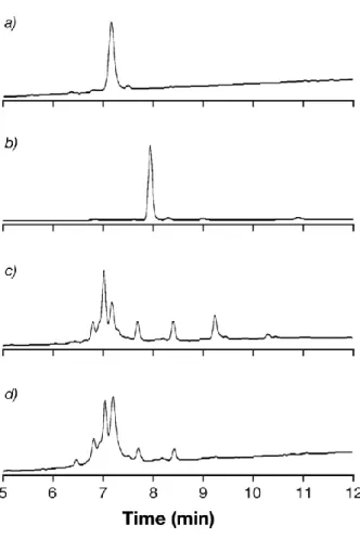
Fig. 10 Reverse Phase Ultra Performance Liquid Chromatography analysis. a) Native
RrgB, b) TLR7aR, c) crude reaction mixture, d) purified reaction mixture
DLS analysis cross-validated the observed features on the conjugate giving information on population distribution by measurement of the peaks polydispersity (PdI) (Tietz, Aldroubi et al. 1991). As expected, an increase of species’ size after conjugation was observed in the scattered light intensity representation. RrgB had a PdI=15% according to a narrow disperse population of macromolecules with hydrodinamic radius of 4.05±0.55 nm, whereas the analysis of RrgB-TLR7aR gave a PdI=35 % with a hydrodynamic radius of 5.4±0.4 nm, 1.35 nm bigger than RrgB (this value was
statistically significant); the higher value of PdI showed a polydisperse population composed of macromolecules with a wide distribution (Fig.11).
Fig.11 Dynamic laser scattering analysis for the Light Intensity representation.
Black peak native RrgB, green peak conjugated RrgB-TLR7aNR
Fluorimetric quantization allowed the estimation of an average conjugation ratio RrgB:TLR7aR. The value of 1:44 led to calculate of the average molecular weight of 131 kDa for RrgB-TLR7aR, agreeing with the apparent molecular weight calculated from SE-UPLC (Fig.12).
Fig.12 Calibration curve for fluorimetric quantization of TLR7aR.
Formulations’ analytical characterizations
RrgB-TLR7aR was alum formulated to be compared with a formulation of the same dose of Ag and TLR7; unfortunately due to the reactivity of a not reactive analogue (TLR7aNR) was used to compare its activity.
Mice were immunized with the following formulations 1)PBS 2)RrgB (2 µg/dose), 3)RrgB (0.2 µg/dose), 4)RrgB-TLR7aR (0.2 µg/dose), 5)RrgB+TLR7aNR (0.2 µg/dose + 0.08 µg/dose).
Formulations 4 and 5 contains the same amount of Ag and TLR7 agonist, they differentiate from how the TLR7 agonist is delivered (through antigen conjugation or simple formulation).
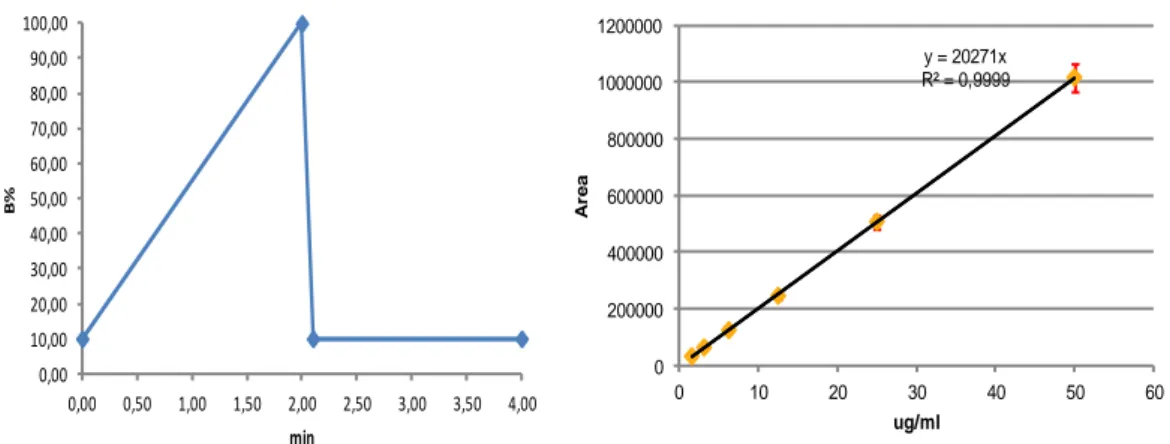
Formulations were characterized for components adsorption, through supernatant and desorbed alum pellet analysis; adsorption was higher than 90% (Fig. 13, 14.15) (Tab.2). Formulations’endotoxin level was lower than 5 EU/ml.
Fig. 13 RP-UPLC gradient for the organic phase variation for TLR7aR and calibration
curve for standard samples of TLR7aR
0,00 10,00 20,00 30,00 40,00 50,00 60,00 70,00 80,00 90,00 100,00 0,00 0,50 1,00 1,50 2,00 2,50 3,00 3,50 4,00 B% min y = 20271x R² = 0,9999 0 200000 400000 600000 800000 1000000 1200000 0 10 20 30 40 50 60 A re a ug/ml
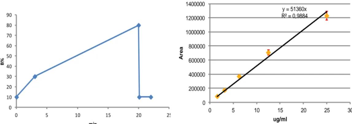
Fig.14 RP-UPLC gradient for the organic phase variation for RrgB and RrgB-TLR7aR
and calibration curve for RrgB standard samples
Fig. 15 Calibration curve of MicroBCA for RrgB-TLR7aR
Tab.2 Formulations composition characterization
0 10 20 30 40 50 60 70 80 90 0 5 10 15 20 25 B% min y = 51360x R² = 0,9884 0 200000 400000 600000 800000 1000000 1200000 1400000 0 5 10 15 20 25 30 A re a ug/ml y = 0,0159x + 0,1318 R² = 0,9987 0 0,2 0,4 0,6 0,8 1 0 20 40 O .D . 5 6 2 n m µg/ml
Groups Composition Supernatant (µg/ml) Not adsorbed % Desorbed (µg/ml) Adsorbed %
1 PBS n.a. n.a. n.a. n.a.
2 RrgB 2µg / alum 0,26±0,01 1,29 18,68±0,03 93,41
3 RrgB 0,2µg / alum 0,08±0,01 3,99 1,96±0,03 97,76
4 RrgB-TLR7aR 0,2µg /alum 0,07±0,01 3,50 1,94±0,01 97,15 5 RrgB 0,2µg + TLR7aNR 0,08µg /alum 0,03±0,00 3,87 0,78±0,01 98,00
In vivo model
Six-week-old, specific-pathogen-free female BALB/c mice from Charles River were immunized intraperitoneally on days 0, 14, 28 with a volume of 100 µL of vaccine formulations (Fig.16). At day 42 samples of sera were obtained from each animal. At day 43, each mouse was challenged intraperitoneally with a mean dose of 160 CFU of
S.pneumoniae TIGR4 variant that was considered as LD100. Blood bacteremia was
evaluated 24 hours after challenge and mortality course monitored for 10 days as reported. In addition OPKA was measured for antibodies functionality.
Antibodies titer
After the 3rd immunization, sera were collected at day 42 and IgG titers were measured
by Luminex assay (Fig. 17). RrgB-TLR7aR gave a 3 times higher IgG titer than group immunized with RrgB+TLR7aNR. The conjugated protein gave comparable titer as 2 µg of RrgB and gave a 6 time higher IgG titer than 0.2 µg of RrgB. When the dose of 0.08 ug of TLR7aNR was administered with RrgB, it showed no adjuvant effect in terms of IgG stimulation and the titer was comparable with the corresponding amount of RrgB alone.
The increase of the IgG titer from RrgB-TLR7aR in comparison to mice that received the physical mixture RrgB+TLR7aNR was clearly due to the co-localization Ag and TLR7 agonist, which enhanced the immune response.
The level of IgG produced was comparable to a 10 fold higher dose of native RrgB, so the conjugation effect led to an Ag dose reduction.
Interestingly the dose of 0.08 µg of TLR7aNR administered to animals of group 5 was ineffective as adjuvant in term of IgG enhancement (tab.3).
Fig.17 Median IgG titers with error bars measurement using protein
coupled-microspheres (Luminex® technology) for the conjugate RrgB-TLR7aR in comparison to the physical mixture (RrgB + TLR7aNR) and the native unconjugated RrgB from 8
mice sera per each group 14 days after the 3rd immunization. RLU stands for Relative
Luminex Units. p values: * < 0.05; **<0.005; ns non-significant statistical difference.
Tab.3 p values for IgG titers of immunized groups
Bacteremia
At day 43 each vaccinated group was infected with a dose of 160 CFU of S.pneumonie i.p. injected (equal to the LD100) and after 24 hours blood bacteremia was measured
(Fig. 18).The RrgB-TLR7aR gave a 3.5 fold lower bacteremia than group vaccinated with the mixture of RrgB+TLR7aNR and gave comparable CFU counts as 2 µg of RrgB. RrgB+TLR7aNR gave a comparable bacteremia as the group vaccinated with 0.2 µg of RrgB without the TLR7 agonist; these groups gave analogous CFU count as mice immunized with PBS . IgG are protective against pathogen proliferation, therefore the bacteremia showed the expected trend observed in the antibodies measurement (tab.4).
Groups 1 2 3 4 5
1 ref 0,0003 0,0003 0,0002 0,0009
2 ref 0,0017 0,3960 0,0027
3 ref 0,0019 0,2470
Fig.18 Median blood bacteremia with error bars measured from 8 mice per each group
at day 43 (24 hours after lethal challenge with 160 CFU of S.pneumoniae TIGR4 variant
i.p. administred) expressed as log10 of the CFUs/ml for the conjugate RrgB-TLR7aR in
comparison to the physical mixture (RrgB + TLR7aNR) and the native unconjugated RrgB. p values: * < 0.05; **<0.005; ns non-significant statistical difference.
Tab.4 p values for blood bacteremia values of immunized groups
Survival
Mice immunized with 0.2 µg of RrgB-TLR7aR after challenge had a median life of 9.5 days, with a residual survival at the end of the observation period of 50% as the animal
Groups 1 2 3 4 5
1 ref 0,0006 0,0014 0,0005 0,0157
2 ref 0,0490 0,9575 0,0086
3 ref 0,0330 0,0661
treated with RrgB at 2 µg per dose (Fig.19). Animals vaccinated with RrgB+TLR7aNR had a median survival of 1.5 days, as the group immunized with RrgB at 0.2 µg per dose. They exhibited a comparable residual survival of 10 and 30% respectively. 90%
of the animal that received PBS died at the 1st day, the remaining 10% died the day after
(Tab.5).
Fig.18 Kaplan-Meier plot for survival estimation of the immunized groups for 10 days
after lethal challenge. 8 mice were immunized per each group for the conjugate RrgB-TLR7aR in comparison to the physical mixture (RrgB + TLR7aNR) and the native
unconjugated.
Survival conclusive confirmed the observed trends from IgG titer and bacteremia.
Tab.5 Median values for survival of immunized groups
Groups Composition Median of survival
1 PBS 1,5
2 RrgB 2 ug / alum 9,0
3 RrB 0,2 ug / alum 2,0
4 RrgB-TLR7a 0,2 ug / alum 9,5
Tab.6 p values for median survival of immunized groups
Opsonophagocytosis killing assay
Post 3 antisera were tested in 6 replicates through in vitro OPKA to assess functionality of antibodies raised in BALB/c mice (Fig. 19). When TIGR4 was incubated with antisera, antibodies were able to kill the bacteria in a concentration dependent manner in immunized groups. Sera of animals immunized with PBS was not able to kill pneumococci (titer <80), differently from the other four groups tested. The RrgB-TLR7aR gave a killing titer of 4210, statistically different from the one observed in RrgB+TLR7aNR (titer 218). RrgB at 2 µg dose and RrgB-TLR7aR at 0.2 µg showed a non significant different killing titer (11365 and 4402 respectively). Similarly, no significant difference was observed between RrgB at 0.2 µg dose and RrgB+TLR7aNR (titer 498 and 218 respectively) antisera (tab. 7).
Groups 1 2 3 4 5
1 ref 0,0283 0,0962 0,0020 0,5371
2 ref 0,0600 0,7343 0,0500
3 ref 0,0600 0,4008
Fig.19 Median with error bars of the opsonophagocytosis killing titers for the
immunized groups obtained from 8 mice per each group. Results are the mean of six replicate and expresses the sera concentration at which the 50% of killing occurred for the conjugate RrgB-TLR7aR in comparison to the physical mixture (RrgB + TLR7aNR)
and the native unconjugated. p values: * < 0.05; **<0.005; ns non-significant statistical difference.
Tab.7 p values for median OPKA titers of immunized groups
Groups 1 2 3 4 5
1 ref 0,0000 0,0001 0,0000 0,0000
2 ref 0,0120 0,3010 0,0020
3 ref 0,0390 0,6970
The chemical behavior of proteins makes bioconjugation reactions difficult to modulate to control the conjugation ratio and the selectivity. Using bifunctional linker or conjugant with highly reactivity moiety the aggregation of the bioconjugate can increase as the product heterogeneity, as can happen with an UV-induced conjugation reaction (Wille-Reece, Flynn et al. 2005).
Aminoacids can undergo into different degradation pathways, the more sensitive are tryptophan (Fig.20), tyrosine (fig.21), phenylalanine (fig.22) and cysteine (fig.23).
Fig.21 Tyrosine light induced degradation
Fig.23 Cysteine light induced degradation
This work was conducted with the aim to adjust a mild and reproducible conjugation approach for a TLR7 agonist and characterize the conjugate.
Reaction’s conditions were developed to enhance protein’s reactivity, lowering the amount of free antigen in the final product, and achieve the highest conjugation ratio. The high number of exposed lysines allowed this model protective antigen to achieve a high derivatization ratio, which led to a high loading capacity, plus its unfolding temperature and good temperature stability provided the possibility to screen several reaction conditions.
The requirements needed for a good model protective antigen were selected based on chemical and biological features. For this purpose, the antigen needed to have an available structure characterization, ready availability with sufficient purity (higher than 80%), high solubility, known chemical and physical stability properties.
The requirements needed for a good model antigen were selected based on chemical and biological features. For this purpose, the antigen needed to have an available structure characterization (i.e. resolved X-rays and known conformation) (fig.24), to evaluate different conjugation approaches, ready availability and sufficient purity (higher than 80%), high solubility, known chemical and physical stability properties.
As biological requirements we focused our attention on the availability of an in vivo model with established and defined readouts that were able to give a robust data panel about immunogenicity, antibody functionality and protection against challenge.
After a screening of different antigens, we chose RrgB from the pilus of
S.pneumoniae(LeMieux, Hava et al. 2006) because it has all the chemical
(Hilleringmann, Giusti et al. 2008; Spraggon, Koesema et al. 2010; El Mortaji, Contreras-Martel et al. 2012) and biological (Barocchi, Ries et al. 2006; Gianfaldoni, Censini et al. 2007; Gentile, Melchiorre et al. 2011; Moschioni, De Angelis et al. 2012) requirements desiderated.
Streptococcus pneumoniae is the major cause of invasive pneumococcal disease and kills over 1.5 million children per year (http://who.int/vaccines/en/pneumococcus.shtml) and the actual vaccination strategy is based on two types of polysaccharide vaccines. One is composed of 23 purified capsular polysaccharides from the bacteria (Pneumovax 23®) (Teele, Klein et al. 1981) and the other one is composed of polysaccharides
conjugated to the carrier protein CRM197 (Prevenar®, a glycoconjugate vaccine) (Black,
Shinefield et al. 2000). These vaccines are serotype dependent and the lack of the actual prophylactic strategy is a serotype-independent one, able to counteract the serotype redistribution by strains not covered by the actually licensed vaccines (Spratt and Greenwood 2000), therefore a protein-based vaccines could overcome this problem. The ideal protein candidate would be a surface exposed protein, common to all serotypes, this perfect antigen has yet to be found but some promising proteins have been already discovered (Barocchi, Censini et al. 2007). The possibility to increase RrgB immunogenicity through the effect of the TLR7 agonist conjugation could lead to a more promising vaccine candidate than the native protein alone and open the
possibility to develop a serotype independent vaccine with a lower complexity than the ones actually licensed
The low differences in molecular weights between RrgB and the various species of RrgB-TLR7aR did not allowed a selective purification approach as for glycoconjugates (Bardotti, Averani et al. 2008), therefore the amount of residual RrgB was ~20% of the total species.
The tested dose of 0.2 µg of RrgB-TLR7aR contained ~0.03 µg of RrgB; because the effect of 0.2 µg of RrgG was almost ineffective, so is improbable that the contribution of 0.03 µg contributed to the observed effects.
RrgB-TLR7aR was characterized with two high sensitive orthogonal techniques as SE-UPLC and DLS, so we can definitely demonstrate that no aggregates are in the conjugate.
An elegant work from Kastenmüller et al. (Kastenmuller, Wille-Reece et al. 2011) demonstrated how the aggregation enhance and maximize the effect of a conjugate OVA-TLR7/8 to promote optimal antigen acquisition and presentation by multiple DCs
subsets in the proinflammatory cytokines fashion and this led to a potent Th1 CD4+ and
CD8+ T cell response. This effect was able to give a significant decrease in the bacterial
load after challenge with Listeria monocytogenes expressing OVA in liver and spleen. That work agreed with the results obtained with the aggregate Gag protein from Wille-Reece (Wille-Wille-Reece, Flynn et al. 2005; Wille-Wille-Reece, Wu et al. 2005). Unfortunately, aggregation is not a very reproducible phenomena, and in some cases, even if the amount of aggregates is the same for different batches, the type of arrayed structures can be very broad (Rosenberg 2006), reducing the consistency between batches and increase the variability of the observed immune response.
The study of Kastenmüller supported the observation made with the UV-catalyzed conjugate used from Wille-Reece (Wille-Reece, Flynn et al. 2005; Wille-Reece, Wu et al. 2005). The UV-irradiation had some inevitable limitations, in terms of reproducibility and selectivity. Some aminoacid can be photoxidated (Kerwin and Remmele 2007) and the protein can be affected by an UV-induced aggregation that can lead to an increased immunogenicity (Rosenberg 2006); moreover the UV exposure can change the TLR7 agonist structure too. Due to these potential issues we used a mild conjugation approach as the reductive ammination reaction.
Previous works (Gianfaldoni, Censini et al. 2007; Gentile, Melchiorre et al. 2011) demonstrated that RrgB needs to be formulated with alum to give protection against lethal challenge of S.pneumoniae; we observed that RrgB-TLR7aR gives no IgG production if is not alum formulated (Fig.25), therefore RrgB was selected as model protective antigen to test the impact of the formulation contemporary with the co-localization.
Fig.25 Effect of alum adsorption on the conjugate RrgB-TLR7aR. Median IgG titers
with error bars obtained with protein coupled-microspheres measurement from 8 mice
per each group sera 14 days after the 3rd immunization. RLU stands for relative luminex
units. p values: ** < 0.005; ***<0.0005.
This study is aimed to make a direct comparison of the Ag and the adjuvant when they are conjugate and sequentially formulated; this comparison can show the different impact on the immune response of how the Ag and the TLR7 agonist are presented to the DCs. To definitely observe the effect of the co-localization Ag and TLR7 agonist, RrgB-TLR7aR was evaluated against the same amount of Ag and TLR7 agonist; for this reason the formulation of RrgB-TLR7aR/Alum contains an equivalent dose of Ag and TLR7 agonist as RrgB+TLR7aNR/alum.
The agreeing observations made from Abs measurement, bacteremia and survival were also supported also by in vitro analysis, to assess antibodies functionality though opsonophagocitosys killing assay (OPKA). When the serum from RrgB 2 µg/dose of was compared with the one from RrgB-TLR7aR, the antibodies functionality was not statistically different and an equal functional response could be achieved also using a 10 fold lower dose of antigen, through the conjugation of the TLR7 agonist to the Ag. Moreover, OPKA demonstrates that the functionality of antibodies is enhanced through conjugation in respect to the mixture RrgB+TLR7aNR. This assay highlighted the evidences from the protection assay that no functional epitopes were damage or masked from the bioconjugation reaction
Bioconjugation reactions lead to a distribution of species that can be very complex because of the high number of the equivalent surface exposed aminoacids that can be modified and the potential loss of conformation. Therefore, the conjugate was fully characterized with orthogonal techniques that allowed a detailed evaluation of species distribution, conjugation ratio, integrity and aggregation status (Stephanopoulos and Francis 2011).
The present study was developed to evaluate the effect of co-localization of Ag-TLR7 agonist and its impact on protection with a non-aggregated conjugate synthesized using mild, controlled and reproducible reaction conditions.
The RrgB-TLR7aR was tested in a mouse model that correlated protection and blood bacteremia to the IgG titre, via to the known protective effect of antibodies against the pathogen infection (Musher 1992).
The possibility to increase the immunogenicity through TLR7 agonist conjugation led to a more promising vaccine candidate than the native protein alone and opens up the possibility of developing a serotype independent vaccine with a lower complexity than those currently licensed (Spratt and Greenwood 2000).
The rational for the observed effect presumably relies on the co-localization of Ag and TLR7 agonist, which is a more effective delivery system than their simple co-administration (Blander 2007). Co-localization can avoid the systemic dispersion of proinflammatory cytokines enabling the TLR7 agonist and the Ag to target the same dendritic cell at same time. It seems possible that the systemic immune activation via TLR signalling does not create a local cytokine and chemokine gradient that is optimal to mobilise immune cells to the site of infection, so conjugation can overcome this problem. This has been demonstrated by Wu et al. using an OVA conjugate that
enhanced mice survival following infection with B.antracis spores. In that specific case protection was due to an aspecific immune stimulation, which could be translated into a more potent and highly specific response when the TLR7 agonist was conjugated to the protective antigen (Wu, Hayashi et al. 2007).
The conjugation induced a specific response that may be enhanced by a synergistic effect of the alum formulation. In the studied case, the use of RrgB led to a 10 fold dose reduction and allowed the immune system to recognize a very low dose of TLR7 agonist, which if it was only formulated resulted ineffective.
The synergistic effect of conjugation can lead to an increase in IgG production, reducing the adjuvant and antigen dose required whilst increasing potency and safety in comparison to the native antigen. Further investigations will need to be accomplished to fully exploit its potentialities and the possibility to synthesize active conjugates, without the need to be aggregated, will lead to a more reproducible drug substance and increases the potentiality for a scale up of this technology on other vaccine’s candidates.
Aluminum adjuvants dose guidelines in
vaccines formulation for pre-clinical
The use of insoluble mineral salts as adjuvants commenced at the beginning of the twentieth century and ever since its licensure, aluminum based suspensions have been most commonly used in a large variety of human and veterinary vaccines. Despite this broad application, there is still a poor understanding not only in terms of mechanism of its action, but also in the physico-chemical properties in relation to the interaction with antigen(s) and/or perturbation of protein antigen structure.(Braun, Clapp et al. 2011; Mbow, De Gregorio et al. 2011)
Some guidelines of tolerated aluminum salts doses are available for clinical research, but there are no clear definitions of dose limits for the preclinical research use.
One of the earliest uses of potassium alum (Al(SO4)2*12H2O) as a vaccine adjuvant
was reported by Glenny et al. in 1926.(Glenny, Pope et al. 1926) The investigators demonstrated that the addition of potassium alum to diphtheria toxoid produced an insoluble compound and depending on the added amount of potassium alum, the remaining filtrate no longer contained the toxoid. When the precipitated diphtheria toxoid was injected into guinea pigs it resulted in a higher antigenic response in comparison to the fluid toxoid.
Since then, aluminum salts have been broadly licensed and the long history of record of clinical data in a large spectrum of age population supports an excellent safety profile and a limited reactogenicity in muscle tissues.
Regulatory agencies define antigen adsorption percentage on the mineral adjuvant and aluminum dose for humans, but there are no clear definitions for limits for both veterinary vaccines and preclinical research grade formulations. European Medicines Agency has developed a general guideline for preclinical research, but it does not express aluminum adjuvant limits.(Use 2005) Here we are we are aiming to give a
rationale for the calculation of aluminum doses in small animal studies at the different stages of pre-clinical research, from early antigen selection to vaccine optimization and preclinical development.
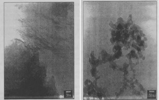
When a solution of aluminum chloride is slowly added to a solution of potassium hydroxide in a buffered environment, a precipitate colloid is obtained and this can be purified to give aluminum oxyhydroxyde (AlOOH, also known as boehmite).(Gupta, Rost et al. 1995) This compound has a crystalline structure with an average adsorption surface of 500 m²/g.(Johnston, Wang et al. 2002)
Fig. 26 Transmission electron microscopy for aluminium oxyhydroxyde (left) and
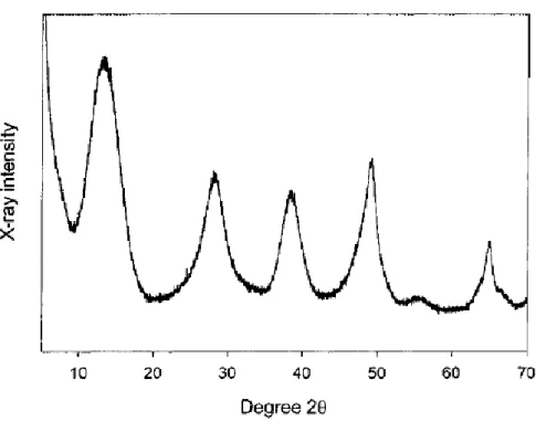
Fig. 27 X-ray diffraction pattern of aluminum hydroxide adjuvant
The precipitated material is a gel composed of particles ranging between 3 and 15 µm, with an average diameter of 10 µm.(Gupta, Rost et al. 1995) The aggregation index and polydispersity of aluminum adjuvant depends on several manufacturing parameters; even if manufacturers use the same pH controlled precipitation procedure, different product profiles can be obtained (in terms of aggregation/polydispersity), e.g. with a different stirring speed or a different reaction vessel geometry.The second most commonly used aluminum salt in vaccines is aluminum phosphate, a complex
heterogeneous salt with the general formula Al(OH)x(PO4)y where the ratio of phosphate
(PO4) and aluminum (Al) is in the range of 0.88-1.0, and the mean particle size is 2 µm,
depending on the pH of preparation procedure.(Burrell, Johnston et al. 2000; Burrell, Johnston et al. 2000) The primary particles are plates having a diameter of
approximately 50 nm.The primary particles form loose, irregular aggregates of 1 to 10 µm. The aggregates are easily dissociated by the shear associated with mixing but re-form readily when the shear is removed.(White and Hem 2000; Hem and Esch 2007)The preparation protocol is similar to the one described earlier where potassium phosphate is used instead of potassium hydroxide. The precipitate is an amorphous material with counter ions inclusions.(Lindblad and Schonberg 2010) Like aluminum hydroxide, aluminum phosphate adjuvant is a gel as well but with a lower adsorptive surface (250 m²/g).(Glemza, Parent et al. 1992) Other aluminum adjuvants like double aluminum salts (i.e. KAl(SO4)2*12H2O) are also prepared by co-precipitation of
aluminum chloride and potassium sulfate at a neutral pH and used in vaccines such as tetanus and diphtheria toxoids vaccine Decavac. More soluble aluminum salts have other uses and are listed in pharmacopeias as oral antacid compounds due to their ability to react with hydrogen ions in the stomach.
Aluminum based adjuvants are prepared to have the rheological properties of a Newtonian gel where the viscosity does not change with the shear stress applied and can be easily injected with a syringe. This is achieved by controlling pH, ionic strength, temperature, mixing and addition speed of reagents, in order to avoid the formation of undesired interferences (i.e. inclusion of amorphous material in the crystals or random crystallization within the amorphous product).
Aluminum adjuvanted vaccines can be prepared by either antigen co-precipitation with aluminum salts or antigen adsorption onto preformed aluminum salt gels. The first method has a great historical relevance in the development of adjuvanted vaccines such as the diphtheria vaccine. However, the co-precipitation procedure gives formulations with low reproducibility and high variability in the immunological response. Antigen(s)
addition to preformed aluminum gels is generally the preferred option in the pre-clinical evaluation of model vaccine antigens. The latter procedure is preferred due to ease of screening, reproducibility and induction of consistent immune responses.(Gupta and B.E. 2000)
Alum has different mechanisms of action, and they are not fully comprehended still. The dilution of the vaccine preparation into the muscle interstitial fluid (MIF) results in an array of potential agonists of the immune cascade (a), including:(1) Al3+(aq); (2) free antigen (AG); (3) particulate adjuvant (ADJ); (4) ADJ with associated AG; (5) AG-Al complex; (6) MIF ligand-AG-Al complex; (7) ADJ with associated MIF ligand; (8) MIF ligand-AG complex; (9) particulate iron (as contaminant of adjuvant) either free or with adsorbed Al/AG and resultant reactive oxygen species (ROS); (10) ADJ with associated MIF ligand-AG complex; (11) ADJ with associated MIF ligand-Al complex. MIF ligands might include biomolecules such as; ATP, albumin, transferrin, citrate, fibrinogen. (b) The array of agonists act upon a number of cell types including, the resident muscle tissue (potentially causing necrotic and/or apoptotic cell death) andinfiltrating innate cells such as, monocytes (potential for AlADJ-induced differentiation to dendritic cells), granulocytes (potential for AlADJ-induced eosinophilia acting directly on B cells), macrophages (are known to persist for long periods close to the injection site and may be characterized by inclusions of AlADJ) and dendritic cells (DC). The latter may be the major antigen presenting cell (APC). (c) There are myriad possible modes of interaction between agonists and innate cells including; (i) toll-like receptor (TLR) binding of AG2, AG-Al complex5, MIF ligand-AG complex8, Al3+(aq) ; (ii) multiple TLR binding of ligand-AG-ADJ4; (iii) phagocytosis of ADJ3, AG-ADJ4, MIF ligand-ADJ7, MIF.
Aluminium dose limits recommendations
and conversion factors
The complex chemical structure of aluminum salts and the gel-like morphology do not allow for a direct quantification. The elemental Al content is generally determined via
indirect methods such as the complexometric titration of Al3+ with EDTA upon sample
dissolution, the ion-coupling plasma analysis or the atomic adsorption analysis. The values can be then converted into a nominal concentration of aluminum salts considering the corresponding minimal formulas, as reported in Tab. 8.
Tab.8 Conversion table for aluminium adjuvants
US Code of Federal Regulations recommends that the amount of aluminum permitted in
a single unit human dose of vaccine should not exceed 0.85 mg of Al3+ per dose if the
level is assayed, or 1.14 mg if determined by calculation on the basis of the amount of aluminum compound added in the preparation. The regulations were last amended in 1981 to increase the permissible level of aluminum to 1.25 mg in biological products, consistently with the WHO and European standards per single human dose of a product as well (Tab. 9).
mg AlOOH
mg Al(OH)3
mg AlPO4 mg AlK(SO4)2*12H2O
Tab.9 Maximum dose allowed of aluminium adjuvants
If different aluminum compounds other than aluminum oxyhydroxyde and aluminum phosphate are used (i.e. double aluminum salts), the total amount of total aluminum salt should not be more than the permitted equivalent amount expressed as potassium alum (e.g. 15 mg of potassium alum correspond to 1.25 mg of aluminum). The limits were empirically selected from safety data, adjuvanticity and the risk/benefit ratio. The levels of aluminum salts can be higher if demonstration of efficacy is proven.(Wolf, Kaplanski et al. 2010)
As already mentioned herewith, there is no defined maximum limit for the allowed content of aluminum adjuvants in veterinary vaccines and this is normally set from a balance between efficacy and local reactogenicity as demonstrated from many licensed vaccines (tab.10)
EU
1.25 mg Al3+ 0.85 mg Al3+ (Assayed) 1.14 mg (calculated)
mg AlOOH 2,778 1,889 2,535
mg Al(OH)3 3,613 2,457 3,296
mg AlPO4 0,000 0,000 5,153
mg AlK(SO4)2*12H2O 22,044 14,990 20,037
Tab.10 Aluminium amount in licensed vaccines
Trade Name Manufacter ug Al dose mg Al per dose mols Al mg Al(OH)3 mg AlPO4 Childhood Vaccine
DtaP Infanrix GSK 625 0,625 0,023 1,808
Certiva NAVA 500,00 0,500 0,019 1,447 Acelimmune Wyeth 230,00 0,230 0,009 0,665 Tripedia Sanofi Aventis 170,00 0,170 0,006 0,492 DTP Avaxim Sanofi Aventis 300,00 0,300 0,011 0,868
Boostrix GSK 500,00 0,500 0,019 0,576 2,260 Hib conj Liq Pedvax Hib Merck 225,00 0,225 0,008 0,651
Pneumo conj Prevenar Wyeth 125,00 0,125 0,005 0,565
DTP-Hib Tetramune Wyeth 850,00 0,850 0,032 2,459 HBV/Hib Recombivax B Merck 225,00 0,225 0,008 0,651
HPV Engerix B GSK 250,00 0,250 0,009 0,723 DT Anatetall Novartis 58,00 0,058 0,002 0,168 Adult Vaccine HPV Cervarix GSK 500,00 0,500 0,019 1,447 HBV Fendrix GSK 500,00 0,500 0,019 2,260 HAV Harix GSK 250,00 0,250 0,009 0,723 VAQTA Merck 450,00 0,450 0,017 1,302 Lyme Lymerix GSK 500,00 0,500 0,019 1,447
Tickborne encephalitis virus Ticovac Masta 170,00 0,170 0,006 0,492
HBV/HAV Twinrix GSK 450,00 0,450 0,017 0,576 2,034
What is an acceptable dose of aluminium
adjuvant for pre-clinical evaluation?
Many experiments have been performed during the last several years to better understand if an average human dose of aluminum adjuvant has an impact on tolerability in preclinical species. Flarend et al. demonstrated that 0.85 mg of Al3+ in the form of aluminum oxyhydroxyde or aluminum phosphate injected intramuscularly in rabbits lead neither to a significant aluminum systemic exposure nor to an acute toxic effect.(Flarend, Hem et al. 1997) In a pharmacokinetic study, the authors showed that aluminum phosphate gives a three fold higher area under the curve (AUC) for a 28 days
serum Al3+ concentration profile respect to aluminum oxyhydroxyde. Most likely, the
crystalline structure of aluminum oxyhydroxyde is more resistant to dissolution in interstitial fluids in comparison to the amorphous aluminum phosphate. Even if
aluminum phosphate gives higher systemic exposure, the increase of Al3+ concentration
in the plasma from a 0.85 mg dose of aluminum adjuvant (maximum allowed human dose) was only 2 ng/ml with both adjuvants respect to the base levels of 30 ng/mL in rabbits. The increased level corresponds to 0.071 ng/ml*day, which does not induce an acute toxic effect. In a rat acute toxicology study,(Titkov and Oganesyan 1995) animals were treated intramuscularly with aluminum chloride and the LD50 (lethal dose able to
kill 50% of the animals) was found to be 400 mg/kg, i.e. 36.42 mg of Al3+, 29 times or
42 times higher than the maximum allowed dose in humans in Europe or in USA respectively.
Even if still controversial, preclinical data on protective immunity indicate that antigens and adjuvants must be delivered together to achieve an enhancement in immunogenicity and protection. From a physico-chemical perspective, the adsorption of recombinant protein antigens results in both stabilization and extension of formulation shelf life in most of the cases. Regardless of the strength of antigen-aluminum salt interactions that
may play a role in immunogenicity,(Hansen, Sokolovska et al. 2007) protein antigen adsorption onto aluminum salts should be generally considered beneficial and the formulation optimization should aim ideally to a 100% adsorption of each vaccine component. Adsorption onto an aluminum-containing adjuvant is generally required in order to potentiate the immune response, generally true for recombinant protein antigens unless there is a tight binding like in the case of the Hepatitis B antigen. The HBsAg antigen has a high adsorptive coefficient and a low degree of elution from aluminum oxyhydroxyde and this has a detrimental effect in the in vivo immunogenicity when compared to a formulation where the aluminum oxyhydroxyde is pretreated with phosphate and the adsorptive coefficient is lowered.(Hansen, Belfast et al. 2009) Even when formulations were specifically designed to avoid antigen adsorption and found to be immunogenic in vivo, a degree of association was suggested based on the confocal microscopy analyses evidencing entrapment of proteins in void spaces within the adjuvant aggregates.(Romero Méndez, Shi et al. 2007) In preclinical studies, the concentration of aluminum salt should already consider the minimum dose required to obtain at least 70-80% adsorption (or association) for each antigen. If more than one protein antigen is used in the vaccine formulation, an alternative way to ensure a complete and long lasting adsorption is to use the “separate adsorption procedure”.(Matheis, Zott et al. 2001) In this procedure antigens can be adsorbed individually under optimal controlled conditions. Upon complete adsorption, the single antigen/aluminum formulations can be combined in one final formulation.
Starting from an indicative 0.85 mg dose of Al3+ that correspond to the maximum dose
of approved human vaccines (Tetramune®, Wyeth Fig.29) a scaling down of aluminum
should be verified. The factor to take into account for scaling down would be simply the ratio of volume of human vaccine and the maximum allowed animal injection volume .(Brito and Singh 2011)
Fig.29 Tetramune vaccine (DT, TT, aP, Hib alum adsorbed) from Wyeth (ex Lederle)
Taking rodents as the elected species for formulation optimization studies and considering a 100 μL as a standard injection volume, the scale-down factor is 5 and the dose of Al3+ becomes 0.17 mg , which represents the maximum dose to begin with (Tab.11).
Tab.11 Recommended injection volumes
The suggested range of aluminum adjuvant in rodent animal model is therefore 150±20
µg of Al3+ in 100 µl of vaccine final formulation (150 µg of Al3+ correspond to 333 µg
Model
body weight (kg) dose (ml/hr)
Mouse
0,03
0.050-0.200
Rat
0,45
0.050-0.200
Rabbit
4
0.100-0.500
Monkey
8
0.100-0.500
Baboon
12
0.100-0.500
of aluminum oxyhydroxyde or 680 µg of aluminum phosphate). The adsorption of each antigenic component in the desired combination needs to be verified in a titration of
Al3+ dose from the maximum suggested (150 µg) to at least 10 fold less (15 µg). The
maximum suggested dose can reasonably ensure an adsorption percentage of 80% or more for each antigen in the final formulation.
The principle of scaling down a human aluminum dose for preclinical species is driven first by tolerability and the use of the minimum required adjuvant dose that is necessary. However, no significant toxicity has been observed by several researchers with a scaled down human dose of aluminum based vaccine in preclinical species. Aluminum salts are effective and safe adjuvants in animal models, from early antigen screening through vaccine development with no major concerns on adverse effects.
Antigen adsorption is the second and the most fundamental parameter to consider in aluminum concentration selection for vaccines based on single or multiple protein components. A prediction of adsorption of proteins on aluminum based adjuvants is possible on the basis of parameters such as isoelectric point of antigens, buffer composition, required antigen loading, and total binding capacity of the aluminum salt employed. The adjuvanted formulation should be designed to get the proper adsorption level, pH and osmolality. A dose of 150 µg of Al3+ is suggested especially in case of early antigen screening where information on adsorptive capacity of proteins on aluminum based adjuvants may not be available. Full physico-chemical characterization should lead to the selection of aluminum concentration at a later stage and the minimum required dose to ensure an 80% adsorption on all components should be considered. The
in vivo profiling of adjuvanted vaccines with titration of aluminum is then fundamental
for an in depth understanding of the relationship between formulation and immune response in the preclinical animal model to support selection of Al3+ dose for clinical advancement.