A
A
l
l
m
m
a
a
M
M
a
a
t
t
e
e
r
r
S
S
t
t
u
u
d
d
i
i
o
o
r
r
u
u
m
m
–
–
U
U
n
n
i
i
v
v
e
e
r
r
s
s
i
i
t
t
à
à
d
d
i
i
B
B
o
o
l
l
o
o
g
g
n
n
a
a
DOTTORATO DI RICERCA IN
CHIMICA
Ciclo XXIX
Settore Concorsuale di afferenza: CHIM/01 Settore Scientifico disciplinare: 03/A1
DEVICE FOR SEPARATION OF STEM CELLS FROM ADULT
TISSUES, SUITABLE FOR REGENERATIVE MEDICINE
RESARCH: FROM THE IDEA TO THE READY-TO-MARKET
STAGE
Presentata da: KRISTEL MARTINELLI
Coordinatore Dottorato
Relatore
Prof. Aldo Roda
Prof. Pierluigi Reschiglian
Dott. Kristel Martinelli
Supervisore: Prof. Pierluigi Reschiglian Curriculum: Scienze Chimiche
Indirizzo: Chimica Analitica
Titolo della Tesi: Device for separation of stem cells from adult tissues, suitable for regenerative medicine research - from the idea to the ready-to-market stage
During the three years of her PhD, Kristel Martinelli focused her research project on design, instrumental development and application of a novel, flow-assisted technology for the separation and selection of cells, in particular stem cells. The importance of this technology concerns the particularly gentle method by which cells are swept down the separation device and are, then separated. This allows full maintenance of the physiological characteristic of the cells, a key point to make cells, and more specifically stem cells, able to be used for further biological characterization or cell culture. The novel technology implements a cluster of patents of the University of Bologna (IT1371772, US 8263359, CA2649234) into an instrumentation that will be addressed to the market. The physical principle underpinning this novel technology is that Earth’s gravity assists the dynamic fractionation of cells suspended in a liquid stream based on differences in physical-morphological properties (size, shape, density, surface features) of the cells. This turns out into a unique tool for non-invasive cell sorting. "Non-invasive" means that cells can be separated at a highly pure level (>90%) just by physical means. This avoids the use of surface immune-markers that can modify cell biology and promote the unpredicted outcome of their molecular characterization.
The candidate focused on the analytical, instrumental aspects of the technology using, first, cell samples, either from cell cultures or from real, raw samples. Using cells of different nature, particular emphasis was given to the application to mesenchymal stem cells and on the relevant search of biocompatibility and
adequacy of the technical solutions chosen for developing the instrumental prototypes into a possible, future product.
Indeed the candidate acquired good experience also in validation and development of flow-assisted separation methods, analytical instrumentation design and development, techniques for cell characterization like flow cytometry and related FACS, magnetic-assisted cell sorting (MACS), immunofluorescence, microscopy, cell culturing and cloning. The possible orientation to a market outcome of the PhD project made the candidate getting acquainted also on strategic marketing, communication techniques and business planning.
During the PhD project, the candidate attended national and international congresses and events, also presenting poster and oral communications, among which, in 2016, a presentation to MEDTEC Europe, Stuttgart (Germany), to the “International Summer School - Innovation and Technology Management in Medical and Pharmaceutical Biotechnology”, of the Bologna Business School, and to the “Y-RICH-Young Research Ideas”, Università di Roma “La Sapienza”. With an entrepreneurship project based on the technology developed during her PhD, the candidate got the final of the “Premio Marzotto 2014”, the Italian, most important competition for startup projects.
The candidate has developed understanding of all the issues involved. She acquired a good mastery of the experimental techniques, demonstrated skills of organization, coordinated well with laboratory colleagues, and showed ability to relate with external collaborators. She also developed “soft skills” that will make her able to be competitive on business-related activities.
In my opinion Kristel Martinelli has carried out a very good work for the thesis.
The Board expresses a very good score on the activity carried out by the candidate during the whole cycle of doctorate and considers her worthy to attain the PhD in Chemistry.
TABLE OF CONTENTS
Introduction & Aim of the study
Chapter1
Cells and Regenerative Medicine………1
Focus on the most interesting source of mesenchymal stem cells: adipose tissue derived MSCs……….7
Chapter2
Cell separation Overview on cell separation………..22Field Flow Fractionation ……….41
Non-equilibrium, Earth gravity-assisted dynamic fractionation (NEEGA-DF)….45 The property of the invention : Stem Sel srl……….52
The Product: Celector® ………..52
Operational mode: the main phases……….67
Software interface Operations………68
Chapter3
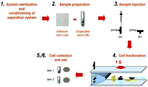
Development of Celector® ………80Aim of the study and Introduction
My PhD research project was focused on the development of a brand new instrumentation for the separation of particles, chiefly of interest for medical and clinical issues, thanks to the advantage related to the preservation of the sample or “minimum manipulation” in the medical field. The competitive advantage, respect to the state of art of current cell separation techniques, is the unique and specific separation method based solely on the morpho-physical proprieties of particles moving inside the flow of a capillary separation channel. The combination of forces developed inside the channel and due to the flow composed of a biocompatible liquid and the Earth Gravitation Field, joined to the geometries of the system and the sample manipulation procedures, allows the separation in time and in space of particles with different physical proprieties. The different population can be collected, characterized and reused for further applications. This feature is particularly relevant when the preservation of the native proprieties of the sample is unavoidable and when the traditional parameters of traditional techniques are not effective or not efficient for the separation of the species contained in a suspension. This is a significant limit when we’re talking about drugs or therapies presenting peculiar characteristics, as the stromal cell population and in particular the mesenchymal stem cells (MSCs), where the complexity of the sample in reason of their “not specialized” state, leads to a really heterogeneous population, preventing the identification by traditional protocols. This heterogeneity can be tapped by the novel instrumentation, first of all in the regenerative medicine field that uses cell based therapy, most of them belonging from stem cell or derived or progenitors cells, samples not to be compromised during the analysis or the separation procedures. MSCs are the most promising stem cell type for cell-based therapies since they are virtually present in all adult tissues and possess tissue regenerative and immunosuppressive properties. MSCs are adult stem cells which can be induced to enter various mesenchymal lineage pathways to differentiate towards the more specialized osteogenic, chondrogenic, myogenic and adipogenic cell lineages. They appear to be particularly suitable for clinical
applications in the fields of cell therapy and tissue reconstruction, for treatments of compromises organs and tissues. Mesenchymal cells are located in all human tissues, but some tissues are particularly rich in MSCs such as the fatty tissue, spinal cord (bone marrow), dental pulp and neonatal tissues. Starting from these sources I studied the behavior of stem cells before, during and after the separation procedure to build up the technology respect the biological requirements of manipulation and optimizing the methods respect the proprieties of stemness of the different fractions resulting from the separation. Moving from this request, I developed a technology that builds on the patented method (IT1371772, US 8263359, CA2649234) for gentle stem cell separation and evolves into an instrumentation serviceable and scalable to be brought on the market and available for “stringent criteria of manipulation” applications. To meet the demands of the market, I considered the whole project in order to insure an organic and coordinated development of the product with the aim to guarantee a fully functional product, thought to be appropriate for the beta-testing and the first placement on the market. Briefly, the project of my research was to transform a method of separation compliant for cell samples into a full automated product usable by not specialized personnel, related with a full protocol of separation portfolio which include a panel of characterization off-&-on-line cell population and subpopulation, ensuring the compliance of the whole process with Class IIb Medical Device certification. This latter aspect worked as the “shadow guideline” moving with the project progress, starting from the suppliers, materials and manufacturing techniques/ procedures, transports, destination of use and environment, and finishing with the biologic cell sample selection and timing, cell characterizations, sterilization and operational methods dependent on methodical daily working rules of the target client. Coordinating all these aspects allowed to gain a first instrumentation boasting the principal features for future medical applications or immediate “low scale” amount of cells in particular therapies.
The resulting product developed in compliance with engineering and biotechnological requirements, merged with industrial and production and
strategies, in order to helpfully supply the Regenerative Medicine sector, is called Celector®.
1
Chapter 1
Stem cells and Regenerative
Medicine
Stem Cells: Basics
Stem cells are a population of precursor cells that are capable of developing into many different cell types in the body. When a stem cell divides, each new cell has the potential either to remain a stem cell or differentiate into another type of cell with a more specialized function. Stem cells are distinguished from other cell types in the body by capability of self- renewal and under certain conditions induced to differentiate into specific cells. In some organs, (for example the bone marrow, or skin), stem cells regularly divide to repair and replace worn out tissues which was discovered in the early 1960s, and knowledge about their characteristics and composition has come a long way. The existence of stem cells was first demonstrated in 1960 by Till and McCulloch in a study on hematopoiesis. The establishment of the concept of hematopoietic stem cells (HSCs) was followed by the discovery of tissue stem cells in other organs in mammals, for example,
2
epithelial stem cells, neural stem cells, and intestinal stem cells1. Stem cells are important for living organisms for many reasons. In the 3 to 5 day old embryo, called a blastocyst, the inner cells give rise to the entire body of the organism, including all of the many specialized cell types and organs such as the heart, lung, skin, sperm, eggs and other tissues. In some adult tissues, such as bone marrow, muscle, and brain, discrete populations of adult stem cells generate replacements for cells that are lost through normal wear and tear, injury, or disease
Stem cells differ from other kinds of cells in the body. All stem cells regardless of their source have three general properties: Stem cells are unspecialized. One of the fundamental properties of a stem cell is that it does not have any tissue-specific structures and cannot work with its neighbors to pump blood through the body (like a heart muscle cell); it cannot carry molecules of oxygen through the bloodstream (like a red blood cell); and it cannot fire electrochemical signals to other cells that allow the body to move or speak (like a nerve cell).
However, unspecialized stem cells can give rise to specialized cells, including heart muscle cells, blood cells, or nerve cells. Stem cells are capable of dividing and renewing themselves for long periods. When cells replicate themselves many times over it is called proliferation. A starting population of stem cells that proliferates for many months in the laboratory can yield millions of cells. If the resulting cells continue to be unspecialized, like the parent stem cells, the cells are said to be capable of long-term self-renewal. Stem cells can give rise to specialized cells. When unspecialized stem cells give rise to specialized cells, the process is called differentiation. Scientists are just beginning to understand the signals inside and outside cells that trigger stem cell differentiation. The internal signals are controlled by a cell's genes, which are interspersed across long strands of DNA,
3
and carry coded instructions for all the structures and functions of a cell. The external signals for cell differentiation include chemicals secreted by other cells, physical contact with neighboring cells, and certain molecules in the microenvironment. A number of experiments have reported that certain adult stem cell types can differentiate into cell types seen in organs or tissues other than those expected from the cells' predicted lineage (that is, brain stem cells that differentiate into blood cells or blood forming cells that differentiate into cardiac muscle cells and so forth). This reported phenomenon is called transdifferentiation.
Types of stem cells
Stem cells can be divided based on their self-renewal and potency2. Self-renewal is the ability to go through numerous cycles of cell division while maintaining the undifferentiated state while potency is the capacity to differentiate into specialized cell types. Based on the potency, stem cells can be divided into five groups. The first type is the totipotent stem cells. These cells can differentiate into embryonic and extraembryonic cell types. These cells are produced by fusion of an egg and sperm cell. The second type is pluripotent stem cells. These cells are the progenies of totipotent cells and can differentiate into almost all cells except extraembryonic cell types. The cell has the potential to differentiate to any of the three germ layers are examples of this type. The third type is the multipotent stem cells which can differentiate into a number of cells, but only those of a closely related family of cells. The fourth type is the oligopotent stem cells. These cells can differentiate into only a few cells, such as lymphoid or myeloid stem cells. Finally, the fifth group is the unipotent cells. Therefore, all types of stem cells have the ability of self-renewal but their potency is different and depends on the source that
2
4
they have arisen from3. Based on their source, stem cell can also be classified as embryonic, fetal, adult, amniotic cord blood and Induced pluripotent.
Stem cell bioprocessing4
The success of stem cell bioprocessing relies on robust and reproducible culture conditions and processes. For stem cell bioprocessing, this includes the scale-up of stem cells to a differentiated end product of sufficient quality and quantity for clinical and commercial goals. Automation and the use of an efficient bioprocess paradigm are imperative for the creation of successful clinical products. The design principles 5pertinent to stem cell bioprocessing can be categorized into three groups: process components; process requirements and process function, as summarized in Figure 1. A combination of generic, ‘off-the-shelf’ and personalized manufacturing paradigms must be considered as no single technology satisfies all requirements6 (Figure 1.1)
3
Yao et al., 2012
4
Dubie et al. Journal of Cell Biology and Genetics, Vol. 4(4), pp. 40-52,,2014
5
Lim et al., 2007
6
5
Figure 1.1 Design principles for stem cell bioprocesses. Source: (Lim et al., 2007).
MSC in Regenerative Therapy
The regenerative potential of MSC isolated from different tissues has been shown to undergo alteration according to the tissue of isolation78. It has been shown that BM-MSC possess a higher potential in giving rise to osteoblasts and chondrocytes9, whereas adipose tissue-derived MSC (AT-MSC) have been shown to contribute more successfully to capillary-like network formation in vitro as well as vasculogenesis in vivo [85, 86]. Umbilical cord blood- (UCB-) MSC also showed a high potency in giving rise to pericytes during vasculogenesis, whereas their potential for osteogenic differentiation has been shown to diminish compared to
7 A. Reinisch, N. A. Hofmann, A. C. Obenauf et al., Blood, vol. 113, no. 26, pp. 6716–6725, 2009 8
N. A. Hofmann, A. Ortner, R. O. Jacamo et al., PLoS ONE, vol. 7, no. 9, Article ID e44468, 2012.
6
BM-MSC 10, which still play as the gold standard for osteogenic differentiation and regeneration. AMN-MSC were also shown to successfully participate in neurogenesis, whereas such a regenerative potential has not been distinguished in UC-MSC11. Amniotic membrane-derived MSC, however, have not been shown to participate in the process of vasculogenesis as successfully as UC-, UCB-, AT-, and BM-MSC did [86]. Despite the fact that DPSC and BM-MSC are regulated by similar factors and they also possess a similar protein expression profile, these populations have been shown to alter significantly in their proliferative capacity in vitro and, more importantly, in their regenerative capacity in vivo12. BM-MSC give rise to bone tissue in the mouse model under treatment as described in studies. The chondrogenic and adipogenic potential of BM-MSC has been higher compared to that of DPSC13. Conversely, the neurogenic differentiation potential of dental mesenchymal stem cells might be more robust compared to that of BMMSC, since these cells possess neural crest origin. BM-, dental pulp- (DP-), and adipose tissue- (AT-) derivedMSC have revealed a greater promise in regenerative therapy since these adult stem cells might promote patientspecific regenerative interventions. MSC are attractive alternatives for regeneration of the injured and/or deficient cells and tissues due to their multipotent differentiation capacity as well as their immunomodulatory and anti-inflammatory properties through cellular crosstalk and production of bioactive molecules. MSC have the unique potential either to directly participate in regeneration and repair processes or to play an immune
10
A. Ardeshirylajimi, M. Mossahebi-Mohammadi, S. Vakilian et al., Cell Proliferation, vol. 48, no.1, pp. 47–58, 2015.
11 E. Y. Kim, K.-B. Lee, and M. K. Kim, BMB Reports, vol. 47, no. 3, pp. 135–140, 2014. 12
S. Shi, P. G. Robey, and S. Gronthos,, Bone, vol. 29, no. 6, pp. 532–539, 2001.
13
W. Zhang, X. F.Walboomers, S. Shi, M. Fan, and J. A. Jansen, Tissue Engineering, vol. 12, no. 10, pp. 2813–2823, 2006.
7
modulatory role to enhance treatment of autoimmune diseases such as type 1 diabetes (T1D).
Focus on the most interesting source of
mesenchymal stem cells: adipose tissue
derived MSCs
In the last decade, rapid evolution in the biology and biotechnology’s fields led to development of different viable cell-based medical applications, which hold a high potential in treatment of several diseases still lacking a specific therapy. In this context, stem cells are the most promising source of cells, mainly because of their limitless avalaibility and easy manipulation (Guilak et al, 2010).
Stem cells can be defined as cells with the capability of generating daughter cells (self-renewal property) and having multi-lineage differentiation capacity (EMA/CAT/571134/2009). Stem cells are able to proliferate in an undifferentiated form and include:
embryonic stem cells derived from blastocysts (hESC); adult and/or somatic stem cell, including:
haematopoietic stem cells (HSCs);
mesenchymal stromal/stem cells (MSCs);
tissue-specific progenitor cells, unipotent cells that can develop into a limited panel of tissues;
8
Among all these types of stem cells, MSCs are the most promising for cell-based therapies since they are virtually present in all adult tissues (14) and possess tissue regenerative (Pittenger et al., 1999) and immunosuppressive properties (Aggarwal et al., 2005).
MSCs are adult stem cells which can be induced to enter various mesenchymal lineage pathways to differentiate towards the more specialized osteogenic, chondrogenic, myogenic and adipogenic cell lineages. Although bone marrow has been considered for years the classical reservoir of MSCs (BM-MSCs), several new sources are currently under investigation. In particular, the adipose tissue has been proven to be an increasingly attractive source of MSCs for mesenchymal tissues regeneration15, since fat is easily obtainable in large quantities and it yields a cells number per gram of tissue which is 500-fold higher than the bone marrow.16
MSCs isolated from different tissues differently reacts to inductive molecules, thus reflecting the characteristics of tissues of origin (Caplan, 2008); however, in culture, BM-MSCs and adipose-derived MSCs (ASCs) share an important combination of features:
1) adherence to plastic 1718 2) morphology19;
14Caplan, 2010; da Silva Meirelles et al., 2006 15Locke et al., 2009
16Fraser et al., 2006
9
3) immunophenotype20; 4) differentiation capacity;
5) immunosuppressive capacity21.
Therefore, also considering the same embryonic mesodermic origin, it is likely to account ASCs as a peripheral MSCs lineage, supporting their use in several therapeutic applications. In particular, ASCs hold high potentials in orthopaedic tissue-engineering field, since they both promote osteogenesis at break sites and increase bone grafts integration22. Moreover, ASCs were shown to possess immunosuppressive and anti-rejection capacities; this finding rationally supports their allogenic use.
MECHANISMS OF ACTION
The therapeutic value of MSCs is based on a number of intrinsic characteristics, briefly listed and discussed below, which are shared by both BM-MSCs and ASCs: 1) differentiation ability; 2) trophic activity; 3) immunomodulatory capacity 19Zuk et al., 2002 20Peroni et al, 2008 21 Puissant et al., 2005; McIntosh et al., 2006 22Tapp et al., 2008
10
1) Differentiation ability
MSCs have been originally isolated and characterized to study their ability to differentiate into a broad spectrum of mesenchymal tissues, such as bone, cartilage, tendon, fat, muscle and marrow stroma. Firsts therapeutic applications were thus proposed, basing on the mere tissue engeneering logic that lineage-oriented stem cells could reconstruct a specific site of application23. However, several pre-clinical studies demonstrated that MSCs-induced functional recovery of treated injured tissues occurs without a substantial differentiation of injected MSCs towards tissue-related phenotypes. Therefore, others mechanisms of action must be involved and differentiation should be considered as a secondary feature.
New insights in MSCs pharmacodynamic depict this multipotent cell lineage as intelligent, injury-site specific, multidrug release system (Caplan, 2010). In fact, MSCs could be recluted by injured organs and, while chemoattracted by the proinflammatory cytokine tumor necrosis factor-α (TNF-α)24, home to sites of inflammation where they secrete a massive amount of bioactive agents, both trophic and immunomodulatory .
2) Trophic activity
It is considered “trophic activity” the MSCs ability to stimulate host regeneration trhough paracrine secretion of a serie of molecules that induce the following physiological responses:
23Wagner et al., 2009 24Ponte et al., 2007
11
a) inhibition of apoptosis with consequent limitation of the damaged field;
b) inhibition of scarring and fibrosis in the site of injury, thus reducing severe post-lesions fibrogenesis;
c) stimulation of angiogenesis;
d) stimulation of proliferation of tissue-specific regenerative progenitors.
Trophic activity of MSCs represent a key feature in bone regeneration and graft survival. In fact, angiogenesis and consequent avalaibility of blood supply are crucial, both for reformation of new structural osseous tissue and for success of engineered scaffolds engraftment.
In addition, MSCs-induced stimulation of tissue progenitors to divide and differentiate into functional regenerative units, represents one of the most important properties underlying organs regeneration.
3) Immunomodulatory capacity
MSCs are known to avoid allogeneic rejection (Ryan et al., 2005); powerful immunomodulatory and antinflammatory properties of this cell lineage are the most important pharmacological rationals justifying their allogeneic uses. Three broad mechanisms contribute to MSCs anti-rejection ability:
a) MSCs are hypoimmunogenic themselves; even if there are still some controversial results about MSCs cell surface expression of major histocompatibility complexes (MHC), many researches suggest that these cells are
12
MHC-II negative (McIntosh et al., 2009; Ryan et al., 2005). Absence of MHC-II gives to MSCs the useful potential to escape host CD4+ T cells recognition;
b) MSCs are able to suppress proliferation and cytokine secretion of natural killer (NK) cells by cell-to-cell direct interaction (Sotiropoulou et al., 2006);
c) MSCs extensively secrete a wide range of bioactive molecules, which create a surrounding immuno-suppressive milieu. Prostaglandin E2 (PGE-2) was found to be a central effector of several MSCs-mediated effects on immune system; in fact, it has been shown that MSCs-secreted PGE-2 has powerful inhibiting activities on dendritic-1 (DC-1), T and NK cells proliferation and secretory profile (Aggarwal and Pittenger, 2005; Sotiropoulou et al., 2006). In the meantime, PGE-2 also increases DC-2 cells secretion of interleukin-10 (IL-10), which, in turn, suppresses the outcome of TNF-α and interferon-γ (IFN- γ), two of the most important proinflammatory cytokines25. Catabolites of tryptophan produced by MSCs, are also bioactive, since they act suppressing both CD4+ and CD8+ T lymphocyte subtypes activation.
In brief, cumulative results show that any immunosurveillance cell coming into the range of MSCs will be suppressed. This feature grants MSCs several abilities, such as escaping host immuno-recognition, inhibiting immunosurveillance at the injury site and preventing autoimmune events to estabilish. Therefore, alloreactivity doesn’t seem to be a major problem for MSCs and their addition to a bone graft should protect it from the host immune system, enhancing its survival probabilities.
13
PRE-CLINICAL STUDIES, CLINICAL TRIALS AND CURRENT APPLICATIONS OF ADIPOSE-DERIVED MSCs
As described above, therapeutic uses of ASCs are supported by two important characteristics of this cell lineage: regenerative properties and immunomodulatory activity. To date, proposed employments for ASCs in tissue repair and regeneration are quite impressive and can be listed following clinical application criteria.
1) Musculoskeletal tissues regeneration; 2) myocardial infarction;
3) applications based on ASCs immunomodulatory properties; 4) gastrointestinal diseases;
5) urogenital system disorders; 6) nervous system diseases; 7) wound healing;
8) plastic surgery and tissue reconstruction; 9) other clinical trials.
1) Musculoskeletal tissue regeneration
Considering the adipose tissue mesodermal origin, application of ASCs to bone and cartilage defects is obvious, along with their uses in tendon and invertebral disk repair.
14
a) repairing of calvarial defects, studied both in rat (26) and rabbit models (Dudas et al., 2006);
b) repairing of rats cleft palatal bone defects27; c) repairing of rabbits tibia proximal epiphysis28;
d) repairing of mice cartilage defects using a human ASCs (hASCs) tissue-engineered cartilage29;
e) primary tendon repair in an in vivo tendon injury model30;
f) intervertebral disc regeneration in small animals model, such as rats and rabbits and in larger animal models, such as goat and canine ;
g) facilitation of spine fusion in rats using allogeneic ASCs isolated both from rat and from human adipose tissue.
For what it concerns data on humans, to date licterature decscribes two important case reports and and one ongoing clinical trial (NCT01218945).
The first is a report of a 7-year-old girl suffering from a widespread calvarial defects after severe head injury31. Due to the limited amount of autologous cancellous bone, autologous ASCs were purified and applied to the calvarial defects toghether with autologous fibrin glue. Three months after the reconstruction, CT-scan showed new bone formation and almost complete calvarial continuity.
26
Cowan et al., 2004; Yoon et al., 2007
27
Conejero et al., 2006
28
de Girolamo et al., 2010
29Dragoo et al., 2003 30Uysal and Mizuno, 2009 31Lendeckel et al., 2004
15
The second reports the orbital floor reconstruction of a 65-year-old male patient who had undergone a hemimaxillectomy due to a large keratocyst. The large defect was reconstruct with a titanium cage, filled with autologous ASCs and betaTCP, that was previously inserted for 6 months in a pouch prepared in the patient’s left rectus abdominis muscle. Success of this reconstruction is mainly to ascribe both to bony neotissue and good vascularization of the titanium scaffold; this result also indicates that ASCs promote intense neovascularization, a crucial feature for grafts survival.
The clinical trial number NCT01218945 concerns the development of engineered synthetic bone grafts, preloaded with hASCs, to repair large osseous defects.
2) Myocardial infarction
Numerous studies in animal models have investigated the ASCs potential for treating myocardial infarctions and chronic heart failure32. ASCs mainly exert their myocardial regenerative effect through secretion of trophic soluble factors33. Again, paracrine activity seems to play a key role in ASCs-mediated therapeutic properties.
In humans, there are two ongoing phase I clinical research studies (NCT00442806 and NCT00426868).
32Hwangbo et al, 2010; Mazo et al., 2010; Bai et al., 2010; Valina et al., 2007 33Bai et al., 2010
16
3) Applications based on ASCs immunomodulatory properties
The capacity of ASCs to regulate a wide spectrum of inflammatory mediators, offers a precious therapeutic tool to treat several clinical conditions needing pharmacological immunosuppresion.
Pre-clinical studies include:
a) treating of mice experimental arthritis with hASCs
b) treating of mice experimental allergic rhinitis with allogenic mASCs;
c) anti-rejection activity in organ transplantation; in a rat liver transplantation model, allogeneic ASCs significantly alleviated acute rejection. This field of application holds great promises for the future of MSCs cell lineages, however, to date, sudies are limited to animal models;
In humans, an encouraging result comes from a study reporting allogeneic infusion of hASCs in six patients who have developed chronic and extensive graft versus host disease (GvHD), after haematopoietic stem cell transplantation34. In addition, allogeneic infusion of hASCs has also been approved to be used for the same application in an ongoing phase II clinical trial (NCT01222039).
4) Gastrointestinal diseases
hASCs have also been shown to be a valuable opportunity to treat patients with intractable enterocutaneous35, perianal and rectovaginal fistulas36, as a result of Crohn’s disease. Four related clinical trials are reported:
34 Song et al., 2007
17
a) safety and efficacy study of autologous cultured hASC for the Crohn's fistula, phase I, completed (NCT00992485 );
b) safety and efficacy study of autologous cultured hASC for the Crohn's fistula, phase II, ongoing (NCT01011244);
c)allogenic hASCs derived from lipoaspirates for the treatment of recto-vaginal fistulas associated to Crohn`s disease, phase I and II, ongoing (NCT00999115); d)treatment of fistulous Crohn's disease by implant of autologous hASCs, phase I and II, ongoing (NCT01157650)
Interestingly, no pre-clinical studies are available for the same indications.
5) Urogenital system disorder
ASCs regenerative properties have also been applied in several urology preclinical researches:
a) treatment of rats stress urinary incontinence 37; b) rats and rabbits bladder reconstruction;
c) treatment of erectile dysfunction in obese type 2 diabetic;
In addition, one case report has been recently published, regarding two patients that receive periurethral injection of autologous ASCs for urinary incontinence, due to post-radical prostatectomy (Yamamoto et al., 2010). This prelminary study showed that periurethral injection of autologous ASCs is a safe and feasible treatment modality for stress urinary incontinence in humans.
36 Garcìa-Olmo et al., 2010
18
6) Nervous system diseases
As shown by pre-clinical results, ASCs trophic activity improves nervous system’s cell replacement and tissue regeneration. Proposed field of application include:
a) improving of brain recovery in rat stroke models -hASCs-;
b) improving of motor function in rat models of spinal cord injury -autologous rASCs-;
c) repairing of injured rats peripheral nerves –hASCs-.
In human, a safety/efficacy phase I and II clinical study is evaluating the feasibility of regenerative therapy with autologous ASC, administered intravenously, in patients with secondary progressive multiple sclerosis who do not respond to regular treatments (NCT01056471).
7) Wound healing
Therapeutic potential of ASCs in wound healing has also been investigated.
In rats mitomycin C-treated healing-impaired wounds, local application of autologous ASCs can induce significant wound healing acceleration38.
Clinical outcome potential was also confirmed in humans. Twenty patients being treated for the side effect of radiotherapy, with severe symtpoms, received autologous ASCs via repeated hypoinvasive computer-assisted injections; this
19
clinical approach led to a systematic improvement or remission of symptoms in all evaluated patients39.
8) plastic surgery and tissue reconstruction
Engineer of adipose tissue finds one of its major expressions in plastic surgery and in tissue reconstruction fields. Four clinical trials are currently reported:
a) phaseIV post-marketing study evaluating the transplantation of autologous fat enriched with ASCs, in patients with functional and cosmetic breast deformities post lumpectomy (NCT00616135);
b) completed phase II and III clinical trials evaluating the safety and efficacy of autologous adipocytes and ASCs, differentiated towards the adipocytes phenotype, to treat depressed scars (NCT00992147);
c) phase I study determining the safety of the autologous ASCs transplantation in the treatment of lipodystrophies (NCT00715546);
d) completed phase III clinical trial investigating safety and efficacy of autologous ASCs for the closure of perianal fistulas in patients without Crohn´s disease (NCT00475410).
9) Other clinical trials
20
For what it concerns ASCs-based ongoing clinical trials, others four human applications are currently under investigation:
a) phase I and II clinical studies determining whether intravenous administration of autologous adipose ASCs is safe and beneficial in patients with type 1 diabetes (NCT00703599);
b) phase I and II trials determining whether intravenous administration of autologous ASCs would account a benefit in the types 2 diabetics management (NCT00703612);
c) completed phase III clinical trial investigating safety and efficacy of autologous ASCs for the closure of perianal fistulas, in patients without Crohn´s disease (NCT00475410);
d) phase I and II studies evaluating safety and feasibility of regenerative therapy with autologous ASCs, administered intramusculary, in patients with critical leg ischemia (NCT01211028).
SAFETY CONCERNS
The use of adult MSCs -including ASCs- in cell-based therapies is considered safer and more functional than use of either hESCs and iPSs. In fact, MCSs are immunocompatible and don’t require genetic manipulation; moreover, their clinical employment doesn’t elicit any ethical controversy.
ASCs are known to undergo malignant transformation during protracted culture in
vitro (20-30 passages); however, for clinical applications, it is unlekely that there
21
Finally, no adverse and rejection reactions were reported in pre-clinical and clinical trials, thus confirming the high safety rate of ASCs.
Finally it can ba considered that MSCs are the major candidates for the future of regenerative therapies. Among several proposed putative sources of MSCs, adipose tissue has been proven to be the most promising because of three intrinsinc features: high yield of stem cells, avalaibility and easy harvesting. In addition, it has been demonstrated the ability of ASCs to suppress specific aspects of immune system, toghether with pre-clinical and clinical studies reporting no rejection -or adverse effect- for allogeneic treatment. The possibility to use unmatched allogeneic ASCs implies that a single lot of cells, derived from one donor, could be transplanted into multiple patients. There are two consequent advantages in that: reduction of the quality control costs and benefit for treated patients, that would be always transplanted with young and healthy selected cells.
Abilities of adult ASCs in promoting bone formation and grafts survival are well established. Even though, focus of investigations surrounding ASCs applications in spine fusion is still limited40. However, the physiological characteristics of ASCs indicate that this cell lineage possesses exciting potentials in the stem cell-based regenerative therapies. For this reason, optimization of both cell growth and choice of scaffold will offer succesful surgical outcomes in several orthopaedic applications.
22
Chapter 2
Cell separation
Overview on cell separation
Cell separation is a powerful tool, which is widely used in many strands of biological and biomedical research and in clinical therapy. For research, the ability to sort cells into distinct populations enables the study of individual cell types isolated from a heterogeneous starting population without (or with greatly reduced) contamination from other cell types. This technology underpins many discoveries in cell biology and is further enabling research in areas as diverse as regenerative medicine, cancer therapy and HIV pathogenesis.
In terms of clinical usage, therapeutic cell separation allows for the introduction of enriched cell populations to a patient with a clinical need for those cells, for example, separation of leukocytes by aphaeresis or enrichment of haematopoietic
23
stem cells by immunomagnetic separation41 42. It also enables the enumeration of cells within an individual’s blood system and can aid repopulation of the immune system, for example, in multiple sclerosis patients who have undergone immunoablation treatment.
Currently, most regenerative treatments based on cell separation are restricted to tissues such as blood and bone marrow43. Recently, however, advances in stem cell therapy, tissue engineering and regenerative medicine are showing the potential for clinical cell-based therapies using cells derived from a variety of tissues, such as adipose and intestine. The use of highly selective cell separation procedures in clinical cell-based treatments has the potential to improve the quality of repair and the subsequent clinical outcome. Because of this potential, there is an increasing usage of these methodologies in the fields of tissue engineering and regenerative medicine, which has resulted in an increasing number of researchers using, or wanting to use, cell separation technologies. These researchers are drawn from a diverse range of backgrounds, not all of whom are necessarily based in biology. Indeed, the increasing demand for cell separation in multiple disciplinary research fields is not restricted to tissue engineering and regenerative medicine; cell sorting is also being used in many other areas such as biochemistry, electrical engineering, physics and materials science.
A multitude of cell separation techniques currently available to researchers are based on three core themes: density, adherence and antibody binding, with many points of crossover between these different themes. New techniques incorporating microfluidics combined with a variety of cellular properties are also in development.
41 Handgretinger R, Lang P, Schumm M, et al. Bone Marrow Transplant 1998; 21: 987–993 42
To LB, Haylock D, Simmons PJ, et al. The biology and clini-cal uses of blood stem cells. Blood 1997; 89: 2233–2258
24
Despite the differences between different cell separation techniques, they share common problems and pitfalls, which can at best hinder research progress and at worst give rise to erroneous data. Many of these technical problems and pitfalls are only applicable to certain techniques, whereas others are universal regardless of the method of separation. Other difficulties can arise in the experimental planning stage, where there can be a lack of understanding in identifying appropriate controls. Finally, there is a potential lack of clarity in the terminology used around cell separation methods, which can lead to confusion and a misunderstanding of the analytical measures required.
This review is written taking cognisance of the diversity of backgrounds and expertise of those researchers wishing to use cell sorting methods. The aim is not to produce a detailed step-by-step guide for each methodology but to offer potential solutions when common difficulties arise and provide clarity in areas of ambiguity related to experimental preparation and terminology.
Cell separation techniques
A large variety of cell separation methods are currently commercially available, these are predominantly based on three methodologies: adherence, density and antibody binding. New techniques are being developed that utilise microfluidic technologies and take advantage of a variety of cellular properties such as elasticity in response to acoustic waves and membrane polarisation in a non-uniform electric field44. 45 However, these techniques are mostly still experimental and not yet available commercially for research. The choice of separation method
44
Petersson F, Åberg L, Swärd-Nilsson A-M, et al. Anal Chem 2007; 79: 5117–5123
45
25
depends upon a variety of factors, and each methodology has benefits and drawbacks that affect its applicability in a given situation. In this section, we will briefly outline the three overall methodologies with specific examples of each.
Adherence
Techniques that utilise cellular adherence are some of the most simple methods used for cell separation and are routinely used when isolating cells from digested or explanted primary tissues (Figure 1.2). An example of simple cell separation by adherence is the isolation of dental pulp stromal cells from whole digested dental pulp. In this technique, enzymatically digested dental pulp is filtered and plated directly onto tissue culture plastic, and following a period of culture, the adherent stromal cells are passaged.46 This technique benefits from being very simple and cheap, but it is not at all specific and relies on the cells of interest adhering and in some instances rapidly proliferating to outcompete other adherent cells in the suspension, such as neurons and monocytes. Adherence can also take time leading to some uncertainty as to the success of a separation. Recently, techniques based on cell adherence, such as differential binding of cells to polymer brushes of varying lengths, grafted to glass surfaces, have been developed and these are currently being refined.However, despite this progress, current uses of adherence sorting are mostly only applicable when cell purity is not of concern and isolation of various subpopulations is not required.
26
Figure 1.2. Diagram detailing cell separation by plastic adherence. (a) Whole tissue is disrupted into a cell suspension by enzymatic or mechanical means or a combination of both (separations of blood or bone marrow aspirate do not require this step). (b) Following disruption, the cells can be passed through a filter to remove cell clumps (c) giving a single-cell suspension, which will be added to (d) an adherent surface, and after a period of culture, (e) adherent cells can be observed.
Density
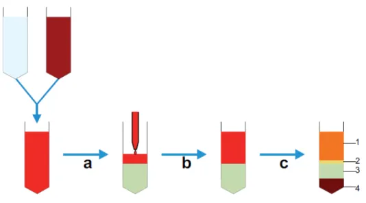
Density-based techniques are now mostly based on the use of centrifugation, although historically sedimentation-based methods have been employed47 .Techniques based on centrifugation are commonly used in many laboratories and are also routinely used clinically. The ability to sort large numbers of cells based on their density, relative to a graduated separation medium (usually sugar based), makes these techniques particularly applicable for separations involving the use of blood (Figure 1.3), which contains 4 × 10⁹ to 6.5 × 10⁹ cells/mL. Indeed, the most
27
commonly used clinical cell separation method is aphaeresis of whole blood to isolate mononuclear cells for treatment of a variety of conditions, including leukaemia48. However, despite the large-scale use of density-based methods, there are still problems with specificity as the differing densities of different cell populations are, in some instances, not large enough to be able to separate out individual cell types. These problems can be overcome by performing repeated centrifugations using differing concentrations of centrifugation medium and differing angular velocities. By using these techniques, it is possible to isolate different cell types from a complex mix, including disrupted solid tissues (Figure 1.4) such as mouse liver. However, although technically feasible, this is still challenging to perform with high specificity. As such, centrifugation methods are generally used if specificity is not absolutely necessary, as in aphaeresis, or as a pre-enrichment stage to remove cells like red blood cells and platelets.
Another density-based method used in laboratory separations is rosetting, which works as a combination between antibody binding and density methods. In this method, unwanted cells are labelled with antibodies that subsequently form complexes with erythrocytes, creating immunorosettes that are much denser than the mononuclear cells of interest. Following centrifugation, these rosettes, containing the labelled unwanted cells, pellet with erythrocytes leaving purified target cells in the mononuclear cell phase.21
28
Figure 1.3 : Diagram detailing whole blood cell separation by density gradient centrifugation. (a) Initially, whole blood is diluted with saline buffer, and (b) this is then carefully layered on top of the centrifugation medium contained in a conical tube avoiding any mixing of the two phases. (c) Following centrifugation, at the appropriate velocity without braking, distinct phases can be observed; 1 – plasma, 2 – interphase containing mononuclear cells, 3 – centrifugation medium and 4 – erythrocytes and granulocytes; cells can then be aspirated from the interphase.
Figure 1.4: Diagram showing separation of solid tissue–derived cells by density gradient centrifugation. Tissues are (a) dissociated and (b) filtered to give (c) a
29
single-cell suspension. (d) This suspension is carefully layered over a centrifugation medium avoiding mixing to give (e) two distinct phases, which can then be centrifuged to give (f) a cell-rich interphase between the centrifugation medium and the cell suspension buffer. (g and h) It is possible to isolate different cell fractions by removing cells from the supernatant or the interphase and then recentrifuging them at different concentrations of centrifugation medium and angular velocities until the desired fractions are obtained.
Methods that sort cells by density are useful techniques to employ when working with tissues that contain a large number of unwanted cells, for example, blood, bone marrow and adipose tissue. This can be either for the isolation of a heterogeneous mix of cells, which can then be used experimentally, or as a pre-enrichment step prior to sorting by other methods.
Antibody binding
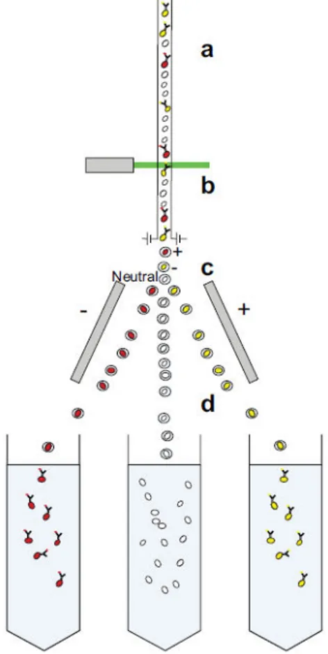
Antibody-binding methods generally refer to the commonly used techniques of fluorescence-activated cell sorting (FACS) and magnetic-activated cell sorting (MACS)4950.51 Both technologies utilise the same cellular properties for separation, namely, cell surface antigens against which antibodies are raised. FACS separation relies on the conjugation of fluorescent labels to these antibodies, whereas MACS uses conjugation to iron oxide containing microbeads. Following binding of conjugated antibodies, FACS and MACS proceed down different routes. FACS separation is achieved by laser excitation of the bound fluorophores, with
49 Bonner WA, Sweet RG, Hulett HR, et al. Rev Sci Instrum 1972; 43: 404–409 50
Miltenyi S, Müller W, Weichel W, et al. Cytometry 1990; 11: 231–238
30
excitation above a threshold level signalling the corresponding cell to be separated (Figure 1.5).
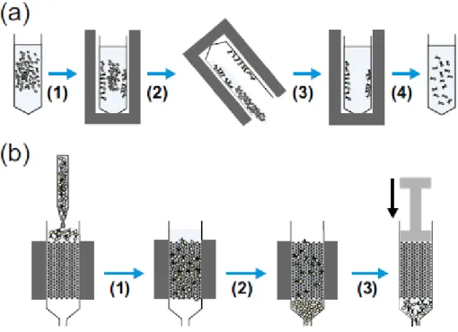
MACS requires the cells to be placed in a magnetic field; unlabelled cells are eluted, and labelled cells are retained in the field until they are removed from the magnet, giving the separated populations (Figure 1.6).
Figure 1.5 Diagram showing cell separation by FACS. Fluorescently labelled single cells from solid or fluid tissues, filtered to remove cell aggregates, are channelled to give a continuous stream of individual cells; (b) these cells then pass through a light source or laser, and the signature of each cell is detected. From this detection, the cells will be determined to be above or below a designated threshold value, and it is decided whether to collect or not collect each cell. (c) This is achieved by electrically charging the droplet
31 each cell is contained within and (d) then by passing it through charged deflector plates that deflect the cells to the appropriate collection tubes.
FACS: fluorescence-activated cell sorting.
Figure 1.6: Diagrams showing the common methods used for magnetic cell separation. (a) Tube-based separation where a magnetically labelled cell suspension held in a conical tube is placed in a (1) magnet causing movement of labelled cells to the sides of the tube towards the magnet. This tube is then (2) inverted (or aspirated), allowing removal of the non-labelled cells before (3) resuspension of the labelled cells and removal from the magnet giving (4) a dispersed suspension of labelled target cells. (b) Column-based separation where a magnetically labelled cell suspension is injected into a column held within a magnet, (1) cells then flow through the column and (2) labelled cells are retained, whereas unlabelled cells are washed out. (3) Following the removal of unlabelled cells, the column is removed from the magnet, and suspension buffer is forced through the column by plunger giving labelled target cells in suspension.
As such, a key difference between MACS and FACS is that MACS can be seen as a bulk method, there is no individual cell analysis, and magnetically tagged cells
32
are retained and non-tagged cells are eluted. FACS, however, analyses each individual cell, which can be tagged with multiple antibodies, whereas MACS is restricted to individual markers (although some kits use enzymatic removal of the microbeads, allowing the cells to be relabelled with a subsequent antibody). This individual cell analysis means that while FACS can be more specific, it is significantly slower than MACS. Sorting that takes several hours by FACS can be achieved in less than 1 h by MACS.
There are other techniques, in addition to FACS and MACS, that utilise antibody binding to enable cell separation, an example of which is rosetting as previously mentioned. However, this is a relatively old technique, and there are many new technologies being developed, which use antibody or cell–ligand binding as the basis for separation. For example, antibodies, immobilised to polymer surfaces, have been used in a microfluidic system to capture circulating tumour cells from whole blood with subsequent release and enumeration. Columns have also been developed with antibody-immobilised surfaces to enrich osteoblastic cells based on CD34 binding. Polymer cryogels with large interconnected pores and surface-immobilised protein A ligands have been used to isolate antibody-labelled CD34+ umbilical cord blood cells in an affinity chromatography–based separation.52 Other methods in development include magnetophoresis, DNA aptamer binding53 and aqueous phase partitioning54. However, despite the variety of antibody-based methods, for the purposes of this review, FACS and MACS will be focussed on due to the experimental nature of these newer techniques.
Antibody-based methods of separation are currently the gold standard for the selection of individual cell populations, and both FACS and MACS can be used to
52 Kumar A and Srivastava A.. Nat Protoc 2010; 5: 1737–1747 53
Xu Y, Phillips JA, Yan JL, et al Anal Chem 2009; 81: 7436–7442
33
isolate cell populations to high purity. Despite this, there are still some problems with FACS and MACS such as the reliance on cell surface markers, which, for most researchers, limits separations to those markers for which antibodies are commercially available. It can also cause problems if the cell type of interest does not have unique markers, making the isolation of a homogeneous population difficult. For example, mesenchymal stem cells (MSCs) express markers associated with many other cell types such as CD90, which is also expressed by primitive haematopoietic stem cells. In addition, the isolation of a viable homogeneous population of cells that contain a unique intracellular marker can also be problematic, as the permeabilisation steps required to stain the marker can damage cell membranes leading to cell death.
Lab-on-a-chip methods
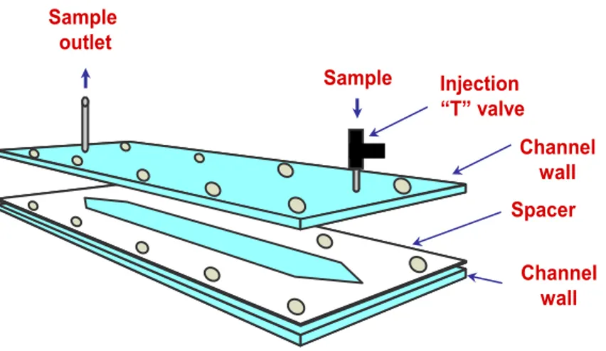
In addition to the traditionally used methodologies for cell separation are several new lab-on-a-chip techniques that operate on a microfluidic scale and utilise a multitude of cellular characteristics to isolate different cell populations in a label-free manner. These techniques are mostly still in the experimental stage, but their development demonstrates the variety of possible ways to separate cells, and they are extensively reviewed by Gossett et al.55 Examples of label-free separation are the use of micro-scale filters or pillars that separate cells based upon size and membrane deformability, as larger cells are prevented from navigating through the filter leading to cell separation.56 Field flow fractionation (FFF) can be used to separate cells along the length of a microfluidic channel by a combination of the parabolic flow within the channel and an external field, such as an electric field or
55
Gossett DR, Weaver WM, Mach AJ, et al.. Anal Bioanal Chem 2010; 397: 3249–3267.
34
gravity. With FFF, particles that are more greatly affected by the external field are forced closer to the channel wall, which is moving more slowly than the centre of the channel and contains more weakly affected particles. Therefore, cell separation occurs because of the effect of the force on the cells and the speed of elution based on the cells’ location in the microfluidic channel.57
Acoustophoresis separates cells based on membrane deformation or elasticity and occurs when a high-pressure sound wave interacts with a cell. This interaction can cause membrane deformation to differing degrees based on the cell’s density and size and leads to the cells being positioned in different parts of the microfluidic channel and therefore able to be separated. Dielectrophoresis can lead to cell separation due to the differential polarisation of particles within a non-uniform electric field. This dipole effect depends on factors such as size and protein content and leads to the attraction or repulsion of the cell away from or towards an electrode. Due to differences in these factors between different cells, it is therefore possible to exert different effects on different cell types within the same field and allow for cell separation.
Label-free lab-on-a-chip isolation methods have great potential to improve cell sorting methods both in a research environment and clinically. However, there are still potential problems associated with these techniques, many of which are general cell sorting problems, which can be applied to the commonly used techniques such as cell clusters, and others that are technique specific. One of the largest problems these techniques currently face is resolving the differences between cell types; for example, with dielectrophoresis, it can be difficult to discern the differences between target and non-target cells. However, perhaps the greatest
35
challenge these techniques face is showing great enough efficacy while overcoming the challenges associated with currently used methods.
Overall, the choice of cell separation methodology is very much dependent upon the initial cell source, the characteristics of the desired cell type and its required purity. Adhesion-based techniques are useful if there is little requirement other than the isolation of adherent cells, and the cell of interest will, if necessary, outcompete other cell types. Centrifugation techniques are useful when dealing with samples with large cell numbers, such as blood, but where specificity is not essential, and are also useful as a pre-enrichment step prior to other separation methods. Antibody-mediated separation methods are the gold standard techniques currently available as they can be used to isolate specific cell populations. However, speed can be an issue, as can costs. Potentially, lab-on-a-chip methods will overcome some of the limitations in the currently used techniques, but, as yet, these are experimental and not accessible to the majority of the researchers performing cell sorting.
Clinical cell therapy
The majority of separations currently performed for clinical cell therapy use cells isolated from tissues such as bone marrow and blood. These separations isolate the mononuclear cells, including the stem cell fraction, and can be used to recapitulate the haematopoietic system of a patient suffering from, for example, chronic myeloid leukaemia, following immune ablation therapy. These separations mostly utilise systems based on centrifugation, such as aphaeresis, as these technologies allow for the isolation of the large numbers of mononuclear cells needed for cell transplantation relatively quickly. MACS can also be used for cell
36
therapy, and the clinically approved MACS-based systems use the same technology as research-grade magnetic sorting; however, these systems are closed and use reagents and fluidic tubing produced under good manufacturing practice (GMP) conditions.58 Use of MACS for clinical cell sorting allows for greater specificity than can be achieved by centrifugation; however, per patient, MACS is more expensive than aphaeresis, and so it is used in circumstances where specificity of the isolated cells is important.
Standard FACS-based systems are not in clinical use for cell therapy, although some flow cytometers can be used for clinical diagnostics59. This is in part due to the difficulty in developing single-use sterile fluidics, the possibility of cross-contamination should multiuse fluidics be employed and problems with batch-to-batch consistency. There are currently methods utilising closed system optical separation in development, but these are not yet in widespread clinical usage. Clinical cell separation is an established field, but it has strict requirements, and there are challenges and difficulties to overcome. The major requirement is to ensure that a consistent, sterile cell population is isolated. Microbial contamination of cell separation products could lead to the infection of the recipient patient, who, in many instances, will be immunocompromised and unable to fight the infection. It is therefore imperative that clinical cell separation products are produced under strict GMP conditions with stringent batch testing. Consistency of the isolated cell population is also very important so as to ensure that the recipient receives the required cell transplant. In addition, rigorous tissue typing should be performed prior to transplantation to avoid human leukocyte antigen (HLA) mismatch and prevent problems such as graft-versus-host disease.
58 Lang P, Schumm M, Taylor G, et al. 1999; 24: 583–589.
59
37
At this time, the major challenge for clinical cell separation is the robust isolation of rare cell populations with multiple surface markers from a large initial pool of cells. Currently, technologies based on centrifugation allow for the isolation of cells from a large initial cell number, and technologies based on MACS can isolate specific populations of cells; however, these technologies use single markers meaning that cells of interest with two or more markers cannot be specifically isolated. Development of high-speed optical cell sorters holds great promise, as these systems could have the speed of an MACS-based system, but with the specificity of an FACS system allowing for more than one parameter to be selected.
Considerations for experimental design
Initial planning and design is key for any experimental strategy, including cell separation, where many factors must first be considered. These factors impact different stages of the separation procedure, but all share a basic set of preliminary requirements. These are the need for a detailed understanding of the cell and tissue types of interest, knowledge of the potential techniques available and the ability to select the correct methodology to yield the desired cell population.
The reason for this required level of understanding is that one cell separation method may be more suitable than another for achieving a given outcome, and different cells react differently to the same conditions. Current methods for cell separation generally offer a balance between purity and recovery. It is therefore important that the separation protocol is designed with this in mind and tailored to suit the desired outcome. For example, if a large number of cells are required, then percentage enrichment may need to be sacrificed; alternatively, for a highly
38
enriched population, the trade-off may be low numbers recovered. Factors to be considered when designing a cell separation strategy are discussed below.
Cost
Cost is a design constraint that is relevant to most separation experiments. Cell separation can be a potentially expensive technology depending on the strategy selected. It may therefore be important to devise a strategy that is not prohibitively expensive by employing cost-saving measures. For example, FACS is a very accurate technique, but it can be slow when sorting rare cells from whole blood, and this consequently increases the running time on the instrument and thus the expense. A way of reducing this time would be to perform an initial erythrocyte lysis step or density gradient centrifugation to remove the erythrocytes, leaving only the mononuclear cells to sort.60 Pretreatment of a sample can thus reduce overall cost and should be considered where cost is an issue.
Methodological difficulties
There are several key technical considerations that must be taken into account before performing a successful cell separation, some of which are universally applicable, while others are more specific to immunomagnetic and immuno-fluorescent cell separation. Figure 6 gives an overview of potential technical problems at each stage during the separation process.
The more universal considerations relate to the quality of the cells, which are being separated, and specifically to the cell isolation process. Antibody-mediated separations also have considerations relating to antibody binding. There can also be specific idiosyncratic problems associated with different commercially available