Alma Mater Studiorum – Università di Bologna
DOTTORATO DI RICERCA IN
BIOLOGIA CELLULARE E MOLECOLARE
Ciclo XXVI
Settore Concorsuale: 05/E2
Settore scientifico-disciplinare: BIO/11
Exploring the biofilm of Streptococcus agalactiae to identify
virulence factors
Presentata da: Nunzia D’Urzo
Coordinatore Dottorato
Relatori
Chiar.mo Prof. Vincenzo Scarlato
Chiar.mo Prof. Vincenzo Scarlato
Dott. Manuele Martinelli
4
Attività di ricerca
Durante il Dottorato di Ricerca mi sono occupata dello studio del biofilm di Streptococcus
agalactiae. In particolare ho valutato l’influenza che ha il pH acido sulla capacità di formazione
di biofilm in vitro di un ampio numero di isolati clinici. Tale analsi mi ha permesso di identificare
una correlazione tra capacità di formazione di biofilm e sierotipo ed in particolare ha evidenziato
che la maggior parte dei ceppi formanti biofilm appartiene all’ipervirulento Sequence Type -17.
Ho valutato il coinvolgimento della capsula, del DNA e delle proteine nella formazione di
biofilm in GBS e ho identificato, mediante spettrometria di massa, proteine espresse
specificamente a pH acido che potrebbero svolgere un ruolo determinante nella prima fase di
adesione del biofilm. I risultati sono descritti nella presente tesi di dottorato ed il corrispondente
manoscritto è stato pubblicato recentemente (D'Urzo et al., 2014).
Parallelamente mi sono occupata della valutazione delle performances di espressione, intra- ed
extracellulare, di proteine ricombinanti da parte di un batterio gram-positivo, Brevibacillus
choshinensis, utilizzando due proteine modello, la GFP e l’-amylasi. Ho inoltre implementato tale sistema di espressione utilizzando, per la prima volta, un plasmide contenente un promotore
inducibile che ha permesso di incrementare le rese fino a 10 volte (D'Urzo et al., 2013). Ho infine
utilizzato Brevibacillus per esprimere il dominio catalitico della tossina A di Clostridium difficile,
possibile componente di un vaccino, che ho caratterizzato dal punto di vista biochimico e
5
Nel periodo del Dottorato di Ricerca sono stata co-autrice dei seguenti lavori scientifici:
1. D'Urzo N, Martinelli M, Pezzicoli A, De Cesare V, Pinto V; Members of DEVANI Study Group, Margarit I, Telford JL, Maione D. “Acidic pH strongly enhances in vitro biofilm
formation by a subset of hypervirulent ST-17 Streptococcus agalactiae strains.” Appl
Environ Microbiol. 2014 Jan 31. [Epub ahead of print]
2. Leuzzi R, Spencer J, Buckley A, Brettoni C, Martinelli M, Tulli L, Marchi S, Luzzi E,
Irvine J, Candlish D, Veggi D, Pansegrau W, Fiaschi L, Savino S, Swennen E, Cakici O,
Oviedo-Orta E, Giraldi M, Baudner B, D'Urzo N, Maione D, Soriani M, Rappuoli R,
Pizza M, Douce GR, Scarselli M. “Protective efficacy induced by recombinant Clostridium difficile toxin fragments” Infect Immun. 2013 Infect Immun. 2013
Aug;81(8):2851-60. doi: 10.1128/IAI.01341-12. Epub 2013 May 28.
3. D'Urzo N, Martinelli M, Nenci C, Brettoni C, Telford JL, Maione D. “High-level
intracellular expression of heterologous proteins in Brevibacillus choshinensis SP3 under the control of a xylose inducible promoter” Microb Cell Fact. 2013 Feb 1;12:12. doi:
10.1186/1475-2859-12-12.
4. D'Urzo N, Malito E, Biancucci M, Bottomley MJ, Maione D, Scarselli M, Martinelli M. “The structure of Clostridium difficile toxin A glucosyltransferase domain bound to
Mn2+ and UDP provides insights into glucosyltransferase activity and product release”.
8
Table of contents
Attività di ricerca ... 4 Table of contents ... 8 Summary ... 11 1. Introduction ... 13 1.1 Streptococcus agalactiae ... 131.2 Group B streptococcal disease ... 14
1.2.1 Neonatal infections... 15
1.2.2 Infections in pregnancy ... 16
1.2.3 Infections in non-pregnant adults ... 17
1.3 Molecular pathogenesis of GBS and major virulence factors ... 19
1.3.1 Capsule ... 21
1.4 Characterization of GBS isolates ... 22
1.4.1 Phenotyping methods and Serotyping ... 22
1.4.2 Genotyping methods ... 23
1.4.3 PCR-based gene profiling and Multilocus sequence typing (MLST) ... 24
1.4.4 Whole-genome sequences comparisons ... 25
1.5. Molecular epidemiology of GBS ... 26
1.5.1. Serotype distribution and MLST-based genetic lineages ... 27
9
1.6.1 The fibrinogen-binding protein FbsB ... 28
1.6.2. HvgA, a cell-wall–anchored protein ... 29
1.6.3 Pili components ... 30
1.6.4. Srr and Spb1 ... 31
1.7 Biofilm formation in GBS ... 32
1.8 Proteomics approach of vaccine candidate identification ... 36
1.9 Thesis overview... 36
2. Material and methods... 38
2.1 Bacterial strains and growth conditions ... 38
2.2 Serotype and Sequence Type identification ... 39
2.2.1 Total genomic DNA isolation ... 39
2.2.2 Serotype and ST-17 identification ... 39
2.3 Growth experiments ... 39
2.4 In vitro biofilm formation ... 40
2.4.1 Standard biofilm formation protocol... 40
2.4.2 New biofilm formation protocol ... 40
2.5 Crystal Violet assay ... 41
2.6 XTT viability assay. ... 42
2.7 Confocal laser scanning microscopy ... 44
2.8 Enzymatic inhibition/eradication of biofilms ... 44
2.9 Biofilm inhibition using sera from immunized rabbit ... 45
10
2.11 Surfome preparation of Streptococcus agalactiae grown in biofilm inducing and
non-inducing conditions ... 46
2.12 Protein Identification by LC-MS/MS ... 48
2.13 Computational Analysis of Identified Proteins ... 49
2.14 Statistics ... 49
3. Results ... 50
3.1 A new in vitro biofilm formation protocol minimizes false positive results ... 50
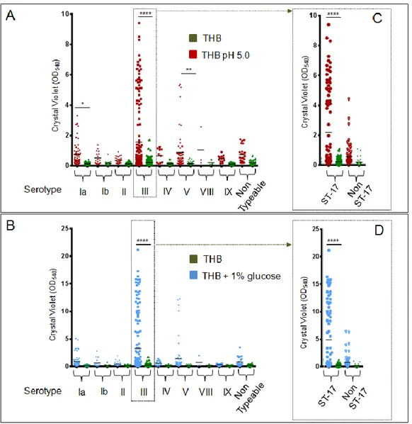
3.2 An acidic pH promotes biofilm formation by Streptococcus agalactiae ... 51
3.3 Most strong biofilm-forming GBS bacteria belong to the hypervirulent ST-17 lineage ... 60
3.4 Role of the capsule expression in GBS biofilm formation ... 63
3.5 Proteins played a significant role in the GBS biofilm formation and maintenance... 65
3.6 Sulfome analysis to identify differentially expressed proteins on the surface of GBS grown in biofilm inducing and non-inducing conditions ... 68
3.7 Antibodies against pilus 2a, but not pilus 2b, inhibit biofilm formation ... 72
4. Discussion... 73
11
Summary
Streptococcus agalactiae, also known as Group B Streptococcus (GBS) is the primary colonizer
of the anogenital mucosa of up to 40% of healthy women and an important cause of invasive
neonatal infections worldwide. Among the 10 known capsular serotypes, GBS type III accounts
for 30-76% of the cases of neonatal meningitis.
Biofilms are dense aggregates of surface-adherent microorganisms embedded in an
exopolysaccharide matrix. Centers for Disease Control and Prevention estimate that 65% of
human bacterial infections involve biofilms (Post et al., 2004). Many species of streptococci are
known to form biofilms; however, the relationship between the pathogenic state and the biofilm
mode of growth has been most clearly established only for the oral streptococci (Cvitkovitch et
al., 2003).
In recent years, the ability of GBS to form biofilm attracted attention for its possible role in
fitness and/or virulence. Here, a new in vitro biofilm formation protocol was developed to
guarantee more stringent conditions, to better discriminate between strong-, low- and non-
biofilm forming strains and reduce ambiguous data interpretation. This protocol was applied to
screen the in vitro biofilm formation ability of more than 350 GBS clinical isolates from pregnant
women and neonatal infections belonging to different serotype, in relation to media composition
and pH.
The results showed the enhancement of GBS biofilm formation in acidic condition and identified
a subset of isolates belonging to serotypes III and V that forms strong biofilms in these
conditions. Interestingly, the best biofilm formers belonged to the serotype III hypervirulent
12
reduction is not enough by itself to ensure biofilm formation. Moreover, the ability of proteinase
K to strongly inhibit biofilm formation and to disaggregate mature biofilms suggested that
proteins play an essential role in promoting GBS biofilm formation and contribute to the biofilm
structural stability. Finally, a set of proteins potentially expressed during the GBS in vitro biofilm
13
1. Introduction
1.1 Streptococcus agalactiae
Streptococcus agalactiae is a Gram-positive bacterium, historically associated to bovine
mastitis and dairy sources under the designation of Streptococcus mastiditis (Lancefield, 1933). It
forms small (3 to 4 mm), grey-white colonies that have a narrow zone of beta hemolysis on blood
agar plate (Figure 1.1). It was first identified as group B streptococci (GBS) in 1933, when
Rebecca Lancefield published her studies on serological differentiation of streptococci
(Lancefield, 1933). GBS was later proposed as an occasional causative agent of puerperal
infections (Lancefield and Hare, 1935), and in 1938 it was recognized as an important human
pathogen responsible for multiple infections (Fry, 1938). It was not until the 1970s that GBS was
acknowledged as a leading cause of neonatal invasive infections (Broughton et al., 1976) and
since the 1990s it has also been increasingly associated with invasive infections in non-pregnant
adults (Farley et al., 1993). Despite its unquestionable importance as a human pathogen, GBS
ismainly a colonizing agent of the gastrointestinal and genitourinary tracts of a significant
proportion of the human population (Schuchat, 1998). GBS strains are classified into ten
serotypes according to immunogenic characteristics of the capsule polysaccharides (Ia, Ib, II, III,
IV, V, VI,VII, VIII and IX). Approximately 10% of strains are non-typeable (Bisharat et al.,
14
Figure 1.1: Streptococcus agalactiae.
A) Scanning Electron Microscopy (SEM) of Streptococcus agalactiae. B) Colonies of Streptococcus agalactiae on a blood agar plate. Note the zone of clear hemolysis.
1.2 Group B streptococcal disease
GBS colonizes the urogenital tract of more 30% of the healthy population and in particular it
colonizes the vagina of 25-40% of healthy women (Dillon, Jr. et al., 1982;Hansen et al.,
2004a;Schuchat, 1998). It has been found in the urethra in both men and women without causing
infections and in the upper respiratory tract. Colonization also is observed in wound and soft
tissue cultures in the absence of obvious infection. Determining the acquisition and transmission
of S. agalactiae can be puzzling, as it is very invasive but produces little inflammation at the
entry site.
This bacterium is an important cause of infection in three populations (Figure 1.2):
Neonates;
Pregnant women;
15
Figure 1.2: Stages of neonatal GBS infection. Adapted from (Doran and Nizet, 2004).
1.2.1 Neonatal infections
GBS is the leading cause of neonatal bacterial diseases in the United States of America;
infection in newborns has been divided in early-onset disease (EOD) and late-onset disease (LOD) depending on the infants’ age and disease manifestations. The onset of GBS infections
that takes place very early in infancy, usually within the first week of life, is designated EOD,
even if the majority of EOD cases occur within the first 24 hours after birth (Schuchat, 1998).
LOD develops between one week and three months of age (Schuchat, 1998). Maternal carriage is
a major risk factor for neonatal GBS disease, which is influenced by the degree of bacterial
colonization; women with heavy colonization are more likely to have symptomatically infected
infants and heavily colonized infants are more likely to develop invasive disease (Lim, DV,
1982). The onset of disease is associated with the presence of GBS in the genital tract of the
mother, and transmission is thought to occur vertically due to an ascending infection during the
course of pregnancy or passage through the birth canal (Schuchat, 1998). Even though perinatal
transmission can occur across intact membranes, both premature and prolonged rupture of
membranes increase the risk of GBS acquisition (Schuchat and Wenger, 1994). Aspiration of
16
rapidly followed by the development of pneumonia (Doran and Nizet, 2004). Breaching of the
pulmonary mucosal barrier leads to the entry of GBS into the bloodstream and to the
development of severe sepsis in some infants (Schuchat, 1998). More than half reported cases of
neonatal GBS disease now occur during the late-onset period (Centers for Disease Control and
Prevention, 2005). The pathogenesis of LOD is less well understood, although some cases also
suggest a maternal source, probably reflecting acquisition of the microorganism during passage
through the birth canal (Schuchat, 1998). Ingestion of contaminated breast milk has also been
proposed as a possible maternal source for LOD (Bingen et al., 1992). Even though nearly 50%
of mothers of infants with LOD were found to carry the same GBS serotype as that causing
infection in their infants, the source of infection in other infants is unclear (Schuchat, 1998).
Nosocomial and horizontal transmission by hospital and community sources are probably
involved in some cases of LOD, but the risk factors are not well understood (Green et al., 1994).
Whereas EOD and LOD can differ in clinical presentation, mode of transmission and risk factors
for disease, the most frequent clinical presentations of invasive disease in neonates are
pneumonia, bacteremia and meningitis (Fernandez et al., 2001;Puopolo et al., 2005).LOD
presents with meningitis and bacteremia without a focus as predominant clinical syndromes;
osteoarticular infections, urinary tract infections, and pneumonia are less frequent (Schuchat,
2006). Meningitis develops when the entry of bacteria into the bloodstream is followed by the
invasion of the cerebrospinal fluid.
1.2.2 Infections in pregnancy
GBS causes a variety of perinatal infections in pregnant women, including both
17
and post-partum wound infections (Pass et al., 1982). It also has been suggested that GBS urinary
tract infections or urinary tract, rectal, or genital colonization in pregnant woman may lead to late
term abortions and preterm and low-birth-weight infants. Pregnant women are colonized at
multiple sites, including rectum, vagina, cervix and throat, but many of them carry GBS in
asymptomatically way (Regan et al., 1991). Approximately 10-30% of pregnant women are
colonized with GBS in the vagina or rectum (Regan et al., 1991), although the colonization can
be transient, chronic or intermittent (Hansen et al., 2004b). As maternal GBS carriage in the
gastrointestinal and/or genital tracts is a prerequisite for EOD, the different prevalence of
maternal GBS colonization could help choose preventive strategies. In most European countries,
the prevalence of GBS carriage among pregnant women varies between 6.5 and 36%, with most
countries reporting colonization rates of 15-20% (Barcaite et al., 2008a;Trijbels-Smeulders et al.,
2004).
1.2.3 Infections in non-pregnant adults
In the past two decades GBS has been also increasingly associated with invasive disease
in non-pregnant adults (Bergseng et al., 2008;Matsubara and Yamamoto, 2009;Phares et al.,
2008;Skoff et al., 2009). Even though colonization among non-pregnant adults is less well
known, vaginal and rectal colonization of healthy young and elderly adults have been reported at
levels (20-34%) similar to those observed during pregnancy (Bliss et al., 2002;Edwards et al.,
2005;Manning et al., 2004;Manning et al., 2002). GBS was also found to be likely transmitted
between sex partners during pregnancy (Foxman et al., 2008), yet multiple transmission modes
may exist (Manning et al., 2004). An increasing number of studies also suggest that limited
18
al., 2010;Oliveira et al., 2006;Sukhnanand et al., 2005), further proposing a framework for GBS
as a possible zoonotic infection, which can have significant public health implications (Manning
et al., 2010). Such infections increase with age, occur more frequently among nursing facility
residents than in the community, and are considered responsible for substantial morbidity and
mortality, with case-fatality rates of nearly 25% (Farley, 2001;Henning et al., 2001). Most cases
occur in individuals with significant underlying conditions; diabetes mellitus being is the most
frequent co-morbidity, typically present in approximately 30% of non-pregnant adults with GBS
disease (Farley et al., 1993;Jackson et al., 1995). Other risk factors have been detailed in recent
years and include liver cirrhosis, heart and neurologic disease, cancer and immunosuppressive
conditions (Jackson et al., 1995). The clinical spectrum of GBS disease in adults is broad,
including more frequently bacteremia with or without sepsis, skin and soft tissue, osteoarticular
and urinary tract infections (Farley et al., 1993). Less frequent clinical presentations include
meningitis and endocarditis that are, however associated with significantly higher morbidity and
mortality (Domingo et al., 1997;Sambola et al., 2002). The possible emergence of GBS as a
respiratory pathogen associated with cystic fibrosis has also been proposed (Eickel et al.,
2009;Sambola et al., 2002). Nosocomial disease is also raising concerns as more than 20% of
patients with GBS invasive infection are thought to have acquired the bacteria from hospital
settings (Jackson et al., 1995). The diversity of clinical presentations and poor outcome of
invasive disease in adults are in support for the complexity of the pathogenesis of GBS
infections. GBS invasive infections are more frequent in the elderly, probably reflecting the
impact of risk factors that increase with age such as co-morbidities, altered integrity of
19
1.3 Molecular pathogenesis of GBS and major virulence factors
Group B Streptococcus infection in human is a complex and multifactorial process which
involves several virulence determinants that contribute to neonatal disease (Figure 1.3). The GBS
pathogenic process can be described in four main steps: I) Colonization of mucosal surfaces II)
Translocation through host cellular barriers III) Evasion of immunological clearance; IV)
Activation of inflammatory response.
Figure 1.3: Schematic representation of the molecular and cellular pathogenesis of GBS (Doran and Nizet, 2004).
Although GBS usually resides as a commensal microorganism in genital and gastrointestinal
tracts, it does have the ability to access several other niches such as the intrauterine compartment
and multiple organs. This indicates that GBS has a survival advantage by being efficiently able to
adapt to different host environments during the course of infection (Rajagopal, 2009). The
development of GBS disease reflects successful bacterial colonization and the capacity to
surface-20
associated and secreted virulence factors that mediate host-cell interactions, including adherence
to host epithelial surfaces, invasion across epithelial and endothelial barriers, and interference
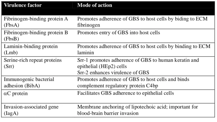
with innate immune clearance mechanisms (Maisey et al., 2008). Table 1 summarizes some key
virulence factors of GBS, their mode of action and mechanism of regulation, detailing their
proposed pathogenic mechanisms, critical for its ability to cause disease.
Table 1.1: Regulation of virulence factor expression (Rajagopal, 2009).
Virulence factor Mode of action
-hemolysin/cytolysin ( -H/C, CylE)
Promotes invasion of host cells and triggers host-cell lysis Impairs cardiac and liver function
Induces inflammatory responses and apoptosis
CAMP factor (Cfb) Forms pores in host-cell membrane; Binds to GPI anchored proteins
Sialic acid capsular polysaccharide (CPS)
Prevents recognition of GBS through molecular mimicry of host-cell surface glycoconjugates
Masks pro- inflammatory cell wall components Superoxide dismutase (SodA) Detoxifies singlet oxygen and superoxide
Pigment (rhamno-polyene) Detoxifies singlet oxygen and superoxide
C5a peptidase (ScpB) Prevents neutrophil recruitment due to cleavage of complement C5a
Promotes adherence by binding to ECM fibronectin and epithelial cells
Serine protease (CspA) Cleaves fibrinogen and chemokines
Impairs neutrophil recruitment and phagocytic killing of GBS
Alanylation of lipotechoic acid Decreases net negative charge on cell surface, repels AMPs
Penicillin-binding protein 1a (PBP1a)
Promotes resistance to AMPs through an unknown mechanism
Pili Promotes resistance to AMPs through an unknown
21
Table 1.1: Regulation of virulence factor expression (continued) (Rajagopal, 2009).
1.3.1 Capsule
The majority of GBS isolates recovered from human infections is encapsulated. Capsule
is a major virulence determinant of GBS, being responsible for resistance to opsonophagocitic
killing and phagocytosis, as well as for the inhibition of complement system clearance (Doran
and Nizet, 2004).
GBS capsular polysaccharides (CPS) are predominantly composed of repeating units containing
four elements: glucose, galactose, N-acetylglucosamine and sialic acid, the terminal sugar on the
side chain of all serotypes. Serotypes VI and VIII are an exception to this composition by lacking
the N-acetylglucosamine and serotype VIII has an additional ramnose residue (Madoff, L. C.,
2006).
Virulence factor Mode of action
Fibrinogen-binding protein A (FbsA)
Promotes adherence of GBS to host cells by biding to ECM fibrinogen
Fibrinogen-binding protein B (FbsB)
Promotes entry of GBS into host cells
Laminin-binding protein (Lmb)
Promotes adherence of GBS to host cells by binding to ECM laminin
Serine-rich repeat proteins (Srr)
Srr-1 promotes adherence of GBS to human keratin and epithelial (HEp2) cells
Srr-2 enhances virulence of GBS Immunogenic bacterial
adhesion (BibA)
Promotes adherence of GBS to host cells and binds complement regulatory protein C4bp
C protein Facilitates GBS adherence to epithelial cells Invasion-associated gene
(IagA)
Membrane anchoring of lipotechoic acid; important for blood-brain barrier invasion
22
The biochemical and immunological properties of the GBS polysaccharide have been extensively
studied. In 1987, the role of the GBS capsule in virulence was evaluated in a rat model of
neonatal infection, by showing that a non-capsulated mutant of GBS presented significantly
reduced virulence as compared to the encapsulated strain (Rubens et al., 1987). The importance
of the capsular sialic acid for bacterial evasion of host mechanisms was also demonstrated when
an encapsulated strain lost its virulence after removal of the sialic acid in a neonatal rat model of
lethal GBS infection (Wessels et al., 1989). According to the chemical composition, structure,
and serological properties, the GBS capsular polysaccharides are classified into ten distinct
serotypes: Ia, Ib, II, III, IV, V, VI, VII, VIII and IX (Feil and Enright, 2004;Slotved et al., 2007).
Moreover, a significant number of strains lack detectable capsule polysaccharide, being
considered non-typeable (NT). Recently a capsular switch among very homogenous clones, as
the hypervirulent CC-17, was reported. It resulted from the replacement of a type III by a type IV
cps locus through exchange of a chromosomal segment (Bellais et al., 2012;Martins et al., 2010).
1.4 Characterization of GBS isolates
Bacterial epidemiologists use typing methods to study the dissemination and population
dynamics of human bacterial pathogens in clinical and environmental settings, including their
transmission patterns and the identification of risk-factors for the control of infectious diseases in
human populations (van et al., 2007).
1.4.1 Phenotyping methods and Serotyping
Bacteriologists have long used phenotypic typing methods to group microorganisms
23
expression of their genotypes (van et al., 2007). Conventional phenotypic methods include
serotyping (based on differences in surface epitopes), phage-typing (based on resistance to
infection by a standard set of bacteriophages), biotyping (according to the different metabolic
capabilities of the cell), bacteriocin typing (the presence or susceptibility to a specific group of
bacteriocins), and antibiotic resistance typing (susceptibility to a panel of antimicrobials).
Although these methods represent a powerful tool to readily identify outbreak isolates in the short
term, are in general inadequate for evolutionary studies and increasingly recognized not to afford
sufficient resolution (Feil and Enright, 2004).
The serological classification of GBS is based upon the identification of capsular
polysaccharides (CPS) and protein antigens (Flores and Ferrieri, 1989;Johnson and Ferrieri,
1984;Lancefield and Freimer, 1966). Many GBS capsular polysaccharide typing methods have
been described (Arakere et al., 1999;Cropp et al., 1974;Holm and Hakansson, 1988;Kiely et al.,
2011;Uh et al., 1997), with the most common methods based on serological tests, i.e.,
immunodiffusion and commercial latex agglutination methods.
1.4.2 Genotyping methods
Genotypic typing methods assess variation in the genomes of bacterial isolates with
respect to composition (presence or absence of plasmids or mobile genetic elements), overall
structure (restriction endonuclease profiles, number and position of repetitive elements), or
precise nucleotide sequence (of genes or intergenic regions) (van et al., 2001). Restriction
Fragment Length Polymorphism (RFLP) is based on DNA digestion with one or more
endonucleases. The resulting restriction pattern of variable length fragments is obtained by
24
endonuclease recognition sites (Maslow et al., 1993). Pulsed-field gel electrophoresis (PFGE)
involves the exposure of chromosomal DNA to endonucleases that recognize only a few sites in
the bacterial genome, generating macrorestriction fragments. However, these methods are more
time-consuming and laborious. More recently, a multilocus sequence-typing (MLST) method,
based on the sequence analysis of 500-bp fragments of seven housekeeping genes, has been
extensively applied to investigate the clonal population structures and genetic lineages of GBS
strains (Jones et al., 2003b).
1.4.3 PCR-based gene profiling and Multilocus sequence typing (MLST)
Polymerase chain reaction (PCR) is a nearly universal typing method, with several
applications in bacterial typing systems, and exhibits an easily adjustable level of discrimination.
Its major advantages include high reproducibility, technical simplicity, wide availability of
equipment and reagents, and rapid turnover time (van et al., 2007). Several PCR-based typing
systems have been used to genotype GBS isolates and include, among other, molecular
serotyping (Kong et al., 2002;Martins et al., 2007), sub-typing within particular serotypes
(Manning et al., 2005), surface protein gene profiling (Creti et al., 2004), detection of mobile
genetic elements (Kong et al., 2003), and of antimicrobial resistance genes (Sutcliffe et al., 1996).
MLST is a sequence-based typing method that involves sequencing of internal fragments of
seven housekeeping genes (Maiden et al., 2013). The sequences are then compared with known
alleles deposited at the MLST database (http://pubmlst.org/sagalactiae), and an allele number is
assigned to each sequence, generating an allelic profile. Each isolate is therefore characterized by
an allelic profile, a seven-integer number that can also be designated by a sequence type (ST)
25
major advantage of MLST over PFGE is the precise, unambiguous and portable nature of the data
obtained, so that the isolates typed in one laboratory can be rapidly compared with all previously
typed strains (Feil and Enright, 2004). In recent years, MLST became increasingly used for the
characterization of bacterial populations because of its ability to infer levels of relatedness
between strains and the reconstruction of evolutionary events (Feil et al., 2004). These questions
have been addressed based on an algorithm, eBURST, that divides an MLST data set into groups
of related isolates by implementing a simple model of clonal expansion and diversification (Feil
et al., 2004). This model predicts that the emergence of clonal complexes (CCs) is due to an
increase in the population of the frequency of the founding genotype, as a consequence of either a
fitness advantage or of random genetic drift. This genotype increases in numbers and by gradual
diversification (point mutation or recombination) gives rise to a clonal complex. In terms of
MLST, the descendants of the founder allelic profile will initially remain unchanged, but over
time variants in one of the seven alleles will arise. These genotypes, which have allelic profiles
that differ from that of the founder at only one of the seven MLST loci, are called single-locus
variants (SLVs). (Feil et al., 2004).
1.4.4 Whole-genome sequences comparisons
The development of efficient and less expensive sequencing methods has produced a
significant number of complete genome sequences of pathogenic microorganisms in recent years.
The comparison of whole-genome sequences offers the possibility to assess genetic differences
within a bacterial species, providing insights on how genetic variability drives the evolution of
virulence mechanisms. The first complete GBS genome sequences were released in 2002 (Glaser
26
predicted coding regions. Both studies revealed substantial similarity with the genomes of the
related human pathogens Streptococcus pyogenes and S. pneumoniae, representing a conserved
backbone between streptococcal species. On the other hand, GBS differed from other
streptococci in genome regions containing known and putative virulence genes, mostly encoding
surface proteins and genes related to mobile elements, suggesting that these regions could be
considered as pathogenicity islands (Glaser et al., 2002). Comparative analysis of multiple
genomes reveal the concept of a “pangenome”, consisting of a core genome shared by all isolates,
accounting for approximately 80% of any single genome, and involved in housekeeping and
regulatory functions, plus a dispensable genome consisting mostly of strain-specific genes.
Again, the abundance of genes associated with mobile and extra-chromosomal elements found in
the variable portion of the genome, supported the hypothesis that the acquisition of the majority
of strain specific traits depends on lateral gene transfer (Tettelin et al., 2005). Furthermore,
genetic heterogeneity among GBS strains, even of the same serotype, revealed that evolution
within genes encoding surface and secreted proteins and those involved in the biosynthesis of the
capsule is mainly due to recombination events leading to gene acquisition, duplication, and
reassortment with the consequent replacement of several genes or to the allelic exchange within a
particular gene. These processes allow GBS to express various combinations of virulence factors,
which are likely to serve as means of adapting to host immunity (Brochet et al., 2006;Tettelin et
al., 2005).
1.5. Molecular epidemiology of GBS
Molecular epidemiology studies has been performed to discriminate genetic lineages in order
27
1.5.1. Serotype distribution and MLST-based genetic lineages
Capsular serotyping has been the classic method used in descriptive epidemiology of S.
agalactiae. Historically, the GBS isolates have been classified into ten different serotypes
according to their capsule polysaccharides (Lindahl et al., 2005;Slotved et al., 2007). Five
serotypes (Ia, Ib, II, III and V) are responsible for most human infections. There are
demographic, geographic, and temporal variations with respect to the predominant serotypes
present in the human population (Blumberg et al., 1996;Hickman et al., 1999;Kieran et al., 1998).
Multiple surveillance studies have indicated that serotype Ia, Ib, III and V are prevalent in the
vagina or perianal region of pregnant women (Barcaite et al., 2008b;Harrison et al., 1998;Phares
et al., 2008;Savoia et al., 2008), whereas serotypes Ia and III are predominant isolates in neonatal
invasive GBS disease, with type III, generally associated with late-onset neonatal disease
(Musser et al., 1989), accounting for 30-76% of cases (Ho et al., 2007;von et al., 2008).
Among serotype III isolates, two main genetic lineages have been identified based on MLST : the
ST-19 clone, frequently found among colonizing isolates (Jones et al., 2003b;Sadowy et al.,
2010) and the ST-17clone, recognized as a hypervirulent clone and strongly associated with
neonatal invasive infections, especially in the late-onset GBS disease (Bisharat et al., 2005;Jones
et al., 2003b;Jones et al., 2006;Lin et al., 2006;Luan et al., 2005). The sequence type ST-17 more
frequently cause meningitis than strains of other STs (Manning et al., 2009) Although the
distinction of lineages within a particular serotype has proved useful, a complete correlation
between capsular type and the genetic lineages as defined by PFGE and MLST was never found
(Brochet et al., 2006;Luan et al., 2005;Manning et al., 2008). The serotype-independent
28
observations support the hypothesis that closely and divergently related clones share the genes
coding for a particular capsular type, suggesting that capsular switching probably occurs in GBS
(Davies et al., 2004;Jones et al., 2003b).
1.6. Secreted or Surface proteins in ST-17 GBS strains
As already described in the Paragraph 1.3, surface and secreted proteins of GBS are likely to
play important roles during different stages of infection, making them promising targets for
vaccines development. Several virulence factors, specific of the CC-17 lineage, were already
identified. These include: (I) FbsB, a fibrinogen-binding protein, (II) HvgA, encoding a
cell-wall–anchored protein, (III) pili components, (IV) genetic variations in the serine-rich repeat
region gene (srr), and V) the surface protein gene (spb-1).
1.6.1 The fibrinogen-binding protein FbsB
FbsA and FbsB are proteins with no structural homology which both bind to human
fibrinogen, mediate the bacterial adhesion to or invasion of epithelial and endothelial cells, and
contribute to the bacterial escape from the immune system (Gutekunst et al., 2004;Jacobsson,
2003;Samen et al., 2006). FbsB has a typical signal peptide but, differently by FbsA, lacks the
LPXTG motif or other wall-anchoring signatures, suggesting that it is not a surface protein but is
secreted into the extracellular medium. The fbs genes and the fbs regulator genes were not
specific of either CC-17 or other CCs strains, but specific gene combinations were related to
particular CCs, indicating that fibrinogen binding is a multigenic process that results from various
gene combinations. Only CC-17 strains contained the fbsA, fbsB, and rgf genes combination. The
29
the rogB gene is missing in the sequenced genome of CC-17 strain COH1 (Tettelin et al., 2005),
and the absence of this gene was also reported in a collection of 20 CC-17 strains (Brochet et al.,
2006). Thus, each CC was characterized by a particular profile of fbs genes and fbs gene
regulators that may account for differences in their fibrinogen-binding abilities. The presence of
the sole fbsA gene was not sufficient to result in strong binding ability to fibrinogen (Rosenau et
al., 2007) and mutants deleted for the fbsA and fbsB genes demonstrated that FbsB protein was
the major fibrinogen binding protein of CC-17 strains (Al et al., 2011a). Indeed, the population of
strains with the significantly highest ability to bind to fibrinogen had both the fbsB and fbsA
genes and belonged to CC-17 phylogenetic lineage (Rosenau et al., 2007).
1.6.2. HvgA, a cell-wall–anchored protein
BibA is an immunogenic surface-associated antigen expressed by GBS that is involved in
virulence. Four allelic variants of this protein have been identified: variant I, found in strains
2603 V/R (V) and 18RS21 (II); variant II, in strains NEM316 (III) and 515 (Ia); variant III, in
strains CJB111 (V), H36B (Ib), and A909 (Ia); and variant IV, in the COH1 (III) strain. The
variant IV was recently identified as a novel ST-17–specific surface-anchored protein, which is
highly prevalent in cases of LOD, and was called hypervirulent GBS adhesin (HvgA, also known
as gbs2018) because mediates GBS neonatal intestinal colonization and crossing of the intestinal
and blood–brain barriers, leading to meningitis, which are key features of LOD (Tazi et al.,
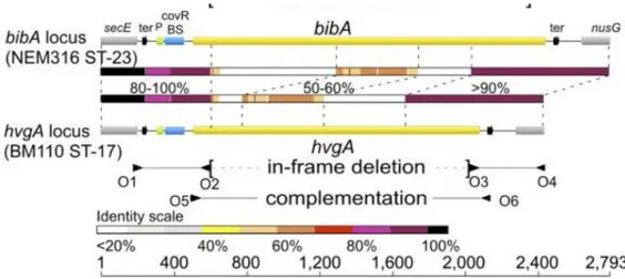
2010). Comparing the structure of the bibA/hvgA locus in GBS strains NEM316 (WT ST-23) and
BM110 (WT ST-17), the nucleotide sequences of the two loci revealed that only the 5′ and 3′
30
internal parts displayed low-level (50–60%) or no significant (<20%) sequence identity (Figure
1.4).
Figure 1.4: Structure of the bibA/hvgA locus in GBS strains NEM316 and BM110 (Tazi A, 2010).
1.6.3 Pili components
The presence of pilus-like structures in GBS was first described in 2005 (Lauer et al.,
2005). The genes encoding pili in GBS are located within two distinct loci in different regions of
the genome, designated pilus-islands 1 and 2 (PI-1 and PI-2), the later presenting two distinct
variants, PI-2a and PI-2b (Rosini et al., 2006a). Pili are composed of three subunits: a backbone
protein (BP), the bona fide pilin, and two ancillary proteins, a pilus associated adhesin and a
component that anchors the pili to the cell wall (Figure 1.5). Both the polymerization and
attachment of the pili to the peptidoglycan cell wall occur by sortase-dependent mechanisms
(Dramsi et al., 2006). Even though PI-1 is not present in all GBS strains, PI-2 is ubiquitously
expressed. Serotype III ST-17 clones are characterized by the presence of PI-2b, serotype V by
the presence of PI-2a. These structures have been recognized to play a role in biofilm formation,
adherence, invasion and translocation of epithelial cells (Konto-Ghiorghi et al., 2009;Rosini et
31
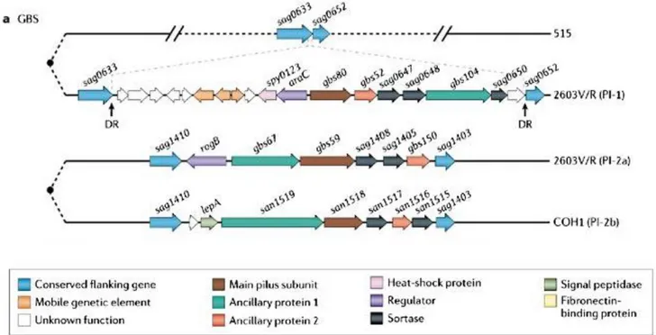
Figure 1.5: Schematic representation of loci that encode group B Streptococcus pili
In the Figure is represented Pilus island 1 (PI-1) in GBS strain 2603V/R and the same region in GBS strain 515, which is pilus negative. The operon is flanked by conserved genes sag0633 and sag0652 and direct repeats (DR). In the lower panel, two alleles of PI-2 flanked by conserved genes sag1410 and sag1403 are depicted: PI-2a from GBS strain 2603V/R, and PI-2b from S. agalactiae strain COH1 (Telford et al., 2006)
1.6.4. Srr and Spb1
Srr family proteins were first characterized in oral streptococci as serine-rich (>35%)
high-molecular-mass glycosylated proteins that are transported across the membrane by a
dedicated SecA2/Y2 secretion system (Chen et al., 2004;Takahashi et al., 2004). In GBS, 2 types
of Srr proteins, known as Srr-1 and Srr-2, have been identified (Seifert et al., 2006). Srr-1 is
surface exposed and highly conserved (>85% nucleotide identity and amino acid identity) among
32
contrast, the expression of Srr-2 that show <20% sequence identity with Srr-1 seems to be
restricted to serotype III and ST-17 strain (Seifert et al., 2006).
The spb1 gene was identified by subtractive hybridization from a serotype III strain of the
putative hypervirulent clone ET1/III-3 (Adderson et al., 2003), and the gene sequence of spb1
indicates characteristics of a surface protein. The Spb1 protein is mainly involved in bacterial
internalization into host cells as a Spb1-negative mutant was significantly reduced in the ability
to invade such cells, but showed little difference in adhering to epithelial cells (Adderson et al.,
2003).
1.7 Biofilm formation in GBS
A bacterial biofilm is composed of groups of bacteria surrounded by an extra cellular
polysaccharide matrix (EPS) (Hall-Stoodley and Stoodley, 2009;Kaur et al., 2009). The
extracellular matrix is composed of water, polysaccharides, proteins, lipids, extracellular DNA,
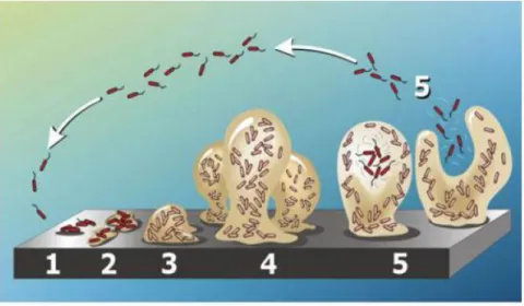
membrane particles and ions (Karatan and Watnick, 2009). Generally, biofilm formation is
characterized by five stages (Figure 1.6): 1. adhesion of bacterial cells to the surface; 2.
production of EPS resulting in more firmly adhered cells; 3. early development of biofilm
architecture; 4. maturation of biofilm architecture; Stage 5, dispersion of single cells from the
33
Figure 1.6: Development of a biofilm as a five-stage process under continuous-flow conditions (e.g. flow cell system) from two dimensions to three dimensions.
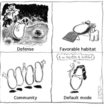
Four plausible driving forces are suggested to act behind bacterial biofilm formation (Jefferson,
2004a): (1) protection from harmful conditions in the host (defense), (2) sequestration to a
nutrient rich area (colonization), (3) utilization of cooperative benefits (community), (4) bacteria
normally grow as biofilms in nature (Figure 1.7). The three dimensional complex of the biofilm is
a coordinated community and allows bacteria to adapt to and survive in host environments
(Hall-Stoodley and (Hall-Stoodley, 2009). Bacteria in biofilms detect environmental changes and respond to
it in order to survive in diverse and stressful conditions (Hall-Stoodley and Stoodley, 2009).
Organisms within biofilms can withstand nutrient deprivation, pH changes, oxygen radicals,
disinfectants, and antibiotics better than planktonic organisms (Jefferson, 2004b). Biofilms
generate resistance to antibiotics by decreasing the antibiotics penetration rate and mediating
bacterial gene expression. Transmission electron micrographs reveal biofilms protect bacteria
34
Figure 1.7: Artistic interpretation of the four driving forces behind bacterial biofilm formation that are discussed in a review by K.K. Jefferson (Jefferson, 2004a).
Bacterial biofilm formation is regulated by different environment signals including mechanical,
nutritional, metabolic and host-derived signals, secondary messenger and protein transcriptional
regulators (Karatan and Watnick, 2009). The majority of the species belonging to the
Streptococcus family have been shown to form biofilm, while just a limited number of studies
have demonstrated GBS biofilm formation in vitro (Borges et al., 2012;Kaur et al.,
2009;Konto-Ghiorghi et al., 2009;Rinaudo et al., 2010a).
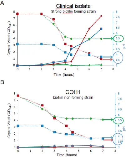
Glucose concentration in culture media were shown to modulate biofilm formation in GBS,
although conflicting data have been reported regarding the biofilm forming capacity of isolates
belonging to different serotypes, and the correlation between biofilm formation and pH (Borges
35
hypothesized that these contradictory results could be due to absence of in vitro protocols that
allowed clearly discriminating between strong and weak biofilm formers and unambiguously
establishing the role of bacterial culture conditions.
Recent studies have demonstrated GBS biofilm formation in vitro (Rinaudo et al., 2010a)
although the data regarding the effect of pH and media composition are controversial. In a recent
study, Yueh-Ren et al. (Ho et al., 2012) found that the low pH condition induced biofilm fomaion
in a nutrient-limited medium (M9YE) but not in THB. Borges S. et al. (Borges et al., 2012), Kaur
et al. (Kaur et al., 2009) and Yang Q. et al. (Yang et al., 2012) found that GBS produced a greater
amount of biofilm at pH 6.5 than at pH 4.2 and Konto Ghiordi et al. (Konto-Ghiorghi et al.,
2009) reported that only LB and RPMI 1640 supplemented with 1% of glucose produced uniform
biofilm and not THB. Manetti et al. (Manetti et al., 2010) observed that, in S. pyogenes, the
presence of glucose resulted in auto acidification of the media and consequently biofilm
formation in GBS. Rinaudo et al. (Rinaudo et al., 2010a) demonstrated that the presence of 1% of
glucose in THB induces biofilm formation in GBS.
Recent studies also suggest that biofilm formation by some GBS strains could have an
important role in host-colonization. The capability of GBS to attach to epithelial cells also
increased in acidic conditions (Tamura et al., 1994). GBS adherence to A549 pulmonary
epithelial cells and vaginal epithelial cells at pH 4.0 was 10 to 20 fold higher than at neutral pH.
Transcription experiments showed there were 317 genes up-regulated and 61 genes
down-regulated when GBS was incubated in pH 5 media compared with pH 7 medium (Santi et al.,
2009). The majority of genes involved in response to environment pH change include genes
36
1.8 Proteomics approach of vaccine candidate identification
A new approach that allows fast and consistent identification of proteins that are exposed on
the bacterial surface has been recently published (Rodriguez-Ortega et al., 2006). The technique,
consisting of the surface digestion of live bacteria with different proteases and analysis by mass
spectrometry, identifies the so-called bacterial “surfome”. This technique was already applied to
analyses the proteins expressed on the surface of a GBS non-biofim forming strain, grown in
standard laboratory condition (Doro et al., 2009). Here, for the first time, surfome analysis is
applied to compare the expression profile of bacteria growth in both planktonic and biofilm like
growth conditions.
1.9 Thesis overview
In the present study, the biofilm formation of more than 350 GBS clinical isolates from
pregnant women and neonatal infections, using a new high-throughput in vitro protocol was
investigated. The isolates were collected during the DEVANI project (Design of a Vaccine
against Neonatal Infections) supported by the European Commission Seventh Framework,
launched on 1 January 2008 (http://www.devaniproject.org).
Specifically, this study was focused on:
1. Develop a new biofilm formation protocol to guarantee more stringent conditions,
reducing unambiguous data interpretation;
2. Screen 366 GBS clinical isolates from pregnant woman and from neonatal infection,
belonging to different serotypes, in relation to media composition and pH;
3. Observe the correlation between biofilm formation and pH decrease in rich and
37
4. Investigate the relationship between capsule amount and biofilm formation at different
pH;
5. Identify the role of proteins, capsule and DNA in biofilm formation and in its structural
stability.
6. Apply the surfome analysis to identify the proteins differentially expressed on the surface
38
2. Material and methods
2.1 Bacterial strains and growth conditions
A total of 366 S. agalactiae isolates of 8 different serotypes (Ia n = 58; Ib n = 18; II n = 28;
III n = 156; IV n = 10; V n = 57; VIII n = 3, IX n = 13) and non-typeable strains (n = 23) were
included in the study. Among these, 357 were vaginorectal isolates obtained from pregnant
women (n = 272) and clinical isolates from neonatal infections (n = 85) in Belgium, Bulgaria, the
Czech Republic, Denmark, Germany, Great Britain, Italy, and Spain. These isolates were
collected during the DEVANI project (Design of a Vaccine Against Neonatal Infections)
supported by the European Commission Seventh Framework, launched on 1 January 2008
(http://www.devaniproject.org).
The overall aim of the DEVANI project was to assess European GBS epidemiology in order to
facilitate the design of a new vaccine capable of conferring broad coverage to immunize neonates
against GBS infections through a durable maternal immune response. Strains CJB111 (type V),
515 (type Ia), COH1 (serotype III) and H36B (serotype Ib), 18RS21 (serotype II), A909 (serotype
Ia), D136C (serotype III) were kindly provided by Dr. Dennis Kasper (Department of
Microbiology and Immunobiology, Harvard Medical School, Boston, MA, USA). 2603 V/R
(serotype V) (Tettelin et al., 2002) strain was obtained from the Istituto Superiore di Sanità. The
COH1 un-encapsulated mutant carries a deletion of the cpsE gene in the capsule locus
(Cieslewicz et al., 2001) and was kindly provided by M. Cieslewicz (Channing Laboratory,
39
2.2 Serotype and Sequence Type identification
2.2.1 Total genomic DNA isolation
For genomic DNA isolation the strains were grown overnight at 37 °C in static conditions. 10 mL
of each culture was centrifuged at 1400 rpm, washed once in PBS and suspended in 300 µL of
TET (20 mM Tris pH 8.0, 2 mM EDTA, 1% Triton X-100) supplemented with 5 µL Mutanolysin
(10 U/µL) and 55 µL of Lysozime (12 mg/mL). The cell pellets were incubated at 37°C for 1
hour and then processed using the Wizard DNA purification Kit (Promega). Final DNA
concentration was assessed by optical density determination at 260 nm.
2.2.2 Serotype and ST-17 identification
GBS strains were typed by latex agglutination method (Strep-B-Latex kit;Copenhagen,
Denmark), as described by Afshar et al.(Afshar et al., 2011). ST-17 identification was performed
for all the 156 serotype III tested strains. PCR amplification and sequencing of the internal
fragments of 7 housekeeping genes, namely, adhP, atr, glcK, glnA, pheS, sdhA, and tkt were
performed as described previously (Jones et al., 2003b). Assignment to ST-17 was performed at
the GBS MLST Web site (http://pubmlst.org/sagalactiae/). The strains showing, at least, an allele
sequence non-correspondent to the ST-17 profile, were classified as non-ST-17.
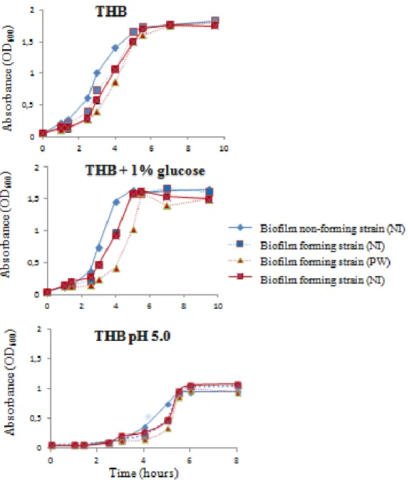
2.3 Growth experiments
Four clinical isolates (three biofilm-forming strains and one non-biofilm-forming strain) grown
overnight at 37°C in Todd-Hewitt broth (THB) at pH 7.8 were diluted to an optical density at 600
40
supplemented with 1% glucose, or pH 5.0 THB. The tubes were then incubated without shaking
at 37°C and the OD600 was measured for 8 to 10 h. Each experiment was performed in triplicate.
2.4 In vitro biofilm formation
2.4.1 Standard biofilm formation protocol
The standard protocol was performed as already described (Rinaudo et al., 2010a). In brief,
Streptococcus agalactiae strains were streaked on blood agar plates and grown at 37 °C for 18
hours. GBS strains, grown overnight in THB, were diluted 1 20 in Todd Hewitt Broth pH 7.8
(THB) or THB supplemented with 1% glucose and used to inoculate (100 µl/well) 96-weel
polystirene microtiter plates (Constar; Corning Inc.; Corning ,NY).
Plates were incubated without shaking at 37°C for 18 h aerobically in 5% CO2. The supernatant
was removed and the wells were subjected to three cycles of washing with 200 µL of
double-distilled H2O (ddH2O) to remove unattached bacteria. A Crystal Violet (CV) assay and a
2,3-bis(2-methoxy-4-nitro-5-sulfophenyl)-5-[(phenylamino)carbonyl]-2H-tetrazolium hydroxide
(XTT) assay were then performed to estimate bacterial biomass and cell viability, respectively.
2.4.2 New biofilm formation protocol
A new protocol for in vitro biofilm formation was set-up. Streptococcus agalactiae strains were
streaked on blood agar plates and grown at 37 °C for 18 hours. Bacterial suspension in Todd
Hewitt Broth pH 7.8 (THB) was prepared at OD600 0.05 and used to inoculate (200 µL/well) a
96-well polystyrene microtiter plates (Constar; Corning Inc.; Corning ,NY). The preliminary
41
already used in the standard biofilm formation protocol. The other media used to investigate the
role of pH in biofilm formation were (i) RPMI GlutaMAX (Gibco-Life Technologies, Milan,
Italy), (ii) RPMI GlutaMAX supplemented with 1% glucose, (iii) RPMI GlutaMAX and THB
both acidified to pH 5.0, and (iv) THB supplemented with 1% glucose and buffered at pH 7.8
with the addition of HEPES (20 to 200 mM) or Tris-HCl (20 to 200 mM). The plate was sealed to
limit oxygen exchange and shaken at 60 rpm at 37°C to reduce bacterial deposition. Following 8
h of adhesion at 37°C, the plates were washed to remove loosely adherent cells and the
supernatant was replaced with 200 µL of fresh medium. After 15 h at 37°C, the medium was
removed and the wells were subjected to three cycles of washing with 200 µL of
phosphate-buffered saline (PBS) to remove unattached bacteria (Figure 2.1). CV and XTT assays were then
performed to estimate bacterial biomass and cell viability, respectively.
2.5 Crystal Violet assay
The wells were stained for 10 min with a 0.5% (wt/vol) solution of Crystal Violet (CV)
(Sigma-Aldrich, Inc., St. Louis, MO). After rinsing with ddH2O, bound dye was released from the
stained cells by using 30% glacial acetic acid. Biofilm formation was quantified by measuring the
OD540 of the solution with a microplate reader (Infinite M200; Tecan). Samples showing an
OD540 higher than 1 were diluted 5 and 20 times in water, and the absorbance reading was
repeated. The measured values were subtracted from the blank and then multiplied by the dilution
factors. Each assay was performed in triplicate. Wells filled with growth medium were included
42
2.6 XTT viability assay.
XTT is a tetrazolium derivative XTT is a tetrazolium derivative cleaved to anorange-colored
formazan product by mitochondrial dehydrogenase inviable cells (D'Urzo et al., 2014;Roehm et
al., 1991). The XTT solution was prepared by dissolving 0.5 mg XTT (Sigma-Aldrich, Inc., St.
Louis, MO) in 1 mL of PBS and then supplementing it with 2.5 µL of a 10 mM menadione stock
solution (dissolved in acetone). 150 µL of XTT-menadione solution was added to each well.
Plates were incubated in the dark for 3 h at 37°C and then centrifuged for 20 min at 4,000 rpm.
100 µL of the supernatant was transferred to the wells of a new 96-well flat-bottom plate, and the
43
Figure 2.1: Protocols of GBS biofilm formation.
Comparison of the protocols used in this study. Standard protocol (on the left) and new protocol (on the right). The details regarding the two protocols are described in Material and Methods (Paragraph 2.4).
44
2.7 Confocal laser scanning microscopy
The GBS biofilms obtained by both the standard protocol (Rinaudo et al., 2010a) and the
new protocol, used in this study, were visualized by Confocal Laser Scanning Microscopy
(CLSM). S. agalactiae strains were inoculated (0.8 mL/well) in a Lab-Tek II eight-well 1.5 cover
glass (VWR, Rochester, NY) containing THB or THB supplemented with 1% glucose and
incubated as already described in the new biofilm formation protocol (Paragraph 2.4.2). Adherent
bacteria were stained for 30 min with LIVE/DEAD BacLight fluorescent stain (Molecular
Probes, Eugene, OR) and fixed with 2% formaldehyde for 30 min at room temperature.
Samples were analyzed with a Zeiss LSM710 confocal microscope by using a Plan-Apochromat
40x/1.3 objective. SYTO 9 fluorescence, corresponding to live bacteria, was acquired in the
green channel (492 to 572 nm), and propidium iodide fluorescence, which does not penetrate
viable bacterial cells, was acquired in the red channel (566 to 719 nm). Images were acquired by
using Zen 2008 software and modified with Volocity (Improvision, Lexington, MA). ImageJ
(http://rsbweb.nih.gov/ij/) and COMSTAT2 (http://www.comstat.dk) were used to evaluate the
biomass and maximum and mean thicknesses of the three-dimensional biofilm images acquired
by CLSM (D'Urzo et al., 2014;Heydorn et al., 2000;Roehm et al., 1991)
2.8 Enzymatic inhibition/eradication of biofilms
The Minimal Inhibition Concentration (MIC), Minimal Biofilm Inhibition Concentration
(MBIC) and Minimal Biofilm Eradication Concentration (MBEC) were measured in 96 -well
polystyrene microtiter plates with 0.4-200 μg/mL proteinase K and a maximum of 200 μg/mL of
DNase. For MBIC determination, the biofilm formation assays were performed as described previously using THB supplemented with 1% glucose and proteinase K (200 μg/mL) or DNase
45
(200 μg/mL). For MBEC determination a 24h-mature biofilm grown in the absence of proteinase
K or Dnase was washed twice with PBS and further incubated in THB supplemented with 1%
glucose containing proteinase K (200 μg/mL) or DNase (200 μg/mL), 3 h at 37°C. Biofilm was
quantified by CV assay, as previously described.
2.9 Biofilm inhibition using sera from immunized rabbit
For the biofilm inhibition assay, rabbit sera for GBS80 (SAG0645) (backbone protein of pilus
island-I), GBS67 (SAG1408) (ancillary protein of PI-2a), and GBS1523 (SAN 1518) (backbone
protein of PI-2b) were tested for their ability to inhibit biofilm formation. Bacterial suspension in
Todd Hewitt Broth pH 7.8 (THB) was prepared at OD600 0.05 and used to inoculate (200
µL/well) a 96-well polystyrene microtiter plates (Constar; Corning Inc.; Corning ,NY) with serial
dilutions of sera. The plate was sealed to limit oxygen exchange and shaken at 60 rpm/min at
37°C to reduce bacteria deposition. Following 8 hours of adhesion at 37°C, media, including any
unattached bacteria, were decanted from the wells, and any remaining planktonic cells were
removed by rinsing with ddH2O. Wells were air dried, and adherent bacteria were subjected to
quantification by colorimetric assays. Sera from immunized New Zealand rabbits (Charles River
Laboratories, Calco Italy) were kindly supply by Rinaudo CD’s group and obtained as previously
reported (Maione et al., 2005;Margarit et al., 2009;Rosini et al., 2006b).
2.10 Quantification of capsular polysaccharides
Capsular polysaccharides from COH1 strain and 4 biofilm forming strains (2 expressing type
46
resorcinol–hydrochloric acid assay as described earlier by Svennerholm (SVENNERHOLM,
1957).
Bacteria were inoculated in 15 mL of THB and THB pH 5.0 and grown to an OD600 of 1.8 and
1.0, respectively. The cells were pelleted, washed twice with PBS, suspended in 0.8 M NaOH and
incubated for 48 h at 37°C. Following neutralization with HCl, the insoluble material was
removed by centrifugation; the supernatant was transferred to an Amicon Ultra -10 (Millipore,
Bedford, MA), concentrated to 0.20 mL, and then perfused two times with 1 mL dH2O.
A final volume of 1.5 mL of supernatant was analyzed to determine the amount of extracted
polysaccharide. Briefly, 500 µL of resorcinol-HCl reagent (2% w/v aq. Resorcinol solution added
to concentrated HCl and 0.1M CuSO4) was added to 500 µL of extracted polysialic acids-sample
which was then heated in a boiling oil bath for 20 min. The released sialic acid
(N-acetylneuraminic acid [NeuNAC]) reacts with resorcinol in the presence of copper sulphate under
reducing conditions to give a blue-purple color. After it cooled to room temperature, the
absorbance at 564 nm was measured. NeuNAC concentrations were calculated from the standard
curves, obtained using NANA standards (range: 5-25 µg/mL) and converted to total GBS
saccharide (conversion factor= molecular weight of NeuNAC/ molecular weight of repeat unit
GBS polysaccharide).
2.11 Surfome preparation of Streptococcus agalactiae grown in biofilm
inducing and non-inducing conditions
Surfome preparation of S. agalactiae live cells was performed as previously described (Doro
et al., 2009) with minor modifications. Briefly, S. agalactiae COH1 and a biofilm forming strain
47
THB) and 1.0 (in THB pH 5.0) was reached. Bacteria were harvested by centrifugation at 4000
rpm g for 10 min, 4°C and washed twice with phosphate buffered saline (PBS). The supernatants
were used for the secretome preparation, as described below. Bacterial pellets were suspended in
700 µL of digestion buffer (33% sucrose and 50 mM Tris pH 7.0) and incubated at 37°C with 10 μg of trypsin (Promega, Madison, USA) for 30 min at 37 °C. Bacterial cells were then spin down
at 4000 rpm for 10 min at 4°C, and the supernatants were filtered through 0.22-μm pore size
filters (Millex, Millipore, Bedford, MA). Protease reactions were stopped with formic acid at
0.1% final concentration. Before analysis, PBS and sucrose were removed by an off-line
desalting procedure using OASIS cartridges (Waters, Milford, MA) following the manufacturer's
protocol. Desalted peptides were concentrated with a Centrivap Concentrator (Labconco, Kansas City, KS) and kept at −20 °C until further analysis.
Cell wall extracts of S. agalactiae COH1 and a biofilm forming strain, grown in THB, THB
pH 5.0 and THB + 1% glucose, were compared both in protein amount both in protein profile
using SDS-PAGE. GBS stains were incubated at 37°C until an OD600 of 1.0 was reached.
Bacteria were harvested by centrifugation at 4000 rpm g for 10 min, 4°C and washed twice with
phosphate buffered saline (PBS). Bacterial pellets were incubated in an ice-cold protoplasting
buffer containing 40% sucrose in 0.1 M K3PO4 (pH 6.2), protease inhibitors and 800U/mL of
mutanolysin (Sigma-Aldrich, Inc., St. Louis, MO) for 30 min at 37°C, as previously described
(Kling et al., 1999). After centrifugation the protein content of the supernatants (cell wall
fractions) were quantified using the BCA assay (Pierce) and the protein profile visualized by
48
2.12 Protein Identification by LC-MS/MS
Peptides were separated by nano-LC on a NanoAcquity UPLC system (Waters) connected to
a SynaptG2 mass spectrometer equipped with a NanoLockSprayTM source (Waters). Samples
were loaded onto a NanoAcquity 5μm Symmetry® C18 trapping column (180μm X 20mm,
Waters) using full loop injection for 2 min at a flow rate of 7.5 μL/min with mobile phase A (2%
acetonitrile, 0.1% formic acid). Peptides were then separated on a NanoAcquity 1.7μm BEH130
C18 analytical column (75μm X 250mm, Waters) using a 90 min gradient of 2–45% mobile phase
B (98% acetonitrile, 0.1% formic acid) at a flow rate of 250 nL/min. The column temperature
was set at 35°C. The reference, [Glu1]-fibrinopeptide was constantly infused B at 600
fmoL/μl,by the NanoAcquity auxiliary pump at a constant flow rate of 400 nL/min and acquired
with an interval of 30 seconds through the reference sprayer of the NanoLockSprayTM source.
The spectra of the eluted peptides were acquired in positive V-mode in a mass range of 50-2,000
m/z using a MSE program for MS/MS with 0.7-s scan times and a collision energy ramp of 20-40
eV for elevated energy scans in the transfer region of the mass spectrometer. All fragmentations
were performed using argon as collision gas. Continuum LCMSE data were processed and
searched against the database of Streptococcus agalactiae COH1 strain
(http://cmr.jcvi.org/tigr-scripts/CMR/CmrHomePage.cgi) using ProteinLynx Global Server version 2.5.2 (Waters).
Methionine oxidation and glutamine and asparagine deamidation were specified as variable
modifications, one trypsin missed-cleavage was allowed, and the default settings in ProteinLynx
Global Server for the precursor ion and fragment ion mass tolerance were used (automatic
setting). The observed mass error tolerance values were typically under 15 ppm. The search