Modulation of the activity of the Locomotor Central
Pattern Generator in the rat spinal cord in vitro
Candidate: Supervisor:
TABLE OF CONTENTS
NOTE
ABSTRACT
INTRODUCTION
1. PHYSIOLOGY OF THE CENTRAL PATTERN GENERATOR (CPG) AND
SPINAL LESIONS
1.1.
The CPG for locomotion
1.2.
Organization of the mammalian CPG
1.3.
Control of locomotion
1.4.
Ontogeny of spinal networks
1.5.
Intrinsic MN activity
1.6.
Evidence for the human CPG
1.7. Spinal cord injury (SCI)
2. STUDYING THE SPINAL PATTERN GENERATION: THE ISOLATED
SPINAL CORD PREPARATION
2.1. The in vitro isolated spinal cord of the neonatal rat
2.2. Spontaneous activity recorded from ventral roots
2.3. Fictive locomotion
2.4. Chemically induced Fictive Locomotion
2.5. Localization of CPG
2.6. Electrically evoked Fictive Locomotion
2.7. Disinhibited Rhythm
3.
MODULATING SPINAL CIRCUITS: A TARGET FOR REHABILITATIVE
PURPOSES
3.1.
Modulation of neural networks
3.2. Rehabilitative approaches
4. THE ROLE OF METABOTROPIC GLUTAMATE RECEPTORS (mGluRs)
4.1. mGluRs in the spinal cord
4.2. Neuroprotective effects of mGluRs in the central nervous system
III
1
3
3
3
4
6
9
11
13
15
17
17
18
20
21
23
25
26
28
28
30
32
32
34
4.3. Group I mGluRs and SCI
4.4. Group II and III mGluRs and SCI
4.5. Chronic central pain: a finely tuned interplay between mGluRs.
5. THE ROLE OF K
+CONDUCTANCE BLOCKERS IN THE SPINAL CORD
5.1. Pharmacological blockade of K
+conductances: 4-aminopyridine (4-AP)
5.2. 4-AP evokes rhythmic antidromic discharges from dorsal roots (DRs)
5.3. Synaptic mechanisms of primary afferent depolarization
5.4. Modulation of CPG by other voltage dependent K
+channel blockers: TEA
6. AIMS OF THE PRESENT STUDY
METHODS see enclosed papers
RESULTS see enclosed papers
DISCUSSION
1. Excitation and oscillations due to group I mGluR activation
2. EPSP and IPSP depression due to group I receptor activation
3. Role of group I mGluRs in fictive locomotion
4. Role of group I mGluRs in disinhibited rhythm
5. Role of group II and III mGluRs in the spinal cord
6. Low micromolar concentrations of 4-AP facilitate fictive locomotion
7. Characteristics of the electrical oscillations evoked by 4-AP on DRs
8. Fictive locomotor patterns generated by TEA
9. Comparisons between effects due to mGluR activity and K
+conductance block
CONCLUSIONS
REFERENCES
ACKNOWLEDGEMENTS
35
37
37
38
38
39
41
43
46
47
48
49
49
50
51
51
52
52
53
56
57
75
NOTE
Part of the data reported in the present thesis has been published in the articles listed
below. In all cases the candidate personally performed the experimental work and data
analysis, and contributed to paper writing.
Taccola G, Nistri A (2005) Fictive locomotor patterns generated by
tetraethylammonium application to the neonatal rat spinal cord. Neuroscience in
press.
Marchetti C, Taccola G, Nistri A (2005) Activation of group I metabotropic glutamate
receptors depresses recurrent inhibition of motoneurons in the neonatal rat spinal cord
in vitro. Exp Brain Res 164:406-410.
Taccola G, Nistri A (2005) Electrophysiological effects of 4-aminopyridine on fictive
locomotor activity of the rat spinal cord in vitro. Acta Neurochir Suppl 93:151-154.
Taccola G, Nistri A (2005) Characteristics of the electrical oscillations evoked by
4-aminopyridine on dorsal root fibers and their relation to fictive locomotor patterns in
the rat
spinal cord in vitro. Neuroscience 132:1187-1197.
Taccola G, Nistri A (2004) Low micromolar concentrations of 4-aminopyridine
facilitate fictive locomotion expressed by the rat spinal cord in vitro. Neuroscience
126:511-520.
Taccola G, Marchetti C, Nistri A (2004) Role of group II and III metabotropic
glutamate receptors in rhythmic patterns of the neonatal rat spinal cord in vitro. Exp
Brain Res 156:495-504.
Taccola G, Marchetti C, Nistri A (2004) Modulation of rhythmic patterns and
cumulative depolarization by group I metabotropic glutamate receptors in the neonatal
rat spinal cord in vitro. Eur J Neurosci 19:533-541.
Marchetti C, Taccola G, Nistri A (2003) Distinct subtypes of group I metabotropic
glutamate receptors on rat spinal neurons mediate complex facilitatory and inhibitory
effects. Eur J Neurosci 18:1873-1883.
Taccola G, Marchetti C, Nistri A (2003) Effect of metabotropic glutamate receptor
activity on rhythmic discharges of the neonatal rat spinal cord in vitro. Exp Brain Res
153:388-393.
ABSTRACT
The present study has investigated the rhythmic properties of spinal networks in the
neonatal rat spinal cord in vitro, by means of intracellular recordings from single
motoneurons (MNs) and extracellular recordings from ventral and dorsal roots (VRs;
DRs).
Distinct subclasses of metabotropic glutamate receptors (mGluRs) on rat spinal
neurons mediated complex facilitatory and inhibitory effects. The class I agonist
DHPG evoked MN depolarization (via the mGluR1 subtype) mostly at network level
and generated sustained, network-dependent oscillations (via the mGluR5 subtype).
DHPG also decreased the amplitude of reflex responses induced by DR stimuli, an
effect unrelated to depolarization but dependent on glycinergic transmission. Single
reflex responses were insensitive to group I mGluRs antagonists, suggesting no phasic
activation of group I receptors during this process. Finally, DHPG depressed the
glycinergic recurrent IPSP, perhaps by impairing the cholinergic input to Renshaw
cells. Thus, the cellular distribution of those mGluRs at strategic circuit connections
may determine the functional outcome of the network in terms of excitation or
inhibition. Activation of class II or III mGluRs had no direct action on MNs although
it strongly blocked evoked synaptic transmission, presumably acting at presynaptic
level.
To extend our understanding of the network-based properties, which enable a
neuronal circuit to produce sustained electrical oscillations, we explored the potential
contribution of mGluRs to generate rhythmic discharges.
During cumulative depolarization or fictive locomotion, spinal mGluRs were
minimally activated by endogenous glutamate, although they could potently modulate
these responses once activated by exogenously applied mGluR agonists. Disinhibited
bursting was associated with the activation of mGluR1 receptors (facilitating network
excitability) and of group II mGluRs (depressing it).
We investigated if the K
+channel blocker 4-aminopyridine (4-AP) could facilitate
spinal locomotor networks in addition to its well-known effect on motor nerve
conduction. 4-AP produced synchronous VR oscillations, which did not develop into
fictive locomotion. These oscillations had network origin, required intact
glutamatergic transmission and were probably amplified via electrotonic coupling.
4-AP slightly increased input resistance of lumbar MNs, without affecting their action
or resting potentials. DR evoked synaptic responses were enhanced by 4-AP without
changes in axon conduction. 4-AP accelerated chemically or electrically induced
fictive locomotion and facilitated the onset of fictive locomotion in the presence of
subthreshold stimuli, that were previously insufficient to activate locomotor patterns.
Thus, although 4-AP per se could not directly activate the locomotor network of the
spinal cord, it could strongly facilitate the locomotor program initiated by
neurochemicals or electrical stimuli.
On DRs, 4-AP induced sustained synchronous oscillations smaller than electrically
evoked synaptic potentials, persistent after sectioning off the ventral region and
preserved in an isolated dorsal quadrant, indicating their dorsal horn origin. 4-AP
oscillations were network mediated via glutamatergic, glycinergic and GABAergic
transmission. Isolated ventral horn areas could not generate 4-AP oscillations,
although their intrinsic, disinhibited bursting was accelerated by the substance.
Activation of fictive locomotion by either application of neurochmicals or stimulus
trains to a single DR reversibly suppressed DR oscillations induced by 4-AP.
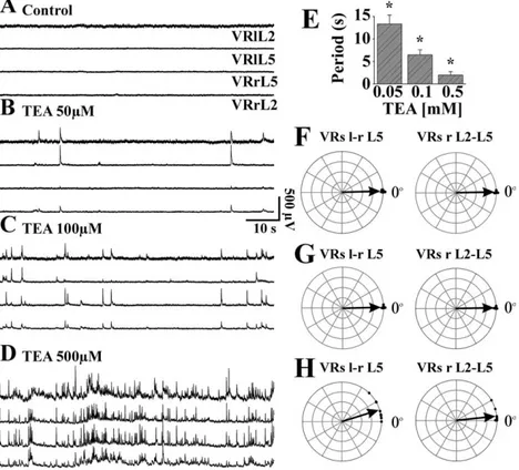
The present electrophysiological investigation also examined whether the broad
spectrum potassium channel blocker tetraethylammonium (TEA) could generate
locomotor-like patterns. Low concentrations of TEA induced irregular, synchronous
discharges incompatible with locomotion. Higher concentrations evoked alternating
discharges between flexor and extensor motor pools, plus a large depolarization of
MNs with spike broadening. The alternating discharges were superimposed on slow,
shallow waves of synchronous depolarization. Rhythmic alternating patterns were
suppressed by blockers of glutamate, GABA
Aand glycine receptors, disclosing a
background of depolarizing bursts inhibited by antagonism of group I mGluRs.
Furthermore, TEA also evoked irregular discharges on DRs. The rhythmic alternating
patterns elicited by TEA on VRs were relatively stereotypic, had limited synergy with
the fictive locomotion induced by DR stimuli, and were not accelerated by 4-AP.
Horizontal section of the spinal cord preserved irregular VR discharges and DR
discharges, demonstrating that the action of TEA on spinal networks was
fundamentally different from that of 4-AP.
INTRODUCTION
1. PHYSIOLOGY OF THE CENTRAL PATTERN GENERATOR (CPG) AND
SPINAL LESIONS
For many species, the automated movements needed for walking, respiration,
scratching, mastication or other rhythmic activities are generated by the remarkable
organization of certain neural networks. For locomotion, one usually refers to the term
“central pattern generator” to indicate a set of spinal neurons responsible for creating
a motor pattern.
1.1. The CPG for locomotion
Early in past century, Thomas Graham-Brown discovered that rhythmic contractions
of hind leg muscles of the spinalized cat could be generated in the absence of any
sensory input from peripheral receptors in the legs. This important observation
demonstrated that networks of nerve cells located entirely in the spinal cord generate
the rhythm for stepping, and established the alternating pattern of activity in flexor
and extensor muscles. The expression `central pattern generator` is an operational
expression to designate an ensemble of neural elements whose properties and
connectivity can give rise to characteristic patterns of rhythmic activity in absence of
external feedback. The expression `fictive patterns` refers to these characteristic
patterns themselves as expressed in paralyzed preparations or in vitro preparations, as
opposed the full `active or real pattern` of movements in vivo.
General principles of the organization of these circuits and their control exerted by
higher brain centers have come from the study of smaller circuits found in
invertebrates and from the studies on CPGs associated with swimming in the lamprey,
tadpole, leech and the molluscs Tritonia and Clione. From investigations of these
networks, we learned that central pattern generation is a complex process that depends
on numerous mechanisms. These mechanisms can be related to cellular properties of
individual nerve cells, properties of the synaptic connections between the nerve cells
and pattern of interconnections between nerve cells (Marder and Bucher, 2001).
As exemplified in Fig. 1, in all classes of vertebrates, the overall locomotor control
system is designed in a similar way, which is not surprising when viewed in an
evolutionary perspective.
Thus, the same mesopontine and diencephalic centres initiate locomotor activity in
lampreys as in primates, via activation of lower brainstem reticulospinal neurons.
These, in turn, activate the spinal CPG which generates the motor pattern, be it
swimming or walking. Sensory feedback acting on the CPG is an integral part of the
control system and helps to adapt the motor pattern to external events.
Figure 1. General control scheme for vertebrate locomotion. The basal ganglia exert a tonic inhibitory influence on different motor centres. Once a pattern of motor behavior is selected the inhibition is released, allowing in this case the locomotor centre in the brainstem to be activated. Locomotion is then initiated by an increased activity in reticulospinal neurons which activate the central spinal network, which in turn produces the locomotor pattern in close interaction with sensory feedback (from Grillner et al., Brain Res Rev 1998).
A remarkable feature that comes from a variety of invertebrate preparations, including
the leech heartbeat system, the crustacean stomatogastric nervous system, Tritonia,
and a variety of insect preparations, is that rhythmic motor patterns can be activated
by a large number of different neuromodulators (Marder and Calabrese, 1996).
Similarly, fictive locomotion patterns in neonatal rat spinal cord in vitro can be
activated by several substances. Therefore, it has been suggested that these substances
are responsible for configuring the spinal cord for different movements (Beato and
Nistri, 1999), activating different spinal networks or switching individual neurons
among distinct functional circuits.
1.2. Organization of the mammalian CPG
Recently, Lafreniere-Roula and McCrea (2005) have examined the features of
spontaneous deletions of bursts of motoneuron (MN) activity that can occur within
either rhythmic alternating flexor and extensor activity during fictive locomotion or
scratching in adult decerebrated cats. Deletions are reductions or absences of one or
more expected rhythmic bursts of activity in multiple agonist MN pools. During
rhythmic fictive locomotion and scratching, the deletions of activity in multiple
agonist MN pools are presumably related to a temporary failure in the operation of the
CPG.
In experiments to explore the issue, the re-emergence of the activity after spontaneous
deletions during fictive locomotion and scratching, occurs as a multiple of the
pre-existing cycle period without cycle resetting or frequency change. This maintenance
of cycle period during deletions suggests that the failure to recruit MNs is not
inextricably linked to the generation of the locomotor or scratching rhythm. This fact
suggests that the organization of locomotor and scratch CPGs presents a separation
between the cells generating rhythm (a sort of internal clock) and the cells distributing
excitation to MNs.
A schematic representation of the proposed architecture for the CPG operating for one
limb pattern is provided by Lafreniere-Roula and McCrea (2005). This consists of
separate networks for rhythm generation and for pattern formation. Pattern formation
modules include circuitry for reciprocal inhibition of antagonist motor pools and for
controlling the activity of subsets of MNs.
The organization in Figure 2 offers an explanation for the deletions associated with
the maintenance of phase of the locomotor rhythm. A spontaneous alteration of
excitability within the pattern formation networks will not affect the rhythm
generation network. In fact, it would simply alter MN activity, resulting in a transient,
complete suppression of activity (or a reduction; partial deletion) in
electroneurographic amplitude, but without producing any rhythm resetting or phase
shift. When the deletion-producing episode disappears, MN activity would return
precisely at the time expected if the deletion had not occurred.
1.3. Control of locomotion
Like in other species, the function of mammalian neural networks for locomotion is
dependent upon different types of external controls (Fig. 3). In the neonatal rat one is
situated in the supraspinal regions, to provide the triggering system for locomotion.
The second system comprising multiple segmental afferents, can modulate pattern
frequency and intensity (Clarac et al., 2004). The third system, represented by
propriospinal pathways, is responsible for the movement coordination among various
parts of the body (Cazalets, 2005).
Supraspinal centers control locomotion via slow and fast descending pathways
impinging on the spinal networks. Fast conducting reticulospinal pathways, that, in
turn, become activated by mesencephalic and diencephalic locomotor regions,
regulate the level of activity within the CPG and can thus induce rapid changes in
locomotion. In addition, slow monoaminergic pathways affect the level of
responsiveness in the spinal circuits. Both control systems appear to function in a very
different way, as the monoamine system sets the level of background tone, whereas
the fast reticulospinal system has the command role (Grillner, 2002).
Figure. 2. Schematic representation of the proposed architecture for the central pattern generator (CPG), which consists of separate networks for rhythm generation (clock) and for pattern formation (i.e., the distribution of excitation and inhibition to MN pools; from Lafreniere-Roula and McCrea, J
Beside the slow and fast descending pathways controlling the locomotor CPG, other
types of input are also needed for a behaviorally-adequate locomotor activity. In fact,
postural adaptation involves cerebellar control via vestibulo-, reticulo- and
rubro-spinal pathways, while visuomotor coordination, for instance to achieve accurate foot
placement in a complex terrain, is mediated via the corticospinal pathways (Grillner,
2002).
At the same time, the brain receives feedback information from spinal locomotor
networks and this appears to be a common feature of spinal pattern generation
throughout phylogeny. For example, Vinay and Grillner (1992) have described the
existence of neurons in the lamprey spinal cord that, with axons ascending to the
brainstem, inform reticulospinal neurons about spinal CPG activity. The same
organization has been previously demonstrated in mammals by Arshavsky et al.
(1984) for the neurons of the ventral spinocerebellar tract. More recently, a
spino-rubral pathway that directly conveys rhythmic signals has been observed in the cat by
Vinay et al. (1993).
An additional way to shape and adapt the CPG operativity is represented by
information coming from the periphery. In fact, during locomotion, afferent inputs
determine several types of reorganization of reflex pathways, which serve not only to
regulate the excitability of local groups of MNs, but also to control the basic operation
of CPG. The mechanisms involved include: the suppression of interneuronal (reflex)
pathways operating at rest, the emergence of new reflex pathways during locomotion,
afferent actions on interneurons that form part of the CPG, presynaptic suppression of
synaptic transmission from afferents and modification of MN membrane currents and
firing properties (McCrea, 2001).
Many authors have confirmed that spinal locomotion can be either induced or
facilitated through ‘non-specific’ sensory stimulation (deGuzman et al., 1991;
Edgerton et al., 1991; Hodgson et al., 1994; Rossignol et al., 1996).
Already at birth, the immature afferents can modulate the fictive locomotion
frequency in vitro, as described by Marchetti et al. (2001), who were even able to
induce a rhythmic activity with strong sensory stimulation. However, in the last few
years there is a growing excitement about the wealth of data on some very specific
types of sensory input which are thought to have direct access to the CPG (Van de
Crommert et al, 1998).
Finally, for the adequate function of the central nervous system various areas of the
body must operate together to ensure either postural or propulsive behaviors during
locomotion. For example, bipedal locomotion is a complex motor behavior which
needs the simultaneous activation of most body parts including trunk and neck,
forelimbs and hindlimbs. Thus, appropriate locomotor and postural activities in
mammals result from the coordinated activation of different spinal networks. Recent
results by Cazalets (2005) suggest that the mammalian spinal cord contains
propriospinal pathways, including local circuits as well as long projection fibers,
through which motor activity propagates along the spinal cord with a specific
temporal pattern and may be involved in coordinating various parts of body.
Figure 3 Automaticity is a key feature of neural motor control systems. Many sources of input to the brain (blue box) and spinal cord (yellow box) are constantly processed as we accommodate to the complexities of an ever-changing physical environment (from Edgerton et al. Annu Rev
1.4. Ontogeny of spinal networks
At birth, although rodents cannot walk because of immature posture, their CPG for
locomotion is already functional. In fact, in vitro preparations from newborn rodents
can express alternating fictive locomotion in the presence of excitatory input as
administration of NMDA plus 5-HT (Vinay et al., 2002). Similar locomotor-like
patterns are also recorded from spinal cords 2 days prior to birth, showing that
left-right, flexor-extensor commands are fully expressed during the last part of embryonic
life (Vinay et al., 2002).
Conversely, analogous experiments done 5 days prior to birth show presence of
synchronous motor patterns only (Kudo and Nishimaru, 1998, Nakayama et al.,
2004). During the perinatal period, important changes occur in the spinal network
neurons in coincidence with the transition from pattern synchrony to alternation.
GABAergic and glycinergic interneurons located in the region dorsomedial to the
lateral motor column are mainly responsible for alternation between homosegmental
MNs. At this stage of development, a shift of E
Cl-towards hyperpolarized values and
a large growth of inhibitory connections between the two sides of the cord, convert
GABA- or glycine-mediated excitation to inhibition (O`Donovan et al., 1998).
Furthermore, in the last embryonic days spinal networks undergo extensive spatial
rearrangement. In fact, both in the chick embryo (Antal et al., 1994) and the rat fetus
(Ma et al., 1992) the transient population of GABA immunoreactive interneurons in
the ventral part of the spinal cord disappears after birth as GABAergic interneurons
are mostly distributed in the dorsal horn. The fate of the ventral horn neurons remains
unclear, although evidence suggests that they can switch from GABA to glycine
(Gonzalez-Forero and Alvarez, 2005) while these transiently express ERG K
+conductances typical of neuronal development (Furlan et al., 2005).
Modifications in the intrinsic membrane properties of MNs are one of the
fundamental characteristics during the transition from embryonic to postnatal age.
These include age-related decrease in input resistance, augmented rheobase, and the
development of repetitive firing properties (Carrascal et al., 2005).
Noteworthy is that the maturation of MNs does not simultaneously proceed in all
motor pools. For instance, ankle flexor MNs acquire repetitive firing properties earlier
than ankle extensor MNs (Vinay et al., 2000).
The action potential properties are markedly changed during the last week of
gestation and immediately after birth. Postnatal differences include a shift of the
threshold for action potential toward more negative potentials, a shortening in the
action potential duration, and a decrease in the length of spike afterhyperpolarization.
(Gao and Ziskind-Conhaim, 1998, Carrascal et al., 2005). The increase in MN
excitability during spinal cord development is due to modifications in many ionic
conductances. The threshold potential for I
Naactivation
is more negative in postnatal
than in embryonic MNs. Modifications in potassium conductances involve a larger
number of leak-potassium channels, a higher amplitude of I
K, a decline in current
density of I
Aand wider postnatal expression of I
K(Ca)(Gao and Ziskind-Conhaim,
1998). Another potassium current whose maturation may be important with regard to
rhythm generation is the hyperpolarization-activated inward current I
h(Takahashi,
1990 b; Kjaerulff and Kiehn, 1996; Kiehn et al., 2000).
Moreover, in the mammalian isolated spinal cord the locomotor rhythmic activity is
blocked by antagonists of L-type Ca
2+channels after P7. This suggests that L-type
channels can make an important contribute to mature motor behavior (Jiang et al.,
1999; Perrier and Hounsgaard, 2000).
Nevertheless, intrinsic properties of mature MNs are also changeable: for instance,
alterations in intrinsic properties may occur as a consequence of tissue damage, thus
making the injured spinal cord closer to the conditions found in the perinatal stage
(Perrier et al., 2000). Nashmi and Fehlings (2001) have reported, following spinal
cord injury, an increased level of expression of channels responsible for I
Acurrent.
In addition, during development, anatomical changes affect the membrane properties
of MNs. At the embryonic stages, in fact, MN somal size increases reaching adult size
at birth and similarly, the dendritic trees growth through neonatal life with consequent
net enlargement of the MN surface area (Carrascal et al., 2005). Age-related
modifications in the kinetics of both GABA and glutamate receptor channels have
also been reported (O`Brien and Berger, 2001; Stegenga and Kalb, 2001) although it
should be noted that GABA and glycine are functionally inhibitory just after birth
(Marchetti et al., 2002).
A key element in the maturation of lumbar networks is the development of
descending pathways in particular serotoninergic projections. At E17 pathways
descending from the brainstem have just reached the lumbar enlargement and, during
the first postnatal week, the growth of serotoninergic axons allows maturation of adult
electrical properties in MNs (Vinay et al., 2002). Conversely, mature networks
resume an embryo-like function following either pharmacological blockade of 5-HT
receptors or when 5-HT receptor activation is prevented by acute spinalization that
severs descending projections (Sillar et al., 1992; Pfieger et al., 2000).
Although electrical coupling among rat spinal cord neurons decreases during the first
postnatal week (Walton and Navarrete, 1991), gap junctions in the adult reappear
following nerve injury (Chang et al., 2000).
In summary then, birth is associated with profound changes in the wiring properties of
spinal networks as well as in the characteristics of intrinsic conductances. These
modifications allow expression of locomotor patterns similar to the adult ones (Kiehn
and Kjaerulff, 1996). The baby rat cannot walk because of skeletal muscle immaturity
(Clarac et al., 1998), and, it can actually generates quadrupedal locomotor patterns if
suspended from the ground.
These observations suggest the use of neonatal spinal cord preparations as a suitable
model to investigate the physiology and pharmacology of locomotor circuits.
1.5. Intrinsic MN activity
MNs are the final convergence pathway for the CPG network, but this does not mean
that they are mere transducers of the synaptic inputs received from premotor network
circuits. In fact, intrinsic properties of spinal MNs determine how converging
neuronal inputs are translated into the final command to muscles. In particular, in the
absence of inputs from the CPG network, MNs can produce coherent synchronous
rhythmic patterns as a result of the activation of some intrinsic membrane
conductances. To observe this type of discharges (which is not locomotor-like
because of lack of alternation) two conditions must be met. The first one is the
activation of NMDA receptors that induce bursting even after pharmacological
blockade of synaptic transmission mediated by action potentials. The second one is
the coordination of neuronal activity across gap junctions.
Such a MN activity expressed by the activation of NMDA receptors is also dependent
on the activation of 5-HT receptors (Sillar and Simmers, 1994; MacLean et al., 1998;
Prime et al, 1999). A different form of sustained MN excitation (independent from
CPGs) is prolonged plateau potentials during which the membrane potential remains
depolarized versus the resting one. The plateau potential allows a cell to possess two
stable membrane potentials: one at rest, without spike activity, and another one at a
more depolarized level, that generates sustained firing. The term of bistability
indicates these two stable membrane potentials (Kiehn and Eken, 1998). Functionally,
when a plateau potential is initiated, a cell can fire action potentials in the absence of
continuous synaptic excitation. Such mechanism might be useful in postural muscles,
where part of a continuous descending synaptic drive could be replaced by
self-supporting membrane properties (Kiehn and Eken, 1998).
In spinal MNs, bistable behavior is a latent property revealed in the presence of
promoting agents. This facilitation is mediated by the activation of muscarinic and
group I mGluRs (Svirkis and Hounsgaard, 1997, Delgado-Lezama et al., 1997;
Svirkis and Hounsgaard, 1998), but also by serotonin (Hounsgaard and Kiehn, 1989)
and noradrenaline (Conway et al., 1988; Lee and Heckman, 1996).
Bistability is originated by a non-inactivating or slowly inactivating inward current. In
spinal MNs this current is predominantly mediated by the influx of Ca
2+through
L-like calcium channels, as it is blocked by the calcium blocker nifedipine (Hounsgaard
and Mintz, 1988; Hounsgaard and Kiehn, 1989; Alaburda et al., 2002). The activity is
terminated when Ca
2+influx activates calcium dependent potassium channels (K
Ca
)
that, in turn, causes a progressive hyperpolarization (Grillner et al., 2000).
It has been proposed that the voltage sensitive persistent inward current in MNs is
normally present, but masked by outward currents (Schwindt and Crill, 1982). Thus,
after pharmacological block of the outward potassium conductance by
neuromodulators, the inward current is capable of generating plateau potentials
(Housgaard and Kiehn, 1985; Hounsgaard and Mintz, 1988). Neuromodulators can
activate intracellular pathways, that target several types of ion channels and, in this
way, regulate outward and inward current generators in parallel.
In addition to the persistent inward current mediated by L-type Ca
2+channels, other
persistent inward currents have been identified for generating plateaux in spinal MNs.
For bistable behavior generation in turtles MNs, many studies have proposed the
involvement of a calcium-activated voltage-dependent sodium current referred to as
I
CAN(Rekling and Feldman 1997; Perrier and Hounsgaard, 1999). However, the
functional significance of I
CANand its presence in the spinal MNs of other species
remain unknown.
Furthermore, the inward current I
h,
that carries a mixture of K
+and Na
+ions in
response to hyperpolarization and that is strongly enhanced by 5-HT (Kjaerulff and
Kiehn, 2001), has been suggested to play a role in generating plateaux (Kiehn and
Eken, 1998). On the contrary, more recent evidence has suggested that I
h,rather than
contributing to bistability, acts as a tonically depolarizing leak conductance (Kiehn et
al., 2000).
The voltage- and tetrodotoxin- sensitive sodium persistent current I
NaPcan initiate and
maintain tonic or burst firing which, in some cases, is sufficient to evoke a plateau
potential (Harris-Warrick, 2002). It has been reported that the pre-Botzinger complex
for respiratory rhythmic activity incorporates an insipiratory pacemaker (kernel of
neurons) possessing a high I
NaP(Del Negro et al., 2002a). In further support of the
data, blocking I
NaPwith the relatively selective blocker riluzole abolishes nauronal
bursting (Del Negro et al., 2002b).
1.6. Evidence for the human CPG
In contrast to the abundance of data in animals, leading to the general assumption of a
CPG underlying the central control of locomotion, little is known about spinal
networks acting like CPGs in primates in general and in humans in particular. Hence,
in the context of human locomotion, an important question arises: is there a CPG in
humans?
Spontaneous, involuntary and alternating stepping-like movements in the lower
extremities have been repeatedly observed by Calancie and his colleagues (1994)
under certain conditions in a subject with a chronic history of neurologically
incomplete spinal cord injury (SCI). These authors pointed out that many of the
recorded movements had the characteristics of the stepping movements seen in a
variety of animal studies, in which the CPG operation has been implicated.
Further evidence suggesting that neural networks responsible for generating rhythmic
locomotor activity can be located in the human spinal cord, comes from experiments
in which specific sites of the spinal cord were electrical stimulated. Dimitrijevic et al.
(1998) have demonstrated that tonic electrical stimulation of human completely
injured spinal cord induces patterned, locomotor-like activity (Fig. 4).
Figure 4. Diagrammatic sketch of the experimental design of this study. In (A), the subject under examination is in the supine position with the stimulating epidural electrode above the lumbar cord. Pairs of surface electrodes for electromyographic (EMG) recording are placed over both quadriceps, adductors, hamstrings, tibial anterior, and triceps surae muscle groups. Diagram of the quadripolar epidural electrode placed within the spinal canal above the posterior lumbar cord structures (B); EMG recording of rhythmic activity from the right lower limb during stimulation of the upper segments of the lumbar cord, with position sensor trace recording movement of the knee during flexion and extension of the lower limb (C ); (from Dimitrijevic et al. Ann N Y Acad Sci 1998)
Gerasimenko et al. (2002) have also used spinal cord stimulation in paraplegic
patients (lesioned at thoracic or cervical level) and found that stimulation of the
ventral surface of the cord initially produces tonic activity in the motor pools of the
leg, eventually giving way to rhythmic activity.
Another example of involuntary stepping movements is given by sleep-related
periodic leg movements. These are stereotypic, periodic, movements involving one or
both lower limbs. As reported by Bixler et al (1982) such movements are not
disease-specific and can also appear in healthy subjects without voluntary trigger.
Moreover, the possible existence of intrinsic spinal networks in humans is shown by
the presence of primitive step-like movements in the externally supported newborn
infants whose stepping is less likely to be under strong cortical control than in the
adult (Forssberg, 1985).
Recently, Yang and his collaborators (2005) showed, for the first time, that infants
can step in a coordinated manner on a split-belt treadmill when the belts run at
remarkably different speeds and when the belts are running in opposite directions.
Since different types of coupling and opposite directions of stepping are
simultaneously possible in both legs, these authors have concluded that the pattern
generator for each leg has some autonomy, even in infants.
The innate character of the CPG is further supported by the well-known presence of
coordinated movements during the prenatal phase. Rayburn (1995), monitoring fetal
movements, has shown that coordination of the whole-body movements is very
similar to the one seen in the newborn infant.
1.7. Spinal cord injury (SCI)
SCI may be defined as a lesion to the spinal cord that compromises, either completely
or incompletely, its major functions (motor, sensory, autonomic and reflex).
Following the initial impact after SCI, there is a cascade of downstream events termed
'secondary injury', which evolves in degenerative events in the spinal cord. These
secondary injury mechanisms include, (but are not limited to), ischemia,
inflammation, free radical-induced cell death, glutamate excitotoxicity, cytoskeletal
degradation and induction of extrinsic and intrinsic apoptotic pathways (Park et al.,
2004).
Nearly all spinal-cord injuries damage both upper and lower MNs. Segmental lower
MNs are damaged or lost in the central gray matter at the injury site and in several
segments above and below the lesion, resulting in flaccid paralysis at the injury level.
Variable injury to the surrounding white matter affects the long tracts, and interrupts
descending pathways coming from supraspinal motor centers. In all lesions, apart
from complete transections, a small doughnut-like rim of white matter remains at the
injury site. This outer white matter allows some axons to
remain intact, but many others cease to function because of segmental demyelination.
While motor function is severely disrupted following SCI, the spinal circuitry,
however, exhibits a great degree of automaticity and plasticity after an injury
(Edgerton et al., 2004). Automaticity describes the concept by which spinal circuits
are able to carry out complex motor tasks without “conscious” supraspinal input.
Plasticity indicates changes at the cellular level in the spinal cord induced in an
activity-dependent manner. These characteristics should be strongly considered as
advantageous in developing therapeutic strategies to assist the recovery of locomotor
function following SCI.
These concepts are important and necessary for the design of locomotor rehabilitation
strategies in patients with spinal cord injuries, especially when different approaches
such as locomotor training, pharmacoterapy and functional electrical stimulation are
combined (Barbeau and Rossignol, 1994).
Pain and spasticity are common sequelae of SCI (Burchiel and Hsu, 2001). Chronic
pain is a major complication to SCI. Epidemiologic studies indicate that
approximately two-thirds of all SCI patients suffer from chronic pain, one-third have
severe pain. Spasticity is defined as both abnormal increase in tone (hypertonus) and
velocity-dependent increased resistance to muscle stretch.
An understanding of these disorders and the therapeutic means to achieve functional
restoration from these complications of SCI are particularly crucial to allow
individuals with para- and quadriplegia the opportunity to live a full, productive, and
comfortable life.
Fig. 5 shows diagrammatic scheme concerning modification of
spinal networks induced by SCI. It is noteworthy that after lesion sensory input
becomes greater than normal, whereas the direct supraspinal influence on locomotion
is greatly reduced
.(B) Figure 5. After a complete SCI, where the supraspinal connections are severed, the spinal circuitry adapts to its altered combination of inputs to facilitate motor output. The locomotion and standing patterns that can be exhibited after training illustrate not only a high level of spinal automaticity but also the spinal cord’s capacity to learn and perform motor tasks. The gray box lists various therapeutic strategies by which functional motor recovery may be enhanced after SCI (from Edgerton et al.
Ann Rev Neurosci
2. STUDYING THE SPINAL PATTERN GENERATION: THE ISOLATED
SPINAL CORD PREPARATION
2.1. The in vitro isolated spinal cord of the neonatal rat
Many experimental preparations from various types of animal have been used to study
the physiology of spinal networks.
Mouse preparations are preferable for defining genetic markers for spinal interneurons
(Carlin et al., 2005; Hinckley et al., 2005), while mouse as much as rat or chicken
preparations are equally suitable for studying the neonatal development of spinal
networks (O`Donovan et al., 1998; Vinay et al., 2002). The turtle spinal cord provides
particular advantages (stability, maturity, etc) for analyzing the properties of various
adult neurons (Alaburda et al., 2002). Lamprey, frog and rat spinal cords have been
traditionally employed for analyzing neuronal networks subserving locomotion
(Grillner et al., 1998; 2000; Clarac et al., 2004). Feline spinal cord preparations have
long been used to describe networks responsible for spinal reflexes because of they
resemble those of the human interneuronal systems involved in mediating simple and
complex reflex responses (Jankowska and Hammar, 2002). Of course, feline spinal
cord preparations in vivo can generate locomotor-like patterns after i.v. injection of
excitatory neuromodulators (Nielsen et al., 2005), thus demonstrating close
similarities with the fictive locomotion process recorded from in vitro rodent
preparations.
In the present project, recording from the rat isolated spinal cord has shown many
analogies with the data available from feline preparations, such as the modulation of
chemically-induced fictive locomotion (Zangger, 1981), the appearance of
synchronous discharges recorded from both ventral and dorsal roots in the presence of
4-AP (Dubuc and Rossignol, 1989 a; b), and MNs plateau properties (Lee and
Heckman, 1999). Furthermore, a recently-developed robotic device has been able to
improve locomotion in spinalized rats after intense training (De Leon et al., 2002)
suggesting that the rodent CPG for locomotion can be reactivated, after lesion, by
training as demonstrated with feline locomotor networks (Rossignol, 2000).
Among them, the most widely adopted is the neonatal rodent spinal cord (Kerkut and
Bagust, 1995; Clarac et al., 2004). Such a mammalian preparation can be used
routinely for several hours when maintained at room temperature in an appropriate
physiological medium, that reproduces the composition of the cerebrospinal fluid and
that is continuously bubbled with a O
2/CO
2mixture, setting the extracellular pH at
7.4.
There is a number of important advantages to justify the widespread use of this
preparation. The first among them is that preparative surgery can be done rapidly and
more easily compared to other mammals. Moreover, all chemical parameters of the
medium are controlled and drugs can be simply added to perfusion bath.
Finally, this mammalian model provides the opportunity to study the operational
mechanisms of the locomotor CPG, which may be important to provide new
approaches to neurorehabilitation of SCI patients.
Because of its accessibility and long-term stability, such a preparation represents a
very advantageous model to evaluate the pharmacological action of drugs proposed
for the symptomatic treatment of spinal cord injured persons.
From the isolated spinal cord of the neonatal rat it is possible to record different types
of rhythmic discharge, namely spontaneous firing, locomotor-like alternated
oscillations, and synchronous bursting.
2.2. Spontaneous activity recorded from ventral roots
Ventral roots (VRs) exhibit ‘‘spontaneous’’ activity, whose occurrence seems to be
inversely proportional to the degree of maturation of the preparation.
Evidence coming from studies on chick embryos has suggested that spontaneous
activity can arise from networks built of either GABA
Aand glycinergic connections
or excitatory amino acid (EAA) and cholinergic connections (O’Donovan et al., 1998
a; b). In embryos, spontaneous VR discharges can alternate between flexor and
extensor MN pools, suggesting the engagement of the spinal circuitry later
responsible for locomotion in adult life. Alternation is ensured by a selective
depolarizing shunt of action potential generation in flexor MNs during extensor
activity (O’Donovan et al., 1998 a, b). Behavioral and electromyographic (EMG)
experiments have shown that the basic synergies of muscle activation that
characterize locomotion, (alternation of antagonists and co-activation of synergists),
emerge early in development and become progressively refined thereafter.
These observations have led to the idea that the circuitry controlling locomotion is
assembled quite early in development. Several important differences exist between
the mechanisms of embryonic motor activity and the production of adult locomotion.
The most important distinction between the two types of behavior is the way in which
the networks are activated. In the adult, locomotion is initiated and controlled by
descending commands from the brain. In the embryo, motor activity is generated
autonomously by spinal circuits in the absence of descending inputs (Landmesser and
O`Donovan, 1984).
Conversely, Nishimaru et al. (1996) have stated that the function of spontaneous
patterns does not seem to be related to the establishment of the specific neuronal
connections necessary for the operation of adult networks. They have proposed that
the presence of these spontaneous patterns could be due to transient synaptic
connections simply involved in spreading excitation within the immature spinal cord.
Nevertheless, spontaneous activity appears to be fundamental in supporting the
development of muscles and joints (Jarvis et al., 1996), the neurochemistry and gene
expression of spinal neurons (Spitzer et al., 1995), the intramuscular branching
patterns of MNs (Dahm and Landmesser, 1998) and the survival of MNs during
development (Pittman and Oppenheim, 1978).
Even the postnatal mouse shows spontaneous rhythmic motor activity as observed in
in vitro spinal cord preparations (Bonnot et al., 1998; Whelan et al., 2000). Such a
spontaneous pattern is characterized by several patterns of VR discharge including
synchronous slow potentials, unilateral rhythmic discharges restricted to one or more
segments of the cord, relatively fast oscillatory depolarizations synchronized over
many segments and also spontaneous bouts of rhythmic alternating discharges which
could be considered as a ‘locomotor-like’ activity.
Using rat preparations, Nishimaru and his colleagues (1996) have been the first to
notice spontaneous movements in fetuses at E15 in utero. Similar synchronous
spontaneous burst activity has been obtained from VR recordings from the in vitro
isolated fetal rat spinal cord.
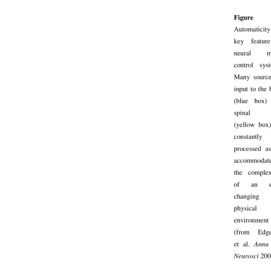
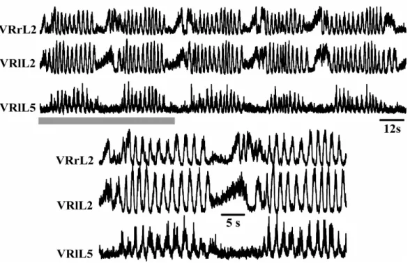
In the neonatal rat spinal cord, we have often observed brief episodes of spontaneous
alternating activity, recorded from VRs during control conditions. Figure 6 shows
examples of alternating activity in the rat isolated lumbar cord (top traces) as well as
in the brain stem spinal cord preparation (bottom traces).
Figure 6. Spontaneous activity recorded from neonatal (P1) rats. In both isolated spinal cord (top traces; G Taccola unpublished data) and brainstem-spinal cord preparation (bottom trace) there are episodes of alternating activity between left-right L2 and left L2-5 (top insert) and between left-right L1 (upper trace; G Taccola and K Ballanyi unpublished data).
2.3. Fictive locomotion
The CPG is responsible for generating locomotor signals, that are electrical discharges
alternating between flexor and extensor motor pools and between left and right MN
within the same segment.
As Figure 7 shows, analogue motor signals are also recorded from isolated rat as well
as mouse spinal cord and are characterized by oscillations alternated between left and
right sides and between lumbar 2 and 5 (L2-L5) pairs of VRs. This phenomenon is
called Fictive Locomotion and is manifested as rhythmic oscillations exclusively from
VRs.
Some studies have left the preparation of the dissected spinal cord still connected to
one or two hind limbs to correlate neuronal activity with muscular output. In these
cases, when a locomotor rhythm is pharmacologically induced, it has been possible to
visualize real alternating movements of the hind limbs (Atsuta et al., 1988; Cazalets et
al., 1992). By keeping one leg attached to the spinal cord, Kiehn and Kjaerulff (1996)
have demonstrated that the rostral lumbar VRs (L1, L2 and L3) contain mostly axons
innervating flexor muscles, whereas the L5 segment contains MNs that determine the
contraction of extensor muscles.
Fictive locomotor signals are evoked by perfusing the isolated mammalian spinal cord
with excitatory agents or by repeated stimuli applied to one DR.
2.4. Chemically induced Fictive Locomotion
Many studies have shown that limb alternation or VR alternated discharges can be
induced by application of a variety of neurotransmitter agonists.
Glutamate and aspartate as well as the EAA agonists NMDA and kainate, induce
locomotor activity with a left-right, alternating pattern (Kudo and Yamada 1987;
Cazalets et al., 1992). AMPA cannot induce fictive locomotion in rats probably due to
desensitization of this receptor class (Dingledine et al., 1999). Moreover, either
NMDA or non-NMDA glutamate receptors are activated during locomotor-like
activity since, when NMDA and non-NMDA receptors are simultaneously blocked,
there is no fictive locomotion (Kudo and Yamada 1987; Cazalets et al.1992).
However, Beato and his colleagues have been able to elicit stable locomotor activity
in the presence of, NMDA or non-NMDA receptor antagonism respectively, showing
that the operation of one glutamate receptor class is sufficient to express locomotor
activity (Beato et al., 1997).
The literature contains other examples of locomotor network operation occurring
independent of NMDA receptor activation. Recently, Cowley and his colleagues
(2005) have demonstrated that, although NMDA receptor activation may appear
essential for locomotor network operation under some experimental conditions,
locomotor-like rhythms can, nevertheless, be generated in the presence of AP5.
5-HT is also able to induce a locomotor pattern: the intensity of the bursts is stronger,
suggesting an activation of a larger pool of MNs, while the rhythm frequency is much
slower (Cazalets et al.1992). The serotonin-induced rhythm can be blocked by a
5-HT
1antagonist (propranolol) and by 5-HT
2antagonists (kentanserin, myenserin and
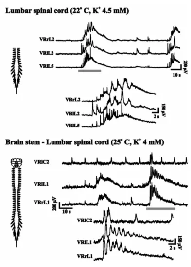
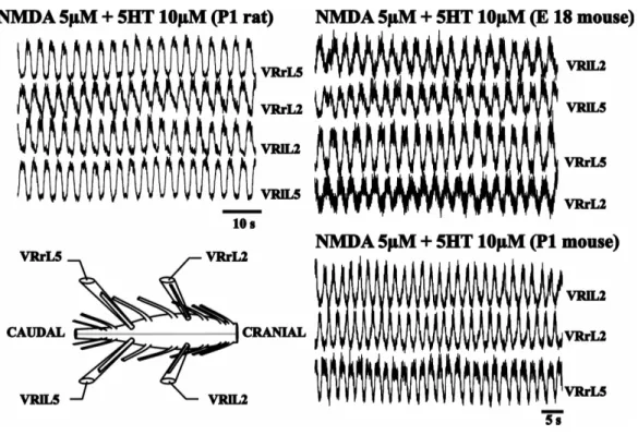
In contrast, if 5-HT and NMDA are applied together, the discharges are much more
regular and the rhythm has a frequency which is intermediate between those obtained
with each substance applied separately: this activity remains very stable for a long
time (Fig 7., Sqalli-Houssaini et al.1993).
Figure 7. Chemically induced fictive locomotion in rat (right) and in mouse (left) preparations (left bottom). Stable rhythmic oscillations alternated between left and right lumbar roots (L2 or L5) and between L2 and L5 homolateral roots are recorded in the presence of NMDA (5µM) and 5HT (10µM; from G Taccola, unpublished data).
Sqalli-Houssaini and Cazalets (2000) have described how bath application of
noradrenaline (NA) elicits an extremely slow alternating motor pattern. The
alternation is clear between right and left sides, but not between flexors and extensors
on the same side. The role of NA could be mimicked by the receptor agonists
methoxamine and phenylephrine, whereas and receptor agonists alone do not elicit
any activity. Kiehn and Kjaerulff (1996) have characterized the fictive rhythms
induced by 5-HT and dopamine. The dopamine-induced rhythm is more irregular and
slower. Kiehn and Kjaerulff (1996) have shown that 5-HT has a tendency to facilitate
flexor drive, whereas dopamine may exaggerate extensor activity.
Acetylcholine (ACh) combined with the acetylcholinesterase inhibitor edrophonium,
usually elicits sustained rhythmic activity characterized by left-right alternation and
non-locomotor co-activation of ipsilateral flexor and extensor discharges (Cowley and
Schmidt, 1994). However, more recent experiments have shown that, if low levels of
cholinergic stimulation are used, transient episodes of rhythmic activity with a
locomotor-like pattern can be evoked (Jordan and McVagh 2004).
The role of GABA has also been studied (Cazalets et al.1994; Cowley and Schmidt
1995; Kremer and Lev-Tov 1998). Bath-applied GABA
Aor GABA
Bagonists decrease
the frequency of the rhythm and, depending upon the dose used, they can completely
suppress activation previously obtained with the NMDA-5HT mixture. Similarly, the
GABA uptake inhibitors nipecotic acid or guvacine, suppress the rhythmic pattern.
The nipecotic response can be reversed by bicuculline or phaclofen.
Bracci and co-workers (1998) have demonstrated that high external K
+induces
alternating motor patterns only within a very narrow range of concentrations. The
alternated pattern can be evoked also in the presence of NMDA, non-NMDA and
5-HT antagonism suggesting that, in the neonatal rat spinal cord, the CPG operation
seems to require a widespread increase in neuronal excitability rather than selective
activation of a distinct subgroup of spinal neurons. A clear answer to this question is
complicated by the fact that the CPG has not been precisely identified in anatomic
terms as discussed below.
2.5. Localization of CPG
Regarding the localization of the CPG responsible for locomotor activity recorded
from in vitro preparations of the mammalian spinal cord, there are conflicting and
contrasting results. Some bath-partition studies point out that the hindlimb locomotor
CPG is not segmentally distributed, but confined to a restricted upper lumbar region
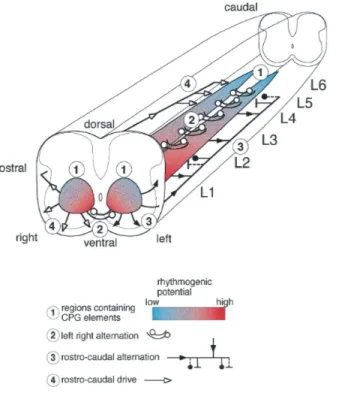
(Cazalets et al., 1995). Nevertheless, the current consensus arising from spinal
ablation experiments, indicates that the rhythmogenic network controlling leg
movements is distributed within all lumbar segments (Fig. 8; Kjaerulff and Kiehn,
1996). The latter view is in accordance with that of Bracci et al. (1996 a) who have
found that the mechanisms for burst triggering require a relatively small neuronal
network, confined to a ventral quadrant, though distributed among all segments.
In the cat and in the rat, activity-dependent neuronal labeling has suggested that the
medial part of the intermediate gray and the area around the central canal are
important for rhythm-generation during locomotion (Viala et al., 1991; Kjaerulff et
al., 1994; Kjaerulff and Kiehn, 1996).
A key element in all locomotor CPGs is the left–right coordination, that ensures that
limbs on opposite sides of the body alternate (walking) or are activated in synchrony
(hopping). The CPG elements controlling left–right coordination necessarily include
commissural interneurons (CINs): these neurons send axons to cross the midline to
form synapses onto MNs and/or other interneurons (including CINs) situated in the
contralateral hemicord.
In the neonatal spinal cord in vitro, Butt and Kiehn (2002) have shown that in the
medial VII, VIII, X laminae of the lumbar cord there is a significant concentration of
interneurons with diverse projections crossing the midline. In a later study, using an
electrophysiological approach, the same authors have identified such neurons as
functionally responsible for left-right coordination of hindlimbs (Butt and Kiehn,
2003).
Figure 8. Summary of CPG localisation. The rhythm-generating network in L1–L6 is shown distributed along the cord as two medial columns (1). The taper and the color gradient indicate the high rostral and lower caudal ability to generate rhythmic activity. The localization of the pathways mediating left/right alternation in the ventral commissure is indicated (2). The pathways mediating rostrocaudal alternation are shown widely distributed in the lateral and ventral funiculus on the left side of the preparation (3 The rostrocaudal drive is indicated on the right side of the cord (4); from Kjaerulff and Kiehn J Neurosci, 1996)
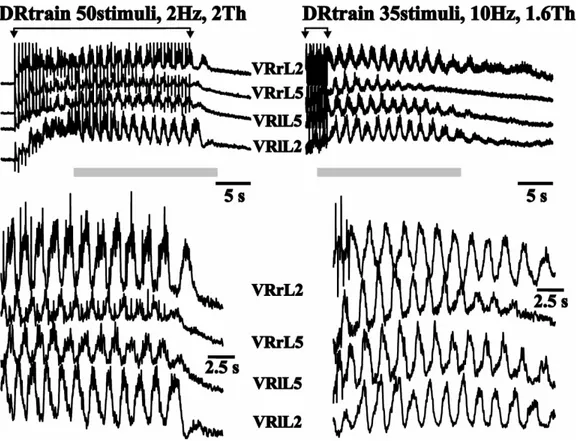
2.6. Electrically evoked Fictive Locomotion
In the neonatal rat spinal cord, rhythmic locomotor-like patterns can be elicited by
bath-applied excitatory substances like NMDA (Kudo & Yamada, 1987), 5-HT
(Cazalets et al., 1992; Beato et al., 1997) or high potassium (Bracci et al., 1998).
Although this approach has yielded important insights into the mode of operation of
the fictive locomotor network, persistent bath application of these excitatory
substances to the entire spinal tissue represents a non-physiological condition.
Marchetti and co-workers (2001) have suggested that, in the neonatal rat spinal cord
in vitro, locomotor-like patterns of activity can be reliably evoked by trains of stimuli
applied to one DR (Fig. 9). This condition might thus mimic physiological activation
of the CPG by sensory inputs. The oscillatory activity induced by DR stimulation
presents the typical phase alternation at segmental and intersegmental level which is a
hallmark of fictive locomotion evoked by chemical substances (Kiehn et al., 1997).
Figure 9. Each panel shows DC records of responses from pairs of VRs (left or right of L2 and L5). Oscillations are in alternation between contralateral VRs and between intersegmental VRs of the same side. In two different preparations, alternating oscillations outlasting the last stimulus are induced by two different protocols of electrical stimulation (number of stimuli, frequency and intensity) applied to L5 right DR (from G Taccola, unpublished data).
Such alternating discharges of MNs are likely to be driven by the CPG and can be
regarded as a locomotor-like pattern turned on by the activation of dorsal afferents
which are known to project to the CPG itself (Hultborn et al., 1998).
Unlike the persistent and stable fictive locomotor pattern elicited by neurochemicals
(Kiehn et al., 1997), the DR-induced one is usually transient. Loss of rhythmicity
might be due to either fatigue in the pathways upstream of the CPG (as observed with
afferent impulses in the presence of strychnine and bicuculline; Bracci et al., 1997) or
stimulus-dependent release of transmitters which inhibit the CPG operation.
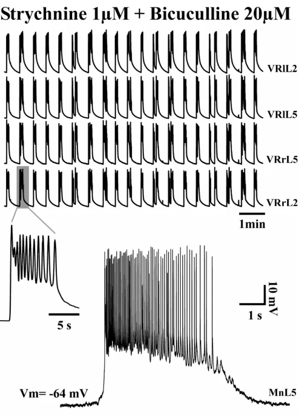
2.7. Disinhibited Rhythm
In the neonatal rat spinal cord, network-driven rhythmic bursting arises spontaneously
after applying saturating concentrations of strychnine and bicuculline, which block
GABA and glycine receptors respectively (Fig. 10). This rhythm, called disinhibited
rhythm, is unphysiological; however, blocking fast, chloride-mediated inhibition
largely simplifies the rhythmogenic network. Furthermore, disinhibited rhythmicity,
by using excitatory connections only, resembles the early type of collective network
bursting (Sernagor et al., 1995).
After dorsal horn removal from the neonatal spinal cord of rat, the remaining network,
localized to the ventral horn, can still perform rhythmic activity in the absence of
synaptic inhibition (Bracci et al., 1996 a).
To comprehend the role of inhibition in the genesis of the fictive locomotor pattern, it
is important to answer the question of whether disinhibited rhythm and fictive
locomotion are generated by the same network (Bracci et al., 1996 a; Nishimaru &
Kudo, 2000). Evidence favors this hypothesis, as Bracci et al. (1996 a; b) have found
that disinhibited bursting shares with locomotor patterns the same sensitivity to block
of either class of ionotropic glutamate receptors (Beato et al., 1997; Bracci et al., 1996
a) and the same anatomical location (Bracci et al., 1996 a; Kjaerulff & Kiehn, 1996).
In addition, this rhythm can be accelerated by 5-HT (Bracci et al., 1996 a), known to
activate fictive locomotor patterns (Cazalets et al., 1992), and a strong synergy
between disinhibited rhythm and locomotor pattern is demonstrated with the use of
the split bath configuration (Beato & Nistri, 1999).
Observations made on the organotypic spinal slice culture have further supported the
possibility that a common network is involved in generation of the two rhythms. In
fact, it has been shown that bursting induced by disinhibition or by increased
excitability are composed of similar waves and involve the same areas of the slice
(Tscherter et al., 2001).
Figure 10. Synchronous rhythmic bursts evoked by pharmacological blockade of synaptic inhibition could be recorded from all lumbar VRs (left, right L2 and L5; top traces). DC-coupled extracellular recording of single burst (left insert) presents similar bursting pattern and structure as observed from intracellular recording of lumbar MN (bottom trace; from G Taccola, unpublished data).