Chronic Local Therapy for Brainstem Tumors
To the Editor:Opinions have been reported about the value and safety of chronic infusions for brainstem tumor therapy. These opinions may influence planning for clinical trials for a disease that relentlessly claims the lives of approximately 200 children yearly in the United States alone.
Median survival for children with diffuse pontine gliomas is approximately 10 months (1). Trials with small numbers of patients from single institutions have generated the consensus that neither chemotherapy nor radiation prolongs survival and that new approaches are needed (8, 9).
Local therapy seems to be a logical response (7). Two strat-egies are available. Both have demonstrated efficacy in animal studies. Both have provided baseline safety data via human trials in supratentorial tumors (5). The methods differ in their rate of intratumoral delivery. Fast infusions pump at rates of 1l/min or more. Convective gradients distribute molecules homogeneously several centimeters from the point of deliv-ery. Slow infusions release drugs from biodegradable im-plants or from pumps at rates of 1l/h or less. Drugs diffuse several millimeters from the point of delivery in normal tis-sues (6, 11, 12, 16).
Conjectures about the clinical irrelevance of slow infusions derive from the opinion that distributive capacities are too small to invade the tumor volumes generally encountered in clinical practice. Some have reported that the modest “statis-tical” gains seen in clinical trials with slow infusions further attest to the irrelevance of this strategy (7, 23).
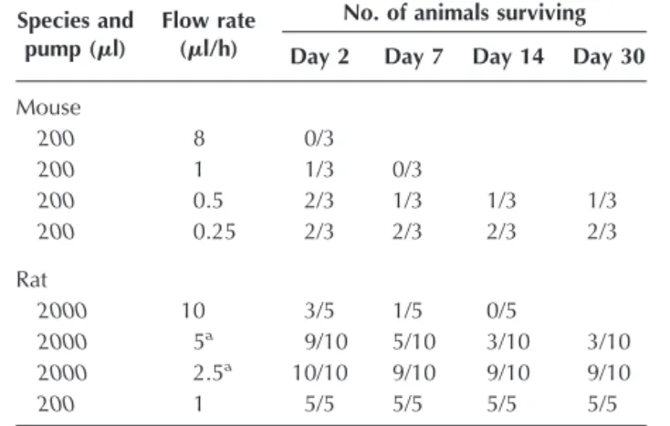
Laboratory models have demonstrated the safety of fast brainstem infusions in normal tissues (15, 19, 21). Questions remain about clinical safety in abnormal tissue. Elegant and technically challenging studies in a rat supratentorial glioma model have demonstrated that intracranial pressure increases with increasing tumor volumes as well as the volume and flow rate of a fast infusion (4). Data in Table C1 show the lethal effects of increasing infusion rates in the brainstems of normal mice and rats. The data on rat infusions have been published (6). We think that it is important to add the unpublished mouse data to further illustrate the stark relationship between morbidity and increasing flow rates. Brainstem infusions of saline in 180- to 220-g Fischer rats at 10l/h were lethal in four of five rats in 7 days. Although a child’s brainstem is significantly larger, fast chronic infusion in a brainstem poten-tially challenged by increased pressures from a glioma could lead to unintended events.
Fast infusions with targeted toxins have shown promise in Phase I trials with supratentorial tumors (13, 17, 20). Increased intracranial pressure, whether related to drug toxicity or tu-mor necrosis, seemed to be manageable. However, there is a lack of information about the management of increased pres-sure in the brainstem (2). Children are a vulnerable population (14). Trials will require an independent committee to monitor
treatment effects (10, 18). With the information at hand, the committee could not distinguish potentially toxic effects of increased pressure from the progression of the disease.
Clinical trials can begin with slow infusions. Theoretical equipoise, the null hypothesis, states that there will be no difference between the infusion of saline, drug escalation, and palliative treatment for a child with a pontine glioma. Infu-sions at 0.42l/h of 1 ml of saline (90 d) have been performed with no apparent toxicity (23). Regardless of the initial skep-ticism about volumes of distribution from slow infusions, the multiple reports of efficacy shown in human trials demon-strate the clinical relevance of this approach in neurosurgical oncology (3, 22, 24–26). Short-term methodological safety has been documented. Side effects, which frequently blunt sys-temic drug trials, have been greatly reduced. Clinical equi-poise considers both the risks and benefits of a new therapy. The safety and efficacy of slow infusions have been achieved with first-generation chemotherapeutics such as carmustine and cisplatin. Randomized controlled trials can continue the search for progress using faster infusion rates and newer generations of chemotherapeutics, including targeted toxins.
Michael Guarnieri Benjamin S. Carson, Sr. Baltimore, Maryland
1. Abbott R, Goh KYC: Brainstem gliomas, in Albright L, Pollack I, Adelson D (eds): Principles and Practices of Pediatric Neurosurgery. New York, Thieme Medical Publishers, 1999, pp 629–640.
2. Avellino AA, Carson BS Sr: Increased intracranial pressure, in Maria BL (ed): Current Management in Child Neurology. New York, B.C. Decker, 2002, pp 481–486.
TABLE C1. Survival and infusion flow rates in rat and mice brainstems
Species and pump (l)
Flow rate (l/h)
No. of animals surviving Day 2 Day 7 Day 14 Day 30 Mouse 200 8 0/3 200 1 1/3 0/3 200 0.5 2/3 1/3 1/3 1/3 200 0.25 2/3 2/3 2/3 2/3 Rat 2000 10 3/5 1/5 0/5 2000 5a 9/10 5/10 3/10 3/10 2000 2.5a 10/10 9/10 9/10 9/10 200 1 5/5 5/5 5/5 5/5
aTreated with 1 mg dexamethasone per day intraperitoneally for 3 days
3. Brem H, Piantadosi S, Purger PC, Walker M, Selker R, Vick NA, Black K, Sisti M, Brem S, Mohr G, Muller P, Morawetz R, Schold SC: Placebo-controlled trial of safety and efficacy of intraoperative Placebo-controlled delivery by biodegradable polymers of chemotherapy for recurrent gliomas. Lancet 345:1008–1012, 1995.
4. Bruce JN, Falavigna A, Johnson JP, Hall JS, Birch BD, Yoon JT, Wu EX, Fine RL, Parsa AT: Intracerebral clysis in a rat glioma model. Neurosurgery 46:683–691, 2000.
5. Carson BS Sr, Guarnieri M: Local therapy for brain tumors. Adv Clin
Neurosci12:89–99, 2002.
6. Carson BS, Wu QZ, Tyler B, Sukay L, Raychaudhuri R, DiMeco F, Clat-terbuck R, Olivi A, Guarnieri M: New approach to tumor therapy for inoperable areas of the brain: Chronic intraparenchymal drug delivery.
J Neurooncol60:151–158, 2002.
7. Dunn IF, Black PMcL: The neurosurgeon as a local oncologist: Cellular and molecular neurosurgery in malignant glioma therapy. Neurosurgery 52: 1411–1424, 2003.
8. Freeman CR: Hyperfractionated radiotherapy for diffuse intrinsic brain stem tumors in children. Pediatr Neurosurg 24:103–110, 1996.
9. Freeman CR, Perilongo G: Chemotherapy for brain stem gliomas. Childs
Nerv Syst15:545–553, 1999.
10. Freidman LM, Furberg CD, DeMets DL: Fundamentals of Clinical Trials. New York, Springer-Verlag, 1998.
11. Fung LK, Ewend MG, Aills A, Sipos EP, Thompson R, Watts M, Colvin OM, Brem H, Saltzman WM: Pharmacokinetics of interstitial delivery of carmustin, 4-hydroperoxycyclophosphamide, and paclitaxel from a biode-gradable polymer implant in the monkey brain. Cancer Res 58:672–684, 1998.
12. Kroin JS, Penn RD: Intracerebral chemotherapy: Chronic microinfusion of cisplatin. Neurosurgery 10:349–354, 1982.
13. Laske DW, Youle RJ, Oldfielfd EH: Tumor regression with regional distri-bution of the targeted toxin TF-CRM107 in patients with malignant brain tumors. Nat Med 3:1362–1368, 1997.
14. Levine RJ: Ethics and Regulations of Clinical Research. New Haven, Yale University Press, 1988.
15. Lonser RR, Walbridge S, Garmestani K, Butman JA, Walters HA, Vortmeyer AO, Morrison PF, Brechbiel MW, Oldfield EH: Successful and safe perfusion of the primate brainstem: In vivo magnetic resonance imaging of macromo-lecular distribution during infusion. J Neurosurg 97:905–913, 2002. 16. Lum JT, Nguyen T, Felpel LP: Drug distribution in solid tissue of the brain
following chronic local perfusion utilizing implanted osmotic minipumps.
J Pharmacol Methods12:141–147, 1984.
17. Mardor Y, Roth Y, Lidar Z, Jonas T, Preffer R, Maier SE, Faibel M, Na D, Hadani M, Orenstein A, Cohen JS, Ram Z: Monitoring response to convection-enhanced taxol delivery in brain tumor patients using diffusion-weighted magnetic resonance imaging. Cancer Res 61:4971–4973, 2001. 18. Meinert CL: Clinical Trials: Design, Conduct, and Analysis. New York, Oxford
University Press, 1986.
19. Occhiogrosso G, Edgar MA, Sandberg DI, Souweidance MM: Prolonged convection-enhanced delivery into the rat brainstem. Neurosurgery 52:388– 394, 2003.
20. Rand RW, Kreitman RJ, Patronas N, Varricchio F, Pastan I, Puri RJ: Intratumoral administration of recombinant circularly permuted interleukin-4-Pseudomonas exotoxin in patients with high-grade gliomas.
Clin Cancer Res6:2157–2165, 2000.
21. Sandberg DI, Edgar MA, Souweidane MM: Convection-enhanced delivery into the rat brainstem. J Neurosurg 96:885–891, 2002.
22. Sheleg SV, Korotkevich EA, Zhavrid EA, Muravskaya GV, Smeyanovich AF, Shanko YG, Yurkshtovich TL, Bychkovsky PB, Belyaev SA: Local chemo-therapy with cisplatin-depot for glioblastoma multiforme. J Neurooncol 60:53–59, 2002.
23. Storm PB, Clatterbuck RE, Liu YJ, Johnson RM, Gillis EM, Guarnieri M, Carson BS Sr: A surgical technique for safely placing a drug delivery catheter into the pons of primates: Preliminary results of carboplatin infu-sion. Neurosurgery 52:1169–1177, 2003.
24. Valtonen S, Timonen U, Toivanen P, Kalimo H, Kivipelto L, Heiskanen O, Unsgaard G, Kuurne T: Interstitial chemotherapy with carmustine-loaded poly-mers for high-grade gliomas: A randomized double-blind study. Neurosurgery 41:44–49, 1997.
25. Wenig BL, Werner JA, Castro DJ, Sridhar KS, Garewal HS, Kehrl W, Pluazanska A, Arndt O, Costantino PD, Mills GM, Dunphy FR, Orenberg EK, Leavitt RD: The role of intratumoral therapy with cisplatin/epinephrine injectable gel in the management of advanced squamous cell carcinoma of the head and neck. Arch Otolaryngol Head Neck Surg 128:880–885, 2002. 26. Westphal M, Hilt DC, Bortey E, Delavault P, Olivares R, Warnke PC, Whitle IR, Jääskeläinen J, Ram Z: A phase 3 trial of local chemotherapy with biodegradable carmustine (BCNU) wafers (Gliadel wafers) in patients with primary malignant glioma. Neuro-oncology 5:79–88, 2003.
In Reply:
Drs. Guarnieri and Carson raise important issues regarding local therapy for brainstem tumors (1). Convection-enhanced drug delivery is rapidly being validated as a safe and useful strategy for delivery of antitumor compounds to brain tumors. This has led to examination of its potential application for tumors in more anatomically hazardous areas, including the brainstem.
Morbidity from local intratumoral infusion can come from either direct drug toxicity, which is influenced by local drug concentration and duration of infusion, or mass effect, which is influenced by volume delivered and preexisting mass effect from the tumor itself. With brainstem lesions, Drs. Guarnieri and Carson are particularly concerned with infusion rates and volumes associated with convection-enhanced delivery and present evidence that increased flow rates can lead to signif-icant morbidity in rodent models. Their work has led them to advocate for more chronic infusions at considerably lower flow rates than those that produce convective forces. Their letter highlights the difficulties in interpreting experimental studies in animals, in which brain volumes are a fraction of human brain volumes.
Given the limitations of animal models, I agree with the need to examine local delivery parameters more fully in clin-ical trials incorporating careful empirclin-ical protocol designs. Intratumoral infusion studies will need to focus on three vari-ables: 1) drug concentration, 2) infusion rate, and 3) duration of infusion (which along with infusion rate determines vol-ume of distribution). It would be prudent to begin these studies slowly, using safe parameters that could be increased gradually. These types of study designs are similar to what has traditionally been used when testing new systemic chemotherapies.
Given the limited treatment options available for patients with malignant brainstem tumors, it is reasonable to propose that these trials begin now. Experienced teams of investigative neurosurgeons, neuro-oncologists, and radiologists will be needed to devise reasonable treatment protocols with careful dose and infusion escalation parameters that will ultimately determine whether convection-enhanced or chronic slow in-fusion can be efficacious for brainstem pathology. These stud-ies would be greatly assisted by noninvasive radiological im-aging to monitor treatment end points for safety and response. Monitoring may be the greatest challenge, because early
clin-ical trials with convection-enhanced delivery have been hin-dered by the difficulties in interpreting radiographic imaging studies of these heterogeneous tumors, which have often un-dergone previous surgical, radiotherapeutic, and chemother-apeutic intervention.
Jeffrey N. Bruce New York, New York
1. Bruce JN, Falavigna A, Johnson JP, Hall JS, Birch BD, Yoon JT, Wu EX, Fine RL, Parsa AT: Intracerebral clysis in a rat glioma model. Neurosurgery 46:683–691, 2000.
DOI: 10.1227/01.NEU.0000117119.32806.AF
The Impact of Provider Volume on Mortality after
Intracranial Tumor Resection and Outcome and Cost of
Craniotomy Performed to Treat Tumors in Regional
Academic Referral Centers
To the Editor:
There is growing evidence that patients with intracranial neo-plasms should be treated in highly specialized neurosurgical centers. This statement was clearly confirmed by the authors of both referenced articles (1, 2), who found direct correlation be-tween the volume of the neurosurgical activity for brain tumors and early outcome, namely, mortality rate and length of in-hospital stay. Several years ago, we reached a similar conclusion in our study of the same problem. However, despite publication in a MEDLINE-cited journal (3), our results seem to be obscured from the neurosurgical community, and therefore, it may be of interest to highlight our study briefly.
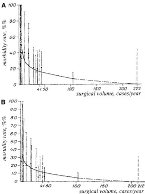
We performed a population-based study of the outcome after surgical removal of primary intracranial tumors in adult citizens of St. Petersburg, Russia. Patients with met-astatic and recurrent neoplasms had been excluded from the analysis. The early outcome in all patients (n⫽ 307) who were treated during 1993 was identified retrospectively, with an emphasis on postoperative morbidity and mortal-ity. There were 148 men and 159 women (mean age, 48 yr). At the time of hospital admission, the condition of 54.7% of the patients was considered to be compensated, 31.9% sub-compensated, and 13.4% decompensated. Gliomas were en-countered most frequently (n⫽ 134), followed by meningi-omas (n ⫽ 87), pituitary adenomas (n ⫽ 39), and acoustic schwannomas (n⫽ 15). Surgical treatment was performed in 13 different neurosurgical departments. The mean vol-ume of the surgical activity, defined as number of brain tumor removals per year, was 23.6 (range, 3–104 opera-tions). The mean morbidity rate constituted 19.5% (range, 6.7–66.7%). The mean mortality rate was 9.4% (range, 0–33.3%). A strong dependence of the morbidity and mor-tality rates on the surgical volume was found (Fig. C1). The mathematically defined breakpoint of both curves corre-sponded to the surgical volume of 41 cases/year.
Compar-ison of the low-volume hospitals (surgical volume ⬍41 cases/yr) and high-volume hospitals (surgical volume⬎41 cases/yr) revealed relatively worse clinical condition of the patients and higher frequency of critical tumor location in the latter group, whereas the mean age did not differ sig-nificantly. The morbidity curve reached its minimal value at the surgical volume of 223 cases/year, whereas the mortal-ity curve reached its minimal value at 212 cases/year.
It seems that our results, which are in complete concordance with the conclusions of the referenced authors, may carry some additional information to the interested reader. First, not only mortality rate but also morbidity rate after removal of brain tumors depends on the volume of surgical activity, which prob-ably reflects the lower average length of in-hospital stay at the high-volume centers found by Long et al. (2). Second, in our study, better results in high-volume neurosurgical departments were obtained despite the relatively worse clinical condition of patients and more frequent critical tumor location. Therefore, better outcome in hospitals with higher surgical volume seems not to correspond to more preferable patient or tumor character-istics. Third, we found that not only the results of surgery for brain tumors but also the quality of the adjunctive neuro-oncological treatment (chemotherapy) is lower in low-volume hospitals (data not shown here).
FIGURE C1. Graphs depicting the morbidity rate (A) and mortality rate
(B) after surgical removal of intracranial tumors versus the volume of sur-gical activity of the neurosursur-gical department (adapted from, Tigliev GS, Ulitin AYu, Chernov MF: The dependence of the results of the surgical treatment of patients with primary intracranial tumors on the volume of surgical activity of the neurosurgical department [exemplified by Saint Petersburg, Russia] [in Russian]. Zh Vopr Neirokhir Im N N Burdenko 2:44–46, 1999 [3]).
Another point that seems to be extremely interesting is that nearly the same surgical volume corresponded to mathemat-ically defined breakpoints of both morbidity and mortality curves in our study (41 cases/yr) and defined for categoriza-tion of low- and high-volume hospitals by Long et al. (2) (50 cases/yr). Moreover, the curves we constructed reached their minimal values at a surgical volume of more than 200 cases/ year, which corresponds to a “high volume of surgical activ-ity” (as defined by authors of both referenced articles) associ-ated with the better outcome. If such similar data were obtained in such different sociological and medical communi-ties, can we consider this similarity as an objective tendency that may be used in the future for better organization of the neurosurgical and neuro-oncological services?
In conclusion, it seems that treatment of patients with in-tracranial neoplasms in the neurosurgical centers with a high volume of surgical activity can provide lower morbidity and mortality rates compared with low-volume hospitals. How-ever, the dependence of other important outcomes of brain tumor management (for example, radicality of tumor removal, long-term patient survival, and quality of life) on the surgical volume is still unknown. Therefore, I am looking forward to further contributions on this topic.
Mikhail F. Chernov Tokyo, Japan
1. Cowan JA Jr, Dimick JB, Leveque J-C, Thompson BG, Upchurch GR Jr, Hoff JT: The impact of provider volume on mortality after intracranial tumor resection. Neurosurgery 52:48–54, 2003.
2. Long DM, Gordon T, Bowman H, Etzel A, Burleyson G, Betchen S, Garonzik IM, Brem H: Outcome and cost of craniotomy performed to treat tumors in regional academic referral centers. Neurosurgery 52:1056–1065, 2003. 3. Tigliev GS, Ulitin AYu, Chernov MF: The dependence of the results of the
surgical treatment of patients with primary intracranial tumors on the volume of surgical activity of the neurosurgical department (exemplified by Saint Petersburg, Russia) [in Russian]. Zh Vopr Neirokhir Im N N
Burdenko2:44–46, 1999.
In Reply:
We appreciate the comments of Dr. Chernov and his pre-vious contributions, which described a volume-outcome effect after surgical removal of primary intracranial tumors in St. Petersburg, Russia. His work reminds us that variation in outcome after the delivery of complex neurosurgical care is not unique to healthcare systems in the United States.
As mentioned in our article (1), “high” caseload volume has been shown to be a consistent marker for superior outcomes after complex surgery. Increasing hospital vol-ume alone will not guarantee better outcomes (even though, on average, these high-caseload centers have better results). We therefore would refrain from applying specific caseload thresholds but rather would focus on the organizational structures and processes of care that improve outcome, including intensive care unit physician staffing, low nurse-to-patient ratios, access to neuroimaging, optimization of
comorbid conditions, and a multidisciplinary approach to care. These organizational components may currently be found more often at high-volume centers. Whether they are unique to high-volume centers is unknown.
John A. Cowan, Jr. B. Gregory Thompson Julian T. Hoff
Ann Arbor, Michigan
1. Cowan JA Jr, Dimick JB, Leveque J-C, Thompson BG, Upchurch GR Jr, Hoff JT: The impact of provider volume on mortality after intracranial tumor resection. Neurosurgery 52:48–54, 2003.
DOI: 10.1227/01.NEU.0000117120.32806.22
Far Posterior Subtemporal Approach to the
Dorsolateral Brainstem and Tentorial Ring: Technique
and Clinical Experience
To the Editor:
It was with great interest that I read the description of the far posterior subtemporal approach (4). It closely resembles another modification of the subtemporal approach, namely, the transpetrous supratranstentorial-anterior approach, which has been used in our clinic since 1992 in more than 70 cases of various tumors of the tentorial notch area (1, 5).
The positioning of the patient, direction of the surgical access, and main steps of the approach we used are the same as de-scribed by the authors. We strongly agree with the importance of the head tilt below the horizontal position (which we prefer to do after opening of the dura), mobilization of the vein of Labbé, use of the lumbar or ventricular drain (to our mind, the latter is preferable in cases with associated hydrocephalus), mediotem-poral retraction for the visualization of the tentorial notch, and laterally directed tentoriotomy, starting from the tentorial edge and proceeding parallel to the superior petrosal sinus. In addi-tion, it should be emphasized that the increase of the retraction force on the basitemporal lobe must be as gradual as possible, with steady evacuation of the cerebrospinal fluid (CSF) after early opening of the basal subarachnoid cisterns. I need not mention the advantages of adequate neuroanesthesia, which pro-vides optimal brain compliance. All of these measures are of paramount importance for the prevention of complications caused by temporal lobe retraction.
Although the risk of injury to the vein of Labbé should not be underestimated, it should also not be overemphasized. We did not observe this complication in any case in our series, because it seems that careful mobilization of the vessel from the subarachnoid space and dural duplication provides suffi-cient mobility for safe retraction of the temporal lobe and further surgical manipulations.
However, it should be noted that the amount of retraction necessary during the subtemporal approach to the region of the tentorial notch depends on the individual peculiarities
of the neuroanatomy, namely, the angle between the tento-rium and the upper plane of the petrous bone. The smaller the angle, the larger the retraction force needed. In this way, the avoidance of complications can be provided by resec-tion of the posterolateral part of the petrous bone up to the level of the external acoustic meatus (Fig. C2). Such limited resection is not associated with the risk of damage of the intrapetrous structures but increases the angle of the surgi-cal access 60% on average (3) and therefore should be kept in mind if the retraction force on the temporal lobe during the subtemporal approach seems to be too high. Although further extension of the resection on the glenoid fossa has been proposed (2), it seems to be unnecessary in the vast majority of cases.
Finally, the authors have provided a good description of the modified subtemporal approach, which represents one of the main routes to the region of the tentorial notch. The only concern I have is about the proposed name of the approach. Because the lateral border between the temporal and occipital lobes is assumed to be the preoccipital notch, which lies a bit posterior to the termination of the vein of Labbé, it seems that “midsubtemporal” rather than “far posterior subtemporal” is a more appropriate definition of the access.
Mikhail F. Chernov Tokyo, Japan
1. Chernov MF: Basal meningiomas with both supra- and infratentorial exten-sion: Clinical picture, diagnosis, management [in Russian]. St. Petersburg, Russian Polenov Neurosurgical Institute, 1995 (dissertation).
2. Goel A: Extended lateral subtemporal approach for petroclival meningio-mas: Report of experience with 24 cases. Br J Neurosurg 13:270–275, 1999. 3. Sindou MP, Fobe JL: Removal of the roof of the external auditory meatus in approaching the tentorial notch through a low temporal craniotomy: Tech-nical note. J Neurosurg 74:520–522, 1991.
4. Smith ER, Chapman PH, Ogilvy CS: Far posterior subtemporal approach to the dorsolateral brainstem and tentorial ring: Technique and clinical expe-rience. Neurosurgery 52:364–369, 2003.
5. Tigliev GS, Chernov MF: The characteristics of the surgical treatment of basal meningiomas with supra- and subtentorial spread [in Russian]. Zh
Vopr Neirokhir Im N N Burdenko1:3–6, 1998.
In Reply:
We thank Dr. Chernov for his thoughtful comments regard-ing the surgical approach to the posterior lateral brainstem region (1). He is indeed correct that it closely resembles the transpetrous supratranstentorial-anterior approach. As pointed out by Dr. Chernov, as familiarity with this approach is gained, one can mobilize the vein of Labbé. This is one of our most feared potential complications; however, if handled with care, the vein can remain intact and complications asso-ciated with venous manipulation can be avoided.
Christopher S. Ogilvy Paul H. Chapman Boston, Massachusetts
1. Smith ER, Chapman PH, Ogilvy CS: Far posterior subtemporal approach to the dorsolateral brainstem and tentorial ring: Technique and clinical expe-rience. Neurosurgery 52:364–369, 2003.
DOI: 10.1227/01.NEU.0000117121.32806.6B
Seizures after Aneurysmal Subarachnoid Hemorrhage
Treated with Coil Embolization
To the Editor:
I read with interest the article by Byrne et al. (1) on seizures after coil embolization of ruptured aneurysms and its relevance to current driving regulations in the United Kingdom. The au-thors successfully demonstrated that the actual rate of epilepsy after subarachnoid hemorrhage in the selected group is lower than previously thought. The interventional technique of treating aneurysms undoubtedly has the advantage of eliminating the inherent risk of epilepsy caused by craniotomy, which is added to that resulting from the subarachnoid hemorrhage itself. How-ever, the authors’ cohort over 10 years may perhaps have a selection bias whereby the patients selected also may be at low risk for epilepsy. Considering an increasing trend in the United Kingdom to treat ruptured aneurysms primarily by interven-tional means, more patients who are at high risk for epilepsy who previously would have been considered unsuitable are now likely to be considered for coiling. Therefore, the current and future patients treated interventionally may not necessarily fol-low the same pattern as demonstrated by the authors. It would be worthwhile to analyze whether the same low rate of epilepsy FIGURE C2. Scheme of the transpetrous supratranstentorial-anterior
approach: tumor in the region of the tentorial notch (1), skin incision (2), burr holes (3), external acoustic meatus (4), resected posterolateral portion of the petrous bone (5), direction of the surgical access (6), and vein of Labbé (7) (adapted from, Chernov MF: Basal meningiomas with both supra and infratentorial extension: Clinical picture, diagnosis, manage-ment [in Russian]. St. Petersburg, Russian Polenov Neurosurgical Insti-tute, 1995 [dissertation] [1]).
continues over the next 1 or 2 decades before any definitive conclusions can be drawn.
In our center, we do not use prophylactic anticonvulsant therapy in any patients with subarachnoid hemorrhage, and we agree entirely with the authors’ conclusion of not admin-istering prophylactic anticonvulsant medications. We also share an experience of a low incidence of epilepsy similar to that noticed by the authors but including the aneurysms treated by surgery. My own personal experience of the last 100 cases of surgically treated acutely ruptured aneurysms shows a 3% incidence of postoperative epilepsy (unpublished obser-vations). Up to half of these patients are those with intracere-bral clots, middle cereintracere-bral territory aneurysms, multiple an-eurysms, and giant or difficult aneurysms considered technically unsuitable for coiling. All of these factors, I believe, are epileptogenic themselves. Unfortunately, the results achieved in any single-institute or single-operator study may not reflect those of the entire population so as to warrant any changes in driving guidelines on a national level.
Recanalization and rebleeding from coiled aneurysms is another important factor that needs to be considered before a person with an apparently protected aneurysm is allowed to drive. It was recently shown that aneurysm recanalization is the major limitation of current Guglielmi detachable coil treat-ment (2). Another recently published study has also clearly established that initially incompletely occluded aneurysms, aneurysms treated in the acute phase after rupture, and the length of follow-up are independent significant predictors of major aneurysm recurrences that may possibly lead to a re-bleed (3). It is reasonable to believe that a rere-bleed during driving may carry significant morbidity and mortality, mak-ing it equally or perhaps more hazardous than a stand-alone seizure. Because most workers would prefer to treat recana-lized or recurred aneurysms before they rebleed, the precise rebleeding rate after acutely coiled aneurysms may never be known. Therefore, unless multicenter studies unequivocally confirm combined rates of long-term angiographic recanaliza-tion or recurrence rate as well as epilepsy after coiling lower than those currently considered acceptable for epilepsy alone, it would be hard to justify modifying the current driving legislation in our country for coiled cases only, as suggested by the authors.
Kishor A. Choudhari Belfast, United Kingdom
1. Byrne JV, Boardman P, Ioannidis I, Adcock J, Traill Z: Seizures after aneu-rysmal subarachnoid hemorrhage treated with coil embolization.
Neuro-surgery52:545–552, 2003.
2. Murayama Y, Nien YL, Duckwiler G, Gobin YP, Jahan R, Frazee J, Martin N, Viñuela F: Guglielmi detachable coil embolization of cerebral aneurysms: 11 years’ experience. J Neurosurg 98:959–966, 2003.
3. Raymond J, Guilbert F, Weill A, Georganos SA, Juravsky L, Lambert A, Lamoureux J, Chagnon M, Roy D: Long-term angiographic recurrences after selective endovascular treatment of aneurysms with detachable coils. Stroke 34:1398–1403, 2003.
In Reply:
Dr. Choudhari makes several interesting observations about current practice and advice to patients about driving motor vehicles after aneurysmal subarachnoid hemorrhage. His comments are well received and welcome. We entirely accept his points concern-ing selection bias and the limitations of sconcern-ingle-center studies such as ours (and his) (1). But the implications of the possibility of rebleed-ing of treated aneurysms require analysis. The experience of the past decade has shown that the vast majority of patients are protected against rebleeding by coil embolization, despite an appreciable an-giographic failure rate. The question is: does the latter warrant imposing driving restrictions more onerous than those for patients managed by surgical clipping?
Dr. Choudhari argues that current evidence is insufficient (in terms of epilepsy and rebleeding risks) to relax the driving restrictions “for coiled cases alone.” But the United Kingdom Driver and Vehicle Licensing Authority restrictions imposed, at the time our article was published, were more restrictive for aneurysmal subarachnoid hemorrhage patients managed by coil embolization than for those treated by craniotomy and clipping (for aneurysms other than at the middle cerebral artery). Guidance updated in January 2000 states that a Group 1 driver (i.e., nonprofessional) treated by embolization should “cease driving until full clinical recovery and undergo annual medical review for 4 years,” whereas the guidance for a pa-tient treated by craniotomy with no deficit states “driving permitted when clinically recovered from craniotomy” (i.e., without subsequent reviews). We are pleased to report that, after our publication, the advice for patients treated by coil embolization has been amended so that patients are now required only to “cease driving until clinically recovered” (2). The lack of clear thinking on this issue is exemplified by the advice to patients who sustain aneurysmal subarachnoid hem-orrhage and whose aneurysm is not treated by either clipping or coiling. Such patients are required to stop driving for 6 months only. We assume that the authority has taken the view that rebleeding after 6 months is infrequent and does not pose a risk to the patient or other road users. We would argue that to impose more onerous driving restrictions on embolized patients requires evidence to show that this form of management makes patients more liable to rebleeding or seizures than those treated by aneu-rysm clipping or managed conservatively.
James V. Byrne Jane Adcock Oxford, England
1. Byrne JV, Boardman P, Ioannidis I, Adcock J, Traill Z: Seizures after aneu-rysmal subarachnoid hemorrhage treated with coil embolization.
Neuro-surgery52:545–552, 2003.
2. Drivers Medical Group, Driver and Vehicle Licensing Agency: At a Glance
Guide to the Current Medical Standards of Fitness to Drive. Swansea, Driver and
Vehicle Licensing Agency, 2003.
Factors Related to Hydrocephalus after Aneurysmal
Subarachnoid Hemorrhage
To the Editor:
We read with interest the recent article by Dorai et al. (1). We were somewhat surprised by their finding that there was a highly significant (P ⬍ 0.001) increase in shunt-dependent hydrocephalus for patients experiencing aneurysmal sub-arachnoid hemorrhage who underwent endovascular obliter-ation of their aneurysms as opposed to those patients under-going microsurgical repair.
It seems that there was no statistical discounting of the fact that the endovascular and microsurgical groups were drasti-cally different in nature, making their comparison by use of a 2
test invalid. The differences in these two groups in terms of presenting grade, Fisher score, and aneurysm location, for example, are not detailed other than to say that “Of patients treated solely with endovascular methods, 38% demonstrated admission Hunt and Hess grades of IV or V, compared with only 12% of patients who underwent surgical treatment.”
It has been shown in multiple reports (including this one) that, overwhelmingly, the major predictors of shunt-dependent hydrocephalus after aneurysmal subarachnoid hemorrhage are Hunt and Hess grade, Fisher grade, and pres-ence of intraventricular blood (2, 4). If the endovascular group had many more patients with higher grades on presentation, this must be accounted for in the analysis. This can be readily performed with a multivariate logistic regression; a simple2 test is not appropriate for this comparison.
Presenting the data in this way is particularly confusing given the fact that the results run counter to those of the few other reports on this subject. In two other prospective random-ized studies, patients undergoing microsurgery were found to have a higher incidence of shunt dependence after aneurysmal subarachnoid hemorrhage compared with the endovascularly treated group, and in one other prospective study, no differ-ence between the groups was found (2, 3, 5).
Although the authors identify the multifactorial nature of shunt dependence in the Discussion and acknowledge that their statistical result with regard to endovascular as opposed to microsurgical treatment is probably attributable to their not discounting for these other predictive factors, leaving the sta-tistical result (endovascular versus surgery, P⬍ 0.001) in the abstract without clarification is misleading. We, too, would welcome a prospective trial to study this issue.
Jonathan L. Brisman Alejandro Berenstein New York, New York
1. Dorai Z, Hynan LS, Kopitnik TA, Samson D: Factors related to hydroceph-alus after aneurysmal subarachnoid hemorrhage. Neurosurgery 52:763–771, 2003.
2. Gruber A, Reinprecht A, Bavinzski G, Czech T, Richling B: Chronic shunt-dependent hydrocephalus after early surgical and early endovascular treat-ment of ruptured intracranial aneurysms. Neurosurgery 44:503–512, 1999.
3. Koivisto T, Vanninen R, Hurskainen H, Saari T, Hernesniemi JA, Vapalahti MP: Outcomes of early endovascular versus surgical treatment of ruptured cerebral aneurysms: A prospective randomized study. Stroke 31:2369–2377, 2000. 4. Vale FL, Bradley EL, Fisher WS III: The relationship of subarachnoid
hem-orrhage and the need for postoperative shunting. J Neurosurg 86:462–466, 1997.
5. Vanninen R, Koivisto T, Saari T, Hernesniemi JA, Vapalahti MP: Ruptured intracranial aneurysms: Acute endovascular treatment with electrolytically de-tachable coils—A prospective randomized study. Radiology 211:325–336, 1999.
DOI: 10.1227/01.NEU.0000117123.32806.F9
Anatomic and Clinical Study of the Orbitopterional
Approach to Anterior Communicating Artery
Aneurysms
To the Editor:
We read with great interest the article by Andaluz et al. (1) showing that modifications to the pterional approach, consist-ing of removconsist-ing the lateral wall and roof of the orbit, widen the exposure and narrow the distance between the surgeon and the operative target. Andaluz et al. nicely present the clinical application of this technical nuance for aneurysms of the anterior communicating artery. We agree that a wider exposure brings the anatomy closer to the surgeon’s hands, reducing the length of the instruments, making it easier to manipulate the instruments, and, most importantly, reducing the amount of retraction on the brain.
We previously reported similar results using a different technique (2). Using a robotic microscope and a computerized system to identify cartesian coordinates, we measured the angle of attack (i.e., the corridor between the dura and brain) as a traditional pterional approach was enlarged into an or-bitozygomatic approach with an additional maxillary osteot-omy and extension. Four different targets were identified during the different approaches (anterior clinoid process, maximal exposure of A2, maximal exposure of M2, and ca-rotid bifurcation). Using these targets as vertices, we defined three triangles that allowed us to calculate the area under the microscope (i.e., working area). Using titanium miniplates to fixate bony osteotomies allowed us to make progressive ex-posures while maintaining the same amount of brain retrac-tion. Angles were measured for the same increment (10 de-grees) as in the angle between the brain and dura (i.e., the projection plane) when the pterional approach was extended into the orbit.
We believe that the concept of a spatial cone developed by Sindou et al. (4) and used by Schwartz et al. (3) and Andaluz et al. (1) is not as critical as the working area. Intuitively, the base of the cone will be wider as bony resection increases. The working area, however, corresponds to the area under the microscope. The larger the working area, the easier it becomes to visualize more distant targets. Using different triangular areas (lateral, superior, and medial), we simulated different tilting movements of the microscope as practiced during surgery.
In conclusion, using a different methodology, Andaluz et al. confirmed that the orbitopterional approach significantly
wid-ens the surgical corridor. They concluded that widening is the key to decreasing complications during surgery for aneurysms of the anterior communicating artery. Their findings and ours are consistent with the primary principles underlying cranial base surgery: wide exposure, proximity to anatomy, and min-imal retraction. The authors have shown the clinical impact and relevance of modifying the pterional approach for the treatment of aneurysms of the anterior communicating artery. L. Fernando Gonzalez Joseph M. Zabramski Phoenix, Arizona
1. Andaluz N, van Loveren HR, Keller JT, Zuccarello M: Anatomic and clinical study of the orbitopterional approach to anterior communicating artery aneurysms. Neurosurgery 52:1140–1149, 2003.
2. Gonzalez LF, Crawford NR, Horgan MA, Deshmukh P, Zabramski JM, Spetzler RF: Working area and angle of attach in three cranial base ap-proaches: Pterional, orbitozygomatic, and maxillary extension of the orbitozygomatic approach. Neurosurgery 50:550–557, 2003.
3. Schwartz MS, Anderson GJ, Horgan MA, Kellogg JX, McMenomey SO, Delashaw JB: Quantification of increased exposure resulting from orbital rim and orbitozygomatic osteotomy via the frontotemporal transsylvian approach. J Neurosurg 91:1020–1026, 1999.
4. Sindou M, Emergy E, Acevedo G, Ben-David U: Respective indications for orbital rim, zygomatic arch and orbito-zygomatic osteotomies in the surgical approach to central skull base lesions: Critical, retrospective review of 146 patients. Acta Neurochir (Wien) 143:967–975, 2001.
In Reply:
It was with great pleasure that we read the letter from Drs. Gonzalez and Zabramski regarding our article (2). In their letter, they reference their cadaveric study (3) that compared the pteri-onal approach with two consecutive maneuvers (i.e., orbitozy-gomatic and maxillary osteotomies) to expand its exposure. In a very compelling and elegant way, they demonstrated, through data collected with the assistance of a robotic microscope and a computerized stereotactic system, a statistically significant incre-ment in the working area afforded by the addition of an orbitozy-gomatic osteotomy to the standard pterional approach. How-ever, the addition of a maxillary osteotomy did not significantly increase exposure. This work clearly highlights the value of cranial base approaches, particularly for the treatment of aneu-rysms of the posterior circulation. In relation to our work, a 10-degree increment in the “angle of attack” was found with the addition of an orbital osteotomy.
In our study, using a different methodology but a similar concept, we focused on the anterior communicating artery region. We achieved an 11-degree increment in the projection plane (equal to the angle of attack by Gonzalez et al.) by taking the midpoint of the anterior communicating artery as a refer-ence. This figure was found not only by Gonzalez et al. (3) but also by Alaywan and Sindou (1), using different landmarks. Unfortunately, we erroneously deleted this datum from the work of Gonzalez et al. during one of our revisions.
In addition to the foregoing, our work on the orbitopterional approach also focused on the extent of exposure afforded in the axial or “circumferential” plane, in which this approach
also demonstrated a statistically significantly improved expo-sure, and the depth of the surgical field, which decreased but was not statistically significant. What is important is that we could find a clinical correlate of our laboratory findings in a series of 40 patients.
We agree with Gonzalez and Zabramski that what matters is the “working area,” which we translate as “useful surgical area exposed” or “effective surgical exposure.” However, its quanti-fication is not an easy task (1–4). The availability of computerized frameless stereotaxy yields a more refined estimate of what is gained in exposure by cranial base approaches. However, other conditions inherent to cadaveric work, such as brain rigidity and degree of brain atrophy, still compromise data acquisition. For these reasons, we decided to rely primarily on fixed, unmodifi-able, and reproducible points (i.e., bony landmarks). Under those premises, the “spatial cone” concept (5) was the most accurate to complement our data.
We envision the ideal comparison between approaches as a volumetric analysis of truncated pyramids with polyhedral bases, with a special interest in the top of the pyramid. The incremental presence of computer-assisted devices in cranial base laboratories may some day bring this near-science-fiction concept into real numbers.
Working area, angle of attack, projection angle, field of view angle, cone of approach, and surgical vector are concepts that convene under the same philosophy of cranial base surgery, that is, minimal brain retraction, better exposure, illumination, and instrument maneuverability. We again thank Drs. Gonzalez and Zabramski for their warm remarks, and we apologize for not including their work in our article.
Norberto Andaluz Harry R. van Loveren Jeffrey T. Keller Mario Zuccarello Cincinnati, Ohio
1. Alaywan M, Sindou M: Fronto-temporal approach with orbito-zygomatic osteotomy: Surgical anatomy. Acta Neurochir (Wien) 104:79–83, 1990. 2. Andaluz N, van Loveren HR, Keller JT, Zuccarello M: Anatomic and clinical
study of the orbitopterional approach to anterior communicating artery aneurysms. Neurosurgery 52:1140–1149, 2003.
3. Gonzalez LF, Crawford NR, Horgan MA, Deshmukh P, Zabramski JM, Spetzler RF: Working area and angle of attack in three cranial base ap-proaches: Pterional, orbitozygomatic, and maxillary extension of the orbitozygomatic approach. Neurosurgery 50:550–557, 2002.
4. Schwartz MS, Anderson GJ, Horgan MA, Kellogg JX, McMenomey SO, Delashaw JB: Quantification of increased exposure resulting from orbital rim and orbitozygomatic osteotomy via the frontotemporal transsylvian approach. J Neurosurg 91:1020–1026, 1999.
5. Sindou M, Emergy E, Acevedo G, Ben-David U: Respective indications for orbital rim, zygomatic arch and orbito-zygomatic osteotomies in the surgical approach to central skull base lesions: Critical, retrospective review of 146 patients. Acta Neurochir (Wien) 143:967–975, 2001.
Trigeminal Neuralgia Associated with a Primitive
Trigeminal Artery Variant: Case Report
To the Editor:
Tamura et al. (5) present a case of trigeminal neuralgia asso-ciated with a primitive (persistent) trigeminal artery (PTA) vari-ant. They conclude that although the PTA variant is frequently associated with intracranial aneurysms, it is extremely rare for the variant to lead to trigeminal neuralgia. They also stress that during microvascular compression surgery, surgeons should be careful to prevent injury to the perforating arteries arising from the PTA variant.
It was Quain (1) who illustrated, as an autopsy study, the first case involving a PTA, and later Sutton (4) did the same as an angiographic study. In 1959, Saltzman (3) proposed an angio-graphic classification for the PTA. He described Type I as a variation in which the vertebrobasilar system distal to the anas-tomosis is supplied by the PTA with an incomplete filling of the posterior communicating artery. Type II has bilateral filling of the superior cerebellar arteries by the PTA, whereas the posterior cerebral arteries receive their blood from the posterior commu-nicating arteries. There is a combination of the two types (inter-mediate type) in which the posterior cerebral artery receives its blood through the posterior communicating artery and the PTA supplies the superior cerebellar arteries on both sides and the posterior cerebral artery on the opposite side. The case presented by Tamura et al. is presumed to be that of a Saltzman Type II.
Which type of PTA may cause trigeminal neuralgia? In one of our recent studies, we found it useful to classify the PTA by its relationship to the abducens nerve, distinguishing the lateral (petrosal) and medial (sphenoidal) variations (2). When the tri-geminal artery courses laterally to the abducens nerve, the artery arises from the posterolateral aspect of the C4 segment of the cavernous carotid and crosses underneath the nerve. The abdu-cens nerve may be displaced superiorly by the PTA. This petrosal variation of the PTA pierces the dura just medial to the sensory root of the trigeminal nerve. This type of PTA may compress the trigeminal nerve. When the PTA courses medial to the abducens nerve, the artery arises from the posteromedial aspect of the C4 segment of the cavernous carotid artery and pierces the dura of the dorsum sellae (sphenoid variation). In cases of PTA with trigeminal neuralgia, the artery should be the lateral (petrosal) type, which will affect the sensory root of the trigeminal nerve. In conclusion, according to Saltzman’s classification, which is based on the blood supply of the PTA (3), and our classification, which is based on the relationship of the PTA with the cranial nerves (2), the case reported by Tamura et al. may be categorized as Saltzman Type II/lateral (petrosal) type PTA.
Ibrahim M. Ziyal Osman E. Özcan Ankara, Turkey
1. Quain R: The Anatomy of the Arteries of the Human Body and Its Applications to
Pathology and Operative Surgery, with a Series of Lithographic Drawings.
Lon-don, Taylor & Walton, 1844.
2. Salas E, Ziyal IM, Sekhar LN, Wright D: Persistent trigeminal artery: An anatomic study. Neurosurgery 43:557–562, 1998.
3. Saltzman GF: Patent primitive trigeminal artery studied by cerebral angiog-raphy. Acta Radiol 51:329–336, 1959.
4. Sutton D: Anomalous carotid basilar anastomosis. Br J Radiol 23:617–619, 1950.
5. Tamura Y, Shimano H, Kuroiwa T, Miki Y: Trigeminal neuralgia associated with a primitive trigeminal artery variant: Case report. Neurosurgery 52: 1217–1220, 2003.
In Reply:
We thank Drs. Ziyal and Özcan for their comments on our article (1). The PTA is a most common anomalous vessel with carotid-basilar anastomoses. Conversely, the PTA variant re-ported by Teal et al. (2) directly supplied the territory of the distal anteroinferior cerebellar artery and/or superior cerebel-lar artery without a basicerebel-lar artery anastomosis. In our patient, indeed, the posterior cerebral arteries were filled by the pos-terior communicating arteries, similar to Saltzman Type II. As Ziyal and Özcan point out, it is thought that the lateral (petro-sal) type of PTA running near the trigeminal nerve may cause trigeminal neuralgia. We consider, however, that the PTA variant will be different embryologically from the PTA con-nected with the basilar artery.
Yoji Tamura Toshihiko Kuroiwa Osaka, Japan
1. Tamura Y, Shimano H, Kuroiwa T, Miki Y: Trigeminal neuralgia associated with primitive trigeminal artery variant: Case report. Neurosurgery 52: 1217–1220, 2003.
2. Teal JS, Rumbaugh CL, Bergeron RT, Scanlan RT, Segall HD: Persistent carotid-superior cerebellar artery anastomosis: A variant of persistent tri-geminal artery. Radiology 103:335–341, 1972.
DOI: 10.1227/01.NEU.0000117125.32806.7E
Brain Metastases Treated with Radiosurgery Alone: An
Alternative to Whole Brain Radiotherapy?
To the Editor:
We read with great interest the recent article by Hasegawa et al. (3) and the comments that followed regarding the treat-ment of brain metastases with stereotactic radiosurgery (SRS) alone. The article is an excellent addition to the growing body of evidence regarding the use of SRS without whole-brain radiation therapy (WBRT). The management of brain metas-tases has been evolving with the broader acceptance and application of SRS. WBRT, which has long been the standard palliative treatment for patients with brain metastases, seems to have been superseded by the results of Radiation Therapy Oncology Group 95-08, in which an advantage in overall survival was noted in a significant percentage of patients with one to three brain metastases who were treated with both SRS and WBRT (7). With the establishment of SRS⫹ WBRT as the putative “standard” therapy, the next question that arises is whether WBRT is necessary as an adjunct to SRS.
The present study reaches many of the same conclusions that have been published, including those reached in a retro-spective, multi-institutional study in which 268 patients were treated with SRS alone and 301 received SRS⫹ WBRT (6). In that series, after adjustment for known prognostic factors, there was not a significant difference in overall survival. Hasegawa et al. noted that patients treated with SRS alone relapsed elsewhere in the brain at a gross rate of 38%. In another retrospective series, patients receiving SRS alone for cerebral metastases who sur-vived for 1 year were free from relapse in the brain only 28% of the time (5). Despite the high rate of new lesions developing in patients treated with SRS alone, however, overall survival seems to be equivalent to SRS⫹ WBRT, because salvage therapies are fairly effective and patients’ extracranial disease is frequently the cause of death (5, 6).
The primary argument for use of SRS alone in the treatment of cerebral metastases is to limit the neurocognitive side ef-fects of radiation therapy. Although there is evidence that WBRT can produce negative neurocognitive sequelae, the available data are not compelling. The two most frequently quoted articles on the topic evaluated a total of 18 affected patients and were published in the late 1980s (1, 2). The patients in the larger series were treated in the late 1970s to the mid-1980s with radiation regimens that were generally not representative of present standards. In fact, 75% of the patients received daily fractions of 5 Gy or greater for some or all of their therapy (2). Despite the widespread perception that WBRT inevitably results in worsened neurocognitive function, the contrary argument can be made that the exclusion of WBRT in patients with brain metastases can lead to inferior cognitive outcomes. Recently published data provide evi-dence that when patients treated with SRS alone relapse, they are frequently symptomatic (71%), and the majority experi-ence a neurological deficit (59%) (4).
Unfortunately, any retrospective analysis of such a complex issue is intriguing at best but cannot be definitive as a result of inherent selection bias. The authors correctly conclude that a randomized trial including a prospective quality of life and neu-rocognitive evaluation is the best way to answer the questions raised.
We are pleased to have the opportunity to inform your read-ership about an open Phase III randomized trial addressing the exact issues previously raised. The American College of Sur-geons Oncology Group (ACOSOG) is a relatively recently formed cooperative trial group. ACOSOG is funded by the Na-tional Cancer Institute to conduct prospective, randomized clin-ical trials evaluating surgclin-ical therapies in the management of patients with malignant tumors. ACOSOG has activated study Z0300, “a phase III randomized trial of the role of WBRT in addition to radiosurgery in the management of patients with one to three cerebral metastases.” Z0300 is the first study opened by the group’s Central Nervous System Organ Site Committee. Z0300 is designed to evaluate the role of WBRT in patients with one to three brain metastases treated with SRS. The primary end point of the study is overall survival, because the previous ret-rospective analyses referenced all showed unavoidable selection
bias. Perhaps equally important, however, are secondary end points that include prospective evaluation of quality of life and neurocognitive function. ACOSOG Z0300 is the first multi-institutional trial to prospectively evaluate the neurocognitive effects of WBRT in which one arm does not receive WBRT. This feature should allow accurate conclusions to be drawn regarding the role of WBRT in the optimal management of patients with one to three brain metastases for not only survival but also quality of life and neurocognitive effects.
The first patient in the trial was enrolled in December 2002, and 34 patients have been enrolled at the time of this writing. At present, more than 20 sites around the country are open to accrue patients. Other institutions are in the process of opening the trial, but additional sites that are interested in helping to answer this important scientific question are encouraged to participate. Fur-ther information regarding the ACOSOG Central Nervous Sys-tem Organ Site Committee or this trial can be obtained by con-tacting ACOSOG (www.acosog.org) directly or Dr. Anthony Asher, Chair, Central Nervous System Organ Site Committee, ACOSOG ([email protected]).
Stuart H. Burri Anthony Asher Charlotte, North Carolina Mark Shaffrey
Charlottesville, Virginia
1. Asai A, Matsutani M, Kohno T, Nakamura O, Tanaka H, Fujimaki T, Funada N, Matsuda T, Nagata K, Takakura K: Subacute brain atrophy after radiation therapy for malignant brain tumor. Cancer 63:1962–1974, 1989.
2. DeAngelis LM, Delattre JY, Posner JB: Radiation-induced dementia in pa-tients cured of brain metastases. Neurology 39:789–796, 1989.
3. Hasegawa T, Kondziolka D, Flickinger JC, Germanwala A, Lunsford LD; Brain metastases treated with radiosurgery alone: An alternative to whole brain radiotherapy? Neurosurgery 52:1318–1326, 2003.
4. Regine WF, Huhn JL, Patchell RA, St. Clair WH, Strottmann J, Meigooni A, Sanders M, Young B: Risk of symptomatic brain tumor recurrence and neurologic deficit after radiosurgery alone in patients with newly diagnosed brain metastases: Results and implications. Int J Radiat Oncol Biol Phys 52:333–338, 2002.
5. Sneed PK, Lamborn KR, Forstner JM, McDermott MW, Chang S, Park E, Gutin PH, Phillips TL, Wara WM, Larson DA: Radiosurgery for brain metastases: Is whole brain radiotherapy necessary? Int J Radiat Oncol Biol
Phys43:549–558, 1999.
6. Sneed PK, Suh JH, Goetsch SJ, Sanghavi SN, Chappel R, Buatti JM, Regime WF, Weltman E, King VJ, Breneman JC, Sperduto PW, Mehta MP: A multi-institutional review of radiosurgery alone vs. radiosurgery with whole brain radiotherapy as the initial management of brain metastases. Int J Radiat
Oncol Biol Phys53:519–526, 2002.
7. Sperduto PW, Scott C, Andrews D, Schell MC, Flanders A, Werner-Wasik M, Demas W, Ryu JK, Gaspar LE, Bahary J, Souhami L, Rotman M, Curan WJ: Stereotactic radiosurgery with whole brain radiation therapy improves survival in patients with brain metastases: Report of Radiation Therapy Oncology Group Phase III Study 95-08. Int J Radiat Oncol Biol Phys 54s:3, 2002.
In Reply:
We strongly support the proposed randomized trial orga-nized by the American College of Surgeons. We hope to participate in this study with the goal of answering questions regarding the optimal use of radiation techniques for patients
with brain metastasis, particularly as they apply to quality of life (1).
Douglas Kondziolka Pittsburgh, Pennsylvania
1. Hasegawa T, Kondziolka D, Flickinger JC, Germanwala A, Lunsford LD; Brain metastases treated with radiosurgery alone: An alternative to whole brain radiotherapy? Neurosurgery 52:1318–1326, 2003.
DOI: 10.1227/01.NEU.0000117126.32806.A5
Intrasphenoidal Encephalocele Associated with
Cerebrospinal Fluid Fistula and Subdural Hematomas:
Technical Case Report
To the Editor:
We read the article of Fraioli et al. (1) with interest. The authors obtained excellent results treating an intrasphenoidal encephalocele associated with a CSF fistula using a transsphe-noidal procedure. There is one technical question that we would like to ask Dr. Fraioli. There is general agreement that dural repair is performed with greater success with a viable graft. If the graft material is fat, the sphenoid mucosa should be removed to allow the revascularization of the fat by feeders coming from the bone. Putting a synthetic dura mater between the bone and the fat compromises the vitality of the graft. Therefore, to support the viable graft, might it not be better to put the synthetic dura mater behind the fat and, in the same session, inflate the pouch?
Diego Mazzatenta Ernesto Pasquini Giorgio Frank Bologna, Italy
1. Fraioli B, Conti C, Lunardi P, Liccardo G, Fraioli MF, Pastore FS: Intrasphenoidal encephalocele associated with cerebrospinal fluid fistula and subdural hematomas: Technical case report. Neurosurgery 52:1487– 1490, 2003.
In Reply:
Our primary purpose in the presented technique of plasty (1) was not to obtain a viable graft. We considered that 1) the CSF leakage was so remarkable that a bilateral subdural he-matoma was induced. This required a sealing method able to promptly establish a satisfactory mechanical obstruction, like the inflated pouch described in our article. The amount of rhinoliquorrhea also advised us against using a previously published technique (2) for transmucosal closure of sphenoi-dal CSF fistulae: we considered this method especially suitable for leaks after transsphenoidal surgery for pituitary adenomas despite an accurate intraoperative plasty; 2) in our opinion, once a prolonged arrest of CSF leakage is obtained, the mech-anisms of spontaneous fibrosis should contribute to consoli-date the sealant apparatus. This probably happened in our
patient, because the favorable result is still lasting (after 3 yr of follow-up).
Bernardo Fraioli
Francesco Saverio Pastore Mario Francesco Fraioli F. Contratti
Rome, Italy
1. Fraioli B, Conti C, Lunardi P, Liccardo G, Fraioli MF, Pastore FS: Intrasphenoidal encephalocele associated with cerebrospinal fluid fistula and subdural hematomas: Technical case report. Neurosurgery 52:1487– 1490, 2003.
2. Fraioli B, Pastore FS, Floris R, Vagnozzi R, Simonetti G, Liccardo G, Giuffré R: Computed tomography-guided transsphenoidal closure of postsurgical CSF fistula: A transmucosal needle technique. Surg Neurol 48:409–412, 1997.
DOI: 10.1227/01.NEU.0000117127.32806.EC
Autotransplantation of Human Carotid Body Cell
Aggregates for Treatment of Parkinson’s Disease
To the Editor:I offer the following comments regarding the interesting article by Arjona et al. (1) concerning the bilateral autotrans-plantation of carotid body (CB) cell aggregates into the stria-tum in six patients with advanced Parkinson’s disease (PD). A moderate neurological improvement was observed in five patients and no change in one. Clinical improvement was better during the first months after the surgery than in the following months. These results confirm the efficacy of auto-implants of CB cell aggregates suggested previously by other authors (2, 4).
In my opinion, this modest improvement was the result of a reduced number of translated CB cells and survival of the graft. First, PD has the highest frequency in older patients, and the regional cerebral blood flow is reduced in the neostriatum (3) and mesencephalic structures (6) of these patients. Second, in situ, all donor tissues of catecholamines (adrenal medulla, fetal mesencephalic tegmentum, cervical sympathetic gan-glion, and CB) are normally very vascularized (5), and once they are implanted into the striatum, they release solely nor-adrenaline and dopamine (Fig. C3). Likewise, it seems that the main product of CB cell aggregates is dopamine (2, 4, 5). Third, in donor tissues, the biosynthesis of catecholamines is related directly to its angioarchitecture and intravascular con-centration of the l-tyrosine and molecular oxygen (5, 8). Do-paminergic and/or noradrenergic cells are situated in direct contact with the basement of the fenestrated capillaries, and through these contacts, the catecholaminergic cells receive l-tyrosine, oxygen, and other essential nutrients. Fourth, the cell population in the CB decreases with the age of the pa-tients, especially after age 50 years, because of atrophy or sclerosis (5). Therefore, CB is not a donor tissue acceptable to treat PD, especially for patients with advanced PD.
For these reasons, unlike the conclusions of Arjona et al., I have doubts about the security and viability of this therapeutic approach for the treatment of PD, because it is possible that in some PD patients, we may find during the surgery a CB with scarce glomus or epithelioid cells and, by contrast, abundant conjunctive tissue. Moreover, I believe that all catecholamine-producing grafts implanted into the striatum must be revas-cularized, because a rapid and efficient revascularization of these grafts (Fig. C3) is an essential prerequisite to improve function and prolong survival of the grafts.
Thereby, on the basis of clinical data suggesting that PD is initiated in the intraparenchymal territory of the posterior perforating arteries caused by atherosclerotic plaques located at the mouths of these collateral branches, we proposed two surgical procedures to treat PD (6, 7): 1) omental transplanta-tion on the interpeduncular fossa to revascularize the dopa-minergic nuclei and surrounding structures in the early stages of PD, and 2) dual catecholamine-producing tissue and omen-tal transplantation in moderate or advanced stages of PD (i.e., implantation of donor tissues into the neostriatum by a
transinsular pathway and omental transplantation on the an-terior perforated space and insular cortex).
Hernando Rafael Mexico City, Mexico
1. Arjona V, Mínguez-Castellanos A, Montoro RJ, Ortega A, Escamilla F, Toledo-Aral JJ, Pardal R, Méndez-Ferrer S, Martin JM, Pérez M, Katati MJ, Valencia E, Garcia T, López-Barneo J: Autotransplantation of human carotid body cell aggregates for treatment of Parkinson’s disease. Neurosurgery 53:321–330, 2003.
2. Barinaga M: Unusual cells may help treat Parkinson’s disease. Science 279:1301, 1998.
3. Leenders KL, Salmon EP, Tyrrell P, Perani D, Brooks DJ, Sager H, Jones T, Marsden D, Frackowiak RS: The nigrostriatal dopaminergic system assessed in vivo by position emission tomography in healthy volunteers subjects and patients with Parkinson’s disease. Arch Neurol 47:1290–1298, 1990. 4. Luquin MR: Implants of carotid body cells as a treatment alternative for
Parkinson’s disease [in Spanish]. Neurologia 14:373–376, 1999.
5. Rafael H: Tejidos donadores de Catecolaminas: Una revisión. Diagnóstico
(Perú)34:42–49, 1995.
6. Rafael H: Mesencephalic ischemia and Parkinson’s disease. J Neurol
Neurosurg Psychiatry(in press).
7. Rafael H, Mego R: Ablative surgery and deep brain stimulation for Parkinson’s disease. Neurosurgery 45:199–200, 1999.
8. Rafael H, Moromizato P, Ayulo V: The dopamine transporter gene and PD in a Chinese population. Neurology 52:429–430, 1999.
DOI: 10.1227/01.NEU.0000117128.32806.08
Arctic Explorers
To the Editor:I very much enjoyed reading the September issue of the Journal, and especially all the rare pictures of Arctic explora-tion. However, I think it is a misconception that Richard E. Byrd was the first to fly over the North Pole in 1926. For years, he and Floyd Bennett were credited as being the first, just 2 days before Roald Amundsen and Umberto Nobile passed over the Pole in a dirigible. Seventy years later, it was reported that Byrd never reached the Pole, and the credit now belongs to Amundsen. This is documented in the permanent exhibit in the American Museum of Natural History in New York, where pictures of Byrd were replaced by pictures of Amund-sen in 1996. Oddly enough, AmundAmund-sen always tried to dimin-ish the substantial contributions of his archenemy Nobile, only to lose his life while flying to rescue him from a crash near Spitsbergen in 1928. A few years previously, Amundsen had had a tumor resected from his thigh in San Francisco by Dr. Söyland (Norwegian), who also implanted radioactive mate-rial in the wound. Amundsen was only 56 years old when he died, but he looked 20 years older. I wonder whether he was suicidal. He was a former medical student and must have sensed what was coming. In 1998, Helge Ingstad, who discov-ered the Viking settlements in L’Anse aux Meadows in New-foundland, told me that years before, Bennett had actually admitted that he and Byrd never passed over the North Pole. Apparently, the powerful Byrd family effectively prevented such rumors from spreading by threatening to sue any com-FIGURE C3. Medical (dopamine agonists) and neurosurgical (grafts and
intrastriatal infusion) treatments for PD in at least five (D1 to D5) dopa-mine receptors. DA, dopadopa-mine; NA, noradrenaline; Apom, apomorphine; Brom, bromocriptine; Perg, pergolide; Lisu, lisuride; AM, adrenal medul-la; SNc, embryonic or fetal substantia nigra; CSG, cervical sympathetic ganglion.
pany who dared publish the truth. This includes the memoirs of the Norwegian-American aviator and Arctic explorer Bernt Balchen, who made the first flight over the South Pole with Byrd in 1929. It is a sad fact that whereas Peary, Cook, Byrd, Amundsen, Nansen, and other leaders of polar ventures be-came immortal, their invaluable companions remained in the shadows and were often forgotten.
Finally, I want to tell you a personal story about Amundsen. When I visited Baffin Island in 1998, I took a trip to Gjoa Haven on King William Land, where Amundsen and his crew spent almost 2 years from 1903 to 1905 trying to navigate the Northwest Passage. Amundsen had introduced strict rules of conduct: his men were not to be engaged in intimacies with the ladies of the Arctic. He detested the local customs of wife swapping and Inuit men offering their wives and daughters “for the price of a rusty nail.” He also warned them that many of the local women might have contracted syphilis from
vis-iting whalers. Amundsen would frequently disappear for sev-eral days to make weather observations and look for the magnetic North Pole.
In Gjoa Haven, I stayed in the small Hotel Amundsen, where I met an elderly Inuit who looked a little different, with a big nose and fair skin. When he realized that I was a Norwegian, he said:
“My grandfather was Norwegian too.” “What was his name?” I said.
“Same as mine; Amundsen.”
Roald Amundsen, I believe, found the magnetic North Pole. Harald Fodstad New York, New York