University of Pisa
Ph.D Course in
Molecular Metabolic and Functional Exploration
of Nervous and Sensorial Systems
President of the Ph.D Course Prof. Luigi Murri
Mild Cognitive Impairment:
Neuropsychological Profile
(M-PSI/08 -Clinical Psychology)
Tutor
Prof. Mario Guazzelli
Co-tutor
Prof. Marco Timpano Sportiello
Candidate
Dr. Davide Maria Cammisuli
To my family (Giuseppe, Mary, Sabrina and Max) for their
continuous support, confidence and estimation to me.
To my psychoterapist Vieva, for the immense treasure of her teaching.
Summary pp. 1 1. The aging process: among normality and pathology pp. 4
1.1 Aging and successful aging pp. 4
1.2 Brain aging pp. 6
1.3 Cognitive decline of healthy elderly people pp. 11 2. Mild Cognitive Impairment: a historical-conceptual
perspective pp. 16
2.1 Introduction
2.2 Historical antecedents
pp. 16 pp. 17 2.3 From Kral’s revolution to Petersen’s diagnostic
criteria of Mild Cognitive Impairment pp. 24
2.4 The studies on slight cognitive decline after Petersen
2.5 Conclusion
pp. 47 pp. 53
3. Mild Cognitive Impairment: clinical features pp. 61
3.1 Epidemiology 3.2 Diagnosis
3.2.1 Neuropsychological assessment 3.2.2 Biological and genetic markers 3.2.3 Neuroimaging tecniques 3.2.4 Neuropathological analysis 3.3 Treatment 3.3.1 Pharmacological treatment 3.3.2 Psychological interventions 3.4 Prognosis pp. 61 pp. 64 pp. 65 pp. 69 pp. 71 pp. 77 pp. 78 pp. 78 pp. 81 pp. 92 4. Research pp. 95
4.1 Aim of the study pp. 95
4.2 Methods pp. 96
4.2.1 Sample pp. 96
4.2.2 Cognitive domains evaluation 4.2.3 Neuropsychological assessment 4.2.4 Statistical analysis pp. 97 pp.100 pp.116 4.3 Results 4.4 Discussion
4.5 Limitations of the study 4.6 Future perspective Bibliography Appendices pp.120 pp.128 pp.137 pp.138 pp.140 pp.166
SUMMARY
Dementia is a highly debilitating disease that is growing in the world because of the progressive aging of the population. Dementia affects more than 25 million people in the world, with around 5 million new cases occurring every year (Brookmeyer et al., 2007). It has been estimated that 26% of women and 21% of men over 85 years of age suffer from several types of dementia, of which approximately 70% are of Alzheimer‟s type (Qui et al., 2009). The prevalence of dementia worldwide was estimated to be 3.9 in the elderly population over 65 years of age (Ferri et al., 2005). The pooled data of population-based studies in Europe suggests that the prevalence of Alzheimer’s Disease (AD) in people over 65 years of age is 6.4% (Lobo et al., 2000) for dementia and 4.4% while the pooled incidence rate for AD is 19.4 per 1000 person-years (Fratiglioni et al., 2000).
Dementia currently represents a social and health emergency because of clinical features of the disease (progressive disability, severe
cognitive impairment, and behavioural disturbances), economic
implications (impact on the Welfare State), and human costs of patients and caregivers. Thus, the boundary state between normal aging and early insanity is an area of research interest for both clinical-diagnostic and therapeutic-rehabilitative reasons.
Literature presents many attempts to classify slight cognitive impairment in order to distinguish between physiological and pathological aging. The concept of slight cognitive impairment in aging appears confused with dementia until Kral (1962), who described senescent forgetfulness by distinguishing a physiological cognitive decline due to aging (“Benign
Senescent Forgetfulness”) from a pathological cognitive decline (“Malignant Senescent Forgetfulness” or “Amnestic Syndrome”) apt to convert into dementia. After Kral, several studies provided different classifications of slight cognitive impairment but they did not completely describe its clinical features. In 1995, Petersen et al. defined the primary diagnostic criteria for Mild Cognitive Impairment (MCI) that were subsequently reviewed (Petersen et al., 1999). In order to specify MCI subtypes, Petersen (2004) proposed a new classification model.
Mild Cognitive Impairment refers to the clinical condition in which individuals show a slight cognitive impairment not severe enough to satisfy dementia diagnostic criteria but greater than expected for age and schooling, without notable interference in daily life activities. It represents the most valuable nosological entity currently adopted by clinicians to diagnose a specific form of slight cognitive impairment, believed to be a high-risk condition for developing dementia (especially Alzheimer‟s Disease) (AD). However, researchers have severely criticized MCI for its marked inaccuracy for both theoretical and clinical reasons (Chertkow et al., 2008). Clinical research did not completely describe the neuropsychological features of MCI and its subtypes, and it did not provide characteristic markers to predict dementia, except those related to memory decline (Perri et al., 2005; 2007).
Starting from this assumption, the research project aims at:
1. describing the historical and conceptual course of mild cognitive impairment and its associated clinical entities;
2. critically analyzing psychodiagnostic procedures and psychometric rules commonly used for diagnosis;
3. pointing out the potential role of cognitive Activation Therapy (AT) for MCI subjects in delaying dementia onset;
4. estimating the proportion and defining the different
neuropsychological profiles of each MCI subtype;
5. investigating the possibility to find neuropsychological markers apt to predict dementia
1. THE AGING PROCESS AMONG NORMALITY AND PATHOLOGY
From a biological perspective aging can be defined as a deterioration of a mature organism, a time-dependent and irreversible changing of each member of species that is unable to respond to environmental stress with more probabilities of dying (Hander, 1960). This deterioration is due to the action of several genes (from 7 to 10 thousand) that affects some
aspects of the aging rhythm1 (Austad, 1997). In the course of the
historical development of biology, scientists have suggested three causal theories of aging to allow a plausible explanation of “why we age”:
- the theory of good for the species;
- the theory of rhythm of life;
- the evolutional theory of aging.
The theory of good for species was based on the erroneous belief that ubiquitous phenomena such as the aging of the species, should have some evolutionary advantages. If aging did not exist, there would not be a generational change apt to favor a better adaptation to the environment. The “theory of life rhythm” stated that metabolism regulates aging and lifespan. The oxidative metabolism produces highly reactive radicals that damage proteins and DNA. The oxidants could be involved in many degenerative changes that characterize aging, such as atherosclerosis, rheumatoid arthritis, cataracts, cardiovascular disease, senile dementia and cancer (Knight, 2000). Within the evolutionary theories of aging (Kitwood and Austad, 2000) it has been suggested that the force of natural selection declines with age. Since all organisms eventually die of disease, accidents or predations, genes beneficial early in life are
favored by natural selection over the genes beneficial later in life2. In
1The genes involved in different physiological processes (proteinous metabolism, thophic activites, DNA reparation mechanisms, inflammatory processes of the brain, and stress) could be implicated in brain aging.
2 For example, imagine a species with an average longevity of 2 years. There is little evolutionary
advantage in having beneficial genes at age 10 because only a small fraction of the population will reach that age. On the contrary, genes that are beneficial at age 1 will be selected by evolution.
other words, the greatest contribution to create a new generation comes from young, not old organisms, and so the power of natural selection fades with age, making it possible for hazardous late-acting genes to exist.
Aging is generally thought as a process of variable duration from individual to individual, resulting from the progressive and irreversible alteration of tissues and systems. However, it must be recognized that aging not only involves the loss of certain physiological functions but also the increase and improvement of others. In fact, if the individual can gradually become biologically vulnerable to death, at the same time,
he can mature in wisdom3 (Meeks and Jeste, 2009) and expertise (Horton et
al., 2008). Research on cognitive aging showed that cognitive domains
maintain residual abilities and improve some abilities which become more active in time (Ratti & Amoretti, 1991).
According to the “integrated theory of aging” (Birren & Schroots, 1996), aging should be defined as an ecological phenomenon that involves chromosomal inheritance and a physical and social environment. Several biological, genetic, educational, occupational, and lifestyle factors can explain how certain people age better than others (“successful aging”) and how many individuals are able to modify objectives and strategies to adapt themselves to environmental demand as best as possible. Psychological gerontology literature has indicated the importance of a multidimensional approach to define successful aging that should consider the following criteria: lifespan, biological and mental health, cognitive
Following the same reasoning, a gene that kills organisms at age 20 will have little impact on organisms bearing it since few will reach such advanced ages.
3 According to Meeks and Jeste (2009), the concept of wisdom includes prosocial attitudes/behaviors,
social decision making/pragmatic knowledge of life, emotional homeostasis, reflection/self-understanding, value relativism/tolerance, acknowledgment, and dealing effectively with uncertainty.
efficacy, social competence, social productivity, and personal control (Baltes and Baltes, 1990).
1.2 BRAIN AGING
In healthy people over 65 years of age, a decrease of brain volume and weight and an increase of ventricular volume and of the other brain areas containing cephalorrhachidian liquor (Caltagirone and Spalletta, 2004) has been documented. The brain areas which are more affected by this phenomenon are frontal lobes and the hippocampus. In particular, in the range from 30-90 years of age, the loss of volume that is charged to the cortex is 40%, to the hippocampus is 35% and to white matter of the encephalon is 26%. All these modifications should be considered as physiological changes because they are commonly present in the general population (Anderton, 2002).
The studies focused on the number of neurons of the central nervous system presented controversial results. However, the majority of the studies agree in retaining that there is a loss of neurons associated to aging in the frontal cortex, amygdala, substantia nigra, and cerebellum. The results of the studies on the number of synapses associated to aging also reported contrasting data. An interesting research clarified that individuals over 65 years of age compared to young adults show a decrease of 20% of synapses in the frontal cortex. So, with advancing age the loss of 60% could be identified as the threshold characterizing AD (Masliah et
al., 1993).
The intracellular modifications represent another important aspect of brain aging. A typology of cellular modification associated to aging is
represented by neural accumulation of lipofuscin4 and neuromelanin5 that
increase with age (Sulzer et al., 2008). Moreover, modifications of protein components of neuronal cytoskeleton increase in intensity with age (Geddes and Matus, 1997). They are defined as:
1. senile plaques and neurofibrillary tangles: these components are present in normal elderly people in small numbers and only in some brain areas (hippocampus, amygdala, entorhinal cortex). The senile
plaques are composed of deposits of β-amyloid protein while the
neurofibrillary tangles are composed of abnormal filaments of tau protein;
2. neuropil threads of gray matter, similar to neurofibrillary tangles. They are present in small numbers in healthy elderly people compared to AD patients;
3. granulovacuolar degeneration: degeneration of hippocampal pyramidal neurons in elderly persons, characterized by apparently empty vacuoles except for dense central granules; this kind of degeneration is often associated to Hirano bodies and it is a specific feature of AD;
4. Hirano bodies: they are structures that can be observed in the
hippocampal pyramidal neurons similar to filaments of
neurofibrillary tangles and granulovacuolar degeneration. They are mainly composed of some proteins associated to microfilaments that tend to increase in number with age and are present in significant quantity in AD patients;
4 Lipofuscin is a pigment composed of residues of oxidation of lipids and proteins. It mostly
affects certain areas of the nervous system such as the inferior olives, the lateral geniculate nuclei, the thalamus and the motor neurons of anterior horn.
5 Neuromelanin is a substance chemically similar to lipofuscin. It is believed to be implicated in
5. cerebral amyloid angiopathy caused by extracellular deposition of β-amyloid in blood vessels of the encephalon. It increases in frequency with age;
6. Lewy bodies: they are abnormal aggregates of proteins that develop inside the nerve cells in PD and in AD. They are characterized by intracellular formations of spherical shape with a single body or with clusters. They can be found in small numbers in the cytoplasm of substantia nigra and locus coreuleus neurons of normal elderly people.
In addition to neuronal alterations, age-related modifications of astrocytes of glial cells have been documented (De Vellis, 2002). Finally, it is very common in elderly people to find neurologically free small vascular ischemic lesions of the prefrontal subcortical circuit (Pugh and Lipsitz, 2002). If these lesions occur repeatedly, they can provoke neuropsychological and geriatric disorders, such as cognitive deficits, depressive syndromes, motor and urinary disorders.
A consequence of an aging brain seems to be the global decrease of the blood flow and cerebral metabolism. The causes of these changes should be sought in some natural phenomena, such as the elasticity of blood vessels, the modification of vascular innervations and of endothelial mechanism, with the final result of a tendency to vasoconstriction (Silverman and Phelps, 2001).
From the point of view of neurotransmission, modifications of serotonergic, noradrenergic, dopaminergic, GABAergic, and cholinergic systems have been documented with advantaging age. They can be due to both reduction of synaptic levels of the neurotransmitter and the
alteration of mechanisms of signal transduction. Preliminary studies (Nobler et al., 1999) showed that both blood flow and neural metabolism can be mediated by the serotonergic system. In particular, a correlation has been suggested between advantaging age and reduction of serotonin receptors density (in the thalamus and mesencephalon for 5-HT; in the
frontal cortex, the hippocampus and raphe nuclei for 5-HT1-A; in the basal
ganglia, and the whole cortex for 5-HT2-a) (Meneses, 1999). The study of
serotonergic systems in healthy elderly people and in patients with dementia is very important because a wide amount of scientific data indicate a strong implication of this system in memory and learning. A modest reduction of the number of noradrenergic neurons of locus coreuleus has been demonstrated with the advantaging of age while a considerable reduction is present in AD; furthermore, the concentration of noradrenalin in the cerebrospinal fluid increases both in elderly people and AD patients (Raskind et al., 1999). The dopamine plays an important role for some cognitive functions such as attention, memory, language and executive functions. The advantaged age seems to be associated to (i) a decline of dopamine levels in the extrapyramidal nuclei, limbic system, frontal and temporal cortex; (ii) a reduction of cellular bodies of dopaminergic neurons in the substantia nigra (Nieoullon, 2002). Other studies documented how the reduction of
dopaminergic receptors density (especially D2) in the caudate nucleus was
associated to cognitive impairment charged to prefrontal cortex (planning and mental flexibility abilities), in addition to motor impairment (Cooper et al., 1996). With regard to physiological aging, a reduction and a no change of the number of GABAergic receptors, but a modification
of GABA subunits (GABAA, GABAB and GABAc) (Cooper et al., 1996), have been
described. In AD patients there has been noted: (i) a reduction of 60-95% of choline acetyltransferase (ChAT) activity which is a reliable index
for the functionality of cholinergic neurons (Becker et al., 1991); (ii) a decrease in the transport of choline; (iii) a decrease in the synthesis of acetylcholine and its release; (iv) a reduction of the number of the nicotinic and presinaptic M2 muscarinic receptors (Pomponi et al., 1990). In particular, a compromission of cholinergic innervations in the hippocampus and in the neocortex with reduction of density of cholinergic neurons of the medial septum and the basalis magnocellular nucleus (Cooper et al., 1996) has been described. The activity of ChAT seems to be reduced in elderly not-demented people, especially in the hippocampus; by contrast, other studies conducted by Positron Emission Tomography (PET) did not demonstrate a correlation between aging and cholinergic systems (Namba et al., 1999).
In literature, functional modifications of the hypothalamus-hypophisis-suprarenal gland (HPA) axis related to aging have been described (Ferrari
et al., 2001). A reduction of glucocorticoids receptors in the
hypothalamus, hippocampus and limbic systems provokes an alteration of the physiological response to stress. In elderly people, an alteration of the suprachiasmatic nucleus with consequent compromission of the circadian rhythm has been described, too (Racchi et al., 2003). With aging there is a modification of the suprarenal gland secretion with an increase of cortisol and a decrease of dehydroepiandrosterone (DHEA). This ratio is particularly high in demented people. A dissociation of the
pattern of adreno-cortical secretion with aging thus exists.
1.3 COGNITIVE DECLINE OF HEALTY ELDERLY PEOPLE
Overall, memory and learning tend to decline while some executive function abilities such as conceptualization and logic reasoning (Cesa
Bianchi et al., 1997) are maintained. Crystallized intelligence components can be preserved while fluid intelligence ones usually decrease (Horn & Cattel, 1966).
With regard to attention, the main differences between people of different ages are quantitative rather than qualitative. With advancing age, speed of information processing decreases: the attentional buffer becomes more rigid and selective; it appears less effective to allocate attentional resources on multiple stimuli. Finally, there is a decrease of attentional resources addressed to problem solving that require controlled processes to elaborate information. By contrast, attentional resources of automatic processes remain stable with advancing age. However, if administered cognitive tasks are designed by considering reduced abilities, differences in performances with elderly healthy individuals are minimal (Comall et al., 1962; Birren, Schaie, 1977; McDown, 1986).
With regard to language, there are no evident changes in the ability of repetition, reading aloud and writing from dictation but there are deficits of speech production with reduction of information content and increased use of pronouns in ambiguous reference and difficulty in retrieving lexical items (Obler, 1980; Hochnandel & Kaplan, 1984; Ulatosha, 1985). Individuals over seventy commit more errors in spontaneous speech than old adults in using different forms of the past, in adopting appropriate articles and possessive pronouns and in sentence syntax. This is more evident if elderly people use sentences with complex grammatical structures that require the use of short-term memory, leading to the assumption that these deficits can be attributed to senile memory decay rather than real syntactic abilities (Keynett & Kemper, 1986).
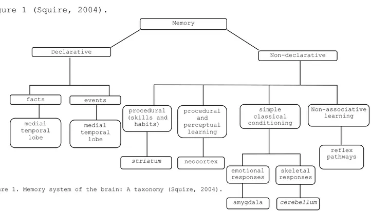
It is generally accepted that there are different forms of memory and multiple memory systems. Forms of memory can be distinguished on the basis of temporal characteristics (e.g. sensory memory, short/long- term), requirements for processing-encoding-retrieval (e.g. recognition/ familiarity) and stimulated domains (e.g. visual-space/verbal). Different memory systems can be distinguished on the basis of investigations of brain function and behavioural evidence (Roedige et al., 2002). A taxonomy from which a considerable amount of data was united is shown in Figure 1 (Squire, 2004).
Figure 1. Memory system of the brain: A taxonomy (Squire, 2004).
In normal aging there is a global decline that mainly involves the speed of information processing, episodic memory and working memory while verbal learning appears to remain stable (Allen et al., 2002; Rönnlund et
al., 2005). Several studies have shown that procedural memory processes
are not particularly influenced by aging (Craik & Jennings, 1992; Jennings & Jacoby, 1993; Light & La Voie, 1993). With regard to declarative memory, age-related deficits are estimated to be more specific for particular facts and events rather than general ones (Prull,
Memory striatum emotional responses skeletal responses reflex pathways neocortex medial temporal lobe medial temporal lobe
Diencephal
on
Declarative Non-declarative Non-associative learning simple classical conditioning procedural (skills and habits) procedural and perceptual learning events facts cerebellum amygdala2000; Gabrieli, 2000; Bunge, 2000; Raz, 2000; 2005). Research suggests that although semantic memory shows a clear failure in patients with various forms of dementia, it remains fairly stable in normal aging (Light, 1992). Data show a considerable maintenance of word meaning in the elderly: knowledge and vocabulary use remains stable until at least 70 years of age (Salthouse, 1991; Light, 1992). Although the loss of word meaning is minimal, elderly people make more errors and are slower than young people in detecting words when they are provided with a description, thus displaying an increase of difficulty in accessing semantic information in a quick and effective manner (Maylor, 1990). Moreover, one of the most frequent difficulties is represented by remembering people‟s names or finding a word temporarily unavailable (Burke et al., 1991; Maylor, 1993). Elderly people have lower performances than the young in verbal fluency tasks (Schaie & Willis, 1993). Further studies outlined the impact of age, gender and education on memory and learning for healthy elderly people. Findings showed a decline of working memory with advantage age, a strong impact of age on visual-verbal association tasks as well as visual-space memory tasks, an increase of forgetfulness and a significant influence of age on visual-space memory tasks (Panza et al., 1996). A considerable amount of recent data support the evidence of memory deficit related to two memory faculties: the power to recall contextual information about past events in time and place (recollection) and the feeling of recognition of past events in absence of contextual information recovery (familiarity). The comparison between young and elderly performances revealed that recollection is impaired in normal aging (Prull et al., 2003). The negative trend for semantic memory after 60 years of age is parallel to episodic memory (Dixon et al., 2004; Lodven et al., 2004). Old adults showed deficits regarding the source of information rather than its
content; age-related deficits are present in cognitive tasks as associating target words with related items or with specific contents (Naveh-Benjamin et al., 2004). It is also demonstrated that older adults are more vulnerable to interference effects created by a misleading prime due to mnestic impoverishment for details (Prull et al., 2005). Differences of performance between young and old adults have been shown in digit span tasks (reading span, listening span and operational span (Boop and Verhaeghen, 2005; Reuter-Lorenz and Sylvester, 2005). Some laboratory studies demonstrated a slight failure in prospective memory tasks (Henry et al., 2004).
According to the “frontal lobe hypothesis of aging” (Dempster, 1992; Hartley, 1993; West, 1996) the prefrontal cortex is the brain area that suffers more than others from the effects of aging. This hypothesis explains the observed behaviours of healthy elderly people, characterized by the reduction of the main three executive subsets: set shifting, information updating and monitoring, and inhibition (Miyake et al., 2000). Elderly people present lower performances than adults on tests that evaluate mental flexibility (set shifting) and creation of plans and strategies (set formation) (Keys and White, 2000). In addition, many studies have shown that during aging there is the reduction of the ability to inhibit a previously learned and dominant response in favour of a more adequate one (Andrés et al., 2008).
2. MILD COGNITIVE IMPAIRMENT: A HISTORICAL-CONCEPTUAL PERSPECTIVE
2.1 INTRODUCTION
The concept of slight cognitive impairment in aging appears confused with dementia until Kral (1962), who described senescent forgetfulness by distinguishing a physiological cognitive decline due to aging (“Benign Senescent Forgetfulness”) from a pathological cognitive decline (“Malignant Senescent Forgetfulness” or “Amnestic Syndrome”) apt to convert into dementia. After Kral, several studies provided different classifications of slight cognitive impairment but they did not completely describe its neuropsychological features. In 1995, Petersen and his co-workers of Mayo Clinic defined the diagnostic criteria for
Mild Cognitive Impairment (MCI) that were subsequently reviewed by Ritchie et al. (2001). In order to specify MCI subtypes, Petersen (2004) created a new classification model.
Literature presents many attempts to classify slight cognitive impairment in order to distinguish between physiological and pathological aging. Every diagnostic category of slight cognitive impairment follows a timeline (Table 1). We would stress that for each category highlighted in bold, scientific studies produced supplementary diagnostic framing and/or new clinical entities over time. They will be treated sequentially in the main category they belong to.
Table 1. Categories of slight cognitive impairment and related clinical entities: A timeline.
2.2 HISTORICAL ANTECEDENTS
Descriptions of cognitive impairment can be traced back to the last centuries. The first author who explained the loss of recent memory was James C. Prichard, who wrote “Treatise on Insanity and other Disorders
affecting the Mind” (1837). He distinguished intellectual insanities from
moral ones. He called “incoherence” or “dementia” a form of intellectual insanity where the first step was “forgetfulness” or “loss of recent memory”, with a preserved power of reasoning, judgement and attention. At the same time, remote memory experiences remained nearly in their original freshness and were able to be recalled when the attention focused on them. The disease accompanied aging and progressed slowly and gradually. The brain deficit did not consist of a past memories forgetfulness but of an attention system inability to receive influences from external agencies.
“Les maladies de la mémoire” (Ribot, 1888) proposed a preliminary classification of amnesia, pointing out those of progressive nature (“les
amnésies progressives”), thought to be a particular cognitive decline apt
to convert into dementia. Ribot explicated the “loi de regréssion” to clarify the gradual loss of memory, by establishing that recent memories are more likely to be lost than more remote ones. The idea that deterioration of psychological functions through mental disorders or neurological impairments retraces in reverse the order of evolutionary development, was drawn on John H. Jackson‟s theory: recently acquired information will be lost before deeply stored older information (Taylor, 1931).
PSEUDODEMENTIA
The term “pseudodementia” was used to describe a syndrome in which some psychiatric disorders could cause cognitive impairment. The impairment is not progressive and it is potentially reversible, if the primary cause (e.g. depression) is treated (e.g. with antidepressants). However, no neuropathological process can be identified or considered sufficient to explain the cognitive deficits. In their study conducted on a sample of 300 psychiatric patients, Madden et al. (1952) noted that some symptoms ordinarily considered to indicate dementia, could be reversed with appropriate therapies. After ten years, Kiloh (1961) described a clinical state in which initially dementia may be very closely mimicked and associated with malingering, reactive or endogenous depression. He considered remote psychiatric disturbances, an abrupt onset of symptoms and antidepressant treatment, as risk factors of pseudodementia. Post (1965) discussed pseudodementia in the elderly but he did not suggest its association with psychiatric syndromes. Lipowski (1967) documented a case of pseudodementia in a young highly educated women with hysterical and depressive symptoms and amnestic syndrome. Folstein et al. (1978) argued that depression can give rise to dementia which, although reversible, has a biological basis and should not be viewed as false dementia. They hypothesized that pseudodementia develops when the neurobiological disturbances of affective disorders are superimposed on a compromised aging brain. Thus, they suggested that the cognitive impairment associated with depressive illness might be accurately described as the “demential syndrome of depression”, rather than a “pseudodementia”. Wells (1979) described 11 patients with a variety of psychiatric disorders, including depression (with or without personality disturbances),
conversion reactions, post-traumatic neurosis and shizoaffective symptoms. He defined pseudodementia as a mimicked or caricatured dementia produced by functional mental illness, personality disorders and post-traumatic neuroses with a previous history of psychiatric illness, symptoms of short duration suffered before a request for help, rapid progression of symptoms, complaints of cognitive deficits, behavioural and cognitive performance inconsistent with the apparent degree of cognitive dysfunction and incapacity to reply to questions. He also proposed a valid checklist to differentiate pseudodementia from dementia. Caine (1981) used the following diagnostic criteria to describe pseudo-demented patients:
1. cognitive impairment with primary mental disorder;
2. superimposed symptoms or similar symptoms to primary organic disorder;
3. reversible disorder;
4. absence of organic pathology.
He adopted the Neuropsychological Screening Test (NST) (Caine, 1981) to evaluate their cognitive impairment. The most impaired performances depended on attention, processing information speed, spontaneous elaboration and analysis of details. This could be observed during the following tasks: verbal learning trial, immediate visual recall, copying, clock drawing procedure, written description, proverb interpretation and the Trail Making Test part B (Reitan, 1971). Reifler (1982) provided arguments to avoid clinical use of pseduodementia, pointing out two related issues:
1. it implied that patients had either an organic or a functional illness, whereas many individuals could have elements of both, especially when depression was superimposed on “true” dementia;
2. the mistaken clinical use of pseudodementia in clinical practice, whereas it was only descriptive.
More recently, Bianchetti and Pezzini (2001) have suggested deleting the term “pseudodementia” and introducing a temporal sequentiality between affective symptoms and cognitive ones (Table 2).
Table 2. Differential diagnosis between mood disorders and cognitive disorders of Type I and Type II (Form: Bianchetti and Pezzini, 2001) (Note: AD, Alzheimer‟s Disease; VD, Vascular Dementia).
On the basis of case reports, McAllister (1983) noted two different categories of pseudodementia: without associated cerebral dysfunction
versus with coexisting cerebral dysfunction. Cognitive impairment was
usually a caricature of dementia with exaggerated memory complaints and it was mainly observed in patients with personality disorders. By contrast, it was entirely indistinguishable from diffuse organic cerebral dysfunctions in other patients. The most common feature of pseudodementia was a relatively acute onset with a duration of symptoms from 6-12 months, characterized by moderate cognitive impairment, psychiatric history (e.g. depressive disorder), age over 50, frequent “do not know” as opposed to “near miss” answers, normal brain activity and structure, as revealed by electroencephalogram (EEG) and computed tomography scan (CT scan), and absence of nocturnal worsening (McAllister, 1985).
SENILE PSYCHOSIS
(PRIMARY DEGENERATIVE DEMENTIA; DEMENTIA AND MILD COGNITIVE IMPAIRMENT DIAGNOSTIC CRITERIA; AGE-RELATED COGNITIVE DECLINE; MILD NEURO-COGNITIVE DISORDER)
Roth (1955) described the diagnostic criteria of senile psychosis by evaluating a total of 472 patients with different mental disorders that had necessitated the institutionalization in Graylingwell Hospital (Chichester, UK). He classified the mental disorders of later life into six different categories: affective psychosis, senile psychosis, late paraphrenia, arteriosclerotic psychosis, acute confusion and other mental disorders. Results suggested that affective psychosis, late paraphrenia and acute confusion were distinct from senile and arteriosclerotic psychosis, thought as the two main causes of progressive dementia in old age. Senile psychosis was defined as «a condition with a history of gradually and continually progressive failure in the common activities of everyday life and a clinical picture dominated by failure of memory and intellect and disorganization of personality, where these were not attributable to specific causes as infection, neoplasm, chronic intoxication or cerebrovascular disease, known to have produced cerebral infarction» (Roth, 1955, p. 283).
In 1980, the American Psychiatric Association (APA) revised diagnostic criteria for senile psychosis, by introducing “Primary Degenerative Dementia” as a diagnostic entity apt to describe the degeneration of dementia in elderly people. The APA recommended limiting the diagnosis of Primary Degenerative Dementia only in cases of notable evidence of progressive and significant decline of cognitive functions and compromised social activities. In 1982, Reisberg et al. published the Global Deterioration Scale (GDS) for the assessment of Primary Degenerative Dementia, identifying three major clinical phases:
an early “forgetfulness” phase with subjective memory complaints and
an intermediate “confusional” phase in which an external observer becomes aware of the deficit;
a late dementia phase in which the patient cannot survive without a
caregiver‟s assistance.
To improve accuracy of Primary Degenerative Dementia, Sulkava et al. (1983) provided more extensive criteria for diagnosis (Table 3) by using a modified Luria-Nebraska Neuropsychological Test (Christensen, 1975; Sulkava and Amberla, 1982) to evaluate the first three criteria.
Table 3. Revised diagnostic criteria for Primary Degenerative Dementia (From: Sulkava et al., 1983).
The psychiatric classification for the diagnosis of dementia formulated by the Diagnostic and Statistical Manual of Mental Disorders 3rd Revision (DSM-III-R) (APA, 1987) provided valuable diagnostic criteria for Mild Cognitive Impairment (see also Zaudig, 1992). Subjects who satisfied criterion A but not criterion B, were included in “Mild Cognitive Impairment Type 1” (MCI Type 1). If DSM-III-R Criteria A and B, but non C were satisfied, the diagnostic label of “Mild Cognitive Impairment Type 2” (MCI Type 2) was applied (Table 4).
Table 4. DSM-III-R criteria for Dementia and Mild Cognitive Impairment (APA, 1987) (Note: ADL, Activities of Daily Living).
Within the DSM-IV section “Additional conditions that may be a focus of clinical attention”, the “Age-Related Cognitive Decline” and the “Mild Neuro-cognitive Disorder” were mentioned (APA, 1994). The first category should be used when the object of clinical attention is a clear decline of cognitive functions in aging, compared to normal limits. Subjects do not remember names or dates and they have difficulty in problem solving. The category of “Age-related Cognitive Decline” should be considered if a specific mental disorder and a neurological condition are excluded (APA, 1994). The diagnosis of “Mild Neuro-cognitive Disorder” was reported by DSM-IV (Table 5).
Table 5. Diagnostic criteria for Mild Neuro-cognitive Disorder (APA, 1994).
2.3. FROM KRAL‟S REVOLUTION TO PETERSEN‟S DIAGNOSTIC CRITERIA OF MILD COGNITIVE IMPAIRMENT
SENESCENT FORGETFULNESS
(BENIGN SENESCENT FORGETFULNESS VERSUS INCIPIENT AMNESTIC SYNDROME)
Even though literature reports different terms referring to mild changes in elderly cognition like “normal senility” (Bleuler, 1924), “normal senescent decline” (Dörken and Kral, 1951), and “mild senescent memory decline” (Kral, 1958), the principal attempt to define the normal tail-end of the continuum normal aging/dementia dates back to 1962, when Kral introduced the term “Benign Senescent Forgetfulness” (BSF) to describe a loss of remote memories in elderly healthy people. As neuropsychiatric consultant at the “Hebrew Old People‟s and Sheltering Home” in Montreal, he conducted two surveys (1956-1957; 1957-1961) to evaluate mental health
among patients and to recommend measures to improve their conditions (Heinik, 2010). In the first survey, based on DSM-I (APA, 1952) and Roth‟s categorization (1955), Kral classified 162 residents according to the following observational criteria:
1. degree of personality preservation, judgment ability and emotional responsiveness;
2. presence (or absence) and type of memory impairment; 3. presence (or absence) of psychotic/neurotic symptoms.
Kral adopted a meticulous comprehensive neurological examination and a psychiatric interview focused on particular aspects of mental functioning (especially memory) with the administration of a modified version of the Wechsler Memory Scale (WMS) (Wechsler, 1945). Based on these criteria, he suggested a classification of the entire population of the Home into five groups:
- Group A (37 persons, 22.8%): well-preserved personality,
appropriate emotional reactions, preserved judgment, no memory impairment, no history or signs of functional psychosis;
- Group B (30 persons, 18.5%): well-preserved personality, preserved
judgment, appropriate emotional reactions, no psychotic signs, “mild memory impairment” characterized by the inability to recall relatively unimportant data from the past, whereas the global experience can be recalled. The same data not recalled at one time, may be retrieved the next time. Remote memory was otherwise not impaired nor was there any clinically ascertainable defect of recent memory and immediate recall. Orientation was largely maintained. Confabulations were absent. Subjects were aware of their shortcomings and tried to compensate them by circumlocutions. Sometimes they might apologize. This group was classified as BSF,
and was found with equal frequency in both sexes. This condition progressed relatively slowly;
- Group C (24 persons, 14.8%): patients without memory deficit but
with a history and/or signs of functional psychosis (affective or paraphrenic type); this group included “neurosis of late maturity” (Kral, 1958);
- Group D (49 persons, 30.2%): typical amnestic syndrome, namely
impairment of recent memory and immediate recall, disorientation (particularly in time), and loss of remote memory. Only some events with a strong emotional charge could still be evoked, although in a distorted fashion. At the beginning, subjects could produce confabulations. The amnestic syndrome varied from moderate to severe, and it was associated with thought, judgment and nominal aphasia impairments;
- Group E (22 persons, 13.6%): amnestic syndrome with additional
psychosis signs, like paranoid delusions, hallucinations,
depressive or manic mood swings.
Group D and E constituted “Incipient Amnestic Syndrome” (IAS), which was more frequent in women than in men. This condition was associated with a high risk of institutionalization and mortality. In the second survey, Kral included results of psychological tests administered to 54 well-preserved residents by his colleague B.T. Wigdor: the verbal scale of the Wechsler Belleview Intelligence Scale I (Wechsler, 1939), the Bender Gestalt Visual Motor Test (Bender, 1938), and a modified version of the Wechsler Memory Scale (Wechsler, 1945). In addition, the Rorschach Test (Rorschach, 1942) was used to test personality structure and level of functioning. According to memory functions, four patient subgroups were formed:
- Group I (25 persons, 48%): without memory deficit or previous depression;
- Group II (13 persons, 13,25%): with mild type of memory impairment
(BSF);
- Group III (9 persons, 17.3%): without memory deficit but with a
history of depression;
- Group IV (5 persons, 9.6%): with signs of Incipient Amnestic
Syndrome (IAS), with intact personal and social functioning. From the psychometric point of view, the findings showed:
- low average of Intelligence Quotients (IQs) of the whole group
(mean 87.5, range 66-105);
- presence of memory impairment in all subjects with the average of
83.7 being under the cut-off of 90, with 98 being the highest score; Group I had higher mean scores (87.7) than Group II (81.7) and III (82.5); Group IV had the lowest mean score (72.2);
- indicative scores of marked impairment in the Bender Gestalt
drawings (total group mean 113, range 41-215).
As Heinik (2010) has suggested, Kral and Wigdor (1961) presented a sample with neuropsychological characteristics very similar to the current construct of Mild Cognitive Impairment (c.f. Petersen et al., 1995; Petersen, 2004):
a. all well-preserved aged people; b. normal level of intelligence;
c. subnormal performance on specific memory test;
d. subnormal performance on a specific perceptual/organization test; e. no significant signs of malignant amnestic syndrome.
MILD COGNITIVE IMPAIRMENT NOT AMOUNTING TO DEMENTIA
DISORDER; MILD MEMORY DISTURBANCE; MEMORY LOSS)
In 1978, the category of “Mild cognitive impairment not amounting to dementia” was formulated by the World Health Organization (WHO) in the International Classification of Disease 9th Revision, as a condition of mild memory disturbance associated with aging. Senile dementia, classified in the “Psychotics Organic States” headlight, occurs after 65 years of age with an alteration of memory, understanding and calculating, compromised abilities to judge and learn, emotional lability, mood disturbances, lowering of ethical values, emergence of new aspects of personality or exaggeration of existing ones, and difficulty in decision-making.
The ICD-10 (10th Revision of the International Classification of Mental and Behavioural Disorders) (WHO, 1990) established diagnostic criteria for “Mild Cognitive Impairment”, by selecting them from those of dementia (Table 6).
Table 6. ICD-10 diagnostic criteria for Dementia and Mild Cognitive Impairment (WHO, 1990).
The diagnosis of MCI excluded the clouding of consciousness (Criterion B) and a significant interference with the activities of daily living. It was divided into three subtypes:
- MCI Type 1 that meets only A1 criterion;
- MCI Type 2 that meets A1 and A2 criteria;
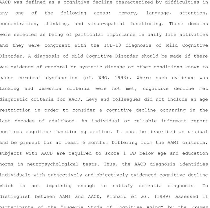
The diagnosis of “Mild Cognitive Disorder” (WHO, 1993) encompassed several clinical conditions such as memory decline, deficits in abstract thinking, difficulty in information processing, compared to a previously supposed higher level of cognitive functioning (Table 7). ICD-10 “Diagnostic Criteria for Research” recommended the differential diagnosis when the cognitive impairment cannot be referred to dementia, delirium and other cognitive disturbances.
Table 7. Diagnostic criteria for Mild Cognitive Disorder (WHO, 1993).
Christensen et al. (1995) assessed 897 community-dwelling elderly people to evaluate the epidemiological evidence of Mild Cognitive Disorder validity. They recommended using the Informant Questionnaire of Cognitive Decline in the Elderly (Jorm and Korten, 1988) to evaluate Criterion B, and a wide neuropsychological battery, including the Symbol Digit Modalities Test (Smith, 1982), a verbal fluency task, the Mini Mental State Examination (Folstein et al., 1975), three items of the Similarities subtest of the Wechsler Adult Intelligence Scale Revised (WAIS-R) (Wechsler, 1981), a sentence verification task, Information subtest similar to that used in the WAIS-R, three items of the Vocabulary form of the WAIS-R, a cube drawing procedure, Face Recognition from the Rivermead Behavioural Memory Test (Wilson et al., 1985), a word recognition memory task, recall of name and address, three individual
words and Visual Reproduction (one item from the Wechsler Memory Scale) (Wechsler, 1945), to evaluate Criterion C. They also suggested an estimation of premorbid intelligence using the National Adult Reading Test (Nelson, 1982).
Mild Cognitive Disorder was restated by the World Health Organization in the “International Statistical Classification of Diseases and Related
Health Problems 10th Revision Version for 2007”. It was defined as «a
disorder characterized by impairment of memory, learning difficulties and reduced ability to concentrate on a task for more than brief periods. There is often a marked feeling of mental fatigue when mental tasks are attempted, and new learning is found to be subjectively difficult even when objectively successful. None of these symptoms is so severe for a dementia or delirium diagnosis. The diagnosis of Mild Cognitive Disorder should be made only in association with a specified physical disorder. The cognitive disorder may precede, accompany, or follow a wide variety of infections and physical disorders, both cerebral and systemic, but direct evidence of cerebral involvement is not necessarily present. It can be differentiated from encephalitic syndrome and post-concussional syndrome by its different aetiology, its more restricted range of generally milder symptoms, and usually shorter duration» (WHO, 2007; F06.7 code).
The classification of “Mild Cognitive Impairment” was formulated as a clinical entity in the “International Classification of Diseases 9th Revision Clinical Modification” (ICD-9-CM) (WHO, 2002), including memory complaints (preferably corroborate), objective memory impairment, relatively preserved general cognition and essentially intact activities of daily living. It did not include head trauma, dehydration, stroke, malnutrition.
“Mild Cognitive Impairment” was not the only diagnostic category of cognitive decline. In fact, diagnostic criteria for “Mild Memory Disturbance” and “Memory Loss” were quoted, too. Mild Memory Disturbance was classified as another specific non-psychotic mental disorder, following organic brain damage (WHO, 2002). Alternatively, Memory Loss was characterized by the organic or psychogenetic loss of the ability to recall information, retrograde and anterograde amnesia, and a temporary or permanent loss of recent memory caused by organic or psychological factors (WHO, 2002). Bowen et al. (1997) had studied the progression to dementia in patients with isolated memory loss, because the history of severe memory loss in patients without other cognitive impairments was poorly understood. Dementia criteria of the DSM-III-R involved impairment in two or more cognitive domains like the NINCDS-ADRDA criteria (criteria proposed by the National Institute of Neurological and Communicative Disorders and Stroke and the Alzheimer's Disease and Related Disorders Association for diagnosis of dementia) for probable Alzheimer‟s Disease (AD). Bowen's longitudinal study circumscribed subjects who did not meet diagnostic criteria for dementia, without other cognitive impairments. The neuropsychological test battery included the Mini Mental State Examination (Folstein et al., 1975), the Mattis Dementia Rating Scale (Mattis, 1988), the Fuld Object Memory Evaluation (Fuld, 1981), an abbreviated form of the Boston Naming Test (Morris et al., 1989), the Wechsler Adult Intelligence Scale Revised (Wechsler, 1981) and the Trail Making Test (Reitan, 1985). All patients scored at least 2 standard deviation (SD) below the average of memory tests, while other areas of cognition were preserved. By detecting conversion rates, the researchers concluded that isolated memory loss was frequently the incipient symptom of AD. According to this evidence, Bowen and colleagues recommend
following isolated memory loss patients over time to verify conversion into dementia.
LIMITED DEMENTIA OR LIMITED COGNITIVE DISTURBANCE
Gurland et al. (1982) formulated criteria for the diagnosis of dementia, by adopting the Geriatric Mental State Examination (Copeland et al., 1976) and the Comprehensive Assessment and Referral Evaluation (Gurland
et al., 1977), a basically semi-structured interview guide with specific
criteria for diagnosis and severity of dementia. The first step was named “Limited Dementia” or “Limited Cognitive Disturbance” and was characterized by mild memory dysfunction not severe enough to satisfy dementia diagnostic criteria and to interfere with the capability of patients to live independently. The diagnostic criteria included:
1. subjective report of memory decline; 2. increased reliance on notes as reminders;
3. occasionally (less than once a week) forgetfulness of name, appointments and misplacing of objects;
4. occasionally (less than once a month) destructive or dangerous memory lapses such as burning cooking or leaving on gas tap;
5. one or two errors on cognitive testing: subject forgets current or past president, exact date, phone number, zip code, dates of marriage or moving to present location.
QUESTIONABLE DEMENTIA
In 1982, two independent tests for cognitive aging and dementia were published. One of these staging measures was the Clinical Dementia Rating (CDR) (Hughes et al., 1982). It identified five stages including a CDR 0.5 termed “Questionable Dementia”, including mild dementia and earlier antecedents, with the following clinical features:
1. mild consistent forgetfulness and partial recollection of events; 2. fully oriented patient;
3. only doubtful or mild impairment on independent functions at usual level of job, shopping, business and financial affairs, volunteer and social groups;
4. life at home, hobbies, intellectual interest well-maintained or only slightly impaired;
5. fully capable of self-care.
Devanand et al. (1997) found that low scores on delayed recall of a modified Mini Mental Examination (Stern et al., 1987), consistent long-term retrieval of the Free and Cued Selective Reminding Test category naming for animals (Buschke, 1984), digit symbol, picture arrangement and block design subtest of the Wechsler Adult Intelligence Scale Revised (Wechsler, 1981), were predictive of the final diagnosis of dementia in a sample of 127 subjects with questionable dementia. Daly et al. (2000) administered the Clinical Dementia Rating (Hughes et al., 1982) and the Mini Mental State Examination (Folstein et al., 1975) to 165 individuals of 65 years of age at baseline and annually for 3 years. The questionable group met the CDR 0.5 criteria at baseline and still had the same evaluation after 3-year follow-up. The researchers found that only three
tests (among seventeen used) were sensitive to differentiate
questionable, normal and converter groups: the California Verbal Learning Test (Delis et al., 1987), the Self-Ordering Test (Petrides and Minler, 1982) and the Trail Making Test (Reitan, 1958). The questionable group showed scores in the normal-converter range. Daly and colleagues suggested using a wider neuropsychological test battery to identify subgroups of individuals within the category of questionable dementia, because only 23 of the 123 subjects met criteria for AD at follow-up.
MILD COGNITIVE DECLINE
Reisberg et al. (1982) created the Global Deterioration Scale (GDS) for the diagnosis of Primary Degenerative Dementia (c.f. APA, 1980), identifying seven stages of cognitive impairment, where stage 0 corresponds to non cognitive decline and stage 7 corresponds to severe cognitive decline. GDS stage 3 termed “Mild Cognitive Decline”, was adopted to describe patients with evidence of memory deficits obtained by a rigorous interview conducted by trained geriatric psychiatrists. GDS Stage 3 established that: concentration deficit may be evident on clinical testing; subjects show decreased facility in remembering names or places and low performance in employment and social demand; co-workers may be aware of patient‟s declining work performance; difficulties in finding words and names also become evident to relatives; patients may get seriously lost when travelling to different places; the impairment provoked by symptoms may be increased by denial; anxiety (from mild to moderate) also accompanies cognitive symptoms. Patients perform 1 SD or more below the average for their age and WAIS vocabulary score on at least 3 of 5 Guild memory subtests (Gilbert and Ferris, 1980). However, patients may still make no errors on the 10-item Mental Status Questionnaire (Pfeiffer, 1975). After the GDS publication, a series of measures were developed to describe the progression of cognitive decline in aging. Among these, the Brief Cognitive Rating Scale (Reisberg and Ferris, 1988) assessed progressive changes along five major axes: (I) concentration, (II) recent memory, (III) remote memory, (IV) orientation, and (V) self-care functioning. These axes were enumerated to be concordant with GDS stages (Reisberg et al., 2008).
Henderson and Huppert (1984) considered “mild dementia” as a rubric for the early stages of several neuropathological disorders without precise diagnostic criteria for diagnosis. The single common feature was a significantly cognitive deficit in relation to a previously supposed higher level of cognitive functioning. The term was adopted as synonymous of Limited Cognitive Disturbance (c.f. Gurland et al., 1982) or of Questionable Dementia (cf. Hughes et al., 1982). Some epidemiological studies had already tried to detect individuals suffering from mild dementia. In the “Newcastle Study”, Kay et al. (1964) had identified mild demented patients and they found that some cerebrovascular and senile diseases were associated with cognitive impairment. The mental deterioration provoked by aging was due to isolation and inadequate diets and it usually determined institutionalization. The study had reported that some individuals developed definite dementia within a 3-year period (Bergman et al., 1971). Nielsen et al. (1977) had found the prevalence of mild dementia at 15.4% among a Danish population: after 15 years the mildly demented group had a higher than expected mortality rate. It was unknown whether any of this group progressed to definite dementia. Because mild dementia was not a distinct diagnostic category, there was not a specific headlight in the DSM-III-R and ICD-10, but only the specification of a severe dementia degree.
AGE-ASSOCIATED MEMORY IMPAIRMENT
(AGE ASSOCIATED MEMORY IMPAIRMENT REVISED CRITERIA; AGE-CONSISTEN MEMORY IMPAIRMENT; LATE-LIFE FORGETFULNESS)
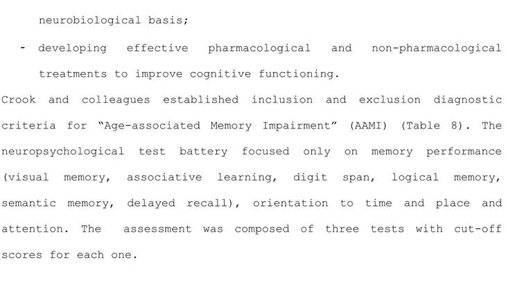
In 1986, the National Institute of Mental Health (NIMH) Work Group proposed specific diagnostic criteria to describe memory loss of elderly healthy people. It tried to analyse the physiological cognitive decline with three objectives:
- understanding the behavioural phenomena of memory decline and their neurobiological basis;
- developing effective pharmacological and non-pharmacological treatments to improve cognitive functioning.
Crook and colleagues established inclusion and exclusion diagnostic criteria for “Age-associated Memory Impairment” (AAMI) (Table 8). The neuropsychological test battery focused only on memory performance (visual memory, associative learning, digit span, logical memory, semantic memory, delayed recall), orientation to time and place and attention. The assessment was composed of three tests with cut-off scores for each one.
Table 8. Diagnostic criteria for Age-associated Memory Impairment (From: Crook et al., 1982) (Note: WMS, Wechsler Memory Scale; AD, Alzheimer‟s Disease).
Blackford and La Rue (1989) modified the NIHM diagnostic criteria for AAMI (Table 9) and quoted a new clinical entity named “Age-consistent
Memory Impairment” (ACMI), according to specific psychometric
concomitants (performance within ± 1 SD of the mean established for age on 75% or more of the tests administered).
Table 9. Revised diagnostic criteria for Age-associated Memory Impairment (From: Blackford and La Rue, 1989) (Note: AAMI, Age-associated Memory Impairment; ACMI, Age-consistent Memory Impairment; LLF, Late-life Forgetfulness; IQ, Intelligence Quotient; WAIS, Wechsler Adult Intelligence Scale, WAIS-R, Wechsler Adult Intelligence Scale-Revised; NINCDS-ADRDA, criteria proposed by the
National Institute of Neurological and Communicative Disorders and
Stroke and the
Alzheimer's Disease and Related Disorders Association
for diagnosis of dementia).In order to describe AAMI patients, they adopted an age range of 50-79, because no upper age limit had been specified by the NIMH work group. A perceived decrease in areas of day-to-day memory functioning had to be verified by standardized self-report memory questionnaires. In addition, “subjective memory complaints” of NIMH criteria did not suggest any means of quantification and they were overinclusive.
Blackford and La Rue distinguished “Late-Life forgetfulness” (LLF) from AAMI and ACMI on the basis of memory performance in four tests of secondary memory. LLF performance was calculated between 1 and 2 SD below the mean established for age on 50% or more of the tests administered.
MINIMAL DEMENTIA
The term “Minimal Dementia” was used in the Cambridge Mental Disorders in the Elderly Examination (CAMDEX) (Roth et al., 1986). It was a structured psychiatric interview intended for use in studies of prevalence and incidence of dementia, especially mild. Patients were graded for dementia severity (minimal, mild, moderate, severe). Minimal Dementia was defined as «a limited and variable impairment of recall, minor and variable errors in orientation, a blunted capacity to follow arguments and solve problems and occasional errors in everyday tasks». The “Hughes Hall Study of the Elderly” in Cambridge identified 44 people with minimal dementia assessed by CAMDEX. At 1-year follow-up, 29 subjects had an objective deterioration of cognitive functioning (O'Connor et al., 1990). At 2-year follow-up, 12 subjects met dementia criteria of such a structured interview (O'Connor et al., 1991; Cooper et al., 1996).
MILD COGNITIVE IMPAIRMENT
The term “Mild Cognitive Impairment” was introduced into literature by Fliker et al. (1991) to describe patients obtaining a score of 3 on the Global Deterioration Scale (GDS) (Reisberg et al., 1982), who were not demented and who exhibited at least two of the following symptoms :
1) getting lost when travelling to an unfamiliar location; 2) decline in work performance apparent to co-workers; 3) word- and name- finding deficit apparent to intimates;
4) relatively little retention of material read in a passage of a book;
5) decreased facility in remembering the names of newly introduced people;
6) losing or misplacing an object of value;
They conducted a full diagnostic evaluation of elderly subjects diagnosed as having MCI, adopting tests of immediate memory (digit span and delayed spatial recall), verbal recall (paragraphs, shopping list), visuospatial recall (delayed spatial recall and misplace objects), visual recognition memory (visual recognition span and facial recognition), remote memory (remote memory questionnaire), language (vocabulary, category retrieval, object naming, object functional recall, object name recognition, object functional recognition, object formation), concept formation (object sorting), visuospatial praxis (digit symbol), visuoperceptual function (road map), psychomotor speed (i.e. finger tapping, driving test, release, travel). Mildly impaired subjects performed significantly more poorly than controls on tests of recent and remote memory, language function, concept formation and visuo-spatial praxis. These results were also revealed by 2-year follow-up, suggesting that most subjects with mild cognitive deficits could manifest the progressive symptoms of dementia. GDS stage 3 was evaluated as a psychometric predictor able to distinguish between benign and more severe deficits of mildly impaired subjects.
The diagnosis of Mild Cognitive Impairment was also formulated on the basis of performance resulting in the neuropsychological battery of the Structured Interview for the Diagnosis of Dementia of the Alzheimer Type, multi-infarct dementia, and dementias of other aetiology according to DSM-III and ICD-10 (SIDAM) (Zaudig, 1992). It was a short diagnostic screening instrument to service each item of DSM-III and ICD-10 criteria for dementia. All items of the SIDAM could be summed up as resulting in the SIDAM score (SISCO), which ranged from 0 (the worst cognitive impairment) to 55 (no cognitive impairment). The diagnosis of Mild
Cognitive Impairment was given with a SISCO score of 34-47 and a Mini Mental State Examination (Folstein et al., 1975) score of 23-27.
In 1995, Petersen et al. created the primary diagnostic criteria for Mild Cognitive Impairment:
a. complaint of defective memory; b. normal activities of daily living; c. normal general cognitive function; d. abnormal memory function for age; e. absence of dementia.
They studied a sample of 75 subjects during a period of five years who reported memory decline from 1.5-2.0 SD below the mean of individuals with similar age and education level. These patients were less able than healthy controls to benefit maximally from the use of semantic cues during the recall task in the Free and Cued Selective Reminding Test (Buschke, 1984); they also showed impairment in delayed recall in the Rey Auditory Verbal Learning Test (Rey, 1964). By contrast, the mean scores in the Mini Mental State Examination (Folstein et al., 1975) were approximately 26 and the measures of general cognitive function in the Wechsler Adult Intelligence Scale Revised (Wechsler, 1981) and in the Dementia Rating Scale (Mattis, 1988) were relatively unaffected (Petersen et al., 1997). The first major study focusing on the clinical characterization and outcome of MCI was published in 1999 by Petersen and colleagues to identify individuals at high risk for severe cognitive decline and progression to dementia. They recruited a sample of 76 subjects with MCI, 243 healthy normal controls and 106 patients with mild AD. All subjects were administered two sets of tests. The first set was used for diagnostic purposes and it included the Wechsler Adult Intelligence Scale Revised (Wechsler, 1981), the Rey Auditory Verbal