UNIVERSITA’ DELLA CALABRIA
Dipartimento di Farmacia e Scienze della Salute e della Nutrizione
Dottorato di Ricerca in Medicina Traslazionale
CICLO XXIX
FoxO3a reactivation restores the sensitivity to the antiestrogen
treatment in tamoxifen resistant breast cancer
Settore Scientifico Disciplinare MED/06
ABSTRACT
Resistance to endocrine treatments is a major clinical challenge in the management of estrogen receptor alpha positive (ER+) breast cancers (BC). Although multiple mechanisms leading to endocrine resistance have been proposed, the poor outcome of this subgroup of BC patients demands additional studies. Here we show that the expression of FoxO3a transcription factor is strongly reduced in ER+ BC MCF-7 cells (wtMCF-7) that developed resistance to Tamoxifen (TamR). On the other hand, FoxO3a silencing (siF3a) was able to counteract Tam induced growth inhibition in wtMCF-7, demonstrating that FoxO3a is a mediator of cell response to Tam.
To analyze the role of FoxO3a in the acquisition of a Tam resistant phenotype, TamR clones bearing an active FoxO3a (F3aAAA), whose expression can be induced by Doxycycline (Dox) were developed. FoxO3a re-activation was able to re-establish the sensitivity of TamR cells to the antiestrogen, inhibiting proliferation and cell cycle progression, as well as restoring Tam dependent apoptotic response. For a closer look at the molecular mechanisms involved, an unbiased proteomics analysis on F3aAAA-inducible TamR cells was conducted, unveiling novel interesting and potential mediators of the anti-proliferative and pro-apoptotic activity of FoxO3a, all worthy of future investigations.
Kaplan-Meier (K-M) survival curves confirmed the relevance of FoxO3a also in a clinical setting, since high levels of the transcription factor strongly correlate to a positive response to tamoxifen therapy. Finally, to assess if FoxO3a reactivation is able to restore the sensitivity to Tam also in vivo, the widely used anti-epileptic drug (AED) Lamotrigine (LTG; Lamictal), which is able to induce FoxO3a expression in TamR cells leading to growth inhibition, was also tested on TamR deriving xenografts tumors, where it showed the same effects observed in vitro.
Altogether, our data indicate that FoxO3a could not only be considered a good prognostic factor in ER+ BC, predicting a positive response to endocrine therapy, but also a key target to be exploited in combination therapy. In this context, LTG might represent a valid candidate to be used as an adjuvant to Tam therapy in patients at risk.
INTRODUCTION
Breast cancer is the most common malignancy in women and represents one of the major causes of death worldwide, making up 21% of all new cancer diagnoses. Each year, almost 1.4 million women are diagnosed with breast cancer, and the disease will be responsible for 450 000 deaths [1]. Survival rates have been steadily extending over the past 50 years, primarily due to improvements in diagnosis and treatment.
The drivers of proliferation in breast cancer are also the phenotypic drug targets; hormone receptors (estrogen, progesterone and HER2 receptors) are commonly overexpressed. Early studies established that steroid hormones are of pivotal importance in directing the growth and development of breast tumours and endocrine treatments, by perturbing the steroid hormone environment of tumor cells, can promote extensive remissions in established tumors and furthermore provide significant patient survival benefits. More than 60% of human breast cancers are estrogen receptor α positive (ERα+) [2]. The presence of ERα is considered a good prognostic factor and correlates with a higher degree of differentiation of the tumour [3, 4] and increases disease-free survival [5]. The earliest approved therapy for the prevention and treatment of ERα+ breast cancer patients was Tamoxifen.
Tamoxifen is a triphenylethylene derivative pharmacologically classified as a selective estrogen receptor modulator (SERM) that acts as a partial antagonist (an agonist in the uterus but an antagonist in the breast), impairing ERα function by competing with 17β-estradiol (E2) for the binding to the
receptor [6]. Binding of the primary human E2, to ERα seals the hydrophobic pocket by the helix-12 domain [7, 8] . ERα then translocates to the nucleus, binds to the estrogen response element (ERE), activates and drives the transcription of estrogen-dependent genes [9]. Tamoxifen is metabolized to 4-hydroxytamoxifen (4-OHT) [10, 11]; , which also binds to ERα. However, this leads to a different conformational change as compared to E2 and the hydrophobic pocket is not sealed by helix-12 [8]. Consequently, 4-OHT blocks ERα activation and reduces cell proliferation by activating apoptosis, and/or autophagy [12].
Thus, the effects of Tamoxifen in breast tissues result from its ability to bind to the ligand-binding domain of the ERα, thereby antagonizing the proliferative potential of estrogens [13, 14], which are responsible for cancer cell growth or proliferation [15]. Unfortunately, clinical application of all endocrine measures examined to date has revealed that their beneficial actions are limited and can eventually be counteracted by the capacity of breast cancer cells to ultimately circumvent the need for steroid hormones, allowing them to grow and progress despite such therapy.
In fact, with time, about 50% of patients with ERα+ breast cancer stop benefiting from Tamoxifen treatment and acquire resistance, leading to disease progression.
Thus, at presentation of breast cancer, current endocrine therapies are not effective in all patients (de novo endocrine resistance) and initially responsive tumors will invariably progress despite such treatments (acquired resistance) resulting in patients relapse and associated poorer survival [16]. The clinical existence of these phenomena, together with substantial experimental evidence, indicates that such a therapeutic approach based on using anti-hormones as single agents may be now regarded as somewhat simplistic. Indeed, an increasing body of data has revealed that the control of endocrine responsive and resistant breast cancer growth is in reality multi-faceted, comprising a complex network of interacting signal transduction pathways impinging on tumor proliferation and cell survival parameters [17, 18] . The identification of the pathways responsible for de novo and acquired endocrine resistance has thus been an important goal for many breast cancer researchers. It is self-evident that any accurate identification of factors indicative of endocrine resistance in advance of therapy would not only prevent unnecessary side effects in unresponsive patients, but would also significantly limit both the time lost during which the disease may progress unchecked by appropriate therapy, and the wastage of financial resources associated with ever escalating costs of drugs. Moreover, knowledge of the causative elements of de novo and acquired endocrine resistance might allow the development of new therapeutic agents and strategies to prevent either the evolution of these conditions or at least delay their appearance, hence severely compromising the disease process and enhancing patient survival.
The potential mechanisms underlying the evolution toward an antiestrogen resistant phenotype have been the object of extensive investigations, but the available data are not univocal. Some authors ascribed the acquisition of resistance to a loss or mutation of the ERα [19], other authors shown that
breast cancer cells which lost sensitivity to anti-hormonal treatment, often retain an ERα+ phenotype with normal ERα functionality [20, 21]. In Tamoxifen resistant breast cancer, ERα has been reported to interact with deregulated growth factor pathways, facilitating the proliferation of resistant cells [22, 23] . The enhancement of growth factor signaling results in the increase of ERK1/2 phosphorylation [24] and in phosphatidylinositol 3-kinase (PI3K)/AKT cell survival pathway activation [25]. Furthermore, the PI3K-Akt signalling pathway has also been demonstrated to play a crucial role in the development of tamoxifen resistance [25-27]. AKT activation by phosphorylation (pAKT) regulates critical cellular activities such as growth, proliferation, differentiation, metabolism and survival as well as tumorigenesis [28]. Importantly, PI3K/AKT signaling is implicated in the pathogenesis of breast cancer. PI3K is activated in response to a variety of extracellular signals through a receptor tyrosine kinase (RTK) such as HER2, epidermal growth factor receptor (EGFR) or insulin-like growth factor 1 receptor (IGF1R). The serine/threonine kinase AKT is a downstream multifunctional kinase, which serves as the central mediator of the pathway [29]. pAKT promotes cellular survival via either direct inactivation by phosphorylation of multiple proapoptotic proteins or by inhibition of the Forkhead box transcription factors that results in decreased expression of proapoptotic proteins [30]. The characteristic attenuation of apoptosis by pAKT has been hypothesized as a major mechanism of resistance to cancer treatment [31]. Indeed, one of the downstream targets of AKT as well as of MAPK, currently attracting a great interest in hormone dependent breast cancer, is Forkhead box class O (FoxO)3a. The FoxO genes encode for the O-subfamily of proteins that belong to the larger family of winged-helix forkhead transcription factors which contains four members (FoxO1a, FoxO3a, FoxO4, and FoxO6), whose functions are negatively regulated by the insulin- PI3K/AKT signaling [32] and MAPK [33]. The expression and activity of FOXO factors are strongly controlled by post-translational modifications such as phosphorylation,
acetylation, methylation and ubiquitination [34]. A major mechanism of regulation of FOXOs consists of phosphorylation by AKT or SGK(serum and glucocorticoid-regulated kinase), on three residues (T32, S253 and S315 of FOXO3), following insulin or growth factor stimulation [30], leading to FOXO inactivation. Similarly, FoxO3a undergoes phosphorylation on residues (S294;S344;S425) [35] upon MAPK activation by growth factors. Indeed, these phosphorylations allow the binding of 14-3-3 proteins to FOXOs and their export from the nucleus to the cytoplasm (reviewed in [36]. Into the cytoplasm FOXOs are sequestrated and maintained in an inactive state, which can be rapidly reversed. In this condition FOXOs can also be ubiquitinated and degraded by proteasomes.
The mitogen-activated protein kinase (MAPK) cascades are evolutionary conserved, intracellular signal transduction pathways that respond to various extracellular stimuli and control a large number of fundamental cellular processes including growth, proliferation, differentiation, motility, stress response, survival and apoptosis [37, 38] . In mammalian cells, the major MAPK are extracellular signal-regulated protein kinase (ERK) [39], p38 MAP kinase [40], and c-Jun N-terminal kinase (JNK) [41, 42]. These MAP kinases are activated by the dual phosphorylations of neighbouring threonine and tyrosine residues in response to various extracellular stimuli [43, 44] . Specifically, p38 and JNK have been implicated in stress responsive signals leading to the initiation of adaptive events such as gene expression, differentiation, metabolism, and apoptosis [45]. ERKs are often activated by growth signals, such as epidermal growth factor (EGF) or platelet-derived growth factor [46]. Activation of ERK has been shown to phosphorylate FOXO proteins, resulting in nuclear exclusion and transcriptional repression. In addition to ERK, direct phosphorylation of FOXO by AKT results in its cytoplasmic retention and inactivation, causing the inhibition of the expression of FOXO-regulated genes, which control the cell cycle, cell death, cell metabolism and oxidative stress [47-49]. Moreover, active MAPK negatively regulates FOXO3 stability via an MDM2-mediated ubiquitin-proteasome pathway [50]. On the contrary, MAPKs signalling are potentially inhibited upon FOXO3
activation, in line with the observations in other studies [51]. This implies that FOXO3 activation results in an overall change in the wiring or activity of several signal transduction networks.
The mechanisms that direct FOXOs to degradation rather than sequestration might be related to the intensity of the signal that triggers nuclear export [34]. In absence of growth factors, FoxOs are mainly located within nuclei and regulate a set of target genes promoting cell cycle arrest, stress resistance, apoptosis, DNA damage repair and metabolism [52]. Increasing interest in FoxOs factors is emerging in the oncologic research field. In particular, in breast cancer, it has been proposed as a bona fide tumour suppressor [53]. In ERα+ breast cancer, several reports have suggested a functional interaction between ERα and FoxO members. E2 have been found to promote ERα binding to FoxO1a, FoxO3a, and FoxO4, which, in turn, regulate ERα-mediated transcription, showing either coactivator or corepressor functions on ERE sites [54, 55] . Noteworthy, a significant enrichment of Forkhead motifs within ERα binding regions was found at very high frequency [56], suggesting a role for FoxOs in determining ERα binding and function [57]. In line with this assumption, we have previously shown that in ERα+ breast cancer cells, nuclear (thus active) FoxO3a behaves as a repressor for ERα-mediated transcription by binding to Forkhead responsive elements on ERα target gene promoters, evidencing a protective role in ERα+ breast cancer [58]. On this basis seems to be important to understand the involvement of FoxO3a in the acquisition of the resistance of breast cancer cells to 4-OHT treatment. Moreover, a deeper knowledge of the molecular mechanisms involved in both 4-OHT resistance acquisition that in the inactivation and nuclear exclusion of FoxO3a will provide additional opportunities for the development of new therapeutic strategies that could increase nuclear FoxO3a content and function.
Here we suggest a possible role for an antiepileptic drugs Lamotrigine (LTG; Lamictal) [59] that have been found to exert anti-cancer activity [60, 61] . LTG has a strong antiproliferative activity on several types of breast cancer cells, including TamR cells, this effect is paralleled by a significant increase of FoxO3a expression and nuclearization. LTG behaves in a similar way also in vivo, where in TamR derived xenografts, induces a dramatic decrease of tumors mass.
MATERIALS AND METHODS
Cell culture, conditions, and treatments
The ER-positive human wild-type (wt) breast cancer epithelial cell line wtMCF-7 was maintained in monolayer culture in Dulbecco’s modified Eagle’s/Ham’s F-12 medium (1:1) (DMEM/F-12), supplemented with 5% fetal bovine serum (FBS), 100 IU/ml penicillin, 100 ng/ml streptomycin, and 0.2 mM L-glutamine. For experimental purposes, cells were synchronized in phenol red-free and serum-free media (PRF-SFM) for 24 h and then, where opportune, switched to PRF-media containing 5% charcoal-treated FBS (PRF-CT) or 5% FBS, in presence or not of Tam (4-OHT) or EGF-1 (both from Sigma-Aldrich, Italy) depending on the experiment. All other media and reagents were purchased from ThermoFisher Scientific (Waltham, MA USA).
Selection Procedure of Tamoxifen resistant MCF-7 cells (TamR)
TamR cells were obtained after long-term cultivation of parent ERα (+) MCF-7 cell line (wtMCF-7) in Tam. wtMCF-7 cells were exposed to increasing concentrations of 4-OHT, starting from 10-9M up to a final concentration of 10-6M 4-OHT. Cells were refeed with fresh growth medium containing the drug every 2-3 days.
Generation of FoxO3a inducible stable clones
TamR/TetOn-AAA clones were generated using the Tet-On Gene Expression System (Clontech, Palo Alto, CA, USA). The Tet-On belongs to a high-level gene expression system that employs a regulator plasmid and a response plasmid to establish a double-stable Tet cell line.
The Tet-On system is a regulatory system that allows activation of gene expression by the addition of the effector substance tetracycline or one of its derivatives, e.g. Dox.
This control circuit was described as a rtTA dependent expression system, because a recombinant tetracycline controlled transcription factor, rtTA, interacts with a rtTA responsive promoter, Ptet, to drive expression of the gene under study. The effector, act at the level of DNA binding rtTA
transcription factors. So, rtTA requires tetracyclines for binding to tetO, and the subsequent fusion of the VP16 (herpes simplex virus protein) activation domain resulted in a reverse-tTA (rtTA) that binds Ptet and activates transcription exclusively in the presence of Dox.
To generate pTRE-F3aAAA inducible plasmid, the cDNA encoding the entire open reading frame of a constitutively active form of the human FoxO3a gene, where the three known AKT phosphorylation sites on FoxO3a have been mutated to alanine (F3aAAA), was excised as a 2kb BamHI-XbaI fragment from the plasmid 1319 pcDNA3 flag FKHRL1 AAA (Addgene, Cambridge, MA, USA, plasmid #10709). The fragment was sub-cloned into the pTRE-zeo vector, harboring a EGFP cassette and a Zeocin resistance gene to allow selection of stably transformed cells in the presence of zeocin. Restriction digestion and sequence analysis verified a correct cloning. The pTRE-F3aAAA plasmid allows the conditional expression of active FoxO3a under the control of a tetracycline-response element (TRE) in the presence of Doxycycline (Dox, Sigma Aldrich) and of a rTetR.
To obtain a stable TamR/TetOn-AAA cell line, TamR cells were first transfected with the regulator plasmid pTet-On, containing the Geneticin (G418, Invitrogen) resistance gene, and constitutively encoding rtTA proteins, using FuGENE® HD Transfection Reagent (Promega Italia s.r.l., MI-Italy) as a transfection reagent.
Several G418 resistant TamR/TetOn stable clones were isolated by single-cell cloning and selected by successful transient transfections with the pTRE-F3aAAA plasmid.
TamR/TetOn selected clones were pooled together and subjected to a second round of transfection with the pTRE-F3aAAA plasmid. G418- and Zeocin-resistant clones were isolated and screened by western blot (WB). TamR/TetOn-AAA clones with low background expression and high Dox-dependent (3 g/ml) induction of FoxO3a protein were selected. In this cellular system, rtTA protein binds to TRE and activates FoxO3AAA transcription in response to Dox in a precise and dose-dependent manner.
Control cell lines (TamR/TetOn-V) were established by stably transfecting the pTRE backbone (vector only) without a cDNA insert.
Pools of TamR/TetOn-AAA and TamR/TetOn-V clones were collected and used in all experiments. Clones were maintained in monolayer culture in Dulbecco’s modified Eagle’s/Ham’s F-12 medium (1:1) (DMEM/F-12), supplemented with 5% (FBS), 100 IU/ml penicillin, 100 ng/ml streptomycin, 0.2 mM L-glutamine, G418 (0.2 mg/ml) and Zeocin (0.1mg/ml).
siRNA-mediated RNA interference
Custom-synthesized siRNA-annealed duplexes (25 bp double- stranded RNA [dsRNA]) were used for effective depletion of FoxO3a (siF3a) transcripts. A scramble siRNA (siScramble) lacking identity with known gene targets was used as a negative control. 106 wtMCF-7 cells were seeded in 60mm Petri dishes in growing medium without antiobiotics. The day after cells were transfected with siF3a (150 pmol/dish) and siScramble (120pmol/dish), using Lipofectamine 2000 (all reagents were from ThermoFisher Scientific). Six hours after transfections, cells were synchronized in PRF-SFM for 24 h and then switched to 5% PRF-CT, in presence or absence of 1µM 4-OHT up to 72h, depending on the experiment.
Plasmids and transient transfections
For key experiments, results were confirmed by transiently over-expressing F3aAAA in TamR cells. 106 TamR cells were plated in 60mm plates and transfected in suspension in GM-PRF with 4g/dish of the 1319 pcDNA3 flag FKHRL1 AAA (F3aAAA), encoding the constitutively active triple mutant of FoxO3a (provided by William Sellers, Addgene plasmid 10709), or the pcDNA3.1 vector (Invitrogen) as control. All the transfections were carried out using FuGENE® HD (DNA/ FuGENE ratio, 2:1). After 6 h, the medium was replaced with fresh PRF-SFM, shifted next day to PRF-CT and treated or not for 1, 2 and 3 days with 1µM 4-OHT.
Total RNA was isolated using TRI-reagent (Ambion) and treated with DNase I (Life Technologies). Two micrograms of total RNA were reverse transcribed with the High-Capacity cDNA Reverse Transcription Kit (Applied Biosystems) according to the manufacturer’s instructions. cDNA was diluted 1:3 in nucleasefree water, and 5 μl were analysed in triplicate by RT-PCR in a iCycler iQ Detection System (Bio-Rad) using SYBR green Universal PCR Master Mix (Bio-Rad) and the following pairs of primers: FoxO3a forward 5′- CAAACCCAGGGCGCTCTT-3′ and reverse 5′- CTCACTCAAG CCCATGTTGC T-3′ (221 bp). Negative controls contained water instead of first-strand cDNA. Each sample was normalized on its 18S rRNA content. The relative gene expression levels were normalized to a calibrator that was chosen to be the basal, untreated sample. The final results were expressed as n-fold differences in gene expression relative to 18S rRNA and the calibrator, calculated using the ΔΔCT method as follows: n-fold = 2−(ΔC T sample – ΔC T calibrator), where the ΔCT values of the sample and calibrator were determined by subtracting the average CT value of the 18S rRNA reference gene from the average CT value of the different genes analysed.
Western blotting (WB) assay
Proteins were extracted using a lysis buffer containing 50 mM Hepes (pH 7,5), 150 mM NaCl, 1,5mM MgCl2, 1mM EGTA, 1mM Glycerine, 1% Triton X-100 plus inhibitors (0.1 mM Na3VO4, 1% PMSF and 20 mg/ml aprotinin). Where opportune, after the collection of cytoplasmic proteins, intact nuclei were lysed with nuclear buffer containing 20 mM HEPES (pH 8), 0.1 mM EDTA, 5 mM MgCl2, 0.5 M NaCl, 20% glycerol, 1% NP-40, plus inhibitors (as above). The protein content was determined using Bradford dye reagent (Bio-Rad). Lysates were separated on an 11% polyacrylamide denaturing gel, transferred to nitrocellulose membranes: proteins of interest were detected with specific polyclonal (p) or monoclonal (m) antibodies (Abs), recognized by HRP (Horse Radish Peroxidase)-coupled secondary Abs, and developed using the Clarity Western ECL Substrate Detection System (BIO-RAD). The following Abs were employed: anti-FoxO3a (75D8), p-FoxO3a (Ser253) (#13129), p-FoxO3a (Ser294) #5538, Her2/ErbB2 (D8F12) XP® (#4290), p44/42 MAPK (ERK1/2) #9102,
Phospho-p44/42 MAPK (Erk1/2) (Thr202/Tyr204) #9101 (all from Cell Signalling, The Netherlands, EU). Anti-phospho-erbB2/Her2 (Tyr1248) (06-229) was purchased from Millipore (Vimodrone, MI-Italy). AKT 1/2/3 (H136) sc-8312 p-AKT 1/2/3 (Ser473) sc-7985 and -Actin (AC-15) sc-69879, GAPDH (FL-335) sc-25778 all from Santa Cruz Biotechnology, Inc. (Heidelberg, Germany). Images were acquired by using an Epson Perfection scanner (Epson).
Proliferation assay
To perform growth curves, 105 cells/well were plated in triplicates in 12-well plates in GM-PRF. After 16h, cells were shifted in PRF-SFM for 24h (day zero) to synchronize the cells in the same cell cycle phase, thus avoiding growth differences among cells. Following starvation cells were shifted in PRF-CT and treated or not with Dox (3g/ml) for 1, 2, and 3 days. Cells were harvested by trypsinization, incubated in a 0.5% trypan blue solution for 10 min at room temperature. Trypan blue negative cells were counted through a Countess II FL Automated Cell Counter (Life Technologies, Italy). Tam (1µM) treatment was refreshed every day to maintain constant levels in the medium.
Immunostaining
wtMCF-7 and TamR cells were seeded in growing medium on coverslips. The day after, cells were fixed with 3% paraformaldehyde and permeabilized with 0.2% Triton X-100. Non-specific sites were blocked with bovine serum albumin (BSA) (3% for 30 min). The blocked samples were incubated for 1 h with FoxO3a (75D8) antibody (2 g/ml), washed with phosphate-buffered saline (PBS) (Invitrogen) and incubated with fluorescein-conjugated goat anti-rabbit IgG (Sigma-Aldrich) secondary antibody. 4’,6-Diamidino-2-phenylindole (DAPI, Sigma-Aldrich, Italy) was used to counterstain the nuclei. FoxO3a subcellular localization and DAPI nuclear staining were examined under a microscope connected to an Olympus camera system dp50. Captures were taken at x400 magnification using ViewFinder™ 7.4.3 Software. The optical densities of stained FoxO3a proteins were analyzed by ImageJ software (NIH, USA).
TUNEL assay
Apoptosis was determined by enzymatic labeling of DNA strand breaks using a Dead End Fluorometric TUNEL System (Promega, Italy) according to the manufacturer's instructions. This system measures the fragmented DNA of apoptotic cells by catalytically incorporating fluorescein-12-dUDP at 3’-OH DNA ends, forming polymeric tail, by means of the recombinant enzyme Terminal Doxynucleotidyl Transferase (rTdT).
3x10⁵ cells were seeded on coverslips in 35 mm Petri dishes and then treated as described for growth experiments. After 72 hours of incubation, coverslips were mounted on slides using Fluoromount mounting medium (Sigma-Aldrich, Italy) and observed under a fluorescence microscope (Olympus BX51, Olympus Italia srl, Milan, Italy). DAPI was used to counterstain the nuclei. Apoptotic cells were photographed at 10x magnification using ViewFinder™ 7.4.3 Software, through an Olympus camera system dp50 and then counted using Image J software (NIH, USA).
Transmission electron microscopy (TEM)
TEM was conducted as previously described [62]. Cells were seeded on coverslips in 60 mm Petri dishes and then treated as described for growth experiments for 48 and 72 hours. At indicated time points, cells were fixed in 3% glutaraldehyde (Sigma- Aldrich, Milan-Italy) solution in 0.1 M phosphate buffer (pH. 7.4) for 2h. Then the samples were post-fixed in osmium tetroxide (3%), dehydrated in graded acetone, and embedded in Araldite (Sigma-Aldrich, Milan-Italy). Ultrathin sections were collected on copper grids and contrasted using both lead citrate and uranyl acetate. The grids were examined in a “Zeiss EM 10” electron microscope.
Cell Cycle Analysis
For cell cycle distribution analysis 106 cells were seeded on coverslips in 60 mm Petri dishes and then treated as described for growth experiments for 24 and 48 hours. Cells were then harvested by trypsinization, resuspended in 0.5 ml of propidium iodide solution and processed as already described
[63]. The DNA content was measured using a FACScan flow cytometer (Becton Dickinson, Mountain View, CA, USA) and the data acquired using BD CellQuest™ Pro Analysis software. Cell cycle profiles were determined using ModFit LT™ (by Verity Software House).
Label-free semi-quantitative proteomics analysis
Cell lysates were prepared for trypsin digestion by sequential reduction of disulphide bonds with TCEP and alkylation with MMTS. Then, the peptides were extracted and prepared for LC-MS/MS. All LC-MS/MS analyses were performed on an LTQ Orbitrap XL mass spectrometer (Thermo Scientific, San Jose, CA) coupled to an Ultimate 3000 RSLCnano system (Thermo Scientific, formerly Dionex, The Netherlands). Xcalibur raw data files acquired on the LTQ-Orbitrap XL were directly imported into Progenesis LCMS software (Waters Corp) for peak detection and alignment. Data were analysed using the Mascot search engine. Five technical replicates were analysed for each sample type [64].
Ingenuity pathway analyses
Pathway and function analyses were generated using Ingenuity Pathway Analysis (IPA) (Ingenuity systems, http://www.ingenuity.com), which assists with proteomics data interpretation via grouping differentially expressed genes or proteins into known functions and pathways. Pathways with a z score >1.5 were considered as significantly activated, and pathways with a z score <-1.5 were considered as significantly inhibited.
In vivo studies
Female 45-day-old athymic nude mice (nu/nu Swiss; Harlan Laboratories, Milan, Italy) were maintained in a sterile environment. At day 0, estradiol pellets (0.72 mg per pellet, 90-day release; Innovative Research of America, Sarasota, FL, USA) were subcutaneously implanted into the
intrascapular region of the mice. The next day, exponentially growing TamR cells (5.0 x10⁶ per mouse) were inoculated subcutaneously in 0.1 ml of Matrigel (BD Biosciences, Bedford, MA). When the tumors reached average ~ 0.2 cm3 (i.e. in about 4 weeks), mice were randomly allocated to estrogen withdrawal plus tamoxifen [n = 5 mice per group; estradiol pellets were removed and tamoxifen pellets (5 mg per pellet, 90-day release) were given to each mouse into the intrascapular region]. After two weeks, mice were divided into two groups, according to treatments administered by intraperitoneal (i.p.) injection for 28 days. The first group of mice (n = 5) was treated daily with 100 µl of vehicle (0.9% NaCl with 0.1% albumin and 0.1% Tween-20), (Sigma-Aldrich, Milan, Italy), the second group of mice (n = 5) was treated daily with 100 µl LTG (20 mg/kg/die). Lamotrigine was dissolved in DMSO at 10 mg/ml, and, before administration, an appropriate volume of resuspension vehicle was added.
TamR derived xenograft tumor growth was monitored twice a week by caliper measurements, along two orthogonal axes: length (L) and width (W). Tumor volumes (in cubic centimeters) were estimated as described [65]. At day 28, animals were sacrificed following the standard protocols and tumors were dissected from the neighboring connective tissue. Specimens of tumors were frozen in nitrogen and stored at - 80°C; the remaining tumor tissues of each sample were fixed in 4 % formalin and embedded in paraffin for the histologic analyses. Animal care, death, and experiments were done in accordance to the principle of the 3Rs and to the institutional guidelines and regulations at the University of Calabria, Italy.The project was approved by the local ethical committee.
Histological Analysis
Formalin-fixed, paraffin-embedded sections of tumour xenografts were cut at 5 µm and allowed to air dry. Deparaffinized, rehydrated sections were stained for 6 min with haematoxylin (Bio-Optica, Milan, Italy), washed in running tap water, and counterstained with eosin Y (Bio-Optica, Milan, Italy). Sections were, then, dehydrated, cleared with xylene, and mounted with resinous mounting medium. The epithelial nature of the tumors was verified by immunostaining (Vectastain Elite ABC
HRP Kit, VECTOR Laboratories, CA, USA) with mouse monoclonal antibody directed against human cytokeratin 18 (Santa Cruz Biotechnology, Milan, Italy), and nuclei were counterstained with hematoxylin. Tumour sections were immune-labelled with FoxO3a (Cell Signaling Technology, Inc) and Ki67 (Santa Cruz Biotechnology, Milan, Italy) which served as a proliferation markers. For negative controls, nonimmune horse serum (included in the Vectastain Elite ABC HRP Kit) was used in place of the primary antibody.
Statistical analysis
All data were expressed as the mean ± standard deviations (SD) of at least three independent experiments. Statistical significances were evaluated using Student’s t test.
RESULTS
FoxO3a expression is downregulated in TamR BCC
In order to assess the role of FoxO3a in the acquisition of antiestrogen resistance, we developed a Tam resistant MCF-7 clone (TamR), by chronic exposure of ER+ BC MCF-7 cells (wtMCF-7) to 4-hydroxytamoxifen (4-OHT), as described in Materials and Methods. The acquired resistance to the antiestrogen was checked on a scheduled basis by the lack of any inhibitory effect on the proliferation of TamR compared to the parental cell line (Fig.1A and B).
FoxO3a expression and subcellular localization was compared in the two cell lines. A significant decrease of both FoxO3a mRNA (Fig.1C) and protein expression, associated to a dramatic reduction of its nuclear localization (Fig.1D), was observed in TamR cells with respect to wtMCF-7. FoxO3a nuclear exclusion (thus inactivation) was confirmed by immunostaining of endogenous FoxO3a, which showed how the nuclear accumulation of the transcription factor observed in wtMCF-7, was almost completely lost in TamR cells (Fig. 1E).
The molecular mechanism underlying FoxO3a downregulation in Tam resistant cells is currently under investigation in our laboratory. Nevertheless, a potential explanation might reside in the hyperactive growth factor signaling observed in Tam resistant cells, which results in the increase of
ERK1/2 phosphorylation [24] and in phosphatidylinositol 3-kinase (PI3-K)/Akt cell survival pathway activation [25]. Indeed, ERK1/2 MAP kinases is hyperphosphorylated in TamR cells, compared to wtMCF-7, under EGF-1 stimulation, resulting in FoxO3a hyperphosphorylation on the MAPK target Ser294 residue, which presumably leads to FoxO3a degradation through the MDM2-mediated ubiquitin-proteasome pathway [50] (ongoing experiments). On the contrary, the pro-survival AKT pathway is more active in wtMCF-7 than in TamR and this is reflected in a lower FoxO3a phosphorylation on the AKT target Ser253 residue (Fig.1F).
In addition, an unbiased proteomics analysis conducted on TamR cells and parental wtMCF-7 cells (Fiorillo M. et al., Oncotarget, in press) revealed a decreased expression in E2F-1 ( -1.2 fold vs wt) and p53 ( -0.5 fold vs wt), both reported as upstream transcriptional regulators of FoxO3a [66]. The role of these two proteins in mediating the transcriptional inhibition of FoxO3a in TamR is also under investigation in our laboratory.
Figure 1. FoxO3a is down-regulated in TamR breast cancer cells
wtMCF-7 (A) and TamR (B) cells were treated or not with 4-OHT 1M for 24, 48 and 72 hours, and processed as described in Materials and Methods. Data are reported as percentage of cell increase over time 0. Results are the mean ± s.d. of at least three independent experiments. *, P < 0.05 vs. untreated. C) FoxO3a transcripts were analysed by real-time PCR in wtMCF-7 and TamR cells cultured in growing medium. Each sample was normalized vs its 18S rRNA content and presented as fold enrichment versus wtMCF-7. Results represent the mean ± s.d. of 3 independent experiment. *, P < 0.01 vs. untreated D) A duplicate set of growing wtMCF-7 and TamR cells was lysed and cytoplasmic and nuclear protein extracts were subjected to WB (30 g/lane) to evaluate the subcellular localization of FoxO3a. GAPDH (cytosolic marker) and Lamin B (nuclear marker) were used as loading controls and to assess the subcellular protein fractionation. E) Immunostaining of FoxO3a expression and localization (green) in wtMCF-7 and TamR growing cells; nuclear integrity was visualized by DAPI (blue). F) Comparison in signal transduction pathway in wtMCF-7 and TamR cells. Cells were seeded in 60mm dishes, starved in PRF-SFM for 16h and then treated or not with 10ng/ml EGF-1 for 15 min. Protein expression were analyzed by WB using indicated antibodies.
Re-expression of FoxO3a in TamR cells restores the sensitivity to the antiestrogen
To investigate if FoxO3a re-expression could restore the sensitivity of TamR cells to the treatment, we developed two Tet-On based inducible clones of TamR (see Materials and Methods), which we will refer to as TamR/TetOn-AAA, i.e. a tetracycline inducible clone expressing the constitutively active triple mutant of FoxO3a (FoxO3aAAA), and the relative control TamR/TetOn-V (containing the empty vector in place of the FoxO3aAAA).
As expected, FoxO3aAAA (F3aAAA) induction by Doxycycline (Dox) inhibited cell growth and restored the sensitivity to 4-OHT treatment only in TamR/TetOn-AAA but not in TamR/TetOn-V cells (Fig.2A and B and data not shown). Interestingly, FoxO3a silencing (siF3a), by reproducing the FoxO3a status in TamR cells, was able to counteract 4-OHT induced growth arrest in wtMCF-7 (Fig. 2E and F).
Figure. 2. FoxO3a restores the sensitivity of Tam resistant BCC to tamoxifen
TamR/TetOn-V (A) and TamR/TetOn-AAA (B) cells were serum starved for 24 h and then switched to 5% PRF-CT plus 1M Tam and treated or not up to 72 h with Dox (3µg/ml). wtMCF-7 cells were transfected with E) a siRNA for FoxO3a (siF3a) or a F) Scramble siRNA (siScramble) as control. After 6 h, cells were serum starved for 16 h and then shifted to 5% PRF-CT +/- Tam (1µM) up to 72 h. Tam treatment was renewed every day. Cells were then harvested by trypsinization and counted using trypan blue dye exclusion assay. Data represent the mean±SD of three independent experiments. *p<0.05 vs. relative treated cells. The error bars indicate SD.
Duplicate experiments were subjected to WB analysis to assess FoxO3a expression in Dox treated TamR/TetOn-V and TamR/TetOn-AAA clones (C and D), and in wtMCF-7 FoxO3a silenced cells (G). -Actin and GAPDH were used as loading controls.
Cell cycle distribution of TamR/TetOn-AAA clones was analyzed through flow cytometry in presence or absence of Dox for 72h. A significant inhibition of cell cycle progression was observed in F3aAAA over-expressing (Dox treated) cells compared to control (Dox untreated) cells, as a result of a relevant increase in the percentage of cells in the G1 phase and the concomitant decrease in the S-phase population (Fig. 3A and B). No relevant change in the G1 and S phase was observed in Dox treated TamR/TetOn-V cells, confirming that the effect is not due to Dox treatment, but, indeed, to FoxO3a overexpression (data not shown).
Cell cycle distribution was also conducted on wtMCF-7 silenced or not for FoxO3a, in presence or absence of 4-OHT for 72 hours. As expected, 4-OHT caused a substantial increase in G1 phase if compared to control (siScramble) cells, dramatically lowering the S phase, indicating a block in G1/S transition. Although the relatively short time of exposure to 4-OHT, a small amount of apoptotic cells (0.03%) was already detectable. Surprisingly, siF3a was able to counteract the antiestrogen effect, restoring the transition from G1 to S phase (Fig. 3D and E).
Figure. 3. Effect of FoxO3a on cell cycle distribution in wtMCF-7 and Tam resistant cells
A-B) TamR/TetOn-AAA cells were treated or not with Dox (3µg/ml) and D-E) wtMCF-7 cells were silenced for FoxO3a as already described in presence or absence of Tam. After 72 h cells were subjected to cell cycle analysis (see Materials and Methods). The percentage of cells in the G0/G1, S, and G2/M phases of the cell cycle are reported. Results are expressed as mean ± SD from three independent experiments. *, p<0.05 vs. relative untreated control, , p<0.05 vs. untreated siScramble.
Duplicate experiments were subjected to WB analysis to assess FoxO3a expression in Dox treated TamR/TetOn-AAA clones (C), and in wtMCF-7 FoxO3a silenced cells (F). -Actin and GAPDH were used as loading controls.
FoxO3a restores the apoptotic response to Tam in TamR cells
In line with these results, the Dox-induced over-expression of F3aAAA was able to restore the normal sensitivity to the antiestrogen treatment, by triggering the apoptotic pathway in TamR/TetOn-AAA cells (Fig. 4 and data not shown), while FoxO3a silencing in wtMCF-7 rescued the cells from 4-OHT dependent apoptosis (data not shown).
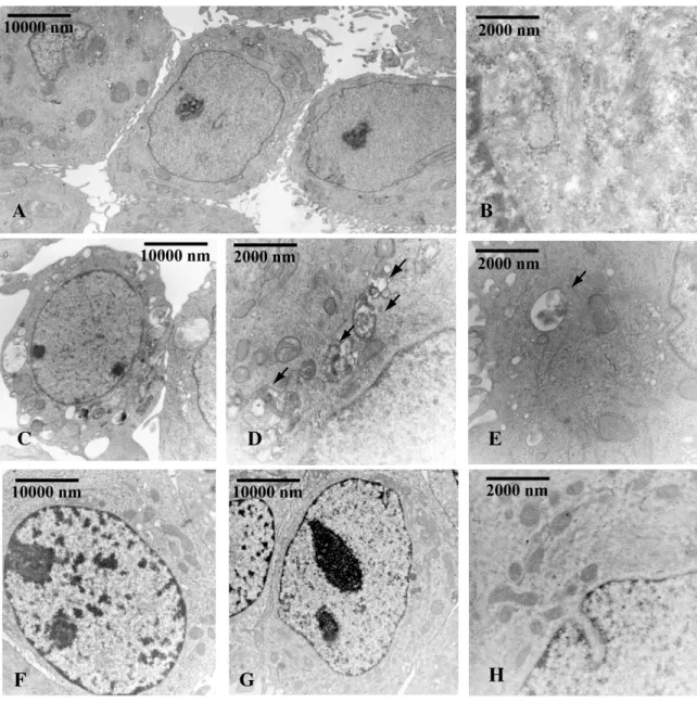
The ultrastructural effects of the F3aAAA over-expression in TamR/TetOn-AAA cells and those of FoxO3a silencing in wtMCF-7 cells have been assessed by TEM analysis. Most of the Dox-treated TamR/TetOn-AAA cells ( 80%), over-expressing F3aAAA, showed clear signs of injury characterized by rarefaction of the nuclear chromatin and cytoplasm, with formation of electron-dense bodies, vacuoles and lipid droplets, while 90% of Dox-treated TamR/TetOn-V cells appeared normal, with complete cell organelles, and well-distributed chromatin, mitochondria and bundles of tonofilaments (Fig. 4A). Moreover, a consistently greater amount of apoptotic cells were found in the surnatants collected from TamR/TetOn-AAA cells compared to controls (data not shown).
TUNEL assay confirmed this result, with Dox treated TamR/TetOn-AAA showing a much higher level of apoptosis compared to Dox treated TamR/TetOn-V (Fig. 4B).
On the other hand, FoxO3a silencing in 4-OHT-treated wtMCF-7 cells resulted in a significant reduction of cell damage (95% of intact cells) respect to 4-OHT-treated siScramble samples (75% of intact cells), maintaining the morphology of control, non-treated, cells (siScramble and siF3a samples, both showing 90% of intact cells), with well-preserved cellular elements. A small percentage of apoptotic cells were only found in 4-OHT treated controls (data not shown). TUNEL assay gave similar results (data not shown).
Figure 4. FoxO3a reactivates apoptosis in Tam resistant cells
A) TamR/TetOn-V and TamR/TetOn-AAA cells were treated with Dox (3µg/ml) for 72 h and processed for TEM analysis. Scale bars: 10 m. Original magnification: x1200. B) TamR/TetOn-V and TamR/TetOn-AAA cells were treated as in (A) and processed for TUNEL assay. DAPI was used to counterstain the nuclei. Apoptotic cells were photographed at 10x magnification and then counted using Image J software. Graph on the right represent the corresponding apoptotic index (% apoptotic cells/total cell number in the field).
Proteomic analysis of TamR cells expressing active FoxO3a: the impact on several proteins controlling G1/S cell cycle phase, apoptosis and growth factor signals
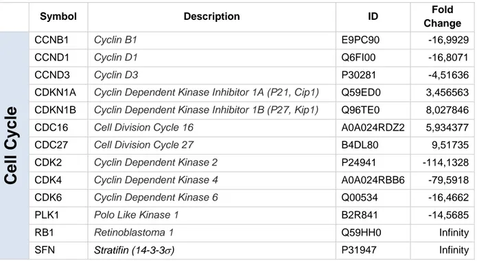
To analyze the molecular mechanisms through which FoxO3a re-expression can restore the response to the antiestrogen, TamR/TetOn-V and TamR/TetOn-AAA cells were next subjected to unbiased proteomics analysis [67]. Among all proteins affected by FoxO3a re-expression in TamR cells, we selected all the modified proteins involved in the regulation of cell cycle, apoptosis and growth factors signals. The results confirm the block of cell cycle in G1/S phase indicating that it may be due to the decrease of several cell cycle regulators such as cyclin D1 and cyclin D3, of CDK4, and CDK2 and an increase in p21Waf1 and p27Kip1 cyclin dependent kinase inhibitors that are involved in G1/S phase transition and RB1. The reduced mitosis is also underlined by the increased expression of CDC16 and CDC27, components of the anaphase promoting complex/cyclosome, and by the reduced expression of PLK1 and by the increase in 14-3-3 expression (Table 1).
Table 1. Some cell cycle controlling proteins regulated modified by nuclear FoxO3a expression in TamR cells (fold change versus vector).
Symbol Description ID Fold
Change
Cell
Cycle
CCNB1 Cyclin B1 E9PC90 -16,9929 CCND1 Cyclin D1 Q6FI00 -16,8071 CCND3 Cyclin D3 P30281 -4,51636CDKN1A Cyclin Dependent Kinase Inhibitor 1A (P21, Cip1) Q59ED0 3,456563 CDKN1B Cyclin Dependent Kinase Inhibitor 1B (P27, Kip1) Q96TE0 8,027846
CDC16 Cell Division Cycle 16 A0A024RDZ2 5,934377
CDC27 Cell Division Cycle 27 B4DL80 9,51735
CDK2 Cyclin Dependent Kinase 2 P24941 -114,1328
CDK4 Cyclin Dependent Kinase 4 A0A024RBB6 -79,5918
CDK6 Cyclin Dependent Kinase 6 Q00534 -16,4662
PLK1 Polo Like Kinase 1 B2R841 -14,5685
RB1 Retinoblastoma 1 Q59HH0 Infinity
Alterations in cell cycle regulation are paralleled by an increase in the expression of pro-apoptotic genes such as AIFM1, BAD, BCLAF1, Caspase 2, 6, 7, and 9, DIABLO and PARP1 indicating restoration of the apoptotic events in response to 4-OHT treatment (Table 2).
Table 2. Some Pro-apoptotic proteins up-regulated by nuclear FoxO3a expression in TamR cells (fold change versus vector).
Symbol Description ID Fold
Change
Apo
pt
osis
AIFM1 Apoptosis inducing factor, mitochondria associated 1 O95831 14,924 BAD BCL2 associated agonist of cell death A0A024R562 4,126 BCLAF1 BCL2 Associated Transcription Factor 1 B7Z8J9 1058,722
CASP2 Caspase 2 P42575 10,106
CASP6 Caspase 6 P55212 14,237
CASP7 Caspase 7 P55210 14,997
CASP8 Caspase 8 B5BU46 Infinity
DIABLO Diablo IAP-binding mitochondrial protein Q9NR28 24,874 PARP1 Poly(ADP-ribose) polymerase 1 A0A024R3T8 Infinity
Consistent with cell growth inhibition and with apoptotic induction, unbiased proteomic analysis revealed a down-regulation of several proteins involved in growth factor signaling (Table 3).
Table 3. Some Growth factors signaling proteins down-regulated by nuclear FoxO3a expression in TamR cells (fold change versus vector).
Symbol Description ID Fold
Change
G
F si
gn
a
ling
AKT1 AKT serine/threonine kinase 1 P31749 -14,180
AKT2 AKT serine/threonine kinase 2 P31751 -441,405
AKT3 AKT serine/threonine kinase 3 Q9Y243 -16,161
IRS1 insulin receptor substrate 1 A0A024R49
9
-10000,000
MTOR mechanistic target of rapamycin P42345 -10000,000
PIK3C3 phosphatidylinositol 3-kinase catalytic subunit type 3 B4DPV9 -40,509 PIK3C2A phosphatidylinositol-4-phosphate 3-kinase catalytic
subunit type 2 alpha L7RRS0 -14,866
PIK3CB phosphatidylinositol-4,5-bisphosphate 3-kinase
catalytic subunit beta P42338 -10000,000
RAF1 Raf-1 proto-oncogene, serine/threonine kinase P04049 -10000,000
Nuclear FoxO3a down regulates three Akt family members and several PI3K catalytic subunit. In addition, other important proteins involved in growth factor pathways, such as IRS1, SHC1 and RAF1, resulted markedly reduced. Altogether, proteomic analysis indicates that nuclear FoxO3a restores the sensitivity to 4-OHT by switching off the hyperactivation of Growth factor signals.
In silico validation of the clinical relevance of FoxO3a in human ER+ BC patients
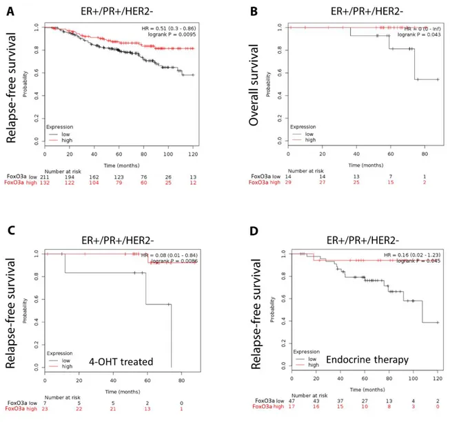
To assess the possible clinical relevance of FoxO3a in the response to endocrine treatment and in the acquisition of tamoxifen resistance, we questioned if its levels (in terms of mRNA) might be indicative of the potential outcome of ER+ human BC patients cohorts, with long-term follow-up (more than 5 years). The Kaplan-Meier (K-M) relapse-free survival (RFS) curve (Fig. 5A) demonstrated a poorer prognosis for Luminal A subtype BC patients expressing low level of FoxO3a mRNA (log-rank test, p=0.0095). The relevance of FoxO3a mRNA expression was also evident by evaluating the overall survival in the Luminal A subtype cohort (Fig. 5B) (log-rank test, p=0.043). Anyway, we have to consider this result as indicative due to the small number of patients. Interestingly, in a small part of the luminal A subtype cohort (n. 30) treated with 4-OHT (Fig. 5C), the K-M curve reported a high probability of RFS in the group expressing high FoxO3a levels (log-rank test, p=0.0086). Even in this case, the results are only indicative due to the small number of patients. We were not able to evaluate the overall survival in the luminal A subtype cohort treated with 4-OHT since only 19 samples were found.
Finally, since low levels of FoxO3a mRNA were associated with disease progression in patients that received endocrine therapy (Fig. 5D), this may probably be indicative of a clinical association with endocrine therapy-resistance. Thus, low levels of FoxO3a could be used to identify high-risk ER+ BC patients that might benefit from potential adjuvant treatments that are able to increase FoxO3a expression, restoring the sensitivity to 4-OHT therapy.
Figure 5. The relapse-free and overall survival of patients according to FoxO3a expression.
All graphs are calculated using microarray data from luminal A subtype cohorts of 343 patients (relapse-free survival, panel A), 43 patients (overall survival, panel B), 30 patients (relapse-free survival 4-OHT treated, panel C), and 64 patients (relapse-free survival endocrine therapy, panel D) determined using an online survival analysis tool. Kaplan-Meier correlations are plotted for high (above median, in red) and low (below median, in black) FoxO3a gene expression. Biased and outlier array data were excluded from the analysis. Hazard-ratios were calculated, at the best auto-selected cut-off, and p-values were calculated using the logrank test and plotted in R. M curves were also generated online using the K-M-plotter (as high-resolution TIFF files), using univariate analysis: http://kmplot.com/analysis/index.php?p = service&cancer = breast. This allowed us to directly perform in silico validation of the clinical relevance of FoxO3a in human BC patients. The most updated version of the database was used for all analyses.
The antiepileptic drug Lamotrigine restores the sensitivity to Tam treatment through FoxO3a re-expression in tamoxifen resistant xenografts: perspective on an off-label use
Ongoing studies in our laboratory suggest that a promising pharmacological candidate for such an adjuvant therapy in patients who result refractory to the antiestrogen treatment is lamotrigine (LTG), a well-known AED which is able to increase FoxO3a expression in BCC (manuscript in preparation). Thus, in order to evaluate if LTG causes FoxO3a induction also in vivo and, consequently, if it is able to restore the sensitivity to the anti-hormonal therapy, the effects of LTG on the development of TamR derived breast carcinomas, was evaluated in mice models. To this aim, female nude mice, bearing TamR cells-derived tumor xenografts into the intrascapular region, were treated with LTG (20 mg/kg/die) on the basis of our in vitro results (manuscript in preparation) and on pertinent literature [68]. Mice well tolerated all in vivo procedures, since no changes in body weight (data not shown), in motor function or in food and water consumption was observed. In addition, no significant difference in the mean weights or histologic features of the major organs (liver, lung, spleen, and kidney) after sacrifice was observed between vehicle-treated mice and those that received LTG treatment. On the other hand, a significant reduction in tumor volume ( 50%) was observed in mice administered with 20 mg/ml LTG (Fig. 6A and B).
To distinguish the xenograft from the mouse tissue, immunohistochemistry was performed on xenograft sections with hematoxylin and eosin which revealed that xenografts were primarily composed of tumor epithelial cells, as also confirmed by immunostaining for the human epithelial cell marker cytokeratin 18 (Cyt18) (Fig. 6C).
The antiproliferative effect mediated by LTG was confirmed by reduced expressions of the proliferation marker Ki-67 in LTG treated xenografts compared to those deriving from control mice (Fig. 6D). These results are congruent with the strong increase in FoxO3a expression observed both in tissue sections and in protein extracts from LTG treated tumors respect to control tumors (Fig. 6D and E). Moreover, Cyclin D1 downregulation associated to FoxO3a upregulation perfectly fits with other authors’ observations [69] and with our proteomics results. Notably, the immunohistochemical
analysis of LTG samples clearly shows a markedly nuclear localization of FoxO3a (Fig. 6D), which is consistent with an antiproliferative effect. Taken together, these results indicate that LTG might represent a useful therapeutic tool to be exploited as an adjuvant treatment in patients who failed to respond to the antiestrogen regimen.
Figure 6. Lamotrigine induces FoxO3a in vivo and inhibits the growth of TamR derived tumor xenografts. A) Xenografts were established with TamR cells in female mice implanted with E2 and, successively, Tam pellets (see Materials and Methods for details). One group was treated with 20 mg/kg/day LTG (n = 5) and a second group with vehicle alone (n = 5). Tumor mass was measured at indicated time points with a caliper. *, P < 0.05, treated versus control group. B) Representative images of explanted tumors at day 28. Scale bar, 0.3 cm. C) Representative tumor sections from mice at 28 days were formalin fixed, paraffin embedded, sectioned, and stained with hematoxylin and eosin Y (H&E) or incubated with antibodies directed against the epithelial marker cytokeratin 18 (Cyt 18). D) Immunostaing were also performed for the proliferation marker Ki67 and for FoxO3a. The insets in C) and D) are representative images of negative control sections, where the primary antibody was replaced by nonimmune serum. E) FoxO3a and CyclinD1expression was assessed in protein extracts from xenografts excised from control mice and LTG treated mice. β-actin was used as loading control.
DISCUSSION
Approximately 70% of BCs do express ER and are subjected to endocrine-based therapies (selective ER modifiers, e.g. tamoxifen, selective ER down-regulators, e.g., fulvestrant, and aromatase inhibitors, e.g., letrozole, anastrozole and excemestane), but the development of endocrine resistance is highly common and it still remains an unsolved problem. Thus, elucidating the molecular mechanisms leading to hormone insensitivity of BC is important to improve the efficacy of endocrine therapy.
To this aim, we addressed our studies on the involvement of tumor suppressor FoxO3a in the acquisition of resistance to tamoxifen in BC. Indeed, several data from our and other’s laboratories, established the existence of a functional interaction between FoxOs and ER in BCC (reviewed in [70]). In particular, FoxO3a seems to have a protective role in ER+ breast tumors [55, 71, 72], therefore, it is reasonable to suppose that FoxO3a deregulation could favor the acquisition of a phenotype resistant to treatments targeting ER, such as tamoxifen.
In line with this assumption, here we report that ER+ cells chronically exposed to Tam (TamR cells) show a strong decrease in FoxO3a mRNA and protein expression and in its nuclear localization, if compared to parental, Tam sensitive, cell lines. Notably, the phenomenon was not peculiar to a specific cell line (i.e. MCF-7), but it was observed in other BCC (e.g. ZR-75, data not shown). The mechanism underlying FoxO3a downregulation in TamR cells does not seem to directly depend on the estrogen/antiestrogen action, nor on epigenetic modifications of FoxO3a regulatory regions that might have been caused by long-term Tam exposure [73] (data not shown).
Two additional mechanisms are currently under investigation in our laboratory: i) the involvement of FoxO3a transcriptional regulators p53 and E2F-1 (reviewed in [66]), whose levels are decreased in TamR cells compared to parental cells, and ii) post-translational modifications of FoxO3a, which results hyper-phosphorylated by hyperactive ERK1/2 MAPK in TamR cells, leading to FoxO3a degradation through the MDM2-mediated ubiquitin-proteasome pathway [50]. These results will be included in a forthcoming article.
Instead, here we questioned if FoxO3a re-activation might restore the sensitivity to Tam in TamR cells. Indeed, the over-expression of constitutively active FoxO3a (F3aAAA) in TamR cells was able to re-establish the anti-proliferative effect of the antiestrogen (Fig. 2A and B), by triggering the apoptotic pathway, as confirmed by TUNEL (Fig. 4B) and Annexin V (data not shown) assays, as well as by TEM observations (Fig. 4A).
Oddly, proteomics results and WB analysis did not reveal any significant change in the levels of the pro-apoptotic factors Bim and Fas-L, whose expression is notoriously regulated by FoxO3a [74], in F3aAAA overexpressing clones compared to relative controls. Neither we were able to obtain the induction of these two proteins in cells transiently expressing F3aAAA (see Materials and Methods), nor their downregulation in parental cells silenced for FoxO3a. However other pro-apoptotic proteins like BAD, PARP, TP53I3 (Tumor Protein P53 Inducible Protein 3) and several caspases were upregulated, some of these even ∞ folds (Tables 1-3). Since none of these other apoptotic markers have ever been reported as FoxO3a target genes, these novel findings will be further investigated. In addition, although FoxO3a seems to be involved in the regulation of autophagy [75] proteomics analysis, nor our TEM observations revealed any significant alteration in mediators of the autophagy response (e.g. p67, Ulk1, PI3K class III, AMBRA, UVRAG, Beclin1, Atg5, LC3 II and LC3B) in F3aAAA expressing TamR cells (data not shown).
The increased apoptosis is paralleled by a down regulation of several cell cycle controlling proteins (Table1) such as Cyclin D1, Cyclin D3, CDK4, and CDK6 all involved in the transition from G1 to S phase of the cycle, and CDK2 that drives the entrance in S phase of the cycle. Moreover, the blockage of cell cycle is ensured by the down regulation of Cyclin B1 involved in the G2 phase and PLK1 (Polo Like kinase 1) involved in the M phase. In addition, nuclear FoxO3a increases the expression of CDC16, CDC27 (both components of the anaphase-promoting complex), and p21Cip1, p27Kip1, RB1 and SFN, all inhibitors of the transition from the G1 to the S phase of cell cycle. Interestingly, proteomic analysis revealed a deeper involvement of FoxO3a in the modulation of cell cycle regulating proteins (Reviewed in [76]). Here we reported that active FoxO3a modifies the
expression of additional cell cycle regulators, at least in TamR breast cancer cells, such as CDC16, CDC27, CDK2, CDK4, CDK6 and SFN that have never been described before and that require a deeper study in the future. The ability of nuclear FoxO3a to restore the sensitivity of breast cancer cell to the hormonal treatment is, additionally, highlighted by the down regulation of several proteins involved in the signaling pathways activated by growth factors. Proteomic analysis revealed a down regulation of several components of the PI3K/AKT pathway, the major inhibitor of FoxO3a activity (Table 3). In addition, the majority of the growth factor signals are down-regulated since both adaptor proteins such as IRS1 and SHC1, and transductional factors such as RAF1 are markedly down regulated in FoxO3AAA expressing TamR breast cancer cells.
FoxO3a involvement in mediating the cellular response to Tam treatment was confirmed by the inhibition of apoptosis and the increased cell proliferation observed in FoxO3a-silenced wtMCF-7 cells, particularly in Tam treated samples (an accurate analysis of the mechanism through which FoxO3a mediates the antiproliferative effect of Tam in wtMCF-7 is ongoing). Indeed, low FoxO3a expression stemming from FoxO3a silencing, reflects somehow the FoxO3a downregulation observed in TamR cells, thus mimicking the behavior of a resistant phenotype.
In agreement with these findings, the K-M survival curves of a population of breast cancer patients subjected to Tam therapy suggest that high levels of FoxO3a strongly correlate with a positive response to the tamoxifen treatment and, consequently, with a long-term relapse free survival. Thus, FoxO3a expression might not only be considered a general favorable prognostic marker in breast cancer [71, 77], but might also be predictive of the potential efficacy of the antiestrogen therapy. Nevertheless, it cannot be excluded that the low levels of FoxO3a observed in ER+ BC patients might be, at least for the subgroup receiving tamoxifen therapy, a consequence of the gradual acquisition of a Tam resistant phenotype (Fig. 4).
Our results confirm the need of developing anti-cancer therapies exploiting FoxO3a in BC patients with acquired resistance to Tam treatment. Indeed, FoxO3a reactivation could represent an adjuvant to Tam therapy by restoring the sensitivity to the antiestrogen.
Several drugs have been reported to increase FoxO3a activity and promote apoptosis by targeting the growth factors, PI3k/PTEN, Akt and MAPK pathways in breast cancer cell lines. Moreover two clinical trials (one using a combination of the pure antiestrogen fulvestrant with a PI3k inhibitor and the other using reparaxin, an IL-8 receptor antagonist) aimed to assess, among the other endpoints, FoxO3a status, just ended [78].
Here we add a new molecule to the list of FoxO3a activating drugs, showing how the well-known AED Lamotrigine (LTG; Lamictal) [59], widely used in the clinic, has a strong anti-proliferative activity on xenografts tumors deriving from TamR cells. The restored response to Tam treatment was paralleled by a significant increased expression and mainly nuclear localization of FoxO3a in vivo. Taken together, our results show that: 1) FoxO3a is phosphorylated, thus inactivated, by hyper-active growth factors signaling in TamR cells; 2) FoxO3a re-expression is able to overcome tamoxifen resistance; 3) low levels of FoxO3a are predictive of the failure of the antiestrogen therapy; 4) the AED LTG might represent a good candidate for adjuvant endocrine therapy in patients refractory to Tam treatment.
Reference
1. Siegel, R., et al., Cancer statistics, 2011: the impact of eliminating socioeconomic and racial
disparities on premature cancer deaths. CA Cancer J Clin, 2011. 61(4): p. 212-36.
2. Keen, J.C. and N.E. Davidson, The biology of breast carcinoma. Cancer, 2003. 97(3 Suppl): p. 825-33. 3. McCarty, K.S., Jr., et al., Correlation of estrogen and progesterone receptors with histologic
differentiation in mammary carcinoma. Cancer, 1980. 46(12 Suppl): p. 2851-8.
4. Mossler, J.A., K.S. McCarty, Jr., and W.W. Johnston, The correlation of cytologic grade and steroid receptor content in effusions of metastatic breast carcinoma. Acta Cytol, 1981. 25(6): p. 653-8. 5. Osborne, C.K., Steroid hormone receptors in breast cancer management. Breast Cancer Res Treat,
1998. 51(3): p. 227-38.
6. Banerjee, S., et al., 17alpha-estradiol-induced VEGF-A expression in rat pituitary tumor cells is mediated through ER independent but PI3K-Akt dependent signaling pathway. Biochem Biophys Res Commun, 2003. 300(1): p. 209-15.
7. Brzozowski, A.M., et al., Molecular basis of agonism and antagonism in the oestrogen receptor. Nature, 1997. 389(6652): p. 753-8.
8. Shiau, A.K., et al., The structural basis of estrogen receptor/coactivator recognition and the
antagonism of this interaction by tamoxifen. Cell, 1998. 95(7): p. 927-37.
9. Kushner, P.J., et al., Estrogen receptor pathways to AP-1. J Steroid Biochem Mol Biol, 2000. 74(5): p. 311-7.
10. Jordan, V.C., et al., A monohydroxylated metabolite of tamoxifen with potent antioestrogenic activity. J Endocrinol, 1977. 75(2): p. 305-16.
11. Allen, K.E., E.R. Clark, and V.C. Jordan, Evidence for the metabolic activation of non-steroidal antioestrogens: a study of structure-activity relationships. Br J Pharmacol, 1980. 71(1): p. 83-91. 12. Clarke, R., J.J. Tyson, and J.M. Dixon, Endocrine resistance in breast cancer--An overview and
13. Ali, S. and R.C. Coombes, Endocrine-responsive breast cancer and strategies for combating
resistance. Nat Rev Cancer, 2002. 2(2): p. 101-12.
14. Elkak, A.E. and K. Mokbel, Pure antiestrogens and breast cancer. Curr Med Res Opin, 2001. 17(4): p. 282-9.
15. Chang, X.Z., et al., Identification of the functional role of peroxiredoxin 6 in the progression of breast cancer. Breast Cancer Res, 2007. 9(6): p. R76.
16. Robertson, J.F., Current role of endocrine therapy in the management of breast cancer. Breast
Cancer, 2002. 9(4): p. 276-81.
17. Nicholson, R.I. and J.M. Gee, Oestrogen and growth factor cross-talk and endocrine insensitivity and acquired resistance in breast cancer. Br J Cancer, 2000. 82(3): p. 501-13.
18. Nicholson, R.I., et al., Modulation of epidermal growth factor receptor in endocrine-resistant,
estrogen-receptor-positive breast cancer. Ann N Y Acad Sci, 2002. 963: p. 104-15.
19. Ring, A. and M. Dowsett, Mechanisms of tamoxifen resistance. Endocr Relat Cancer, 2004. 11(4): p. 643-58.
20. Brunner, N., et al., MCF7/LCC2: a 4-hydroxytamoxifen resistant human breast cancer variant that retains sensitivity to the steroidal antiestrogen ICI 182,780. Cancer Res, 1993. 53(14): p. 3229-32. 21. Johnston, S.R., et al., Changes in estrogen receptor, progesterone receptor, and pS2 expression in
tamoxifen-resistant human breast cancer. Cancer Res, 1995. 55(15): p. 3331-8.
22. Nicholson, R.I., et al., Growth factor signalling in endocrine and anti-growth factor resistant breast cancer. Rev Endocr Metab Disord, 2007. 8(3): p. 241-53.
23. Musgrove, E.A. and R.L. Sutherland, Biological determinants of endocrine resistance in breast
cancer. Nat Rev Cancer, 2009. 9(9): p. 631-43.
24. Gee, J.M., et al., Phosphorylation of ERK1/2 mitogen-activated protein kinase is associated with poor response to anti-hormonal therapy and decreased patient survival in clinical breast cancer. Int J Cancer, 2001. 95(4): p. 247-54.
25. Campbell, R.A., et al., Phosphatidylinositol 3-kinase/AKT-mediated activation of estrogen receptor alpha: a new model for anti-estrogen resistance. J Biol Chem, 2001. 276(13): p. 9817-24.
26. Faridi, J., et al., Expression of constitutively active Akt-3 in MCF-7 breast cancer cells reverses the
estrogen and tamoxifen responsivity of these cells in vivo. Clin Cancer Res, 2003. 9(8): p. 2933-9. 27. DeGraffenried, L.A., et al., Eicosapentaenoic acid restores tamoxifen sensitivity in breast cancer cells
with high Akt activity. Ann Oncol, 2003. 14(7): p. 1051-6.
28. Kandel, E.S. and N. Hay, The regulation and activities of the multifunctional serine/threonine kinase Akt/PKB. Exp Cell Res, 1999. 253(1): p. 210-29.
29. Franke, T.F., et al., Direct regulation of the Akt proto-oncogene product by
phosphatidylinositol-3,4-bisphosphate. Science, 1997. 275(5300): p. 665-8.
30. Brunet, A., et al., Akt promotes cell survival by phosphorylating and inhibiting a Forkhead transcription factor. Cell, 1999. 96(6): p. 857-68.
31. Osborne, C.K., et al., Estrogen receptor: current understanding of its activation and modulation. Clin
Cancer Res, 2001. 7(12 Suppl): p. 4338s-4342s; discussion 4411s-4412s.
32. Greer, E.L. and A. Brunet, FOXO transcription factors in ageing and cancer. Acta Physiol (Oxf), 2008. 192(1): p. 19-28.
33. Wang, X., W.R. Chen, and D. Xing, A pathway from JNK through decreased ERK and Akt activities for FOXO3a nuclear translocation in response to UV irradiation. J Cell Physiol, 2012. 227(3): p. 1168-78. 34. Calnan, D.R. and A. Brunet, The FoxO code. Oncogene, 2008. 27(16): p. 2276-88.
35. Coomans de Brachene, A. and J.B. Demoulin, FOXO transcription factors in cancer development and
therapy. Cell Mol Life Sci, 2016. 73(6): p. 1159-72.
36. Van Der Heide, L.P., M.F. Hoekman, and M.P. Smidt, The ins and outs of FoxO shuttling:
mechanisms of FoxO translocation and transcriptional regulation. Biochem J, 2004. 380(Pt 2): p. 297-309.
37. Shaul, Y.D. and R. Seger, The MEK/ERK cascade: from signaling specificity to diverse functions. Biochim Biophys Acta, 2007. 1773(8): p. 1213-26.
38. Pimienta, G. and J. Pascual, Canonical and alternative MAPK signaling. Cell Cycle, 2007. 6(21): p. 2628-32.
39. Schaeffer, H.J. and M.J. Weber, Mitogen-activated protein kinases: specific messages from
ubiquitous messengers. Mol Cell Biol, 1999. 19(4): p. 2435-44.
40. Han, J. and R.J. Ulevitch, Emerging targets for anti-inflammatory therapy. Nat Cell Biol, 1999. 1(2): p. E39-40.
41. Davis, R.J., Signal transduction by the JNK group of MAP kinases. Cell, 2000. 103(2): p. 239-52. 42. Robinson, M.J. and M.H. Cobb, Mitogen-activated protein kinase pathways. Curr Opin Cell Biol,
1997. 9(2): p. 180-6.
43. Woessmann, W., Y.H. Meng, and N.F. Mivechi, An essential role for mitogen-activated protein kinases, ERKs, in preventing heat-induced cell death. J Cell Biochem, 1999. 74(4): p. 648-62. 44. Kyriakis, J.M. and J. Avruch, Sounding the alarm: protein kinase cascades activated by stress and
inflammation. J Biol Chem, 1996. 271(40): p. 24313-6.
45. Ono, K. and J. Han, The p38 signal transduction pathway: activation and function. Cell Signal, 2000. 12(1): p. 1-13.
46. Lubinus, M., et al., Independent effects of platelet-derived growth factor isoforms on
mitogen-activated protein kinase activation and mitogenesis in human dermal fibroblasts. J Biol Chem, 1994. 269(13): p. 9822-5.
47. Cappellini, A., et al., The phosphoinositide 3-kinase/Akt pathway regulates cell cycle progression of HL60 human leukemia cells through cytoplasmic relocalization of the cyclin-dependent kinase inhibitor p27(Kip1) and control of cyclin D1 expression. Leukemia, 2003. 17(11): p. 2157-67. 48. Uddin, S., et al., Role of phosphatidylinositol 3'-kinase/AKT pathway in diffuse large B-cell
lymphoma survival. Blood, 2006. 108(13): p. 4178-86.
49. Stahl, M., et al., The forkhead transcription factor FoxO regulates transcription of p27Kip1 and Bim in response to IL-2. J Immunol, 2002. 168(10): p. 5024-31.
50. Yang, J.Y., et al., ERK promotes tumorigenesis by inhibiting FOXO3a via MDM2-mediated
degradation. Nat Cell Biol, 2008. 10(2): p. 138-48.
51. van den Heuvel, A.P., A. Schulze, and B.M. Burgering, Direct control of caveolin-1 expression by FOXO transcription factors. Biochem J, 2005. 385(Pt 3): p. 795-802.