University of Naples “Federico II”
SCUOLA POLITECNICA E DELLE SCIENZE DI BASE
AREA DIDATTICA DI SCIENZE MATEMATICHE, FISICHE E NATURALI
PhD in Biology (XXXI Cycle)
PhD Coordinator: Salvatore Cozzolino
Analysis of the genes involved in floral symmetry
of the orchid Orchis italica
PhD Student: PhD Supervisor:
Maria Carmen Valoroso Prof. Serena Aceto
1
Summary
Abstract ... 3
CHAPTER 1 – The molecular basis of flower symmetry ... 4
1. Introduction ... 4
1.1 The Orchidaceae family: a focus on Orchis italica ... 4
1.2 The molecular basis of bilateral symmetry of the flower ... 6
1.2.1The TCP family: CYCLOIDEA and DICHOTOMA ... 7
1.2.2 The MYB family: RADIALIS, DIVARICATA and their interacting factor ... 9
1.2.3 The molecular network underlying the establishment of bilateral symmetry in A. majus... 13
1.3 Aim of the work ... 14
2. Results and Discussion ... 15
2.1 Identification and structure of DIV- and RAD-like genes in orchids ... 15
• Identification of DIV- and RAD-like transcripts ... 15
• Genome organization of the orchid DIV- and RAD-like genes ... 16
2.2 Phylogenetic and evolutionary analysis of the orchid DIV- and RAD-like genes ... 17
• Phylogenetic analysis ... 17
2.3 Identification of the DRIF-like genes of O. italica ... 20
2.4 Expression analysis of the DIV-, RAD- and DRIF-like genes of O. italica ... 20
2.5 Protein interaction among DIV, RAD and DRIF1 in O. italica ... 25
CHAPTER 2 – The MADS-box genes ... 27
1. Introduction ... 27
1.1 The MADS-box genes: from flower development to bilateral symmetry ... 27
1.2 The ABCDE model of flower development ... 28
• The orchid code and the Homeotic Orchid Tepal (HOT) model ... 31
1.3 Aim of work... 34
2. Results and Discussion ... 35
2.1 Identification of the MADS-box genes of O. italica ... 35
2
2.1.2 Class II MADS-box genes ... 37
• The MIKC* genes ... 37
• The SOC, SVP, ANR1, AGL12 and OsMADS32 genes ... 38
• The AGL6-class genes ... 38
• The C- and D- class genes ... 43
• The E-class genes ... 44
2.2 Protein-protein interaction between B-class MADS-box and MYB transcription factors ... 46
Conclusions ... 48
Material and Methods ... 49
1. Plant material ... 49
2. Identification of the orchid MYB and MADS-box transcripts ... 49
• Identification of the orchid DIV and RAD-like genes ... 49
• Identification of the DRIF and AGL6 transcripts of O. italica ... 50
• Identification of the MADS-box transcripts of O. italica ... 50
3. Analysis of the Orchis italica transcripts ... 50
4. Gene structure analysis ... 51
• Analysis of DIV- and RAD-like genes ... 51
5. Phylogenetic and evolutionary analysis ... 52
• Analysis of the DIV- and RAD-like genes ... 52
• Analysis of the MADS-box genes ... 52
6. Expression Analysis ... 53
• In situ hybridization of the DIV RAD and DRIF1 transcripts ... 53
• Real Time PCR ... 53
7. Yeast two-hybrid analysis ... 54
Appendix ... 55
3
Abstract
Orchis italica è un’orchidea selvatica mediterranea appartenente alla sottofamiglia
Orchidoideae delle Orchidaceae, una delle più vaste famiglie di piante a fiore. Lo scopo di questo lavoro è lo studio dei geni coinvolti nella determinazione della simmetria bilaterale del fiore di O. italica. Studi sulla determinazione della simmetria fiorale effettuati sulla specie modello Antirrhinum majus hanno evidenziato che cinque geni, due appartenenti alla famiglia TCP (CYC e DICH) e tre appartenenti alla famiglia MYB di fattori di trascrizione (DIV, RAD e DRIF), svolgono un ruolo cruciale nella formazione del fiore zigomorfo. In A.
majus queste proteine interagiscono tra di loro attraverso un meccanismo antagonistico che
permette la formazione nel fiore di una regione dorsale e una ventrale. In O. italica, l’analisi del trascrittoma dell’infiorescenza matura ha evidenziato la presenza di 8 trascritti DIV-like, 4 trascritti RAD-like e due trascritti DRIF1/2. L’organizzazione genomica dei geni DIV-like e
RAD-like presenta un singolo introne, fatta eccezione per un unico gene RAD-like che risulta
essere privo di introni. L’analisi evolutiva ha evidenziato che sulle regioni codificanti dei geni
DIV-like e RAD-like agisce selezione purificante. I geni DIV, RAD e DRIF di O. italica hanno
un’espressione conservata rispetto ad A. majus. L’analisi delle interazioni proteiche tra i fattori di trascrizione MYB di O. italica ha dimostrato che il modello alla base della determinazione della simmetria bilaterale sembra essere conservato anche in orchidea. I geni MADS-box di tipo II MIKCC, classificati in cinque classi funzionali ABCDE, hanno un
ruolo cruciale della determinazione della struttura e dell’organizzazione fiorale. In particolare, in orchidea, secondo il modello “orchid code”, i geni MADS-box appartenenti alla classe B hanno un ruolo fondamentale nella formazione della struttura zigomorfa del fiore di orchidea. Nell’infiorescenza matura di O. italica sono espressi 29 trascritti MADS-box appartenenti al tipo I e al tipo II. I geni MADS-box di O. italica hanno un profilo di espressione in linea con il modello di sviluppo fiorale “fading borders”.
Poiché entrambe le famiglie geniche, MADS-box e MYB, sono alla base della determinazione fiorale è stato effettuato uno studio sull’interazione proteica tra i fattori di trascrizione MYB (DIV, RAD e DRIF) e le proteine MADS-box di classe B di O. italica. L’analisi condotta ha evidenziato che le singole proteine MADS-box appartenenti alla classe B non sono in grado di interagire con i fattori di trascrizione MYB.
4
CHAPTER 1 – The molecular basis of flower symmetry
1. Introduction
1.1 The Orchidaceae family: a focus on Orchis italica
The origin and diversification of angiosperms, defined by Charles Darwin as “an abominable mystery”, has been subject of many studies during the last 100 years [1].The emergence of molecular techniques has improved the understanding of angiosperm phylogeny and evolution [2-5]. Among the flowering plants, the monocot family Orchidaceae is one of the most species-rich and widespread in the world, adapted to different habitats (epiphytic and terrestrial) and with highly specialized reproductive strategies. This family includes more than 28,000 species divided in five subfamilies: Apostasioideae, Cypripedioideae, Vanilloideae, Epidendroideae and Orchidoideae [6]. Each subfamily includes numerous tribes and subtribes [7, 8] (Fig. 1). The origin of Orchidaceae has been placed 112 million years ago (Mya). The different subfamilies started their diversification 90 Mya and the separation between Orchidoideae and Epidendroideae happened 64 Mya [9].
Despite the high morphological difference among the flowers of the orchid species, they share common features. The orchid flower is generally characterised by bilateral symmetry (zygomorphy) and it is composed by three outer tepals, two lateral inner tepals and an inner medial tepal, called labellum (or lip), highly diversified [10]. The orchid’s reproductive structure, called gynostemium or column, is composed by the fusion of male (stamen/anther) and female (pistil/stigma) tissue. The pollen grains (pollinia) are located at the top of the column, while at its base is located the ovary, whose maturation is triggered by pollination [10, 11] (Fig. 2A). In addition, many orchid species show a resupinate flower, where the pedicel and ovary undergo a 180° degree twisting process resulting in the ventral position of the lip in the mature flower [12] (Fig. 2B).
The shape and pigmentation of the lip and its abaxial orientation in the opposite part of the fertile anther after resupination suggest that these characters are adaptations to specific pollinators [6, 13].
5
Fig. 1 Phylogenetic tree of the Orchidaceae family. Modified from Aceto et al. (2011) [6]. On the right there are images of representative orchid species of the different subfamilies.
Fig. 2 The orchid flower structure and the resupination of the flower. (A) Reprinted from Aceto et
al. (2011) [6]. Structure of an orchid flower with a detail of the reproductive structures. (B) Reprinted from Valoroso et. al (2017) [14]. The diagram shows the position of the organs of the first (pink triangle) and second
6
Orchis italica is an orchid species belonging to the subfamily Orchidoideae. It is one of most
widespread Mediterranean orchids, characterized by a white-purple cluster inflorescence (Fig. 3A) and a lip with white-purple flaps that assume an anthropomorphic form (Fig. 3B).
Fig. 3 Orchis italica. Reprinted from Valoroso et al. (2017) [14]. (A) The inflorescence and (B) the flower of
Orchis italica.
1.2 The molecular basis of bilateral symmetry of the flower
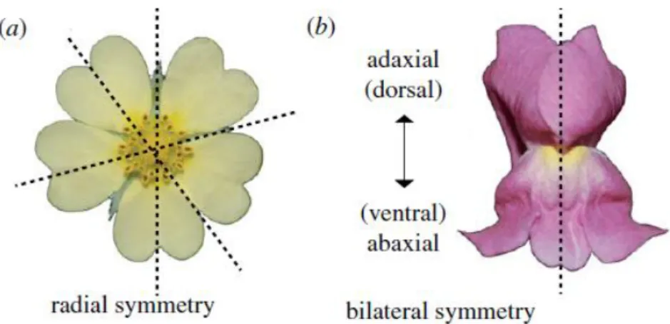
During the evolution of flowering plants, actinomorphy (radial symmetry) (Fig. 4) represents the ancestral state of flower symmetry. The first changes from radial symmetry have evolved during the first angiosperm radiation (Turonian age, late Cretaceous), with the appearance of asymmetric flowers that can be considered “precursors” of zygomorphic flowers [15, 16]. The analysis of fossil records indicates that zygomorphy evolved in several plant lineages during the same period, as well as the rise of some bee families, supporting the idea that there was a coevolution between flower structure, symmetry and specific pollinating insects [17].
7
Fig. 4 Difference between radial and bilateral symmetry of the flower. Reprinted from Hileman
(2014) [18]. (a) Flower with radial symmetry characterized by multiple planes of mirror image symmetry; (b)
flower with bilateral symmetry characterized by a single plane of mirror image symmetry.
The molecular basis underlying the establishment of bilateral symmetry of the flower has been dissected in the model plant Antirrhinum majus (snapdragon), where a key role is played by the genes CYCLOIDEA (CYC), DICHOTOMA (DICH), RADIALIS (RAD),
DIVARICATA (DIV) and DIV and RAD INTERACTING FACTOR 1 (DRIF1).
1.2.1The TCP family: CYCLOIDEA and DICHOTOMA
TCPs are plant-specific transcription factors whose name derives from four proteins that share the TCP domain: TEOSINTE BRANCHED 1 (Tb1) from Zea mays [19], CYCLOIDEA (CYC) from Antirrhinum majus [20] and PROLIFERATING CELL FACTORS 1 and 2 (PCF1 and PCF2) from Oryza sativa [21]. The TCP factors contain two conserved regions: the TCP and the R domain. The predicted structure of the TCP domain is a basic helix–loop–helix (bHLH) composed of 21 amino acids with DNA binding function [22]. The R domain is rich of polar residues (arginine, lysine and glutamic acid) and, together with the TCP domain, forms coiled-coil structures (leucine zippers) [22-24].
Based on the sequence of the TCP domain, the members of this family are divided in two different groups: TCP-P (class I) and TCP-C (class II) [22, 25, 26]. These two classes have different but overlapping consensus binding sites that are GGNCCCAC for class I and G(T/C)GGNCCC for class II [27-29]. Both these consensus motifs are over-represented in the promoters of cell cycle and protein synthesis related genes [28, 30, 31]. The TCP-C factors are further divided into two clades: ECE (CYC/TB1-like), characterized by both the TCP and R domain, and CINCINNATA (CIN), generally showing only the TCP domain [25,
8
26]. Phylogenetic analysis of the ECE clade shows that it is divided in three subgroups (CYC1, CYC2 and CYC3), raised after duplication events from the ancestral ECE sequence [32].
The transcription factors encoded by the TCP genes are involved many processes of plant growth and development [18, 22, 33]. An important role in the establishment of bilateral symmetry is played by the ECE clade, in particular by the CYC and DICH genes [18, 26, 32]. During the evolution of zygomorphy, duplications and mutations within the subgroup CYC2 have probably facilitated the transition from radial to bilateral flower symmetry [34]. In A. majus the CYC and DICH genes are involved in the establishment of the lateral and dorsal part of the flower. The snapdragon wild-type flower (bilateral symmetry) has one ventral, two dorsal and two lateral petals (Fig. 5A). In the wild type flower, CYC is expressed in the dorsal and lateral domains and its expression is stable until the late stage of development [20]. The loss of function cyc mutant (Fig. 5B), named semipeloric, has five or six petals, four with ventral identity and two with combination of lateral and dorsal identity. In this mutant, the bilateral plan of symmetry is misaligned respect to the dorsal-ventral axis. The expression pattern of CYC and the phenotype of the semipeloric cyc mutant have suggested the involvement of this gene in the establishment of the dorsal and lateral identity and in the organ placement of the snapdragon flower [20]. The DICH gene is expressed in the dorsal domain, but differently from CYC, it is restricted to the most dorsal part of the flower. The dich mutants show partial loss of the dorsal petals asymmetry [35]. These evidences have suggested that also the DICH gene is related to the establishment of the identity and asymmetry of the dorsal petals [35].The role of CYC and DICH in the dorsal and lateral asymmetry of A. majus has been further confirmed by the phenotype of the peloric double mutants cyc:dich (Fig. 5C), radially symmetric and with ventral identity of all petals [20].
9
Fig. 5 Wild type and mutant flower of Antirrhinum majus. Reprinted from Luo et al. (1996) [20]. (A) Wild-type, (B) semipeloric cyc mutant and (C) peloric cyc:dich double mutant flower of A. majus, their different petals and diagram of their symmetry.
1.2.2 The MYB family: RADIALIS, DIVARICATA and their interacting factor
The MYB transcription factors have been described in all the eukaryotic organisms [36, 37]. The first MYB gene isolated in plants, more than 30 years ago, was COLORED1 from Zea
mays, involved in the anthocyanin synthesis [38]. The MYB transcription factors are
characterized by the presence of MYB repeats (R), from one to four or more. Each repeat is long 52 amino acids and contains three regularly spaced residues of tryptophan, or other aliphatic amino acids, that form a hydrophobic core [39, 40]. Each repeat adopts a helix-turn-helix conformation with DNA binding and protein-protein interaction function [39, 41]. Based on the number of R repeats, the MYB proteins are divided into three groups [39, 42]:
1. MYB3R: showing three MYB repeats (R1, R2, R3), involved in the control of cyclins during the late G2 and M phase of the cell cycle.
2. R2R3-MYB: with two similar MYB repeats (R2-R3) and very variable C-terminus. Frequently, the C-terminus contains a transcriptional activation or repression domain composed by residues of serine and threonine. These proteins represent the largest
10
part of the plant MYB factors and are implicated in many functions, from metabolism regulation to biosynthesis and floral organ determination.
3. Other MYB-type: this group includes all the other forms of MYB proteins with a variable number of R repeats. Based on their structure, they can be divided into four sub-groups:
➢ R-MYB, with a single R repeat, involved in the chromatin and histone metabolism;
➢ The second sub-group includes proteins related to the evolutionary old R1/R2 class, involved in the circadian clock control;
➢ GARP, involved in the control of organ polarity;
➢ 4R-MYB that contains four repeats of R1/R2 domains.
In A. majus, three genes related to the establishment of bilateral symmetry belong to the MYB family: DIV, RAD and DRIF1.
DIV belongs the R2R3-class of MYB proteins and contains two similar MYB domains [14, 43]. The DIV transcription factor is implicated in the ventralization of the snapdragon flower and it is expressed in all the whorls of the flower. In the loss of function div mutant (radial symmetry) the ventral petals adopt the shape of dorsal and lateral petals (Fig. 6) [43, 44]. The div:cyc mutant has radial symmetry and the ventral petals have lateral identity, as in the single cyc mutant. The div:dich mutant has the ventral petals similar to the lateral petals; however, the dorsal petals resemble the phenotype of the dich single mutant. Finally, in the triple mutant div:cyc:dich (Fig. 6) all the petals have lateral identity, suggesting that the flower domain influenced by DIV is extended to all the flower and that both genes, CYC and
11
Fig. 6 Antirrhinum majus div mutants. Reprinted from Galego and Almeida (2002) [43]. Floral diagram of the wild-type and div mutant (first line) and cyc:dich double and cyc:dich:div triple mutant (second line) of A.
majus.
RAD belongs to the third group of MYB proteins. It is a small protein that contains only one MYB domain and is expressed in the dorsal part of the snapdragon flower [14, 45]. The phenotype of different snapdragon single and double mutants reveals the involvement of the RAD gene in the dorsalization of the flower and its connection with the activity of the
CYC gene. Indeed, all the rad mutants have petals that resemble the wild type ventral ones,
with only the dorsal half of each petal showing dorsal lateral identity (Fig. 7). In addition, the double mutant cyc:rad shows a fully ventralized phenotype, while the double mutant rad:div shows the features of both the single mutants [45].
The DRIF proteins are MYB-like transcription factors that display a MYB-like domain at the N-terminus and a domain with unknown function at the C-terminus [46]. Phylogenetic analysis has shown that DRIFs can be divided into two sub-groups, where the group 1 contains both DRIF1 and DRIF2 of A. majus, involved in the establishment of bilateral symmetry.
12
Fig. 7 The different snapdragon rad mutants. Reprinted from Corley et al. (2005) [45]. (a) A. majus
wild-type flower; (b-g) different rad mutants, from the weaker to the strongest; (h) the peloric cyc:dich mutant.
Previous studies conducted in tomato have shown that the DRIF proteins can interact with DIV and RAD [47]. More recently, these interactions have been highlighted also in A. majus. In particular, both DIV and RAD can interact with DRIF1 and DRIF2 and the cellular localization of these latter proteins depends on the expression of the RAD gene. If the RAD gene is expressed, the DRIF proteins are present in the cytoplasm and form a complex with the RAD protein. If RAD is not expressed, the DRIF proteins are localized in the nucleus and form a complex with the DIV protein [46].
13 1.2.3 The molecular network underlying the establishment of bilateral symmetry in A. majus
At the basis of the establishment of bilateral symmetry of A. majus there is an antagonistic mechanism due to the presence of the DRIF proteins, shown in Fig. 8 [27]. The CYC gene is expressed in all the dorsal parts of the flower, as well as the RAD gene, whereas DICH is expressed only in the more dorsal part [20, 27, 45]. The DIV and DRIF1 genes are both expressed in all the whorls of the flower [43, 46].
In the dorsal part of the flower, the CYC and DICH proteins are able to induce the transcription of the RAD gene, binding its promoter and intron regions [27]. Once activated, the RAD protein goes in the cytoplasm of the cell and here binds the DRIF1 protein, a co-activator of DIV. In this way, in the dorsal part of the flower DIV is expressed but it is not active because DRIF1 is sequestered by RAD in the cytoplasm. In absence of the active DIV function, the proteins CYC, DICH and RAD can induce the dorsalization of the flower. On the contrary, in the ventral part of the flower CYC and DICH are not expressed. In this way, the RAD protein is not expressed and DRIF1 is able to move inside the nucleus, where it binds and activates the DIV protein that can induce the ventralization of the flower.
Fig. 8 Molecular network underlying the establishment of bilateral symmetry in the flower of
14
1.3 Aim of the work
The aim of my PhD project was the study of the molecular basis of the orchid flower development, paying particular attention to the genes involved in floral symmetry of a non-model orchid species, the Mediterranean orchid Orchis italica. Starting from the knowledge of the molecular network of interactions described in the model plant A. majus, the goal of my PhD research project was to verify the existence of evolutionary conserved modules of interaction underlying the flower symmetry in distantly related flowering plants (O. italica and
A. majus).
At the beginning of my PhD research project, the orchid TCP genes had just been described in O. italica [48] and subsequently in Phalaenopsis equestris [49]. Unfortunately, in P.
equestris the role in flower symmetry of the three CYC-like genes identified was only
marginally investigated, whereas in O. italica only a small, conserved fragment of the TB1 (CYC-like) gene was reported. In addition, the orchid MYB genes DIV, RAD and DRIF had never been studied. For these reasons, I decided to undertake the analysis starting from the search of the DIV, RAD and DRIF genes expressed in the inflorescence of O. italica. More in details, the first part of my work can be divided in different points:
• Identification of the DIV-, RAD- and DRIF-like transcripts expressed in the available inflorescence transcriptome of O. italica [48] by in silico analysis;
• Analysis of the gene structure of the DIV- and RAD-like genes of O. italica and P.
equestris;
• Phylogenetic and evolutionary analysis of the orchid DIV- and RAD-like genes;
• Expression analysis of the DIV, RAD and DRIF1 genes of O. italica by real time PCR and in situ hybridization;
• Study of the protein interactions among DIV, RAD and DRIF1 of O. italica by yeast two-hybrid analysis.
15
2. Results and Discussion
2.1 Identification and structure of DIV- and RAD-like genes in orchids
• Identification of DIV- and RAD-like transcripts
The analysis of the inflorescence transcriptome of O. italica reveals the presence of 8 DIV- and 4 RAD-like transcripts.
The 8 DIV-like transcripts contain two conserved MYB domains. Their length ranges from 737 to 1901 bp and they encode for proteins ranging from 175 to 297 amino acids (Tab. 1 - Appendix). Two DIV-like transcripts, OITA_23026 and OITA_13252, are subjected to alternative splicing (Fig. 9). In particular, the alternative transcript of OITA_23026 includes a 111 bp fragment (whose ends are 5’-GT and AG-3’) that generates a premature stop codon within the coding sequence (CDS), leading to the formation of a 117 amino acids protein with a single MYB domain. The two transcripts of OITA_13252 differ in the 5’-UTR, 31 nucleotides upstream the translation start codon, for the presence/absence of a fragment of 88 bp (with 5’-GT and AG-3’ ends) whose presence does not change the protein product.
Fig. 9 Alternatively spliced isoforms of DIV-like transcripts of O. italica. Reprinted from Valoroso
et al. (2017) [14]. The agarose gel electrophoresis shows different PCR amplified fragments of the alternative
isoforms of two DIV-like transcripts. 1) GeneRuler 100 bp DNA ladder (Thermo Scientific); 2) the two different isoforms OITA_13252 and OITA_13252_AS; 3) fragment of OITA_13252; 4) fragment of OITA_23026_AS; 5) the two different isoforms OITA_23026 and OITA_23026_AS. The red asterisks indicate the alternatively spliced isoforms. Forward primers that specifically amplify the intron-retaining isoforms were designed in the region covering the exon–intron junction [14].
16
The in silico analysis of the transcriptome of the orchid Ophrys sphegodes revealed the presence of four DIV-like transcripts. Even though they are not full length, these transcripts were included in the further analyses because Ophrys belongs to the same subfamily of O.
italica (Orchidoideae). It is possible that the low number of DIV-like transcripts found in Ophrys is due to the sequencing and assembly strategies used to obtain the transcriptome
of this species [50]. In addition, the recently released orchid genomes of P. equestris [51] and D. catenatum [52] were analysed, showing the presence of 7 and 8 DIV-like transcripts, respectively (Tab. 1 - Appendix). This result shows that the number of DIV-like genes identified in the present study reflects the real number of DIV-like copies in orchids and that this number is similar to that found in other species (e.g., 8 DIV-like genes in Dipsacales [53]).
The four O. italica RAD-like transcripts present a single MYB domain, a length ranging from 543 to 420 bp (Tab. 1 - Appendix) and encode for proteins ranging from 82 to 94 amino acids. The in silico analysis of the Ophrys transcriptome showed the presence of a single
RAD-like transcript, whereas the search conducted on the genomes of P. equestris and D. catenatum revealed the presence of 5 RAD-like genes (Tab. 1 - Appendix). As for the
DIV-like genes, the number of RAD-DIV-like genes seems to be quite conserved within orchids and similar to that of other species (e.g. six genes in A. majus and Arabidopsis thaliana [54]). The difference relative to the lower number of RAD-like transcripts found in O. italica compared to the other orchid species could be due to the very low expression level of the fifth RAD-like gene, resulting in the absence of its transcript in the assembled transcriptome of O. italica. In alternative, it is possible that in O. italica there are only four RAD-like genes. As currently the assembled genome of O. italica is not available, it is impossible to discriminate between the two hypotheses.
• Genome organization of the orchid DIV- and RAD-like genes
The orchid DIV-like genes of O. italica, P. equestris and D. catenatum include a single intron (Fig. 10C). In O. italica intron position is conserved relative to P. equestris and D. catenatum, with canonical donor and acceptor splicing sites. Intron length of the different DIV-like genes of O. italica varies in a range from 76 to 10,000 bp, as in P. equestris and D. catenatum (Tab. 1 - Appendix). Due to their length, introns of the genes OITA_13233 (5,000 bp),
OITA_8681 (6,000 bp) and OITA_12910 (10,000 bp) were only partially sequenced (Tab.
17
Three out of the four RAD-like genes of O. italica have a single intron (Fig. 10C). In addition, a premature stop codon in OITA_32153 shifts the intron position to the 3’ UTR. Among the examined orchids, this feature, where intron is located within the 3’UTR, with a premature stop codon, is peculiar of O. italica. Although it seems to be an intermediate condition between the intron presence and absence, the orthologs of OITA_32153 in P. equestris and
D. catenatum present a canonical intron within the coding region, suggesting that this intron
shift is a derived character of O. italica. The presence of RAD-like genes with a single intron within the CDS, within the 3’UTR or intronless has been described also in other species, as
A. majus and A. thaliana [54]. These evidences support the possible evolution of the
RAD-like subfamily from an ancestral gene organization with two exons to a more recent structure without introns. However, it cannot be excluded the independent origin of these two gene structures in the different lineages.
The analysis of the three orchid genomes currently released (P. equestris, D. catenatum and the basal species Apostasia shenzenica) has shown that they are very rich in transposable elements, generally localized within the large intron sequences [51, 55]. CENSOR analysis revealed traces of transposable/repetitive elements within the introns of the DIV-like genes OITA_13252 and OITA_13233, of their orthologs of P. equestris and D.
catenatum and in all introns of the RAD-like genes. The large number of
repetitive/transposable elements within the orchid genomes might have a functional significance in the regulation of gene expression. It is possible that the presence of these elements can drive the antisense transcription and/or promote heterochromatin formation, reducing the transcriptional levels [56].
2.2 Phylogenetic and evolutionary analysis of the orchid DIV- and
RAD-like genes
• Phylogenetic analysis
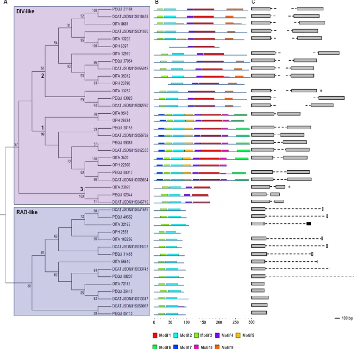
The Maximum Likelihood (ML) tree shown in Fig. 10A is obtained from the DIV- and RAD-like amino acid alignment of the orchid sequences identified by in silico analysis. Both the DIV-like and RAD-like groups present a high bootstrap support value (87%).
Within the DIV-like lineage, the ML tree shows the presence of three subgroups (1-3), all including sequences of O. italica, P. equestris and D. catenatum, whereas the sequences of Ophrys are included only in the subgroups 1 and 2. Two well-supported subgroups are detected within the RAD-like group: the largest subgroup includes the sequences of all the
18
species considered in this study, the smaller one includes only two sequences, one of P.
equestris and one of D. catenatum.
The presence in almost all the subgroups of the ML tree of the DIV- and RAD-like sequences of all the orchid species considered in this study demonstrates that the duplication that has originated the different DIV- and RAD-like genes occurred before the divergence of Orchidoideae and Epidendroideae (64 Mya). In addition, the ML tree topology is in agreement with the results previously reported in Dipsacales [53], suggesting that the origin of these ortholog groups predates the monocot/dicot divergence (140-150 Mya [2]).
The analysis of the conserved domains (Fig. 10B) identified nine amino acid motifs whose distribution reflects the partition of the orchid DIV- and RAD-like genes into three and two subgroups, respectively. The motifs 1, 2 and 3 are part of the MYB DNA binding domain, while the others have unknown function.
Evolutionary analysis was conducted on the coding region of the orchid DIV- and RAD-like genes. The ratio between the mean nonsynonymous and synonymous substitution rates () shows that on these genes is acting purifying selection (<1). Different evolutionary models were compared to verify the existence of variations of the ratios among the different branches of the ML tree of the DIV- and RAD-like genes. The results obtained from evolutionary analysis are summarized in Tab. 2-5 (Appendix).
The one-ratio model assumes an equal for all the branches of the tree, whereas the two- and three-ratio models consider two and three different values, respectively. Within the
DIV-like genes, the three-ratio model fits the data better than the two-ratio models, whereas
within the RAD-like genes the two-ratio model is statistically more supported than the one-ratio model.
The clade model infers different selective pressures on a proportion of sites within a specific branch of the tree. The clade model 2 is statistically more supported than its null model, showing that 34% of sites of the DIV-like subgroup 2 has a different (0.01754) from that of the other subgroups (0.03487). On the contrary, within the RAD-like group, the presence of sites with different selective pressures is not statistically supported.
The sites and branch-sites models assume positive selection in specific sites and branches of the tree. Signals of positive selection are not detected either within the DIV- or RAD-like coding sequences.
19
Although evolutionary analysis highlights strong purifying selection acting on the orchid DIV- and RAD-like coding sequences, the three ortholog groups of the DIV-like genes have different evolutionary rates, whereas the selective constraints of the RAD-like genes are more uniform.
Fig. 10 Phylogeny and genomic organization of the orchid DIV- and RAD-like genes. Reprinted from Valoroso et al. (2017) [14]. (A) ML phylogenetic tree; (B) diagram of the conserved domains; (C) genomic organization and intron size/position.
20
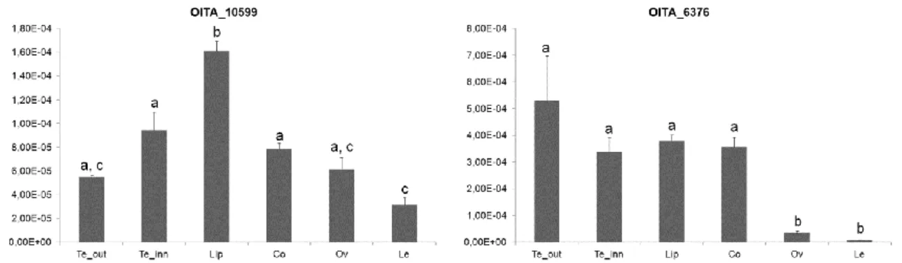
2.3 Identification of the DRIF-like genes of O. italica
Within the inflorescence transcriptome of O. italica there are two transcripts, OITA_10599 and OITA_6376, encoding for DRIF-like proteins. The transcript OITA_10599 (1,250 bp) encodes for a protein product of 258 amino acids that shows 56% identity with the DRIF1 protein of A. majus. The transcript OITA_6376 (2,083 bp) encodes for a protein product of 305 amino acids showing 39% identity with the DRIF2 protein of A. majus. The BLASTP analysis of both proteins reveals a higher identity score (65% for OITA_10599 and 56% for OITA_6376) with uncharacterized proteins; however, due to the poorly conserved sequences of the DRIF proteins, we have assumed OITA_10599 as the putative ortholog of
DRIF1 of A. majus. A recent study has revealed the presence of a larger number of
DRIF-like genes in dicots and monocots. For example, there are 6 DRIF-DRIF-like genes in A. majus and in Oryza sativa [57]. Currently, in silico genome and transcriptome analyses have been undertaken to verify the copy number of the DRIF-like genes in orchids.
2.4 Expression analysis of the DIV-, RAD- and DRIF-like genes of O. italica
O. italica is a wild species very difficult to propagate in vitro. In addition, it is not possible to
perform functional studies based on knock-out techniques. For these reasons, the analysis of gene expression is an important tool to infer gene function in this non-model species. In
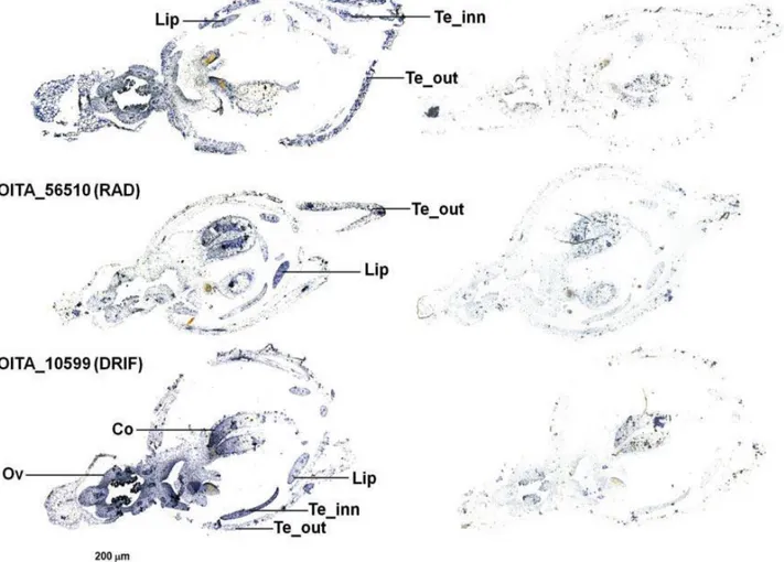
O. italica, the orthologs of the DIV, RAD and DRIF1 genes of A. majus are OITA_9548, OITA_56510 and OITA_10599, respectively. Figure 11 shows the RNA in situ hybridization
of these transcripts in the early floral tissues of O. italica. OITA_9548 (DIV) and OITA_10599 (DRIF1) are localized in all the floral tissues, whereas OITA_56510 (RAD) is expressed in the lip and outer tepals. This pattern is similar to that described in A. majus, where DIV and
DRIF1 are expressed in all the parts and RAD is expressed in the dorsal domain of the
snapdragon flower [43, 44]. In analogy with Antirrhinum, the observed expression pattern suggests that in the lip of O. italica OITA_10599 (DRIF1) could bind OITA_56510 (RAD), thus inhibiting the formation of the DIV-DRIF complex and the subsequent activation of ventralization genes. In O. italica, RAD and DRIF1 could also interact in outer tepals (where both are expressed), thus inhibiting ventralization as in the lip. The expression of RAD (expressed in the dorsal domains of the snapdragon) in the ventral part of the flower of O.
italica is due to resupination, the 180° twist of the median inner tepal (lip - dorsal structure)
that becomes a ventral structure (Fig. 1). The expression pattern of DIV, RAD and DRIF1 in
21
experiments (Fig. 12-14). The expression data suggest a model of interaction among DIV, RAD and DRIF1 to determine the bilateral symmetry of the flower generally conserved between Antirrhinum and O. italica, with some differences due to the peculiarities of the orchid flower. In particular, this model should be integrated with the expression pattern of some MADS-box genes (e.g. AP3/DEF and AGL6) involved in the formation of the orchid lip [58-60]. The role of the MADS-box genes in the formation of the orchid perianth will be analysed in the Chapter 2.
After anthesis, OITA_10599 (DRIF1) is expressed in all floral tissues and has a significantly higher expression in the lip, whereas the OITA_6376 (DRIF2) is expressed at similar levels in all tissues except the ovary and leaf, where it is very weakly expressed (Fig. 14).
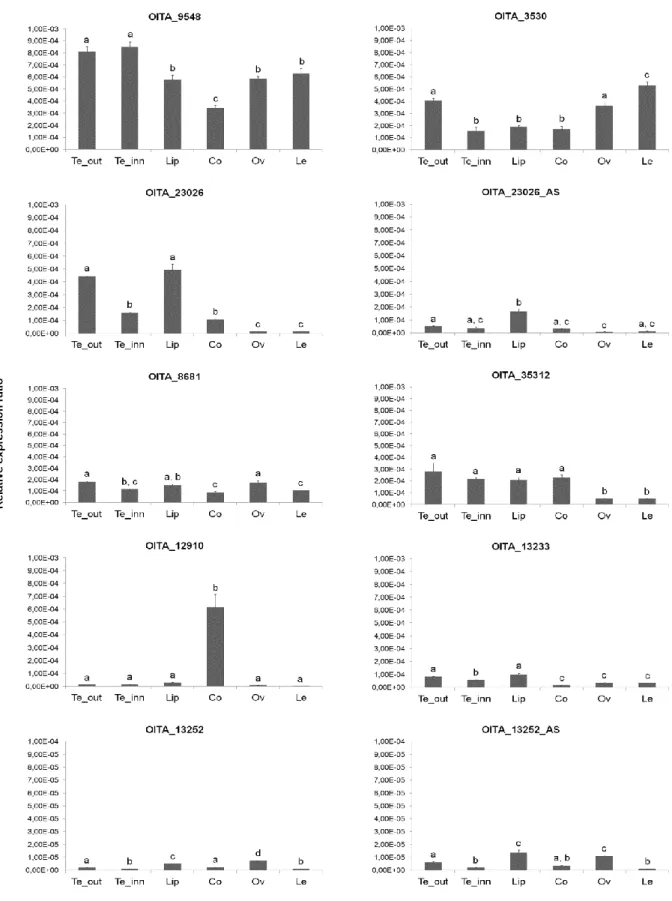
In the mature flower, some DIV-like transcripts of O. italica show overlapping profiles and others have very different expression patterns (Fig. 12). In addition to its expression in the perianth, OITA_9548 is detectable in the column, ovary and leaf. Although at lower levels, also OITA_3530 is expressed in all floral organs, as well as OITA_8681, OITA_35312 and
OITA_13233, whereas OITA_12910 is mostly expressed in the column. The two isoforms of
OITA_23026 differ in their level of expression, significantly lower in all tissues (excluding leaf) for OITA_23026_AS, the isoform that retains the intron. Also the two isoforms of
OITA_13252 show different expression profiles. In this case OITA_13252_AS, the isoform
that retains the intron in the 5’ UTR, is expressed at higher levels in all tissues (excluding leaf) than OITA_13252 (Fig 12). The expression levels of the DIV-like genes in O. italica seem to be inversely correlated with their intron size, in line with previous results obtained from genome analysis and gene expression of other orchid species [51]. This feature could be due to the presence of repetitive/transposable elements within the large introns of the
DIV-like genes that can negatively regulate their expression levels [56].
Although with different levels, the expression patterns of the RAD-like transcripts in the perianth tissues after anthesis are similar (Fig 13), being detectable mainly in the outer tepals and lip and with lower expression in the ovary. OITA_32153 is also highly expressed in the column and leaf, where also OITA_56510 is expressed. The similar expression pattern of the RAD-like genes of O. italica suggests their redundant function in the tissues of the perianth and more specialized roles in reproductive and vegetative tissues.
All the DIV-like genes of O. italica are mainly expressed in outer and inner tepals and lip, suggesting a possible redundant function in these tissues. However, a possible pleiotropic role of some transcripts, as previously reported in other species [53], is supported by their expression also in the column, ovary and leaves. The expression profile of the two
22
OITA_13252 isoforms are similar, even though OITA_13252_AS, that retains the intron in
the 5’ UTR, is expressed at higher levels. This difference could be due to a transcriptional regulatory role of the small intron in the 5’ UTR, whose presence might increase the transcription efficiency. Interestingly, the expression pattern of the DIV-like isoform
OITA_23026_AS (that encodes for a protein with a single MYB domain) is similar to that of
the RAD-like gene OITA_103296, leading to hypothesize that this isoform might be an evolutionary intermediate step towards the RAD-like genes, possibly evolved from a DIV-like gene after the loss of exon 2 through alternative splicing.
Fig. 11 RNA in situ hybridization of the DIV-, RAD- and DRIF1 transcripts on early floral tissues of O. italica. Reprinted from Valoroso et al. (2017) [14]. RNA in situ hybridization with the antisense (left) and sense (right) probes of the transcripts OITA_9548 (DIV), OITA_56510 (RAD) and OITA_10599 (DRIF1). Te_out, outer tepal; Te_inn, inner tepal; Co, column; Ov, ovary.
23
Fig. 12 Expression pattern of the DIV-like genes of O. italica. Reprinted from Valoroso et al. (2017) [14]. Relative expression pattern of the DIV-like transcripts in the different tissues of O. italica. Te_out, outer tepal; Te_inn, inner tepal; Co, column; Le, leaf. The letters above the bars indicate statistically significant groups, as assessed by the Tukey HSD post-hoc test.
24
Fig. 13 Expression pattern of the RAD-like genes of O. italica. Reprinted from Valoroso et al. (2017) [14]. Relative expression pattern of the RAD-like transcripts in the different tissues of O. italica. Te_out, outer tepal; Te_inn, inner tepal; Co, column; Le, leaf. The letters above the bars indicate statistically significant groups, as assessed by the Tukey HSD post-hoc test.
Fig. 14 Expression pattern of the DRIF1/2-like genes of O. italica. Reprinted from Valoroso et al.
(2017) [14]. Relative expression pattern of the DIV-like transcripts in the different tissues of O. italica. Te_out,
outer tepal; Te_inn, inner tepal; Co, column; Le, leaf. The letters above the bars indicate statistically significant groups, as assessed by the Tukey HSD post-hoc test.
25
The results discussed in the previous paragraphs (Chapter 1, from 2.1 to 2.4) were published in:
Valoroso MC, De Paolo S, Iazzetti G, Aceto S (2017). Transcriptome-Wide Identification
and Expression Analysis of DIVARICATA- and RADIALIS-Like Genes of the Mediterranean Orchid Orchis italica. Genome Biology and Evolution, Volume 9, Issue 6, 1 June 2017, Pages 1418–1431, https://doi.org/10.1093/gbe/evx101.
2.5 Protein interaction among DIV, RAD and DRIF1 in O. italica
In order to confirm if the model of interaction of DIV, RAD and DRIF1 of A. majus is conserved in O. italica, a “yeast two hybrid system assay” (Y2H) was conducted in collaboration with Prof. Maria Manuela Ribeiro Costa, at the Plant Functional Biology Center, University of Minho (Braga, Portugal).
The open reading frame (ORF) of the DIV (OITA_9548), RAD (OITA_56510) and DRIF1
(OITA_10599) genes were cloned into two vectors containing the GAL4 DNA-binding or
activation domain, respectively. The interactions were assayed in both the protein fusion forms. When the protein (in this case DIV) is able to promote the reporter gene transcription, it was assayed only the protein form fused to GAL4 activation domain.
The results of the Y2H screening and the quantitative analysis of the interaction levels are shown in Fig. 15. In O. italica, DRIF1 establishes a strong interaction both with the DIV and RAD proteins, whereas the RAD and DIV proteins do not interact each other. The ability of DRIF to interact with DIV and RAD has been demonstrated in Antirrhinum and tomato. This interaction has a key role in the establishment of different developmental pathways (snapdragon flower symmetry and tomato fruit development) [46, 47]. Our Y2H results demonstrate that these protein interactions are conserved also within monocot species, as orchids, where their role in the establishment of flower symmetry is supported also by the
26
Fig. 15 The DIV, RAD and DRIF1 protein-protein interactions in O. italica. (A) Protein interaction between DRIF1BD - DIVAD, DRIF1 BD - RADAD, RAD BD - DIVAD. (-W-L, medium lacking the tryptophan and
leucine; -W-L-H, medium lacking tryptophan, leucine and histidine. The dilution factor applied for the yeast inoculate is indicated with 1:10, 1:100 and 1:1000). (B) Quantitative analysis of the interaction levels by β-galactosidase assay. BD, binding domain; AD, activation domain.
27
CHAPTER 2 – The MADS-box genes
1. Introduction
1.1 The MADS-box genes: from flower development to bilateral symmetry
A crucial role in the evolution of flower architecture and in the control of flowering time is played by the MADS-box genes family. The MADS-box genes, known also as plant homeotic genes, are found in almost all eukaryotes and encode for transcription factors that contain a conserved DNA-binding domain called MADS domain [61]. This family takes its name from the first four MADS-box loci found in different species: MINICHROMOSOME
MAINTENANCE 1 (MCMI) of Saccharomyces cerevisiae, AGAMOUS (AG) of Arabidopsis thaliana, DEFICIENS (DEF) of Antirrhinum majus and SERUM RESPONSE FACTOR (SRF)
of Homo sapiens [62].
The MADS-box transcription factors originated from the topoisomerase II subunit A [6, 63, 64] and are divided into two lineages, type-I and type-II, that differ in genomic organization, developmental role, evolutionary rate and level of functional redundancy. The type-I MADS-box proteins (Fig. 16A) contain the MADS domain and are divided in M-alpha (Mα), M-beta (Mβ) and M-gamma (Mγ) due to the sequence divergence of their C-terminus [65]. They are involved in the development of seed, embryo and female gametophyte [66]. The type-II is the most studied class of MADS-box genes due to its involvement in different plant developmental processes, including flower formation. These transcription factors are characterized by three conserved domains and a variable domain that form the so-called MIKC structure (Fig. 16B-C) [6]:
• The MADS-box domain (M) at the N-terminus; • The intervening domain (I);
• The keratin domain (K); • The variable C-terminus.
The MADS-box is the DNA-binding domain that forms an α-helix structure capable, together with a partner protein, to recognize the CArG-box motif (CC[A/T]6GG) in the promoter of the
target genes [67-69]; the I and K domains are involved in the protein-protein interaction and in the formation of protein complexes [70, 71]; the variable C-domain has a role in the formation of protein complexes functioning as trans-activator domains [62, 71].
28
A duplication event involving the 5’ region of the exon encoding for the K domain, followed by neofunctionalization, gave rise to the two classes of MIKC-type MADS-box genes: MIKCC
and MIKC* [71, 72]. The MIKC* genes (sometimes called M) are involved in the male gametophyte development [73], while the MIKCC, the most studied, play different roles in
various processes of plant growth and in the establishment and maintenance of floral organs [6].
Fig. 16 The MADS-box protein structure. (A) Structure of a type-I (B) type II MIKC* and (C) type II
MIKCC protein.
The MIKCC MADS-box genes involved in flower formation are divided in five functional
classes (from A to E) and their activity is described by the ABCDE model of flower development [74, 75].
1.2 The ABCDE model of flower development
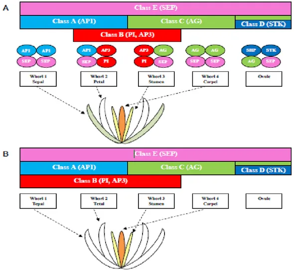
The first molecular model proposed to explain the identity and development of floral organs of the model plant A. thaliana dates back to 1991: the ABC model of flower development [76]. Since then, the model was improved, modified and extended to many plant species and currently it is known as the ABCDE model [75, 77-80]. The identity of floral organs depends on the expression and interaction of floral homeotic genes described by the ABCDE model [81, 82].
The wild-type flower of A. thaliana is composed by four concentric whorls where the floral organs develop: sepals in the outermost whorl 1, petals in the whorl 2, stamens in the whorl
29
3 and carpels in the innermost whorl 4 [83]. Excluding APETALA2 (AP2), all the genes involved in the ABCDE model belong to the MADS-box family. According to this model (Fig. 16A), in A. thaliana, the A-class genes APETALA1 (AP1) and AP2 specify the sepal identity. Together, the A- and B-class genes APETALA3 (AP3) and PISTILLATA (PI) specify the petal identity. The stamens in the third whorl are specified by the combination of the B-class genes and AGAMOUS (AG) of class C. The C-class alone determines formation of the carpels in the fourth whorl. The D-class genes SEEDSTICK (STK) and
SHATTERPROOF1/2 (SHP1/2) are involved in the determination of ovule and carpel
identity. Finally, the E-class genes SEPALLATA 1-4 (SEP1-4) are expressed in all the whorls and act redundantly to specify all the floral organs together with the other gene classes. The MADS-box genes involved in the ABCDE model of flower development work forming homo- and heterodimers that regulate specific expression programmes in the different floral whorls [62]. This model of interactions is called floral quartet. According to this model, the complexes AP1/AP1/SEP/SEP, AP1/SEP/AP3/PI, AG/SEP/AP3/PI and AG/AG/SEP/SEP regulate the formation of the floral structures within whorls 1, 2, 3 and 4, respectively [75, 84].
Although very useful, the plant models A. thaliana and A. majus are highly derived species, whose developmental pathways could be very different from those of other angiosperm species. The “classic” ABCDE floral quartet model is applicable to different plant species (mainly eudicots) where it is well conserved [85-88]; however, analyses of non-model species (mainly monocots, as orchids) have revealed differences probably due to the different structure of the flower [89]. For example, in orchids the class B MADS-box genes show an expression profile expanded to the first floral whorl, explaining the presence of petaloid sepals (Fig. 16B) [90-93].
30
Fig. 16 The ABCDE model of flower development. Reprinted from Aceto et al. (2011) [6]. (A) The ABCDE model and floral quartet in A. thaliana; (B) modified ABCDE model in orchids.
Current advances in transcriptome and genome sequencing are highlighting the molecular programs that underly floral evolution and development of non-model species. For many species belonging to basal angiosperms, magnoliids and basal eudicots, the “fading borders model” proposes a gradient of the expression levels of floral homeotic genes to explain the flower morphology. In particular, low expression levels at the boundary of the domain of a given gene belonging to the class A, B and C overlap with the expression of a different homeotic gene in the adjacent domain (Fig. 17).
31
Fig. 17 The differences between the ABCE model and the fading borders model. Reprinted from
Chanderbali et al. (2015) [94]. (A) Classic and (B) fading borders model of flower development.
• The orchid code and the Homeotic Orchid Tepal (HOT) model
In orchids, the MADS-box involved in the formation of the perianth are well characterized, in particular the B-class MADS-box genes [6, 59, 91-93, 95-98]. This class of genes originated after the duplication from an ancestral gene containing a paleoAP3 motif, giving rise to two lineages: the AP3/DEF lineage (from APETALA3 of A. thaliana and DEFICIENS of A. majus) and the PI/GLO lineage (from PISTILLATA of A. thaliana and GLOBOSA of A. majus) [99, 100]. Two subsequent duplication events within the orchid AP3/DEF genes have played an important role in the evolutionary origin of the current structure of the orchid flower explained by the so-called “orchid code” theory (Fig. 18) [101, 102]. The orchid code theory proposes that the identity of orchid perianth is established through the formation of protein complexes among the four different orchid AP3/DEF proteins and one PI/GLO-like protein [6]. The orchid AP3/DEF genes are divided in 4 clades with different expression patterns in the
32
organs of the perianth (Fig. 18). Genes of clade 1 and 2 are expressed in all tepals and drive the formation of the outer tepals; high levels of clade 1 and 2 proteins and low levels of clade 3 and 4 proteins determine the formation of inner lateral tepals; high levels of clade 3 and 4 proteins and low levels of clade 1 and 3 proteins induce the formation of the lip (Fig. 18) [103-105].The orchid code theory proposes an evolutionary reconstruction of the perianth origin from an ancestral orchid flower with radial symmetry to the current flower with bilateral symmetry. The ancestral orchid flower had undifferentiated tepals; after the first duplication of the AP3/DEF gene, two gene clades were formed: 1/2 and 3/4, with the origin of a perianth composed of outer and inner tepals; after the second duplication, the four gene clades gave origin to the zygomorphic orchid flower with outer and inner tepals and a differentiated lip [102].
Fig. 18 Duplications of the orchid B-class MADS-box genes, their expression and evolution of flower symmetry. Reprinted from Aceto et al. (2011) [6].
33
The regulation of perianth morphogenesis in orchids is explained more in details by the recently proposed “Homeotic Orchid Tepal (HOT)” model (Fig. 19) [98]. In this model, the
PI/GLO and AP3/DEF clades 1 and 2 determine the formation of the outer tepals, while the
combination of PI/GLO and AP3/DEF clades 1-2-3 induces the formation of the lateral inner tepals. The PI/GLO and AP3/DEF clades 3 and 4, together with the MADS-box AGL6-like genes, contribute to the lip morphogenesis [98].
Fig. 19 The Homeotic Orchid Tepal (HOT) model. Reprinted from Pan et al. (2011). Expression of the MADS-box genes at early (A) and late (B) stage of orchid development.
34
1.3 Aim of work
When I started my PhD project, previous studies had analysed the structure, expression and evolution of some MADS-box genes of O. italica, in particular the B-class genes PI/GLO and
AP3/DEF and the C- and D-class genes AG and STK [6, 59, 91, 92, 95, 106-108].
As evidences were accumulating supporting an involvement of the MADS-box genes in the establishment of bilateral symmetry during the development of orchid flower (orchid code and HOT model), I decided to perform a transcriptome-wide characterization of the MADS-box genes expressed in the inflorescence of O. italica and to verify if some of them could interact with the MYB transcription factors DIV, RAD and DRIF1, potentially involved in the zygomorphy of the orchid flower.
More in details, the second part of my work can be divided in different points:
• Identification of all the MADS-box genes expressed in the inflorescence transcriptome of O. italica by in silico analysis;
• Phylogenetic analysis of the orchid MADS-box genes;
• Expression analysis of the MADS-box genes in the floral tissues of O. italica by real time PCR;
• Evolutionary analysis of the MADS-box genes most expressed in the floral tissues of
O. italica;
• Study of the protein interaction between the B-class MADS-box proteins and the MYB factors DIV, RAD and DRIF1 of O. italica by yeast two hybrid analysis.
35
2. Results and Discussion
2.1 Identification of the MADS-box genes of O. italica
The analysis of the inflorescence transcriptome of O. italica reveals the presence of twenty-nine transcripts encoding for MADS-box proteins. Respect to the number of MADS-box genes reported in the orchid species with a released genome assembly (P. equestris, D.
catenatum and A. shenzenica) the number of MADS-box isolated in this study is lower
because were considered only the transcripts expressed in the inflorescence of O. italica [51, 52, 109]. BLAST and phylogenetic analysis show that in O. italica are expressed both class I and class II MADS-box genes (Fig. 20).
Fig. 20 Maximum likelihood tree of the orchid MADS-box proteins. The collapsed branches include MADS-box proteins of O. italica, P. equestris and A. shenzenica. The red asterisks indicate the number of sequences identified in O. italica. The numbers indicate the statistical support of the branches.
36 2.1.1 Class I MADS-box genes
Previous studies proposed that the orchid genomes lack the class I Mβ genes [109]. In the transcriptome of O. italica are expressed four class I transcripts, two belonging to the Mα type, one to the Mγ and one to the Mβ (Fig. 21). This result demonstrates the presence of Mβ genes in orchids, as recently reported also in P. aphrodite [110]. The class I MADS-box transcripts of O. italica are poorly expressed in all the floral tissues, as expected based on their role in embryo and endosperm maturation [66] and in agreement with their expression levels in the orchid A. shenzenica [109]. Although at low levels, they are expressed in all the floral tissues, also in the column (Fig. 22), at whose top are located the pollen grains. The expression in this tissue is in line with the pattern observed the orchid P. aphrodite [110] and suggests the involvement of the class I MADS-box genes in the development of the orchid pollinia.
Fig. 21 Maximum likelihood tree of the orchid MADS-box class I proteins. The numbers indicate the statistic support of the branches. The arrows indicate the O. italica MADS-box class I proteins.
37
Fig. 22 Expression pattern of the class I MADS-box genes of O. italica. Relative expression of the class I transcripts of O. italica in different floral tissues. Te_out, outer tepal; Te_inn, inner tepal; Co, column; Ov, ovary. The letters above the bars indicate statistically significant groups, as assessed by the Tukey HSD post-hoc test.
2.1.2 Class II MADS-box genes
The class II MADS-box genes are divided into two sub-classes: MIKC* and MIKCC [71]. The
analysis of the inflorescence transcriptome of O. italica reveals the presence of one MIKC* and twenty-four MIKCC transcripts.
• The MIKC* genes
The MIKC* transcript expressed in the inflorescence of O. italica belongs to the P-subclade. The MIKC* genes are involved in the development of gametophyte [111, 112] and the MIKC* transcript of O. italica is highly expressed in the column (Fig. 23), as in the orchids E. pusilla [60] and P. aphrodite [110], supporting its function in the development of the orchid gametophyte.
Fig. 23 Expression pattern of the MIKC* gene of O. italica. Relative expression of the class II MIKC* transcript of O. italica in different floral tissues. Te_out, outer tepal; Te_inn, inner tepal; Co, column; Ov, ovary. The letters above the bars indicate statistically significant groups, as assessed by the Tukey HSD post-hoc test.
38
• The SOC, SVP, ANR1, AGL12 and OsMADS32 genes
Some MIKCC genes are involved in the regulation of flowering (as SOC1 and SVP) and root
development (as AGL12 and ANR1) [113, 114]. In the inflorescence of O. italica are expressed two genes involved in the regulation of flowering (SOC and SVP), two related to the root development AGL12 and ANR1 and one involved in the seed development
(OsMADS32) [115, 116]. As expected, all these transcripts have a low expression in all the
floral tissues (Fig. 24), as reported also in other orchids [60, 117, 118].
Fig. 24 Expression pattern of the MIKCC genes of O. italica. Relative expression of the SOC, SVP,
ANR, OsMADS32, AGL12 transcripts of O. italica in different floral tissues. Te_out, outer tepal; Te_inn, inner
tepal; Co, column; Ov, ovary. The letters above the bars indicate statistically significant groups, as assessed by the Tukey HSD post-hoc test.
• The AGL6-class genes
Recent studies in Oncidium and P. equestris [101] have highlighted the role of the MADS-box gene AGL6 in the formation of the orchid lip. In many orchids there are three AGL6 transcripts divided in two clades, one that includes also other monocot species and the other
39
one that includes only orchid species [110, 119]. As in other orchid species, in O. italica there are three AGL6-like transcripts. Phylogenetic analysis of the AGL6 transcripts of O.
italica shows that they belong to the two clades previously reported in orchids. The topology
of tree reveals the formation of two different paralog groups probably due to a duplication within the orchid-specific AGL6 clade (Fig. 25). Both groups include transcripts of basal orchid species (Apostasioideae, Cypripedioideae or Vanilloideae) in addition to Epidendroideae and Orchidoideae.
Analysis of the expression pattern of these transcripts (Fig. 26) reveals an overlapping profile of expression of AGL6_OITA_8204 and AGL6_OITA_1386. Both transcripts are expressed at high level in lip and lower level in outer tepals. This result suggests that the paralogs AGL6_OITA_8204 and AGL6_OITA_1386 probably have a redundant functional role in the formation of lip in O. italica. In addition, AGL6_OITA_4335 presents a high expression in outer tepals and ovary, suggesting a role in the reproductive structures. The evolutionary analysis of the orchid AGL6 coding sequences reveals no evidence of positive selection or relaxation of selective constrains (Tab. 6-7 - Appendix).
40
Fig. 25 Maximum likelihood tree of the AGL6 proteins. The numbers indicate the statistic support of the branches. The arrows indicate the O. italica AGL6 proteins.
Fig. 26 Expression pattern of the AGL6 genes of O. italica. Relative expression of the AGL6 class transcripts of O. italica in different floral tissues. Te_out, outer tepal; Te_inn, inner tepal; Co, column; Ov, ovary. The letters above the bars indicate statistically significant groups, as assessed by the Tukey HSD post-hoc test.
41 • The A-Class genes
In the inflorescence transcriptome of O. italica there are four transcripts belonging to the two monocot FUL-like groups previously identified in other species [120]. An orchid-specific duplication expanded the number of the orchid AP1/FUL genes producing four, well supported AP1/FUL-like clades (Fig. 27). Three AP1/FUL-like transcripts of O. italica (OITA_9283_AP1, OITA_11046_AP1, OITA_2508_AP1) encode for proteins that present the complete FUL-like motif LPPWML at the C-terminus, while in OITA_3679_AP1 it is absents. This is not a peculiar feature of O. italica and it is reported also in other species belonging to Epidendroideae [96] and Apostasioideae. As the divergence of the C-terminus is shared by members of the AP1/FUL proteins belonging to the basal Apostasioideae subfamily, [121] it is possible that it is an ancient condition, raised early during the orchid evolution.
The expression analysis of the AP1/FUL genes OITA_3679_AP1, OITA_9283_AP1 and
OITA_2508_AP1 of O. italica (Fig. 28) reveals a pattern very similar to EpMADS10, 11 and 12 of E. pusilla [60, 122]. They show a high expression in column and ovary and are
expressed also in outer and inner tepals. Contrastingly, OITA_11046_AP1 has a high expression in outer and inner tepals and a weak expression in column and ovary (Fig. 28). The evolutionary analysis reveals absence of positive selection within the AP1/FUL coding sequences of orchids; however, relaxation of purifying selection is evident when the orchid
AP1/FUL genes (=0.23) are compared with the orchid MADS-box genes of the related
functional classes SEP (=0.11) and AGL6 (=0.14) (Tab. 6-7 - Appendix). This result is in agreement with the previous report of diversifying selection in the orchid AP1/FUL genes [122] and suggests a possible functional diversification after duplication of these orchid genes, supported also by the divergence of the C-terminus sequence of some members of this clade and by the peculiar expression profile of OITA_11046_AP1.
42
Fig. 27 Maximum likelihood tree of the AP1/FUL proteins. The numbers indicate the statistic support of the branches. The arrows indicate the O. italica MADS-box class A proteins.
43
Fig. 28 Expression pattern of the class A genes of O. italica. Relative expression of the A class transcripts of O. italica in different floral tissues. Te_out, outer tepal; Te_inn, inner tepal; Co, column; Ov, ovary. The letters above the bars indicate statistically significant groups, as assessed by the Tukey HSD post-hoc test.
• The C- and D- class genes
Previous studies have identified one C- (AG) and one D-class (STK) gene in O. italica [108]. In addition to these, in the present study two additional AG transcripts have been identified:
OITA_1784_AG and OITA_16614_AG. Three C-class and one D-class genes are present
also in E. pusilla [60, 122], suggesting that this is the number of AG/STK genes also in O.
italica.
Both the transcripts encode for proteins containing the AG-motif I and II at the C-terminus. The OITA_1784_AG shows 69% amino acid identity with the C-class OitaAG and 62% with the D-class OitaSTK, whereas OITA_16614_AG shows 71% identity with OitaAG and 52% with OitaSTK. Also in the transcriptome of Ophrys sphegodes (Orchidoideae) there are transcripts encoding for AG proteins similar (84% identity) to those of O. italica.
The expression analysis reveals that the newly identified AG transcripts of O. italica have a high level of expression in ovary and column (Fig. 29), in agreement with the canonical expression pattern of the C-class genes in other orchid species and with their role in the development of female reproductive structures [58, 96, 123].
44
Fig. 29 Expression pattern of the class C genes of O. italica. Relative expression of the C class transcripts of O. italica in different floral tissues. Te_out, outer tepal; Te_inn, inner tepal; Co, column; Ov, ovary. The letters above the bars indicate statistically significant groups, as assessed by the Tukey HSD post-hoc test.
• The E-class genes
The E-class of MADS-box genes are involved in the establishment of all the floral organs in
Arabidopsis thaliana. In orchids there are four clades of SEP-like genes raised from
orchid-specific duplications within the two SEP monocot clades [60, 96, 121]. In the inflorescence transcriptome of O. italica there are only two transcripts (OITA_7010_SEP and
OITA_1006_SEP) that encode for SEP proteins, both containing the SEP I and II motifs.
Phylogenetic analysis (Fig. 30) reveals that one transcript of O. italica belongs to the SEP clade 3 (also including EpMADS9 and PeSEP3) while the other belongs to the SEP clade 4 (also including EpMADS7 and PeSEP4). BLAST search in the transcriptome of Ophrys
sphegodes [50] shows that also in this species only two SEP-like transcripts are detectable,
with significant hits and sequence identity to the SEP transcripts isolated in O. italica. In addition, in Habenaria radiata (Orchidoideae) a recent study reports the presence of only two SEP-like transcripts in the floral buds [124]. Taken together, all these evidences suggest that in the Orchidoideae subfamily there are only two SEP genes or that only two SEP genes are expressed in the inflorescence. Due to the absence of an available assembled genome of Orchidoideae, it is not possible to discriminate between these two hypotheses.
The expression pattern of the SEP transcripts of O. italica (Fig. 31) shows that both are expressed in all the organs of the perianth and that OITA_7010_SEP is expressed also in column and ovary, as reported in other orchids as H. radiata [124], P. equestris [96] and E.
pusilla [60, 122].
The evolutionary analysis reveals a strong and homogeneous purifying selection acting on the SEP orchid genes (Tab. 6-7 - Appendix).
45
Fig. 30 Maximum likelihood tree of the SEP proteins. The numbers indicate the statistic support of the branches. The arrows indicate the O. italica MADS-box class E proteins.

![Fig. 6 Antirrhinum majus div mutants. Reprinted from Galego and Almeida (2002) [43]. Floral diagram of the wild-type and div mutant (first line) and cyc:dich double and cyc:dich:div triple mutant (second line) of A](https://thumb-eu.123doks.com/thumbv2/123dokorg/2768559.1209/12.892.310.583.105.386/antirrhinum-mutants-reprinted-galego-almeida-floral-diagram-mutant.webp)
![Fig. 7 The different snapdragon rad mutants. Reprinted from Corley et al. (2005) [45]](https://thumb-eu.123doks.com/thumbv2/123dokorg/2768559.1209/13.892.247.642.100.814/fig-different-snapdragon-rad-mutants-reprinted-corley-et.webp)