Università Politecnica delle Marche
Research Doctorate in Life and Environmental Sciences
Curriculum of Biomolecular SciencesCycle XXX
Development of new molecular tools for the
characterization of human Granulosa cells: new
implications for the research on human infertility
Supervisor:
Prof. Elisabetta Giorgini Co-Supervisor: Dr. Giorgia Gioacchini Company Supervisor: Dr. Andrea Borini PhD candidate: Valentina Notarstefano
Supervisor
Prof. Elisabetta Giorgini, PhD Laboratory of Infrared Spectroscopy
Department of Life and Environmental Sciences Università Politecnica delle Marche
Ancona, Italy
Co-Supervisor
Dr. Giorgia Gioacchini, PhD
Laboratory of Developmental and Reproductive Biology Department of Life and Environmental Sciences Università Politecnica delle Marche
Ancona, Italy
Company Supervisor Dr. Andrea Borini, PhD, MD 9.baby Family and Fertility Center Bologna, Italy
A. E. G.
Vis unita fortior
CONTENTS
SUMMARY
ISOMMARIO
VIIINTRODUCTION
1 1. Folliculogenesis 1 2. The importance of GCs 63. GCs and Assisted Reproductive Technology 9
4. Factors impacting on GCs functions 11
5. Vibrational spectroscopies 13
References 17
C
HAPTER1
The molecular and functional characterization of
human Granulosa cells by FTIR
Microspectroscopy correlates with oocyte fate
29Introduction 31 Methods 33 Results 43 Conclusions 57 References 61
C
HAPTER2
Validation of an air-dehydration protocol for
human Granulosa cells for FTIRM studies
65Introduction 67
Experimental section 71
Results 78
Discussion 79
C
HAPTER3
Metabolic changes in cultured hGCs: an
innovative FTIRM approach
83Introduction 85 Experimental section 87 Results 92 Discussion 100 References 104
C
HAPTER4
A new insight on aging effects in human
Granulosa cells: a multidisciplinary FTIR imaging
spectroscopy and qPCR approach
107Introduction 109 Experimental section 111 Results 118 Discussion 125 References 131
C
HAPTER5
Ovarian endometriosis affects Granulosa Cells
Endocannabinoid system
137 Introduction 139 Experimental section 141 Results 146 Discussion 151 References 156C
HAPTER6
Could unilateral endometriosis affect the
contralateral ovary? New insights from a
multidisciplinary study
161 Introduction 163 Experimental section 165 Results 174 Discussion 185 References 191C
HAPTER7
How do plasticizers BPA and DGB alter the
macromolecular composition of luteinized GCs?
199Introduction 201 Experimental section 202 Results 207 Discussion 213 References 218
CONCLUSIONS
223ACKNOWLEDGEMENTS
227SUMMARY
Granulosa cells (GCs) play several key roles in folliculogenesis, such as the production of estradiol during follicular growth, the regulation of the advance of meiosis steps and the transcriptional activity in the oocyte, the production of essential nutrients used as energy source during oocyte maturation, the accumulation of secreted metabolites, and the secretion of progesterone after ovulation.
The aim of the PhD project was to investigate human GCs in a multidisciplinary approach, in order to correlate the information on these cells to several aspects related to infertility, to improve the knowledge on mechanisms involved in folliculogenesis and ovarian functions and understand how these processes are impaired by endogenous and exogenous factors.
For all the studies, luteinized GCs were retrieved from patients enrolled in an
in vitro fertilization program, at Tecnobios Procreazione Fertility Center. The
work was organised in seven steps, including establishing protocols, as the basis of targeted studies.
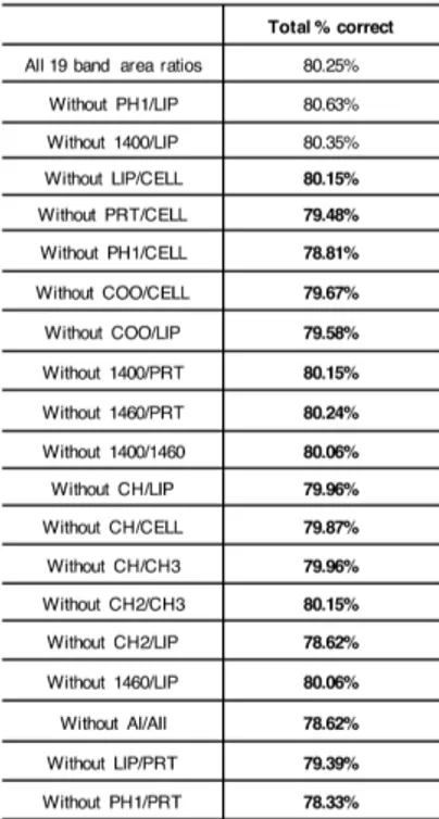
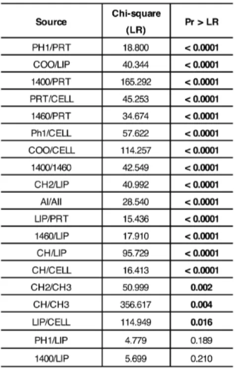
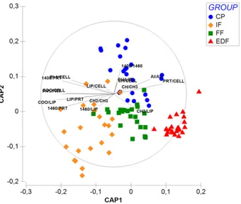
I. Fourier Transform InfraRed Microspectroscopy (FTIRM) analysis of Granulosa Cells: a promising approach for the assessment of good quality human oocytes. The aim of the present section was to find an innovative method for evaluating the quality of human oocytes, by studying GCs. To do this, 55 GCs samples, retrieved from consenting patients, were subjected to FTIRM measurements. IR data were pre-processed and then analysed using 'feature selection' procedures and
univariate/multivariate statistical analyses. Four experimental groups were selected: CP (clinical pregnancy), GCs from oocytes, which gave clinical pregnancy; FF (fertilization fail), GCs from oocytes, which failed fertilization; EDF (embryo development fail), GCs from oocytes, which failed embryo development, and IF (implantation fail), GCs from oocytes, which failed implantation. The ‘feature selection’ procedures allowed the identification of 17 spectral biomarkers, which showed to be correlated with the corresponding oocyte clinical outcome in an accurate and reliable way.
II. Validation of air-dehydration protocol of GCs for FTIRM. The aim of the present section was to establish a reliable preparation protocol for GCs for FTIRM studies, based on air-drying of GCs on CaF2 supports. To do this, FTIRM measurements were performed on GCs, and second derivative spectra of ‘wet’ (analysed in physiological condition in a microfluidic device) and ‘dried’ (deposited onto CaF2 supports and air-dried for 30min) GCs were compared. A satisfactory correspondence in the spectral profile was observed, proving that the air-dehydration protocol is a suitable method to prepare GCs for FTIRM analysis.
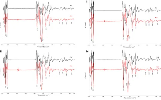
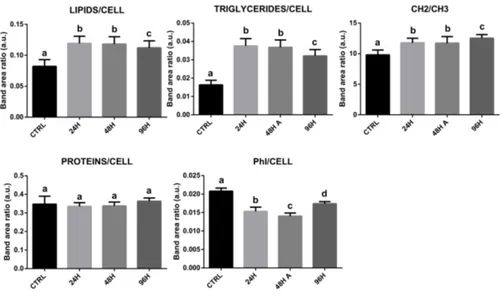
III. Metabolic changes in cultured hGCs: an innovative FTIRM approach. The aim of the present section was the evaluation of the biochemical alterations induced by in vitro culture in GCs. FTIRM measurements were performed on GCs collected from 17 patients immediately after their collection and cultured for 24–48–96 h. the results obtained from FTIRM data showed that lipid metabolism was altered in all cultured
GCs, in terms of total content of triglycerides and fatty acids; also, the amount of phosphorylation was altered. Proteins and carbohydrates, in contrast, were not affected.
IV. A new insight on aging effects in human Granulosa cells: a multidisciplinary FTIR and qPCR approach. The aim was to analyse the biochemical modifications induced by aging on GCs. FTIRM was performed on air-dried GCs divided into two groups: Young (31.2±2.20; n°10) and Old (42.5±1.84; n°10). The results obtained showed that aging affects the biochemical composition of GCs, evidencing a process of age-related oxidative stress: increased lipid content, C=O ester moieties, and =CH groups; decreased protein content, and impaired secondary structure. Results of gene expression analysis were consistent with IR data: an increase of igfbp3, foxo3, and a decrease of igf1, sirt1 and sod2 was found.
V. Ovarian endometriosis affects Granulosa Cells Endocannabinoid system (ECS). The aim of the present work was to analyse the expression of molecules associated with the ECS in GCs from patients affected by ovarian endometriosis. To do this, FTIR imaging and quantitative PCR (qPCR) were performed, on GCs previously divided into two groups: CTRL (n°10; women with non-ovarian infertility) and ENDO (n°10; women with ovarian endometriosis). The obtained results showed that endometriosis alters the gene expression of several molecules involved in ECS (trpv1, gpr55, cnr1, cnr2, nape-pld, faah), and impairs lipid (pparα, pparγ, srebp1, fasn) and carbohydrate (glut1
and glut9) metabolisms. FTIR imaging confirmed the alterations in GCs metabolism.
VI. Could unilateral ovarian endometriosis affect the contralateral ovary? New insights from a multidisciplinary study. The aim was to evaluate how unilateral ovarian endometriosis (UOE) affects the functionality of both the ovaries. FTIRM, Raman MicroSpectroscopy (RMS) and qPCR were performed on GCs previously divided into three groups: CTRL (GCs from 10 healthy women), CONTRAL (GCs from contralateral ovary of 10 women with UOE;) and ENDO (GCs from affected ovary of 10 women with UOE;). FTIRM and RMS data consistently revealed alterations, in both the ovaries, in lipid content and characteristics, protein composition and secondary structure, carbohydrates, and nucleic acids. qPCR confirmed an impairment of lipid (pparα, pparγ and fasn increase) and carbohydrate (glut1 and
glut9 decrease) metabolisms, and methylation process (dnmt1 and dnmt3a increase), in both the ovaries.
VII. How do plasticizers BPA and DGB alter the macromolecular composition of luteinized GCs? The aim was the investigation of the effects of bisphenol-A (BPA) and diethylene glycol dibenzoate (DGB) (1–10–100ng/ml), on GCs. FTIRM measurements were performed on GCs (n°15 women, 32±3.3 years), treated with BPA/DGB for 48h, and air-dried for IR measurements. The results showed that BPA exposure affected the biochemical composition of GCs, in terms of lipids amount and characteristics; proteins decreased and showed
different amino acid composition; phosphate groups from phospholipids and nucleic acids increased. DGB impacts on GCs differently: unsaturation and esterification rates in lipids decreased; proteins and phosphates amounts were altered differently according to dose.
The information obtained on GCs contribute to the understanding of the mechanisms of impairment of folliculogenesis, by a multidisciplinary approach that mainly consisted of supporting spectroscopic analysis with standard qPCR. Thanks to the results, an innovative approach to evaluate oocyte quality by spectral features of GCs was proposed, suggesting the reliability of FTIRM as a clinical feasible and easy diagnostic tool in assisted reproduction routine, in a possible near future.
SOMMARIO
Le cellule della Granulosa (GCs) svolgono molti fondamentali chiave durante il processo della follicologenesi: ad esempio, sono coinvolte nella produzione di estradiolo durante la crescita follicolare, nella regolazione dell’avanzamento degli step della meiosi e dell’attività trascrizionale dell’ovocita, nella produzione di nutrienti essenziali usati come fonte energetica durante la maturazione dell’ovocita, nell’accumulo di metaboliti secreti dall’ovocita, e nella secrezione di progesterone dopo l’ovulazione.
Lo scopo del presente progetto di Dottorato è stato quello di analizzare le GCs umane applicando un approccio multidisciplinare, che correlasse le informazioni ottenute su queste cellule con diversi aspetti legati all’infertilità, per fornire nuove informazioni sui meccanismi coinvolti nella follicologenesi e nelle funzioni ovariche, e per comprendere se e come questi processi possono essere alterati da fattori endogeni ed esogeni.
Per tutti gli studi condotti, le GCs luteinizzate sono state recuperate da pazienti reclutate in un programma di fecondazione in vitro presso il centro per la fertilità Tecnobios Procreazione. Il lavoro è stato organizzato in sette step, a partire dalla definizione dei protocolli, fino agli studi applicati.
I. Fourier Transform InfraRed Microspectroscopy (FTIRM) analysis of Granulosa Cells: a promising approach for the assessment of good quality human oocytes. (Analisi tramite Fourier Transform InfraRed Microspectroscopy (FTIRM) delle cellule della Granulosa: un approccio promettente per la valutazione della qualità degli ovociti
umani). Lo scopo di questa sezione era trovare un metodo innovativo per valutare la qualità degli ovociti umani. 55 campioni di GCs sono stati analizzati tramite FTIRM; i dati ottenuti sono stati processati e, tramite delle procedure di ‘feature selection’, è stata ottenuta la ‘biomarker signature’ del sistema analizzato. Infine, l’applicazione di metodi di statistica multivariata ha permesso di validare il modello, mentre la statistica univariata è stata utilizzata per investigare ulteriormente il significato biologico dei biomarker spettrali individuati. Sono stati selezionati quattro gruppi sperimentali: CP (clinical pregnancy), GCs da ovociti che hanno portato a gravidanza clinica; FF (fertilization fail), GCs da ovociti che non sono stati fecondati; EDF (embryo development fail), GCs da ovociti che non si sono sviluppati correttamente in embrioni; IF (implantation fail), GCs da ovociti che si sono sviluppati in embrioni, ma che hanno fallito l’impianto. Correlando le informazioni spettrali ottenute dall’analisi delle GCs con l’esito clinico dell’ovocita corrispondente, sono stati identificati 17 biomarker spettrali, che si sono rivelati in grado di correlare le GCs con la qualità degli ovociti in maniera accurata e affidabile.
II. Validation of air-dehydration protocol of GCs for FTIRM. (Validazione di un protocollo di disidratazione all’aria delle GCs per la valutazione tramite FTIRM). Lo scopo di questa sezione era quello di stabilire un protocollo affidabile per la preparazione delle GCs per l’analisi tramite FTIRM (Microspettroscopia FTIR), basato sulla
disidratazione delle cellule all’aria su finestre ottiche di CaF2. Per far questo, sono stati confrontati gli spettri in derivata seconda delle GCs ‘wet’ (analizzate in condizioni fisiologiche grazie all’utilizzo di in dispositivo di microfluidica) e ‘dried’ (depositate su supporti di CaF2 e lasciate disidratare all’aria per 30 minuti). È stata osservata una soddisfacente corrispondenza fra i profili spettrali di GCs ‘wet’ e ‘dried’, suggerendo che il protocollo di disidratazione all’aria delle GCs può essere considerato in metodo adeguato per le analisi tramite FTIRM.
III. Metabolic changes in cultured hGCs: an innovative FTIRM approach. (Alterazioni metaboliche in GCs umane mantenute in coltura: un innovativo approccio con FTIRM). Lo scopo della presente sezione era stabilire se la coltura in vitro delle GCs in coltura potesse determinare nelle cellule delle alterazioni biochimiche. Per fare questo, le GCs sono state prelevate da 17 pazienti consenzienti e sono state analizzate tramite FTIRM immediatamente dopo il pick-up, o dopo coltura per 24, 48 e 96 ore. L’analisi spettrale ha rivelato come a tutti i tempi di coltura, le GCs mostravano un’alterazione del metabolismo lipidico, in termini di contenuto totale di trigliceridi e acidi grassi; inoltre, il tasso di fosforilazione risultava alterato. Al contrario, il mantenimento in coltura delle GCs non ha determinato effetti né su proteine, né su carboidrati.
IV. A new insight on aging effects in human Granulosa cells: a multidisciplinary FTIR and qPCR approach. (Una nuova prospettiva
sugli effetti dell’invecchiamento sulle GCs umane: un approccio multidisciplinare, condotto con FTIR e qPCR). Lo scopo di questa sezione era la determinazione delle modificazioni biochimiche indotte dall’invecchiamento sulle GCs. Per far questo, le GCs ottenute da 20 pazienti consenzienti, sono state disidratate all’aria e suddivise in due gruppi sperimentali: ‘Young’ (età di 31.2±2.20) e ‘Old’ (età di 42.5±1.84). Inoltre, da aliquote delle stesse GCs è stato estratto l’RNA totale per l’espressione genica, eseguita tramite qPCR. L’analisi vibrazionale ha messo in luce variazioni nella composizione biochimica delle GCs, causate dall’invecchiamento; in particolare, è emerso un meccanismo di attivazione di stress ossidativo, individuabile grazie all’aumento del contenuto lipidico totale, dei gruppi carbonilici degli esteri, e dei gruppi =CH. Inoltre, l’età delle pazienti ha modificato il pattern proteico, in termini di contenuto totale e di struttura secondaria. I risultati dell’analisi di espressione genica si sono rivelati coerenti con l’analisi vibrazionale: in particolare, sono stati riscontrati un aumento dell’espressione di igfbp3 e foxo3, e una diminuzione di
igf1, sirt1 e sod2.
V. Ovarian endometriosis affects Granulosa Cells Endocannabinoid system (ECS). (L’endometriosi ovarica alter ail Sistema endocannabinoide (ECS) delle GCs). Lo scopo di questa sezione era identificare se e come l’endometriosi ovarica potesse alterare l’espressione di molecole dell’ECS nelle GCs di pazienti affette da questa malattia. Per questo studio, l’FTIR imaging e la qPCR sono
state accoppiate per studiare GCs suddivise in due gruppi sperimentali: CTRL (10 pazienti senza diagnosi di infertilità ovarica) e ENDO (10 pazienti con diagnosi di endometriosi ovarica). Entrambe le tecniche utilizzate hanno mostrato profonde alterazioni dell’ECS nelle GCs prelevate da pazienti con endometriosi ovarica; l’espressione genica di molte molecole coinvolte nel funzionamento dell’ECS (trpv1,
gpr55, cnr1, cnr2, nape-pld, faah) hanno mostrato alterazioni
significative causate dall’endometriosi; inoltre, anche i metabolismi di lipidi e carboidrati sono risultati modificati (pparα, pparγ, srebp1, fasn per i lipidi; glut1 e glut9 per i carboidrati). L’analisi spettroscopica tramite FTIR imaging ha confermato le alterazioni nelle GCs già evidenziate con qPCR.
VI. Could unilateral ovarian endometriosis affect the contralateral ovary? New insights from a multidisciplinary study. (Può l’endometriosi ovarica unilaterale intaccare l’ovario controlaterale? Nuove prospettive da uno studio multidisciplinare). Lo scopo del presente lavoro era valutare come l’endometriosi ovarica unilaterale potesse influenzare negativamente la funzionalità di entrambi gli ovari della paziente affetta dalla malattia. L’approccio multidisciplinare applicato in questo studio ha visto l’utilizzo di due spettroscopie vibrazionali (FTIRM e RMS, Raman Microspectroscopy) e dell’analisi di espressione genica tramite qPCR. Le GCs sono state suddivise in tre gruppi sperimentali: CTRL (GCs da 10 donne senza diagnosi di infertilità ovarica), CONTRAL (GCs dall’ovario controlaterale di 10
donne con endometriosi ovarica unilaterale) e ENDO (dall’ovario colpito dalla malattia di 10 donne con endometriosi ovarica unilaterale). I dati ottenuti dall’analisi con FTIRM e RMS hanno rivelato in maniera coerente che in entrambi gli ovari, erano presenti profonde alterazioni in termini di contenuto lipidico, caratteristiche degli acidi grassi, composizione proteica e struttura secondaria, carboidrati e acidi nucleici. L’analisi di espressione genica tramite qPCR ha confermato un’alterazione del metabolismo lipidico (aumento di pparα, pparγ e fasn), del metabolismo dei carboidrati (diminuzione di glut1 e glut9) e del processo di metilazione del DNA (aumento di
dnmt1 e dnmt3a), in entrambi gli ovari.
VII. How do plasticizers BPA and DGB alter the macromolecular composition of luteinized GCs? (In che modo i plastificanti BPA e DGB alterano la composizione macromolecolare delle GCs luteinizzate?) Lo scopo del presente lavoro era indagare gli effetti del bisfenolo A (BPA) e del dietilene glicole dibenzoato (DGB), utilizzati alle dosi 1–10–100ng/ml, sulle GCs umane. Le GCs ottenute da 15 pazienti età 32±3.3 anni, sono state analizzate tramite FTIRM dopo trattamento in vitro con BPA/DGB per 48 ore, e poi disidratate su supporti di CaF2 per le misurazioni IR. L’analisi dei dati ottenuti con FTIRM hanno rivelato che il BPA altera profondamente la composizione biochimica delle GCs, in termini di contenuto lipidico, caratteristiche lipidiche, quantitativo proteico, composizione amminoacidica, contenuto di gruppi fosfato e acidi nucleici. Il DGB
altera le GCs in maniera diversa, modificando i livelli di insaturazione e esterificazione dei lipidi, il contenuto di gruppi fosfato e il contenuto proteico.
Le informazioni ottenute tramite questi lavori sulle GCs possono contribuire ad aumentare la conoscenza sui meccanismi che danneggiano la follicologenesi, tramite un approccio multidisciplinare che ha previsto l’accoppiamento di analisi spettroscopiche e qPCR standard. Grazie ai risultati ottenuti, è stato proposto un metodo innovativo per la valutazione della qualità degli ovociti umani, basato sulle caratteristiche spettrali delle GCs, che ha suggerito la possibilità di utilizzo dell’FTIRM come strumento diagnostico affidabile e di facile utilizzo nella routine delle cliniche di riproduzione assistita, in un prossimo futuro.
INTRODUCTION
1.
F
OLLICULOGENESISFolliculogenesis is the process in which a recruited primordial follicle grows and develops into a specialized Graafian follicle, which has two potential fates, to either ovulate its egg to be fertilized, or to die by atresia (Figure 1). The first phase of folliculogenesis, named ‘pre-antral’ or ‘gonadotropin-independent’ phase, sees the growth and differentiation of the oocyte, and is mainly modulated by locally produced growth factors. In the second, named ‘antral’ or ‘gonadotropin-dependent’ phase, the size of the follicle tremendously increases, under the modulation of follicle stimulating hormone (FSH) and luteinizing hormone (LH) (1).
Pre-antral Follicles
Primordial Follicle. A primordial follicle consists of a small (~15 µm of
diameter) oocyte arrested in the dictyotene stage of meiosis, a single layer of squamous granulosa cells (GCs), and a thin basal lamina (1). Some of these follicles are recruited to grow, in a process that continues during a woman life, until the depletion of the primordial follicles pool (menopause) (2). The transition of a primordial follicle to a primary one sees the acquisition of a cuboidal shape by GCs (3). Furthermore, the oocyte starts to grow (4).
Primary Follicle. A primary follicle consists of a growing oocyte, a single
stage of primary follicle, several crucial events occur, such as the expression of FSH receptors in GCs (5), the formation of gap junctions between the oocyte and granulosa cells (6), and zona pellucida (ZP) deposition by the oocyte (1,7).
Secondary Follicle. In the secondary follicle, the oocyte is encircled by a
complete ZP, 2-8 layers of GCs, and a theca layer disposed radially around the follicle (1). Thanks to the process of angiogenesis, the secondary follicle is exposed to blood-circulating hormones, such as FSH, LH, and insulin.
Figure 1. Diagram of developing follicles showing steps in the gonadotropin-independent stages of folliculogenesis, from recruitment of a primordial follicle into the growing pool through its development to the antrum or tertiary (cavitation) stage (From Mescher AL: Junqueira’s Basic Histology: Text and Atlas, 12th Edition:
http://www.accessmedicine.com. Copyright ã McGraw-Hill companies, Inc. All rights reserved).
Antral or Graafian Follicles
One of the key processes characterizing the transition from pre-antral to antral phase is cavitation, consisting in a gradual accumulation of a fluid between GCs, which fills the forming antrum, when the follicle has reached ~400 µm of diameter (8). The follicle that has reached this phase is called tertiary, or Graafian (Figure 2).
Figure 2. Cross-sectional diagram of a typical healthy Graafian follicle showing the organization of its various cell types (From Erickson GF. Primary cultures of ovarian cells in serum-free medium as models of hormone- dependent differentiation. Mol Cell Endocrinol. 1983;29:21-49.)
During this phase of cavitation, another important process takes place, the differentiation of theca cells, which become steroid-secreting and express LH receptors (9). At this point of folliculogenesis, the oocyte does not grow anymore, as it has reached its full size and its complete development, while the follicle continues to increase its size, sometimes to over 2 cm in diameter (1).
Graafian follicles form a heterogeneous pool of large follicles (400 µm to >2 cm at ovulation), characterized by the presence of the antrum, and are divided into two categories, healthy and atretic, based whether apoptosis occurs or not in GCs. A healthy Graafian follicle is composed of multiple layers of cells: (i) the theca externa, which induces contractions potentially involved in ovulation and atresia (8); (ii) the theca interna, which is composed of 5-8 layers of cells; (iii) GCs, which are disposed in the follicle creating an outer membrane, a periantral domain, a cumulus domain, and a corona radiata domain (Figure 3).
Figure 3. Diagram of the structure-function heterogeneity of the GCs in a healthy Graafian follicle. Depending on their position in the follicle, GCs become different from one another, also in terms of patterns of proliferation and differentiation (From Erickson GF. The graafian follicle: a functional definition. In Adashi EY, ed. Ovulation: evolving scientific and clinical concepts. New York: Springer-Verlag; 2000.)
GCs’ populations are different not only on the basis of their position, but also in terms of their cellular functions (10): GCs from corona radiata, cumulus and periantral domain are committed to divide during all the Graafian follicle development, while GCs from the membrana domain stop the mitotic divisions and are able to respond to FSH.
In the pool of Graafian follicles, the dominant one is selected during the late luteal phase, by an unknown mechanism, determining the atresia of the other tertiary follicles (11). Among the features characterizing the dominant Graafian follicle, there is a high mitotic rate of GCs and, hence, a rapid growth of the entire follicle (11). It was suggested that this growth relies on the secondary rise in plasma of FSH, which also increases in the follicular fluid of the dominant follicle (12). The high concentrations of FHS in the follicular fluid determines an increase in the synthesis of estradiol, receptors for progesterone and LH, and the acquisition of the ability to produce progesterone.
Ovulation
Ovulation consists of the rupture of the surface of the ovary by the pre-ovulatory follicle, and by the release of the oocyte-cumulus complex. Oocyte
meiosis, which has been arrested, starts again, under the modulation of the pre-ovulatory surge of LH (1). The resumption of meiosis is associated with a mucification of cumulus cells (CCs), which determines an expansion of the cumulus-oocyte complex and makes possible the transport of the oocyte into the Fallopian tube (13).
Luteogenesis
After ovulation, the follicle becomes corpus luteum: theca interstitial cells and GCs undergo a profound modification and are now named theca lutein and granulosa lutein cells, respectively. The luteogenesis phase is divided into luteinisation and luteolysis. In the first step, a large amount of progesterone and estradiol is produced, thanks to the induction of the expression of the steroidogenic acute regulatory protein (StAR), cytochrome P450 family 11 subfamily A member 1 (CYP11A1), hydroxy-delta-5-steroid dehydrogenase, 3 beta- and steroid delta-isomerase 1 (3β-HSD), cytochrome P450 family 17 subfamily A member 1 (CYP17A1), and cytochrome P450 family 19 subfamily A member 1 (CYP19A1, aromatase) in GCs (14). Luteolysis is determined by the gradual apoptosis in lutein cells.
2.
T
HE IMPORTANCE OFGC
SThe process of acquisition of oocyte competence, defined as the ability of an oocyte to be fertilized and to develop to the blastocyst stage, strictly depends on the follicular microenvironment (15). The progression through all
the folliculogenesis steps heavily rely upon bi-directional interactions between germ cells and the surrounding somatic cells (16). GCs are known to support oocyte growth (17), control the advance of meiosis steps (18), and regulate the oocyte transcriptional activity (19). Furthermore, GCs are responsible for many important follicular functions, such as the production of estradiol during follicular growth, the production of essential nutrients used as an energy source during oocyte maturation (through carbohydrate and lipid metabolism, glucose employment, and lipid oxidation and storage in lipid droplets), the accumulation of oocyte secreted metabolites and the secretion of progesterone after ovulation (20–23). In turn, the oocyte modulates several aspects of GC development including proliferation, differentiation, and extracellular matrix and steroid hormone production (22). Some of the most important functions of GCs are given below.
Glucose metabolism. Glucose is fundamental for mammalian oocytes, but
cannot be utilized unless previously transformed into pyruvate by GCs; conversely, GCs display an active glucose metabolism, which provides ATP and metabolites that can be readily utilised by the oocyte, such as pyruvate and lactate (24). Glucose is taken up by GCs via sodium-coupled glucose transporters (SGLTs) or through facilitative glucose transporters (GLUTs) (25).
Lipid metabolism. Among the main functions of GCs, lipid metabolism,
consisting of lipid oxidation and storage in lipid droplets, is a crucial factor for the proper follicle development (22). The utilisation of triglycerides and fatty acids for metabolic needs relies on the lipolysis of triacylglycerol within lipid
droplets catalysed by lipases, necessary to oocyte development (26). Fatty acids produced by lipolysis are metabolised by the LH surge-induced b-oxidation for the production of ATP (26).
Amino acids and proteins. Amino acids are the substrates for the synthesis
of proteins, nucleotides, GSH, glycoproteins, and several signalling molecules, and are crucial for pH and osmolarity regulation, as heavy metal chelators and as energy substrates (27). It is reported in literature that denuded oocytes have reduced ability to uptake some amino acids (alanine, glycine, histidine and lysine), due to the key role played by GCs and CCs in transferring these molecules to the oocytes (28,29). Together with the building blocks of proteins, also some post-translational modifications rely upon the bidirectional communication of oocytes and GCs: for example, the oocyte protein phosphorylation pattern is modulated by GCs, as proven by culturing oocytes with or without follicular cells (30,31).
Steroid hormones. Sex steroids are crucial for growth and differentiation
of reproductive tissues and in the maintenance of fertility, for the follicular and luteal phases, and for uterine receptivity and embryo implantation (32); the de novo production of these hormones from cholesterol, progestins, androgens and estrogens takes place in a sequential manner, following the so-called ‘two-cell, two- gonadotropin model’, which describes the role of theca cells and GCs in the production of steroids, as a result of the different response of these two cell types to FSH and LH (33). The FSH-induced synthesis of sex steroids is modulated by the oocyte (21,34). Progesterone synthesis involves StAR, p450 cholesterol side-chain cleavage enzyme (p450), and
3β-hydroxysteroid dehydrogenase (35). Although both theca cells and GCs produce this hormone, the first cell type mainly transforms progesterone into androstenedione, in order to transfer it, as it is or after conversion into testosterone, to GCs, for the synthesis of estradiol (36). The conversion of androgens derived from theca cells into estradiol is catalysed by cytochrome P450 aromatase (37,38). The surge of LH accompanying ovulation transforms theca cells from androgen producing cells to progesterone-producing ones (39).
3.
GC
S ANDA
SSISTEDR
EPRODUCTIVET
ECHNOLOGYWith the above concepts in mind, it is clear that an optimal oocyte development heavily relies on a proper follicle development. The Assisted Reproductive Technology (ART) field is active in the search for factors affecting oocyte quality, since less than 7% of oocytes retrieved by in vitro fertilization (IVF) develop into a normal embryo (40). In ART clinical routine, oocyte selection is based on the morphological features of the cytoplasm, meiotic spindle, zona pellucida, polar body and CCs (41). All these criteria for grading and screening oocytes are subjective and controversial and seem not to be related to the intrinsic competence of the oocyte (42–45). Moreover, the current knowledge of the mechanisms determining oocyte quality is still insufficient: what is known is that the relationship between the oocyte and its surrounding somatic cells is more complex than previously thought, and represents a determining factor for later developmental competence, crucial for the success of ART procedures (3). In this light, the improvement of the
oocyte quality assessment, via the identification of markers to be added to the morphological evaluation, must be considered as critical both to achieve higher success rates in ART and to design better therapies for infertility.
Helpful information can be derived from the study of GCs, which play a dominant role in regulating oocyte development and in maintaining the appropriate microenvironment for the acquisition of its competence (16); furthermore, the use of GCs and CCs represents a non-invasive method, since these cells are normally discharged in the ART routine.
In recent years, several studies have focused on finding genes in GCs and CCs that could be correlated with oocyte and embryo quality, but there is a lack of consensus on these selected markers (46): the different study designs and inclusion/exclusion criteria used may influence the results, for example in terms of age (47,48), stimulation protocols (49–51), smoking habits (52,53), and infertility diagnosis (54), as illustrated by the following examples. A study conducted in 2004 by McKenzie and colleagues highlighted that PTGS2, HAS2 and GREM1 gene expression is correlated to embryo morphological and physiological characteristics (55). The analysis of the gene expression profile of CCs derived from oocytes that failed in vitro fertilization revealed the differential expression of 160 genes, with respect to normal embryos (56). Hamel and colleagues found 115 genes differentially expressed between GCs/CCs derived from follicles that resulted in a pregnancy, and GCs/CCs from oocytes that produced embryos that failed early embryo development; among them, 3β-HSD and CYP19A1 were more expressed in GCs and CCs from follicles that led to pregnancy (57). Some other genes whose expression
was reported to be correlated to oocyte maturation, embryo development, or pregnancy are for example: EFNB2 (ephrin B2), CAMK1D (calcium/calmodulin dependent protein kinase ID), GSTA4 (glutathione S-transferase alpha 4) and GSR (glutathione-disulfide reductase) displayed an upregulation in follicles that led to pregnancy (58); PTGS2 (cyclooxygenase 2) and GREM1 (gremlin 1) were upregulated when associated to good quality oocytes, while BDNF (brain-derived neurotrophic factor) was lower when fertilisation was normal (59); BCL2L11 (BCL2 like 11), PCK1 (phosphoenolpyruvate carboxykinase 1) and NFIB (nuclear factor I B) in CCs displayed a significant correlation with embryo potential and successful pregnancy (60). Conversely, Papler and colleagues performed a genome-wide gene expression analysis and did not find gene expression signatures predicting embryo implantation and oocyte fertilization (61).
4.
F
ACTORS IMPACTING ONGC
S FUNCTIONSGCs and aging. Female reproductive aging is the process by which female
fecundity is reduced monthly after the age of 30 years and is characterized by a gradual decline both in quality and quantity of the oocytes present in the follicles in the ovarian cortex (62). Female age is one of the key factors affecting clinical outcomes also in in vitro fertilisation (IVF) studies (63). A retrospective study of 2017 on the effects induced by the increasing age on the success rates of ART technology, reported that 38.7% of the cycles performed on women aged 30-35 years, led to clinical pregnancy and 33.3% to live births,
while among women aged 40–44 years, only 23.6% of cycles led to clinical pregnancies and 14.8% to live births (64). Nowadays, subfertility related to female aging has become an important issue for most industrialized societies, mainly because of the increasing delay in childbearing (65,66). Age-induced oxidative stress acts though the impairment of the molecular machinery regulating the response to age-induced stress also in GCs, hence it is considered as a cause of the decline of fertility (67).
GCs and endometriosis. Endometriosis, a chronic gynaecological disease
characterised by the presence of epithelial, glandular and stromal endometrial cells in extra-uterine districts (68), is reported to be associated with infertility in 30-50% of women with this diagnosis (69). Among the factors suggested as possible causes of endometriosis-associated infertility, pelvic adhesions, luteinized unruptured follicles, immunological alterations, progesterone resistance, and impairment of folliculogenesis, ovulation, ovum transport, fertilization, and implantation are reported in literature (70,71). Besides these factors, several studies have highlighted that the factor that impacts the most on the subfertility/infertility of women affected by endometriosis is the poor oocyte quality (72). Due to the deep connections between oocyte and GCs, these follicular cells were studied by several groups, highlighting that endometriosis impairs GCs cell cycle (71), increases GCs apoptotic rate (73), reduces antioxidant ability in women with this pathology (74,75), and determines a reduction of the P450 aromatase expression in GCs (76).
GCs and endocrine disruptors. Plasticizers are chemical compounds
(77). Some of them are known to have a hormone-like activity, functioning as endocrine disrupting chemicals (EDCs). EDCs have been reported to interfere with the endocrine system and disturb the normal function of tissues and organs, producing adverse developmental, reproductive, neurological, cardiovascular, metabolic and immune effects (78). For example, bisphenol-A (BPA), one of the most used plasticizer (79–81), is an oestrogen-like compound, since it is able to stimulate the activity of the nuclear oestrogen receptors (ERs), ERa and ERb (82). In the field of reproduction, BPA was reported to determine implantation failure, lower antral follicle counts, alterations in oviduct and uterine morphology, and abnormal oestrous cyclicity (83). BPA was found in the follicular fluid of women undergoing IVF procedures, raising many concerns, since the presence of EDCs in the fragile microenvironment of the ovarian follicle may be a risk factor for impaired oocyte development, as well as for a correct embryo development (84). Several studies carried out on mammals, showed that BPA affects rat (85), swine (86,87) and murine (88) GCs steroidogenesis; furthermore, BPA exerts a negative effect also on human GCs in vitro: Mansur and colleagues reported that BPA determines a significant decrease in oestradiol and progesterone biosynthesis (89).
5.
V
IBRATIONAL SPECTROSCOPIESFourier Transform Infrared (FTIR) and Raman spectroscopies are fast, label-free vibrational techniques widely applied in life sciences, for
investigating the study of non-homogeneous biological samples, such as cells and tissues, in a non-invasive, label-free and highly specific way.
The analysis of the position, intensity and width of both FTIR and Raman spectral bands allows characterisation, at a molecular level, of functional groups, bonding types, and molecular conformations of the most relevant biological molecules (proteins, lipids, carbohydrates and nucleic acids) and detect the biochemical alterations in tissues and cells affected by various pathologies, or by external agents (90–94). FTIR and Raman spectroscopies are usually considered complementary tools, due to the rule of mutual exclusion, which states that in a symmetric molecule with a centre of inversion, normal vibrational modes cannot be both Infrared and Raman active. Therefore, the parallel use of both of them enables virtually complete information on the spectral features of the investigated samples to be obtained and reliable spectral markers attributable to specific pathologies or alterations to be defined (95,96).
Fourier Transform Infrared spectroscopy allows the examination of the interaction between matter and electromagnetic radiation with an appropriate continuous range of frequencies. In particular, in the mid-IR region (4000–400 cm-1), fundamental vibrations, mainly stretching and bending of chemical bonds, can be detected. The coupling of IR spectrometers with visible microscopes has led to the successful use of this technique for many diagnostic purposes in the life sciences, allowing the detection of subtle biochemical changes caused by specific pathologies, such as tissues affected by tumoral pathologies and stem cells at various differentiation steps (97–99). FTIR
microspectroscopy is successfully applied to investigate tissue samples, since the technique enables the analysis of large sample areas in short time and permits the identification of pathological areas present in the tissue (100–103). To do this, thin sections of samples are required (monolayer cell cultures or 5 mm thick tissues from surgical resection), and IR maps are acquired on previously selected zones. In this way, it is possible to characterize specific subcellular details, giving the possibility to mutually relate the vibrational local features with the morphology of the different compartments of the sample (104–106). Due to the large number of spectral data composing a single map, several programs and statistical tools have been developed to perform multivariate statistical analysis to highlight similarities and differences in the pool of spectra (107–109).
In contrast to FTIR spectroscopy, Raman spectroscopy uses monochromatic light to exploit the phenomena of inelastic scattering, in order to probe the chemical content of a sample (110). Raman spectrometers have also been coupled with visible microscopes, enabling applications of Raman microspectroscopy (RMS) to study biological materials, in several research fields, including of biomedicine, pharmacology, and toxicology (111). RMS spectra contain information about chemical, biological and physical changes of cellular biomolecules, giving a molecular fingerprint of the studied sample (112,113), and RMS has the advantage that it can be performed on cells under physiological conditions, due to the weak contribution of water, with no need for staining (114).
The application of vibrational spectroscopies in several fields of medicine has also included reproductive biology. In particular, the biochemical composition of female gametes, both from oviparous and mammal species, has been investigated (115). Carnevali and colleagues performed FTIRM on zebrafish (Danio rerio) ovary sections and single oocytes, in order to characterize the spectroscopic features related to oocyte maturational processes: vibrational IR features were correlated with Selman classification, and the spectral patterns and distribution of vitellogenin and lipovitellin in III and IV oocytes were highlighted (104). The same research group also proposed a study targeted to evidence the effects of Lactobacillus rhamnosus and melatonin on the reproductive capabilities of zebrafish, via a multidisciplinary approach that coupled FTIRM and qPCR analyses (105,116). Focal plane array (FPA)-FTIR was applied to investigate aging effects on human oocytes: the chemical maps acquired on single oocytes evidenced a different morphological distribution of lipids, proteins and nucleic acids (117). In literature, the use of RMS on female gametes is described in only few studies, focused on describing the molecular architecture of oocytes from several species (118–120), following the changes occurring during maturation (121), on investigating age-induced effects on mouse oocytes (122), and on analysing the structure of the zona pellucida of ovine oocytes after vitrification (123).
Despite the numerous studies focused on the analysis of GCs, no applications of FTIR and Raman microspectroscopies have been reported to date.
R
EFERENCES1. Conti M, Chang RJ. Folliculogenesis, Ovulation, and Luteogenesis. In: Endocrinology: Adult and Pediatric. Elsevier Inc.; 2016. p. 2179– 2191.e3.
2. Hansen KR, Knowlton NS, Thyer AC, Charleston JS, Soules MR, Klein NA. A new model of reproductive aging: the decline in ovarian non-growing follicle number from birth to menopause. Hum Reprod 2008;23(3):699–708.
3. Suh CS, Sonntag B, Erickson GF. The ovarian life cycle: A contemporary view. Rev Endocr Metab Disord 2002;3(1):5–12. 4. Gougeon A, Chainy GB. Morphometric studies of small follicles in
ovaries of women at different ages. J Reprod Fertil 1987;81(2):433–42. 5. Oktay K, Briggs D, Gosden RG. Ontogeny of follicle-stimulating
hormone receptor gene expression in isolated human ovarian follicles. J Clin Endocrinol Metab 1997;82(11):3748–51.
6. Kidder GM, Mhawi AA. Gap junctions and ovarian folliculogenesis. Reproduction 2002;123(5):613–20.
7. Moos J, Faundes D, Kopf GS, Schultz RM. Composition of the human zona pellucida and modifications following fertilization. Hum Reprod 1995;10(9):2467–71.
8. Erickson GF. The Graafian Follicle: A Functional Definition. In: Adashi EY, editor. Ovulation: Evolving Scientific and Clinical Concepts. New York, NY: Springer New York; 2000. p. 31–48.
9. Erickson GF, Magoffin DA, Dyer CA, Hofeditz C. The ovarian androgen producing cells: a review of structure/function relationships. Endocr Rev 1985;6(3):371–99.
10. Erickson GF, Shimasaki S. The role of the oocyte in folliculogenesis. Trends Endocrinol Metab 2000;11(5):193–8.
11. Gougeon A. Dynamics of follicular growth in the human: a model from preliminary results. Hum Reprod 1986;1(2):81–7.
12. McNatty KP, Hunter WM, MacNeilly AS, Sawers RS. Changes in the concentration of pituitary and steroid hormones in the follicular fluid of human graafian follicles throughout the menstrual cycle. J
Endocrinol 1975;64(3):555–71.
13. Eppig JJ. Oocyte-somatic cell communication in the ovarian follicles of mammals. Semin Dev Biol 1994;5(1):51–9.
14. Carr BR, MacDonald PC, Simpson ER. The role of lipoproteins in the regulation of progesterone secretion by the human corpus luteum. Fertil Steril 1982;38(3):303–11.
15. Al-Edani T, Assou S, Ferrières A, Bringer Deutsch S, Gala A, Lecellier CH, et al. Female aging alters expression of human cumulus cells genes that are essential for oocyte quality. Biomed Res Int 2014;2014:1–10. 16. Kidder GM, Vanderhyden BC. Bidirectional communication between
oocytes and follicle cells: ensuring oocyte developmental competence. Can J Physiol Pharmacol 2010;88(4):399–413.
17. Brower PT, Schultz RM. Intercellular communication between granulosa cells and mouse oocytes: Existence and possible nutritional role during oocyte growth. Dev Biol 1982;90(1):144–53.
18. Eppig JJ. Intercommunication between mammalian oocytes and companion somatic cells. BioEssays 1991;13(11):569–74.
19. De La Fuente R, Eppig JJ. Transcriptional activity of the mouse oocyte genome: companion granulosa cells modulate transcription and chromatin remodeling. Dev Biol 2001;229(1):224–36.
20. Chronowska E. High-throughput analysis of ovarian granulosa cell transcriptome. Biomed Res Int 2014;2014.
21. Gilchrist RB, Ritter LJ, Armstrong DT. Oocyte-somatic cell interactions during follicle development in mammals. Anim Reprod Sci 2004;82–83:431–46.
22. Gilchrist RB, Lane M, Thompson JG. Oocyte-secreted factors: Regulators of cumulus cell function and oocyte quality. Hum Reprod Update 2008;14(2):159–77.
23. Downs SM, Mosey JL, Klinger J. Fatty acid oxidation and meiotic resumption in mouse oocytes. Mol Reprod Dev 2009;76(9):844–53. 24. Sutton-Mcdowall ML, Gilchrist RB, Thompson JG. The pivotal role
of glucose metabolism in determining oocyte developmental competence. Reproduction 2010;139(4):685–95.
et al. Nomenclature of the GLUT/SLC2A family of sugar/polyol transport facilitators. Am J Physiol Endocrinol Metab 2002;282(4):E974-6.
26. Dunning KR, Russell DL, Robker RL. Lipids and oocyte developmental competence: The role of fatty acids and β-oxidation. Reproduction 2014;148(1).
27. Collado-Fernandez E, Picton HM, Dumollard Ré. Metabolism throughout follicle and oocyte development in mammals. Int J Dev Biol 2012;56(10–12):799–808.
28. Eppig JJ, Pendola FL, Wigglesworth K, Pendola JK. Mouse Oocytes Regulate Metabolic Cooperativity Between Granulosa Cells and Oocytes: Amino Acid Transport. Biol Reprod 2005;73:351–7.
29. Pelland AMD, Corbett HE, Baltz JM. Amino Acid transport mechanisms in mouse oocytes during growth and meiotic maturation. Biol Reprod 2009;81(6):1041–54.
30. Colonna R, Cecconi S, Tatone C, Mangia F, Buccione R. Somatic cell-oocyte interactions in mouse oogenesis: Stage specific regulation of mouse oocyte protein phosphorylation by granulosa cells. Dev Biol 1989;133:305–8.
31. Cecconi S, Tatone C, Buccione R, Mangia F, Colonna R. Granulosa cell-oocyte interactions: The phosphorylation of specific proteins in mouse oocytes at the germinal vesicle stage is dependent upon the differentiative state of companion somatic cells. J Exp Zool 1991;258(2):249–54.
32. Messinis IE, Messini CI, Dafopoulos K. Novel aspects of the endocrinology of the menstrual cycle. Reprod Biomed Online 2014;28(6):714–22.
33. Drummond AE. The role of steroids in follicular growth. Reprod Biol Endocrinol 2006;4(1):16.
34. Vanderhyden BC, Tonary AM. Differential regulation of progesterone and estradiol production by mouse cumulus and mural granulosa cells by A factor(s) secreted by the oocyte. Biol Reprod 1995;53(6):1243– 50.
Reproduction 2002;123(3):333–9.
36. Erickson GF, Magoffin DA, Dyer CA, Hofeditz C. The ovarian androgen producing cells: a review of structure/function relationships. Endocr Rev 1985;6(3):371–99.
37. Fitzpatrick SL, Carlone DL, Robker RL, Richards JS. Expression of aromatase in the ovary: down-regulation of mRNA by the ovulatory luteinizing hormone surge. Steroids 1997;62(1):197–206.
38. Simpson ER, Mahendroo MS, Means GD, Kilgore MW, Hinshelwood MM, Graham-Lorence S, et al. Aromatase cytochrome P450, the enzyme responsible for estrogen biosynthesis. Endocr Rev 1994;15(3):342–55.
39. Magoffin DA. Ovarian theca cell. Int J Biochem Cell Biol 2005;37(7):1344–9.
40. Patrizio P, Sakkas D. From oocyte to baby: a clinical evaluation of the biological efficiency of in vitro fertilization. Fertil Steril 2009;91(4):1061–6.
41. Balaban B, Urman B. Effect of oocyte morphology on embryo development and implantation. Reprod Biomed Online 2006;12(5):608–15.
42. Guerif F, Lemseffer M, Leger J, Bidault R, Cadoret V, Chavez C, et al. Does early morphology provide additional selection power to blastocyst selection for transfer? Reprod Biomed Online 2010;21(4):510–9.
43. Balaban B, Urman B, Sertac A, Alatas C, Aksoy S, Mercan R. Oocyte morphology does not affect fertilization rate, embryo quality and implantation rate after intracytoplasmic sperm injection. Hum Reprod 1998;13(12):3431–3.
44. Serhal PF, Ranieri DM, Kinis A, Marchant S, Davies M, Khadum IM. Oocyte morphology predicts outcome of intracytoplasmic sperm injection. Hum Reprod 1997;12(6):1267–70.
45. Ruvolo G, Fattouh RR, Bosco L, Brucculeri AM, Cittadini E. New molecular markers for the evaluation of gamete quality. J Assist Reprod Genet 2013;30(2):207–12.
oocyte and embryo quality. Fertil Steril 2013;99(4):979–97.
47. Tatone C, Amicarelli F. The aging ovary - The poor granulosa cells. Fertil Steril 2013;99(1):12–7.
48. Broekmans FJ, Soules MR, Fauser BC. Ovarian Aging: Mechanisms and Clinical Consequences. Endocr Rev 2009;30(5):465–93.
49. Adriaenssens T, Wathlet S, Segers I, Verheyen G, De Vos A, Van der Elst J, et al. Cumulus cell gene expression is associated with oocyte developmental quality and influenced by patient and treatment characteristics. Hum Reprod 2010;25(5):1259–70.
50. Hamamah S, Matha V, Berthenet C, Anahory T, Loup V, Dechaud H, et al. Comparative protein expression profiling in human cumulus cells in relation to oocyte fertilization and ovarian stimulation protocol. Reprod Biomed Online 2006;13(6):807–14.
51. Haas J, Ophir L, Barzilay E, Yerushalmi GM, Yung Y, Kedem A, et al. GnRH Agonist vs. hCG for Triggering of Ovulation – Differential Effects on Gene Expression in Human Granulosa Cells. PLoS One 2014;9(3):e90359.
52. Sadeu JC, Foster WG. The cigarette smoke constituent benzo[a]pyrene disrupts metabolic enzyme, and apoptosis pathway member gene expression in ovarian follicles. Reprod Toxicol 2013;40:52–9.
53. Gannon AM, Stämpfli MR, Foster WG. Cigarette Smoke Exposure Elicits Increased Autophagy and Dysregulation of Mitochondrial Dynamics in Murine Granulosa Cells1. Biol Reprod 2013;88(3):63. 54. González-Fernández R, Peña Ó, Hernández J, Martín-Vasallo P,
Palumbo A, Ávila J. FSH receptor, KL1/2, P450, and PAPP genes in granulosa-lutein cells from in vitro fertilization patients show a different expression pattern depending on the infertility diagnosis. Fertil Steril 2010;94(1):99–104.
55. McKenzie LJ, Pangas SA, Carson SA, Kovanci E, Cisneros P, Buster JE, et al. Human cumulus granulosa cell gene expression: a predictor of fertilization and embryo selection in women undergoing IVF. Hum Reprod 2004;19(12):2869–74.
56. Zhang X, Jafari N, Barnes RB, Confino E, Milad M, Kazer RR. Studies of gene expression in human cumulus cells indicate pentraxin 3 as a
possible marker for oocyte quality. Fertil Steril 2005;83 Suppl 1:1169– 79.
57. Hamel M, Dufort I, Robert C, Gravel C, Leveille M-C, Leader A, et al. Identification of differentially expressed markers in human follicular cells associated with competent oocytes. Hum Reprod 2008;23(5):1118– 27.
58. Wathlet S, Adriaenssens T, Segers I, Verheyen G, Van Landuyt L, Coucke W, et al. Pregnancy Prediction in Single Embryo Transfer Cycles after ICSI Using QPCR: Validation in Oocytes from the Same Cohort. PLoS One 2013;8(4):e54226.
59. Anderson RA, Sciorio R, Kinnell H, Bayne RAL, Thong KJ, de Sousa PA, et al. Cumulus gene expression as a predictor of human oocyte fertilisation, embryo development and competence to establish a pregnancy. Reproduction 2009;138(4):629–37.
60. Assou S, Haouzi D, Mahmoud K, Aouacheria A, Guillemin Y, Pantesco V, et al. A non-invasive test for assessing embryo potential by gene expression profiles of human cumulus cells: A proof of concept study. Mol Hum Reprod 2008;14(12):711–9.
61. Papler TB, Bokal EV, Lovrecic L, Kopitar AN, Maver A. No specific gene expression signature in human granulosa and cumulus cells for prediction of oocyte fertilisation and embryo implantation. PLoS One 2015;10(3):1–13.
62. Kupka M, D’Hooghe T, Ferraretti AP, de Mouzon J, Erb K, Castilla JA, et al. Assisted reproductive technology in Europe, 2011: results generated from European registers by ESHRE. Hum Reprod 2016;31(2):dev319.
63. Tatone C. Cellular and molecular aspects of ovarian follicle ageing. Hum Reprod Updat 2008;14(2):131–42.
64. O’Brien YM, Ryan M, Martyn F, Wingfield MB. A retrospective study of the effect of increasing age on success rates of assisted reproductive technology. Int J Gynecol Obstet 2017;138(1):42–6.
65. te Velde ER, Pearson PL. The variability of female reproductive aging. Hum Reprod Update 2002;8(2):141–54.
al. Fertility and ageing. Hum Reprod Update 2005;11(3):261–76. 67. Ávila J, González-Fernández R, Rotoli D, Hernández J, Palumbo A.
Oxidative Stress in Granulosa-Lutein Cells From In Vitro Fertilization Patients. Reprod Sci 2016;23(12):1656–61.
68. Benagiano G, Brosens I. REVIEW: The history of endometriosis: identifying the disease. Hum Reprod 1991;6(7):963–8.
69. Checa, MA; Gonzalez-Comadran, M; Agramunt, S; Carreras R. Fertility and Endometriosis. Clin Obstet Gynecol 2017;60(3):497–502. 70. Tanbo T, Fedorcsak P. Endometriosis-associated infertility: aspects of
pathophysiological mechanisms and treatment options. Acta Obstet Gynecol Scand 2017;96(6):659–67.
71. Toya M, Saito H, Ohta N, Saito T, Kaneko T, Hiroi M. Moderate and severe endometriosis is associated with alterations in the cell cycle of granulosa cells in patients undergoing in vitro fertilization and embryo transfer. Fertil Steril 2000;73(2):344–50.
72. Sanchez AM, Vanni VS, Bartiromo L, Papaleo E, Zilberberg E, Candiani M, et al. Is the oocyte quality affected by endometriosis? A review of the literature. J Ovarian Res 2017;10(43).
73. Fujino K, Yamashita Y, Hayashi A, Asano M, Morishima S, Ohmichi M. Survivin gene expression in granulosa cells from infertile patients undergoing in vitro fertilization–embryo transfer. Fertil Steril 2008;89(1):60–5.
74. Carvalho LFP, Abrão MS, Biscotti C, Sharma R, Nutter B, Falcone T. Oxidative Cell Injury as a Predictor of Endometriosis Progression. Reprod Sci 2013;20(6):688–98.
75. Ngô C, Ché C, Nicco C, Weill B, Chapron C, Dé F, et al. Reactive Oxygen Species Controls Endometriosis Progression. Am J Pathol 2009;175(1):225–34.
76. Sanchez AM, Somigliana E, Vercellini P, Pagliardini L, Candiani M, Vigano P. Endometriosis as a detrimental condition for granulosa cell steroidogenesis and development: From molecular alterations to clinical impact. J Steroid Biochem Mol Biol 2016;155:35–46.
77. Horn O, Nalli S, Cooper D, Nicell J. Plasticizer metabolites in the environment. Water Res 2004;38(17):3693–8.
78. Schug TT, Janesick A, Blumberg B, Heindel JJ. Endocrine disrupting chemicals and disease susceptibility. J Steroid Biochem Mol Biol 2011;127(3–5):204–15.
79. Kang J-H, Kondo F, Katayama Y. Human exposure to bisphenol A. Toxicology 2006;226(2–3):79–89.
80. Crain DA, Eriksen M, Iguchi T, Jobling S, Laufer H, LeBlanc GA, et al. An ecological assessment of bisphenol-A: Evidence from comparative biology. Reprod Toxicol 2007;24(2):225–39.
81. Rubin BS. Bisphenol A: An endocrine disruptor with widespread exposure and multiple effects. J Steroid Biochem Mol Biol 2011;127(1– 2):27–34.
82. Matthews JB, Twomey K, Zacharewski TR. In Vitro and in Vivo Interactions of Bisphenol A and Its Metabolite, Bisphenol A Glucuronide, with Estrogen Receptors α and β. Chem Res Toxicol 2001;14(2):149–57.
83. Ziv-Gal A, Flaws JA. Evidence for bisphenol A-induced female infertility: a review (2007–2016). Fertil Steril 2016;106(4):827–56. 84. Ikezuki Y, Tsutsumi O, Takai Y, Kamei Y, Taketani Y. Determination
of bisphenol A concentrations in human biological fluids reveals significant early prenatal exposure. Hum Reprod 2002;17(11):2839–41. 85. Zhou W, Liu J, Liao L, Han S, Liu J. Effect of bisphenol A on steroid
hormone production in rat ovarian theca-interstitial and granulosa cells. Mol Cell Endocrinol 2008;283(1–2):12–8.
86. Mlynarčíková A, Kolena J, Ficková M, Scsuková S. Alterations in steroid hormone production by porcine ovarian granulosa cells caused by bisphenol A and bisphenol A dimethacrylate. Mol Cell Endocrinol 2005;244(1–2):57–62.
87. Grasselli F, Baratta L, Baioni L, Bussolati S, Ramoni R, Grolli S, et al. Bisphenol A disrupts granulosa cell function. Domest Anim Endocrinol 2010;39(1):34–9.
88. Peretz J, Gupta RK, Singh J, Hernández-Ochoa I, Flaws JA. Bisphenol A impairs follicle growth, inhibits steroidogenesis, and downregulates rate-limiting enzymes in the estradiol biosynthesis pathway. Toxicol Sci 2011;119(1):209–17.
89. Mansur A, Adir M, Yerushalmi G, Hourvitz A, Gitman H, Yung Y, et al. Does BPA alter steroid hormone synthesis in human granulosa cells in vitro? Hum Reprod 2016;31(7):1562–9.
90. Stuart BH. Infrared Spectroscopy: Fundamentals and Applications. 2005.
91. Pilling M, Gardner P. Fundamental developments in infrared spectroscopic imaging for biomedical applications. Chem Soc Rev 2016;45(7):1935–57.
92. Barth A. Infrared spectroscopy of proteins. Biochim Biophys Acta - Bioenerg 2007;1767(9):1073–101.
93. Mantsch HH, Chapman D. Infrared Spectroscopy of Biomolecules. New York, NY: Wiley-Liss; 1996.
94. Lyng F, Faoláin E, Conroy J, Meade A, Knief P, Duffy B, et al. Vibrational spectroscopy for cervical cancer pathology, from biochemical analysis to diagnostic tool. Exp Mol Pathol 2007;82:121– 9.
95. Larkin P. Infrared and Raman Spectroscopy. Elsevier; 2011.
96. Owens GL, Gajjar K, Trevisan J, Fogarty SW, Taylor SE, Da Gama-Rose B, et al. Vibrational biospectroscopy coupled with multivariate analysis extracts potentially diagnostic features in blood plasma/serum of ovarian cancer patients. J Biophotonics 2014;7(3–4):200–9.
97. Tosi G, Balercia P, Conti C, Ferraris P, Giorgini E, Lo Muzio L, et al. Microimaging FT-IR of head and neck tumors. V. Odontogenic cystic lesions. Vib Spectrosc 2011;57(1):140–7.
98. Giorgini E, Conti C, Ferraris P, Sabbatini S, Tosi G, Centonze M, et al. FT-IR microscopic analysis on human dental pulp stem cells. Vib Spectrosc 2011;57(1):30–4.
99. Movasaghi Z, Rehman S, Rehman I ur. Fourier Transform Infrared (FTIR) Spectroscopy of Biological Tissues. Appl Spectrosc Rev 2008;43(2):134–79.
100. Krafft C, Sergo V. Biomedical applications of Raman and infrared spectroscopy to diagnose tissues. Spectroscopy 2006;20(5–6):195–218. 101. Gazi E, Gardner P, Lockyer NP, Hart C a, Brown MD, Clarke NW. Direct evidence of lipid translocation between adipocytes and prostate
cancer cells with imaging FTIR microspectroscopy. J Lipid Res 2007;48(8):1846–56.
102. Giorgini E, Sabbatini S, Conti C, Rubini C, Rocchetti R, Re M, et al. Vibrational mapping of sinonasal lesions by Fourier transform infrared imaging spectroscopy. J Biomed Opt 2015;20(12):125003.
103. Baker MJ, Trevisan J, Bassan P, Bhargava R, Butler HJ, Dorling KM, et al. Using Fourier transform IR spectroscopy to analyze biological materials. Nat Protoc 2014;9(8):1771–91.
104. Carnevali O, Conti C, Ferraris P, Garavaglia MG, Gioacchini G, Giorgini E, et al. FT-IR Microspectroscopy on molecular building of Zebrafish oocytes. J Mol Struct 2009;938(1–3):207–13.
105. Giorgini E, Conti C, Ferraris P, Sabbatini S, Tosi G, Rubini C, et al. Effects of Lactobacillus rhamnosus on zebrafish oocyte maturation: An FTIR imaging and biochemical analysis. Anal Bioanal Chem 2010;398(7–8):3063–72.
106. Gioacchini G, Giorgini E, Merrifield DL, Hardiman G, Borini A, Vaccari L, et al. Probiotics Can Induce Follicle Maturational Competence: The Danio rerio Case. Biol Reprod 2012;86(3):65. 107. Walsh MJ, Hammiche A, Fellous TG, Nicholson JM, Cotte M, Susini
J, et al. Tracking the cell hierarchy in the human intestine using biochemical signatures derived by mid-infrared microspectroscopy. Stem Cell Res 2009;3(1):15–27.
108. Bonnier F, Byrne HJ. Understanding the molecular information contained in principal component analysis of vibrational spectra of biological systems. Analyst 2012;137(2):322–32.
109. Byrne HJ, Knief P, Keating ME, Bonnier F. Spectral pre and post processing for infrared and Raman spectroscopy of biological tissues and cells. Chem Soc Rev 2016;45(7):1865–78.
110. Keating ME, Byrne HJ. Raman spectroscopy in nanomedicine: current status and future perspective. Nanomedicine (Lond) 2013;8(8):1335– 51.
111. Butler HJ, Ashton L, Bird B, Cinque G, Curtis K, Dorney J, et al. Using Raman spectroscopy to characterize biological materials. Nat Protoc 2016;11(4):664–87.
112. Matthäus C, Bird B, Miljković M, Chernenko T, Romeo M. Infrared and Raman Microscopy in Cell Biology. Methods Cell Biol 2008;89(8):275–308.
113. Chan J, Fore S, Wachsmann-Hogiu S, Huser T. Raman spectroscopy and microscopy of individual cells and cellular components. Laser Photonics Rev 2008;2(5):325–49.
114. Notingher I, Verrier S, Romanska H, Bishop AE, Polak JM, Hench LL. In situ characterisation of living cells by Raman spectroscopy. Spectroscopy 2002;16(2):43–51.
115. Giorgini E, Gioacchini G, Sabbatini S, Conti C, Vaccari L, Borini A, et al. Vibrational characterization of female gametes: a comparative study. Analyst 2014;139(20):5049–60.
116. Giorgini E, Gioacchini G, Conti C, Ferraris P, Sabbatini S, Tosi G, et al. The role of melatonin on zebrafish follicle development: An FT-IR imaging approach. Vib Spectrosc 2012;62:279–85.
117. Gioacchini G, Giorgini E, Vaccari L, Ferraris P, Sabbatini S, Bianchi V, et al. A new approach to evaluate aging effects on human oocytes: Fourier transform infrared imaging spectroscopy study. Fertil Steril 2014;101(1):120–7.
118. Wood BR, Chernenko T, Matthäus C, Diem M, Chong C, Bernhard U, et al. Shedding new light on the molecular architecture of oocytes using a combination of synchrotron fourier transform-infrared and raman spectroscopic mapping. Anal Chem 2008;80(23):9065–72. 119. Rusciano G, Pesce G, Salemme M, Selvaggi L, Vaccaro C, Sasso A, et
al. Raman spectroscopy of Xenopus laevis oocytes. Methods 2010;51(1):27–36.
120. Heraud P, Marzec KM, Zhang Q, Yuen WS, Carroll J, Wood BR. Label-free in vivo Raman microspectroscopic imaging of the macromolecular architecture of oocytes. Sci Rep 2017;7(1):8945. 121. Davidson B, Murray AA, Elfick A, Spears N. Raman
Micro-Spectroscopy Can Be Used to Investigate the Developmental Stage of the Mouse Oocyte. PLoS One 2013;8(7).
122. Bogliolo L, Murrone O, Di Emidio G, Piccinini M, Ariu F, Ledda S, et al. Raman spectroscopy-based approach to detect aging-related
oxidative damage in the mouse oocyte. J Assist Reprod Genet 2013;30(7):877–82.
123. Bogliolo L, Ledda S, Innocenzi P, Ariu F, Bebbere D, Rosati I, et al. Raman microspectroscopy as a non-invasive tool to assess the vitrification-induced changes of ovine oocyte zona pellucida. Cryobiology 2012;64(3):267–72.
C
HAPTER1
T
HE MOLECULAR AND FUNCTIONALCHARACTERIZATION OF HUMAN
G
RANULOSA CELLS BYFTIR
M
ICROSPECTROSCOPY CORRELATES WITH OOCYTE FATEGIOACCHINI G,NOTARSTEFANO V,SERENI E,ZACÀ C,
GIORGINI E,VACCARI L,CARNEVALI O,BORINI A


![Bibliografia [1] J. H. T. Luong, K. B. Male, J. D. Glennon,](data:image/gif;base64,R0lGODlhAQABAIAAAP///wAAACH5BAEAAAAALAAAAAABAAEAAAICRAEAOw==)