UNIVERSITA’ POLITECNICA DELLE MARCHE
SCUOLA DI DOTTORATO DI RICERCA XIII CICLO
FACOLTA’ DI MEDICINA E CHIRURGIA
Curriculum in Oncologia, nuove tecnologie in Chirurgia
Dipartimento di Scienze Cliniche Specialistiche ed Odontostomatologiche
CELLULAR EXPRESSION AND TRAFFICKING
OF STRESS-INDUCED MOLECULES IN CANCER
Tutor: Dottoranda:
Prof.ssa Franca Saccucci Dott.ssa Francesca Leoni
TABLE OF CONTENTS
PAGECHAPTER 1: INTRODUCTION
11.1 The stress response
21.2 The heat shock proteins (HSPs) family
21.3 The cytoplasmic Hsp72/HSPA1A and the importance of its location
31.3.1 Intracellular Hsp72 overexpression
31.3.2 Surface Hsp72 overexpression
41.3.3 Extracellular Hsp72
41.3.4 Extracellular Hsp72 in vesicles
51.3.5 Mechanisms of Hsp72 release
61.4 Grp78 and the Endoplasmic Reticulum stress response
91.4.1 PERK
101.4.2 ATF6
101.4.3 IRE1
111.4.4 UPR IN CANCER
11AIM OF THE THESIS
14CHAPTER 2: MATERIALS AND METHODS
162.1 Tissue culture
162.2 Blood processing: plasma collection and cell separation
172.3 Cell viability and apoptosis determination
182.4 Gene expression analysis
212.5 Flowcytometric analysis
222.6 Fluorescence Microscopy methods
242.7 Exosome and microvesicles purification and characterization
262.7 Cellular and extracellular vesicles protein analysis
272.9 ELISA
292.10 Statistical analysis
30CHAPTER 3: CYTOPLASMIC STRESS RESPONSE
Introduction
31Materials and methods
32Results
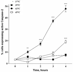
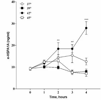
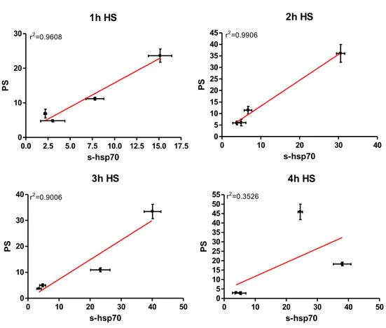
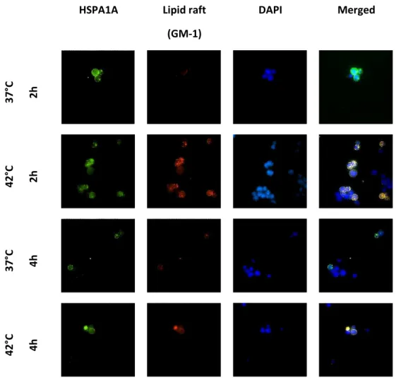
33 Elevated temperatures induces apoptosis and then necrosis 33 HSPA1A intracellular and extracellular levels are modified with heat shock 36 Appearance of surface HSPA1A correlates with PS 38 Surface HSPA1A co-localises with lipid rafts and LAMP-1 42
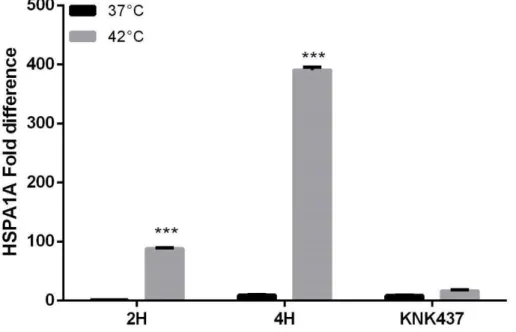
HSPA1A gene expression is induced with heat shock and heat shock factor- 1 inhibitor KNK347 deplete HSP1A1A up-regulation.
46 HSPA1A de novo protein synthesis affect protein secretion but not membrane
insertion
47 Exosome secretion rate did not increase significantly but internal exosome HSPA1A
increase
50 Purified i ro esi les fro stressed ells do ’t i rease their release rate with
heat shock
55 Exosomes purified from stressed cells activate immunocompetent cells 57
Discussion
60CHAPTER 4: ENDOPLASMIC RETICULUM STRESS RESPONSE
Introduction
63Materials and Methods
64Results
65 Endoplasmic reticulum stress markers are activated in Stressed Jurkat cells 65 Surface Grp78 protein expression change with heat shock 67
Discussion
69CHAPTER 5: A HAEMATOLOGICAL MODEL TO STUDY UPR: THE
MYELODYSPLASTIC SYNDROME
Introduction
70Materials and Methods
71Results
72 HSPA1A and UPR gene expression in patients with myelodisplastic syndrome (MDS) 72 HERPUD 2 gene expression and the long non-coding RNA XLOC_006043 are highly
modified in MDS patients
75 Exosome and microvesicles release in MDS patients 76 Exosomes purified from MDS patients stimulate macrophage differentiation 77 Lymphocyte activation is influenced by exosome treatment 79
Discussion
80CONCLUSIONS
831
CHAPTER 1: INTRODUCTION
Beside their canonical chaperone function, Heat Shock Protein (HSPs) have been
considered stress proteins and cellular signals in communication between cells and their
interaction with the immune system need to be addressed more in detail. Although there
are increasing evidences that HSPs are associated with plasma membranes and can be
actively secreted from stressed cells, there is no agreement in the literature about the
mechanism of secretion and the type of stress needed for chaperone induction.
Experimental data suggest that HSP movement can be stress and cell type dependent. The
work presented here aimed to explore the cytoplasmic stress and the endoplasmic
reticulum stress in haematological malignancies and explore possible ways of cellular
communication of stress to distant cells, through the investigation of mechanisms of Hsp
surface presentation to plasma membrane and release in response to cellular stress such
as heat shock. The investigation is focused in the role of extracellular vesicles such as
exosomes in cellular crosstalk. The work concentrates in the analysis of the cytoplasmic
inducible form of the Hsp72 family, HSPA1A, and the corresponding endoplasmic reticulum
protein, Grp78 together with other endoplasmic reticulum stress-related proteins.
2
1.1 The stress response
The cellular stress response is variety of molecular changes that cells experience in
response to environmental stresses such as high temperatures, toxins and oxidative stress,
but also internal stresses such as diseases. The mechanisms activated in cells are adaptive
and aim to protect the whole organism against unfavourable environmental conditions,
through short term responses that control cell damage, and through longer term responses
that promote apoptosis of the damaged cells and protect the whole organism. Stress
response mechanisms operate in every cellular compartment and many molecules are
specifically activated. Between all, chaperones plays a crucial role in cellular stress
response. Chaperones are present in every cellular compartment but the cytoplasmic stress
response, with Hsp72 activation, and the endoplasmic reticulum stress response, with
activation chaperones belonging to the Unfolded Protein response Pathway, are the most
important ones. The present study focus its attention on the cytoplasmic stress response,
looking at the Hsp72/HSPA1A induction and movement to the extracellular space and the
endoplasmic reticulum chaperone Grp78 and its related proteins, looking at unfolded
protein response (UPR) activation.
1.2 The heat shock proteins (HSPs) family
Heat Shock proteins (HSPs) are evolutionary conserved protein crucial for cell viability and
involved in many cellular processes, and their expression depends on their association with
other components and the stress applied. HSPs are well known as chaperones: they refold
denatured or damaged proteins in response to many cellular stresses: heat, oxidative
stress, chemicals treatments, UV radiation, physiological and psychological stress and when
the protein are too damaged, they deliver the impaired proteins to degradation by
ubiquitination or lysosomic proteolysis [1]. Although there are constitutively expressed
chaperones that regulate normal protein folding, most of the HSPs are induced in response
to stress, where a higher amount of intracellular proteins are likely to be denaturated.
HSPs have been classified into families, according to their approximate molecular weight.
The five main groups are Hsp100, Hsp90, Hsp70, Hsp60 and the small HSPs that can be
expressed in the cytoplasm or in the cellular organelles like mitochondria or endoplasmic
reticulum. Members of a given family also possess several other features in common: they
show, for example, a high degree of sequence homology among different species [2].
3
Heat shock protein nomenclature is quite complicated: Hsp70 for example can be named
in several ways depending on the cell compartment where is expressed and whether is
constitutively expressed or induced by stress: Hsp72-1, Hsp72, HSPA1, Hsp72-2, Grp78,
HSC70 and GRP75.
Hence a new nomenclature system established that the gene names can be used for their
correspondent protein product: the systematic gene symbol that have been assigned by
the HUGO gene nomenclature committee and is the one used in the National Centre of
Biotechnology Information Entrez Gene database for the heat shock genes [3].
The present study will focus the attention on two chaperones: the cytoplasmatic inducible
form of Hsp72, referring to the HSPA1A gene product (the new protein name is HSPA1A)
[3] and the endoplasmic reticulum Hsp72 also called Grp78.
1.3 The cytoplasmic Hsp72/HSPA1A and the importance of its location
Hsp72 can be expressed at low levels in normal cells both in vitro and in-vivo and their
expression and release can be modified with several physiological stresses. However
patients with different types of disease or transformed cell lines often present abnormal
levels of the protein and strictly associate with the disease like various auto-inflammatory
diseases and cancer: this aberrant protein synthesis or release in diseases suggests that
heat shock proteins participate in the development of the disease or at least is a direct
consequence [1]. In particular Hsp72 has been found to be over-expressed in several
human malignant cell lines, in cancer tissues and in the serum of patients [1, 4]. Hsp72
seems to be implicated in tumour cell proliferation, differentiation, invasion, metastasis,
death, and recognition by the immune system [1, 5-7]. Hsp72 in cancer cells possess dual
contrasting effects depending on the cell localization:
1.3.1 Intracellular Hsp72 overexpression
Intracellular Hsp72 overexpression promotes cancer development by suppression of
various anticancer mechanisms, like apoptosis, senescence as well as by facilitating
expression of metastatic genes. Hsp72 is a potent anti-apoptotic protein since it interferes
at several levels in the different apoptotic pathways and stimulates cell survival of cancer
cells. Hsp72 over-expression appears to inhibit FAS-induced apoptosis [8]. Heat shock
4
proteins bind to pro-caspases, inhibiting their activation [9-11] but interfere with several
caspase-independent pathways as well [12]. Hsp72 depletion is sufficient to induce
apoptosis through caspase-3 activation in the absence of any additional stress stimulus [6,
13].
1.3.2 Surface Hsp72 overexpression
When Hsp72 is overexpressed on the cell surface it facilitate tumour rejection by immune
system, though innate and adaptive immunity. Hsp72 surface expression has been found
in many types of cancer such as colon, lung, pancreas, mammary head and neck and in
metastases derived from them [1, 14]; high level of surface Hsp72 has also been found in
bone marrow from haematological malignancies including acute and chronic myeloid
leukemia [15]. Tumour cells that have high levels of surface Hsp72 have an increased
immunogenicity compared to a low expressing cells line since they are a target recognised
by NK cells [16]. Hsp72 has been demonstrated to be embedded in the cell membrane and
the part of the Hsp72 protein that extrudes from the membrane triggers the NK mediated
immune response. Incubation of NK cells with cytokines and soluble Hsp72 or TKD peptide,
enhance cell surface density of activating NK cells receptors including CD94 [17] which
seems to interact with Hsp72 on the surface of tumour released exosomes [18]. The
assumption that cells expressing high levels of surface Hsp72 are more sensitive to NK
attack is under investigation to test potential therapies to combine with standard
chemotherapy [19, 20].
1.3.3 Extracellular Hsp72
Released Hsp72, in combination with the tumour antigens are up-taken by the antigen
presenting cells (APC), processed via proteasome TAP-dependent pathway and cross
presented on the MHC class I complex [21-23]. The role of Hsp72 is to facilitate the
internalization, the processing and the cross presentation of the tumour antigens to the
MHC class I on APC. Hsp72 purified from tumours have the ability to cross-present tumour
antigens to APCs and activate the immune response: MHC class I presentation determines
an antigen specific CD8 T-cell mediated initiation of the immune response in in vitro mouse
models [22, 24-26].
5
Hsp a ts as a pote tial da ger sig al interacting with the immune response as the
danger model propose [27]. However, despite the classic danger model by Matzinger
assuming that endogenous components of the cells are released in the extracellular
compartment as a result of necrosis or tissue damage, Hsp72 can be actively secreted from
non-dying cells [28]. This would allow the Hsp72 protein to deliver a different message to
the organism and cause a differential immune response [28].
Extracellular Hsp72 is not just important for immune system activation but also for cellular
protection in response to stress. A direct administration of the protein to cells confered
cytoprotection when heat shock was applied [29]. Hsp72 release from glial cells has been
showed in conjuntion with an enhanced neuronal stress tolerance [30] and a direct external
Hsp72 administration rendered the cells more likely to survive injury than their naïve
counterparts [31] and has an hypothermic and somnogenic effects in brain tissues [32].
Extracellular Hsp72 has a protective effect during sepsis [33] suggesting that there is an
effective cellular protection against stress. These phenomena have great potential
significance in the development of neuroprotective therapeutic strategies utilizing the heat
shock protein response.
1.3.4 Extracellular Hsp72 in vesicles
It is currently unclear whether Hsp72 released as free soluble protein, in combination with
membranes or in detergent soluble membrane vesicles or even all of the above. There are
more evidences that demonstrate that Hsp72 can be actively released from pancreatic and
colon tumour cell lines associated with membrane structures called exosomes [18].
Exosomes in some cellular models are efficient transport vesicles for Hsp72 from the
endosomal compartment into the extracellular space [18, 34]. Studies have also shown that
membrane associated released Hsp72 activate the macrophages via TNF-
α release ith a
much higher efficiency than the soluble form [35]. The exosomal surface appears to reflect
the markers of the tumour cell from which it was derived, with the same relative levels of
Hsp72/Bag4: exosomes surface with high level of Hsp72/Bag4 stimulated the migration of
CD94+ NK cells towards the surface Hsp72 positive tumour cells and increased the
cytoloytic activity of these NK cells [18]. Membrane associated Hsp72 released from cells
has also been demonstrated to activate with more efficiency than the correspondent free
form in order to activate the macrophage, measured as TNF-
α release [35].
6
1.3.5 Mechanisms of Hsp72 release
According to the literature, several cells line models have demonstrated the active release
of Hsp72. However, the results obtained regarding the mechanisms involved are
contrasting and several proposed way have been considered.
Necrosis vs Apoptosis
Early evidences showed that extracellular Hsp72 is a result of necrotic cell death: Hsp72
released from dying cells is a potent inflammatory inducer together with many other cell
product released in the extracellular compartment, like cytokines [36, 37].
Classical release pathways
Some groups support the idea that Hsp72 is released following the classical release
pathway mainly providing with data where Hsp72 release is blocked by classical pathway
transport inhibitors such as Monensin or Brefeldin-A [35]. Despite those findings many
other works have shown the opposite results demonstrating that Hsp72 re
lease ould ’t
be blocked with the classical-release pathway inhibitors. Considering that Hsp72 is a
leaderless protei , so it ould ’t e essar eed to go for pro essi g through the ER a d
Golgi as the classical pathway require, it is more likely that the release mechanism follows
a non-classical pathway.
Non-classical release: lipid rafts associated release
Hsp72 associates with detergent resistant micro-domains enriched in cholesterol and
sphingolipids which form a distinct, liquid ordered phase in the lipid bilayer of membrane
that serves as major assembly and sorting platforms for signal transduction complexes: the
structures are also called lipid rafts. Hsp72 forms ion channels in in-vitro lipid models
resembling the plasma membrane structures [38]. In-vitro models also showed that Hsp72
can interact with phosphatidylserine (PS), a lipid component of the LR structures [39], and
that it can bind to the lipid raft component ganglioside-M1 (GM-1) [40]. Heat shocked cells
express surface Hsp72 in relation with lipid rafts and secrete them in the extracellular
environment [41]. Monensin and Brefeldin A, drugs used to perturb classical protein traffic,
did not reduce this effect, determining the release of Hsp72 from cells is independent of
the classical secretory route [41-43]. When a cholesterol disrupting agent, methyl-
β-7
cyclodextrin, was added to the cells Hsp72 release was abrogated, suggesting that Hsp72
localises preferentially in lipid rafts whose integrity is required for active release [41, 43].
Extracellular Hsp72 was demonstrated to bind the macrophage plasma membrane,
specifically on its lipid raft microdomain, which caused a disruption of the lipid rafts and
abrogated the Hsp72-mediated increase in phagocytosis and enhanced the processing and
presentation of internalised antigens [44]. Endogenous Hsp72, up-regulated following a
bacterial attack, also binds to the surface of macrophages through the TLR-4 and CD14
receptors: these associations seems to take place within the lipid rafts structures [45]. All
these evidences support the idea that Hsp72 can directly bind to membranes by interacting
with lipids. Recently it has being raised the hypothesis, mainly using thermal stress models,
that cellular stress is detected by the plasma membrane which modify its fluidity and
transduce the stress signal to intracellular pathways involving activation of RAF and PI3K
with activation the heat shock factor with consequent HSPs over-expression and
membrane insertion [46]; in other words plasma membrane could be considered a cellular
stress thermometer and HSPs
ould a t as e ra e sta ilisers. It’s i teresti g to
underline that membrane defects are associated with various physiological states such as
ageing and are simultaneous with a deregulation of heat shock protein synthesis [46].
Exosomes associated release
Exosomes represent a specific subtype of extracellular vesicles, between 30 and 100 nm in
diameter, especially rich in cytosolic and membrane proteins, nucleic acids such as
microRNAs, hormones and growth factors [47]. The exosomes are isolated, as well as from
cell cultures, from many biological fluids such as blood, saliva, urine and breast milk. Today
they represent one of the latest and more promising discoveries regarding intercellular
communication [47, 48] as they are involved in many processes such as regulation of
immune system, infectious and autoimmune diseases and cancer [49]. Exosomes are
distinguished from other extracellular vesicles [50] for their cellular origin rather than by
their size or their shape. Exosomes are vesicles that are released from cells following fusion
of multivesicular bodies (MVBs) with the cell membrane [51, 52], while microvesicles are
defines as vesicles that form from the plasma membrane. The MVBs are organelles that
originate from early endosomes; they are enriched of intraluminal vesicles, (ILVs ) that form
following endosomal membrane invagination. The MVBs can be considered a sort of late
8
endosomes involved in the endocytic pathway receptor mediated. Exosomes contribute to
the release of Hsp72 from human peripheral blood mononuclear cells (PBMCs) during basal
conditions and in response to heat shock cellular stress. Vesicles structure consist of lipid
raft regions embedded with ligands common to the original cell membrane
.
Exosome
vesicles released from cancer cells showed the presence of surface Hsp72 in combination
with lipid rafts [18, 53] demonstrating that a possible combination of the proposed release
pathways could occur in different cell models. Hsp72 can be released from heat shocked
PBMCs via an exosome-dependent pathway [18, 34]. Exosomes were produced by
B-lymphoblastoid and Jurkat cell lines where a time-dependent increase in Hsp72 could be
identified. The Hsp72 was located within the exosome lumen following heat stress with no
interaction of Hsp72 with the exosome surface, thus suggesting that such exosomes may
not interact with cells through cell-surface Hsp72-receptors and demonstrating that Hsp72
levels increase in proportion to the degree of stress [54]. However different cellular models
give differential results. Tumour-derived exosomes containing high levels of Hsp72 in their
lumen stimulate NK activation. Examination of the plasma membrane of tumour cells and
of the secreted exosomes revealed selective Hsp72/Bag-4 expression, high levels on the
exosomal surface of Colo+ pancreas and CX+ colon carcinoma tumour sublines and low
levels on Colo- and CX- tumour sublines. Results show identical Hsp72/Bag-4 content in
exosomal lumen however on the surface of exosomes the Hsp72/Bag-4 content reflected
the tumour cell membranes from which they were derived. Colo+ and CX+ tumour cells
expressing high levels of Hp72/Bag-4 were found to be susceptible to cytolytic attack by
CD94+ NK cells after stimulation with IL-2 plus TKD, due to the Hsp72/Bag-4
surface-positive exosomes initiating migration in CD94+ NK cells [18].
Lysosome pathway
Hsp72 does not have the consensus secretory signal, so it cannot pass through the plasma
membrane using any classical mechanism lead to hypothesise different release pathways,
one of which is the endolysomal pathway. Hsp72 has been showed to bind intracellularly
with lysosomes membranes in Hsp72 over-expressing colon carcinoma cells and
immortalized murine embryonic fibroblasts (MEFs); the Hsp72 protects the lysosomal
membrane from permeabilization and cell death induced by tumor necrosis factor,
etoposide and H
2O
2suggesting a lysosomes membrane stabilization role of Hsp72 [5].
9
Mambula et al demonstrated that Hsp72 release from human prostate cancer cells
following heat shock, passes through the endolysosomal compartment: inhibition of
lysosomal transport using a lysomotropic compound, blocks Hsp72 secretion. Purified
lysosomal compartment possesses high level of Hsp72. The increase of surface Hsp72 after
heat shock highly correlates with increase of surface LAMP1, a lysosome marker. The
release is quick and occurs before any detectable increase in gene expression suggesting
that heat shock directly induces the secretion of Hsp72 without activation of the heat shock
factors and seems to be an independent aspect of heat shock response [42].
1.4 Grp78 and the Endoplasmic Reticulum stress response
The endoplasmic reticulum (ER) is an organelle responsible for homeostasis of intracellular
calcium, for membrane lipids biosynthesis, folding and transport of proteins. Protein
folding is a mechanism which is extremely sensitive to cellular environment alterations.
Changes in Ca2 + levels, changes in redox state, increased protein synthesis, pathogenic
and inflammatory stimuli can all alter protein folding. When cellular environment change
misfolded proteins accumulate and form aggregates that are toxic to cells: this condition is
defi ed as ER stress". The Unfolded Protein Response (UPR) is the system by which the ER
respond to stress and is activated by the accumulation of misfolded proteins [55]. The
activation of the UPR involves transient attenuation of protein synthesis, increased protein
trafficking through the ER, increased protein folding and transport, activation of pathways
involved in protein degradation (ERAD
– ER Associated degradation). If these adaptive
mechanisms are unable to solve the problem, the cells go through apoptotic pathway.
The activation of this pathway occurs not only in normal cells, but also in cancer cells.
Therefore, depending on the context, it is not surprising that the activation of the UPR
contributes both to increased survival and cellular apoptosis.
When misfolded proteins accumulate in the ER lumen, two key events are necessary for
activation of UPR: first, these misfolded protein aggregates bind and sequester the
chaperone BIP (immunoglobulin heavy chain binding protein), also known as Grp78 [55].
Secondly, the consequent reduced levels of Grp78 is a clear pathway activation signal that
induce the transcription of BIP, as well as other genes coding for chaperones [55, 56]. This
response takes place via the activation of three transmembrane receptors:
10
• Pa reati ER ki ase PERK
• A ti ati g Tra s riptio Fa tor ATF
• I ositol-Requirng Enzyme 1 (IRE1).
1.4.1 PERK
PERK (Pancreatic ER Kinase) is a type I transmembrane kinase that becomes activated by
dimerization and autophosphorylation, after dissociation with Grp78. The activation of this
kinase causes phosphorylation of
eIF α, respo si le for protein translation inhibition [57].
Activation of PERK-
eIF α redu es the glo al tra slatio of RNA, ut on the other hand,
increases the translation of various mRNA, including ATF4 and ATF5. ATF4 is a transcription
factor that induces the expression of genes involved in ER function, in redox reactions, in
stress response and protein secretion [58]. ATF4 is also associated with CHOP (C/EBP
homologous protein) another transcription factor that induce apoptosis
ER-stress-mediated, both in vivo and in vitro [59]. CHOP transcription is inhibited in the initial phases
of the UPR activation, while it is induced when the stress become chronic.
Therefore, under
stress conditions, PERK is immediately activated and is function is to try to prevent cell
damage and promote survival. If stress persists, ATF4 induces transcription of CHOP that
induces programmed cell death [60].
1.4.2 ATF6
The activating transcription factor 6 (ATF6) is a transmembrane glycoprotein, whose
luminal domain detects protein misfolding. In mammals is present with two isoforms,
α
and
β, both expressed ubiquitously in all the tissues. The cytoplasmic portion of ATF6 act
as transcription factor because it contains a DNA binding domain. Following the Grp78
dissociation, ATF6 moves to the Golgi apparatus, where it is activated by a proteolytic
cleavage by two serine proteases, sp1 and sp2 [61]. Active ATF6 moves to the nucleus and
it induces transcription of genes coding for the chaperones, which have an ER response
element (ERSE) in their promoter [62]. This determines an ER increased folding capacity,
helping to restore initial homeostasis.
11
IRE1 (Inositol Requiring Enzyme-1) is activated after detachment from GRP78, by
dimerization and autophosphorylation. The activation of IRE1 is also affected the fluidity of
the membrane, which is modified when oxidative stress occurs. XBP1 is the IRE1 substrate.
In normal conditions XBP1 levels are very low; they increase when ER occurs due to ATF6
induction. In the presence of its substrate, IRE1 cut by splicing the XBP1 mRNA, forming its
active form (XBP1s) that enters the nucleus and determines the activation of target genes
[62, 63] that determine an increase degradation of misfolded proteins accumulated in the
ER [64]. Protein degradation could then represent the third stage of the response UPR,
following translation block and increase of chaperone synthesis. In addition, XBP1
overexpression induces many genes involved in the secretory pathway and determines the
expansion of the ER. However
IRE α activation is attenuated in case of chronic stress,
through a mechanism not fully established [65, 66]. In addition to this mechanism, which
promotes cell survival, IRE1 can also have a pro-apoptotic role by JNK74 kinase activation.
Under normal conditions, the receptors (PERK, ATF6, IRE1) remain inactive through binding
with the chaperone Glucose Regulated Protein 78 (Grp78). In stress conditions Grp78
dissociates from them and determines the activation, inducing UPR. In the first instance
there is the activation of PERK and ATF6 that try to reduce the stress. Subsequently, the
activation of IRE1 appears to have a crucial role in setting up pro-apoptotic signals. If the
ER stress persists, PERK and IRE1 pathways converge, enhancing their pro-apoptotic effect,
mediated by CHOP and JNK60.
1.4.4 UPR IN CANCER
UPR activation has been found in many human diseases and in mouse models. Cell death
is the physiological consequence of chronic stress of the ER, and is the key of the
pathogenesis of many diseases, including metabolic diseases, inflammation,
neurodegenerative diseases and cancer [55]. Cancer cells are stimulated to produce large
amount of proteins in a short time, therefore they are very dependent on the correct
function of UPR system. UPR is also important in tumor pathology: it is indeed necessary
for cancer cell growth in a hypoxic environment. The inactivation of PERK pathway, impairs
cell survival in hypoxia [67]. PERK also promotes the proliferation and growth of cancer
cells, limiting the DNA damage from oxidative stress, through ATF4 [68]. Thus, the PERK
signaling cascade, phosphorylated
eIF α, ATF is esse tial for cancer proliferation.
12
The activation of UPR in cancer cells is due to intrinsic and extrinsic factors [69]. The
hyper-activation of oncogenes (such as HRAS, MYC, BRCA1 and PTEN) and the loss of tumor
suppressor function, increases the synthesis and translocation of proteins in the
endoplasmic reticulum, due to the high metabolic demand during neoplastic
transformation [70-72]. Consequently UPR pathway is activated to increase the protein
folding capacity. In addition, the activation of UPR is required to promote the expansion of
the ER for division and transmission to the cells during mitosis [73]. In addition, the hostile
environment caused by the rapid proliferation of tumor cells, determines a strong
endoplasmic reticulum stress of cancer cells, which results in activation of UPR. In solid
tumors, there is a hypoxic environment and a lack of nutrients, such as glucose, due to the
rapid growth of the mass and thus poor vascularization.
PERK pathway in carcinogenesis
PERK/phosphorylated eIF
α/ATF4 pathway plays a key role in cancer cell survival. The
inactivation of PERK, alter the possibility of cell survival hypoxic environment [67]. PERK
also promotes cell proliferation by limiting, through ATF4, DNA oxidative damage. The
function of CHOP in oncogenesis is to date unknown, however it is repeatedly confirmed
that the induction of CHOP in response to a prolonged ER stress, causes pre-malignant cell
death, and prevent neoplastic progression [69]. CHOP deletion increases the incidence of
malignant lung tumours in mouse models KRAS-induced, suggesting an oncosuppressive
role of CHOP [74].
ATF6 pathway in carcinogenesis
The main ATF6 target is Grp78/BIP activation, which plays an important role in protein
folding and assembly, in regulating Ca2 + levels in the ER and controlling the activation of
transmembrane sensors of stress [69]. It has been shown that Grp78/BIP activation in
cancer cells protects them from apoptosis and from immune response [75]. By contrast
Grp78/BIP suppression inhibits tumor cell growth, metastases progression and
development, both in vivo and in vitro [76, 77]. Furthermore Grp78/BIP may be considered
a marker of cell malignancy: in normal conditions is localized exclusively in ER, while in
malignant cells, where it is hyper-expressed, can also be detected on the cell surface. In
various tumor sites, such as lung, bladder, stomach and breast, overexpression of
13
Grp78/BIP confers resistance to chemotherapeutic agents, as well as its suppression
sensitizes cancer cells to pharmacological treatment [78].
IRE1α pathway in carcinogenesis
IRE1
α - XBP1 pathway is also important for cell survival and tumour growth in hypoxic
environment, because it induces the transcription of proangiogenic factors, such as
vascular and endothelial growth factors [79]. In a glioma mouse model, IRE1
α inhibition
reduces of tumour growth, angiogenesis and blood perfusion [80]. XBP1 deletion reduces
the tumour formation, and increase cell sensitivity to hypoxia [81]. On the other hand,
there are studies that demonstrate an oncosuppressive role of IRE
α/XBP1 pathway: in
many human tumours is was found IRE1
α mutations [82], some of which result in a loss of
kinase and endoribonucleasic function [83]. In addition, the loss of XBP1 function promotes
oncogenesis [84].
14
AIM OF THE THESIS
The work presented here aimed to explore the cytoplasmic stress and the endoplasmic
reticulum stress in haematological malignancies and explore possible ways of cellular
communication of stress to distant cells. The work was performed in vitro using a cell line
of leukemic malignant cells. More work has been done in vivo, collecting samples from a
haematological disease which is in a pre-malignant state, the Myelodysplastic Syndrome
(MDS). We chose a haematological model because much literature has been collected
regarding solids tumours and stress response but much work still need to be done
regarding neoplastic blood diseases. We chose a disease model that is a pre-malignant
state because there is no literature about it, while only few data are already collected for
malignant haematological diseases such as acute lymphoid leukaemia or acute myeloid
leukaemia and it could be interesting to compare the cytoplasmic stress response and
endoplasmic reticulum stress response between the two conditions. We chose to start the
investigation studying a family of proteins that more than others are sensitive to stress: the
chaperones. These proteins are responsible for protein folding, hence fundamental for a
correct growth and cell division: in the absence of chaperones, cells including cancer ones,
could not divide. Hsp72 is a molecular chaperone and, in addition to being intracellular, it
has been localised to the extracellular environment and the plasma membrane [28, 39, 85,
86]. Grp78 is the corresponding endoplasmic reticulum chaperone and is induced upon
stress condition and as Hsp72, can translocate to the plasma membrane and outside cells.
Outside the cell Hsp72 has been proposed to have several additional functions, including
activation of innate and adaptive immune systems [28, 87], as well as having
anti-inflammatory activity [86]. In addition, altered expression of both HSPs, both inside and
outside the cell, has been reported in several diseases including many types of cancer
[88-91]. It is therefore likely that the translocation of heat shock proteins from inside the cell
to the extracellular environment have important consequences. Several workers have
demonstrated that Hsp72 can be released from cells, constitutively or after a specific stress
depending on the cell type [16, 42, 43, 92-96]. The different routes for HSPA1A insertion
into the plasma membrane and how that is linked to secretion is not at present fully
understood. In this study we investigated Hsp72 cell surface presentation and secretion
15
after heat stress induced apoptosis, possible mechanisms that control this movement and
discuss the significance of different release mechanisms.
Chaperone location is also crucial for other cellular mechanisms such as immune system
modulation, disease progression, and spreading, for this reason HSPs could represent
useful targets in cancer treatment. Understanding these mechanisms, specially looking at
pre malignant state and see differences between advanced malignant state could represent
an important step, for a better definition of cancer pathogenesis, and also in the future, for
the development of customized therapies.
16
Chapter 2: MATERIALS AND METHODS
2.1 Tissue culture
Cell culture conditions and freezing
The cells lines that were used in this studies were both derived from blood malignancies: the E6.1 Jurkat is human leukemic T-cell lymphoblast cell, U937 is human Caucasian histiocytic lymphoma cell line and HUT78 is a cutaneous T cell lymphoma. All of them are suspension cells, which need the same cell culture media, therefore they possess similar procedures for resuscitation and culture. Briefly the cells were purchased frozen and the cryovials containing the cells were removed from the cryostat, soaked with alcohol and quickly placed in a 37°C waterbath. The vial was quickly opened and the content was place in a 25cm2 flask containing 5ml of pre-warmed RPMI 1940 media with 10% serum. The cells were then counted with the haemocytometer and cell density was adjusted to 3-9x10⁵cells ml-1 for E6.1 Jurkat cells and 2-9x10⁵cells ml-1 for U937 cells. Cells were kept at 37°C in the incubator with 5% CO2. A cell viability test was performed after 24hours using trypan blue assay on haemocytometer. Both cell lines were passaged every 3 days and viability was always tested by trypan blue assay.
All cell lines were frozen according similar procedures: cell suspension was kept at low density, between 3-5x10⁵cells ml-1 for E6-1 and 2-5x10⁵ cells ml-1 for U937 cells. At this stage the cells were in the log phase of the growth curve therefore they were actively growing. Cell suspension was centrifuged at low speed of 400g for 3 min at 25°C to minimise any damage and chilled freeze media, prepared as described above, was added to each cell pellet, transferred in cryovials and slowly freezed in liquid nitrogen vapour phase for about 2 hours. The liquid nitrogen vapour phase provides a gentle freezing of 1 to 3°C/min. The cryovials were then placed in the cryostat, stored in liquid nitrogen until needed.
U937 transformation into macrophages
U937 cells is a monocytic suspension cell line, however phorbol 12-myristate 13-acetate (PMA) treatment activate and transform U937 cells into macrophages. Cells within 48 hours gradually have a cell cycle arrest, began to adhere to the bottom of the flask, increase their size and present a granules in the inside of the cells, some can also present protrusions of the cytoplasm. Transformation of U937 into macrophages was achieved using a suspension of cells actively growing in the log phase of growth. Cells were counted; viability was tested by trypan blue, and then centrifuged at 500g for 3 min at 25°C. Cell pellet was resuspended at concentration of 5x10⁵ cells*ml-1 in RPMI medium with 10% heat-inactivated FBS, containing 10 ng/ml phorbol
12-17
myristate 13-acetate (PMA). Cells were plated out in 12-well cell culture plates at 1 ml/well and incubated for 48 hours to enable differentiation of cells. After 48 hours the media was removed and cells rinsed twice with 10 % heat-inactivated-RPMI then 1 ml/well fresh heat-inactivated media was added. U937 macrophages were then ready for experimental treatments.Cell preparation for experiments
The day before each treatment E6-1, HUT78 and U937 cells were counted and plated into 12, 48, 96 well plates depending on the experiment design. Cells were always plated at concentration of 5x10⁵ cells*ml-1. Regarding the activated macrophages, cells were usually plated 48 hours before into 12-well cell culture plates at concentration of 5 X 10⁵ cells/ml (1 ml/well); prior to treatments, once the activation had occurred, cells were washed once with 10 % heat-inactivated-RPMI taking care not to resuspend the attached activated macrophages, then 1 ml 10 % heat-inactivated RPMI was added together with the treatments, and incubated for the desired time.
2.8 . Blood processing: plasma collection and cell separation
Whole blood collection
The blood provided for these studies came from either patients with myelodysplastic syndrome (MDS) involved in a medical study or from voluntary healthy people. Local research ethics committee approval was obtained for these studies, and consent forms were completed by each patient or volunteer. Blood samples were collected by venepuncture in 7ml K2 EDTA vacutainers.
Plasma collection for exosomes purification
Plasma was separated from whole blood by centrifugation at 2000xg for 10 min. Platelets were removed by an additional centrifugation at 2500xg for 15 min. The collected plasma was aliquoted and stored at -80°C for further purifications and analysis.
Cell separation and conservation
Whole blood was washed with two volumes of D-PBS and centrifuged at 500g for 5 min at 25°C. The supernatant was discarded andthe red blood cells were lysed using 1x lysing buffer. The lysed whole blood was then centrifuged at 500g for 5 min and the supernatant discarded. The cell pellet was washed with DPBS and the cells counted using the Trypan Blue exclusion method on a haemocytometer. Cells were centrifuged at 500g for 5 min, supernatant was discarded and cell pellets were frozen and kept at -80°C.
18
2.9 . Cell viability and apoptosis determination
Microscopic analysis
Visualization of cells under a microscope allowed to make a qualitative and preliminary analysis of the samples and highlighted the macro changes occurred in cells. From cell morphology it was possible to discriminate between viable cells, necrotic cells with signs of shrinking and nuclei condensation and apoptotic cells that could be identified for their distinctive blebbings also called as apoptotic bodies.
Trypan blue exclusion assay
Trypan Blue is a non-permeable cell membrane DNA dye that was used to discriminate between viable and dead cells and also help the visualization of cell morphology. Viable cells do not take up the dye because of their membrane integrity, whereas necrotic cells have a loss of membrane integrity that allows the dye to stain the DNA inside the cells: therefore viable cells would appear clear white whereas dead cells would show up blue. Haemocytometer was prepared and cover-slip was put in place; 10 μl of the ell suspension was mixed to 10 μl of trypan blue solution, then 10 μl of the suspe sio i ture as loaded to oth ha ers of the hae o to eter arefully touching the edge of the cover-slip with the pipette tip and allow each chamber to fill by capillary action. Cells were counted in the 0.04 mm centre square and four 0.04 mm corner squares, as highlighted in the Figure 1, and a separate count of viable and non-viable cells was performed.
Viability and proliferation assay by MTS
The number of viable cells in proliferation was tested using a colorimetric method from Promega: CellTiter ® MTS Aqueous solution. The solution was composed of a tetrazolium compound called MTS (3-(4,5-dimethylthiazol-2yl)-5-(3-carcoxymethoxyphenyl)-2-(4-sulfophenyl)-2H-tetrazolium) and an electron coupling agent PES (Phenazyl-Etho-Sulphate). MTS solution is bio-reduced by dehydrogenases enzymes, which are peculiar in metabolically active cells, into formazan which is soluble and coloured in tissue culture. Therefore absorbance at 490 nm can be read directly into a 96 well plate and the quantity of formazan produced is directly proportional to the number of actively living cells in culture. Briefly MTS working solution was added to the 96 well plates where μl of ells ere seeded the da efore at ell de sit as pre iousl des ri ed. Cells ere undergone the desired treatment and, after the required times, MTS working solution was added. Cells were kept at 37°C with 5% CO2 for the required time (each cell type requires different incubation times (2.5 hours for Jurkat). Absorbance at 490 nm was recorded and comparison with
19
control was performed: usually several controls were included such as media control, positive control with viable cells and negative control with dead cells (killed by microwave irradiation for 10 sec).Necrosis detection with Propidium Iodide fluorescent staining
Propidium Iodide binds to the DNA by intercalating between bases with a steichiometry of one dye per 4-5 base pair. PI is membrane impermeable and is generally exclude from viable cells, for instance PI is commonly used to identify necrotic dead cells. The cells were cultured, and treated in a 96 well plate at concentration of 0.2-0.7x10⁶cells*ml-1. The volume of cell used for the assay is μl per ell for a 9 ell plate. The propidiu iodide sto k solutio as kept at -20°C at 100 μg/ l o e tratio a d a orki g solutio of μg/ l i PBS as prepared efore ea h a al sis. The working solutio as the added to the ell suspe sio : μl per ell for a 9 ell plate. The solution was mixed and incubated at room temperature for 20 min in the dark. The PI fluorescence of the cell suspension was read at excitation 535nm emission 617nm. Suitable controls were added in each experiment: a positive control of necrotic cells obtained irradiating the cell with microwaves for 15 sec, negative control of media only and viable cells.
Controls for apoptotic analysis
The following controls have been used in each assay: viable cell control, necrotic cells control obtained by microwave irradiation for 15 seconds and an apoptosis control by camptothecin treatment for 4 hours.
Caspase-3 fluorimetric assay
Caspase-3 is an effector caspase that plays a key role in several apoptosis pathways, and it has been shown to cleave poly-(ADP ribose) polymerase (PARP), DNA-dependent protein kinase (DNA-PK), topoisomerases, and protein kinase C. The kit chosen for the analysis, from Anaspec, uses the inhibitor (Z-DEVD)
, which is conjugated with the fluorogenic indicator Rh110. When caspase-3 is cleaved it binds to the (Z-DEVD)
2-Rh110 that at this condition generates the Rh110 (Rhodamine 110), a fluorophore that can be detected at excitation/emission=496 nm/520 nm and relative quantitation compared to the no treated control was performed. Usually a positive control of cells treated with camptothecin was included.
20
Active caspase-3 was also tested using a polyclonal antibody-FITC conjugated directed against the active cleaved caspase-3. Cells were harvested and washed with wash buffer, centrifuged at 500g for 5 min at 25°C and supernatant discarded. Cell pellet was then carefully resuspended in fix/perm solution to allow the cells to fix and permeabilize; the solution was incubated for 20 min at 4°C. The cell suspension was then washed with wash buffer, centrifuged at 500g for 5 min at 25°C and the super ata t re o ed. Cell pellet as i u ated ith μl/ 6cells*ml-1 caspase-3-FITC conjugated antibody and incubated for 40 min at 4°C in dark. The unbound antibody was washed with wash buffer, centrifuged at 500g for 5 min at 25°C, the supernatant removed and resuspended with fresh D-PBS. Cells were analysed on the Millipore guava easyCyte in the FITC channel and gating of the caspase-3 positive population was performed in each analysis.Annexin-V FC assay
Phophatidylserine (PS) externalization form the inside of the plasma membrane to the outside of the cell is one of the early event that characterizes apoptotic cell death. Annexin V is a protein that binds to the PS and therefore is a marker for early apoptotic events. The detection of early stage of apoptosis was performed by flow cytometry using the Annexin V/PI kit by BD-Bioscience. Cells were harvested, washed with D-PBS and centrifuged at 500g for 5 min at 25°C; supernatant was discarded and cell pellet was resuspended with 1x binding buffer, cells counted and concentration adjusted to be all 1x10⁵cells*ml-1. Cell suspension was incubated with Annexin V solution for 15 min at °C i the dark. Prior the a al sis Propidiu Iodide PI μg/ l solutio as added to the ell suspension and samples were then analysed straight away by flow cytometry: Annexin V was detected at ex/em=490/520nm and PI was detected using a >520nm longpass filter; 10000 events were recorded for each samples and compensation controls were applied for each experiment.
Caspase-2 FC assay
Active caspase-2 detection was performed using the fluorescent labelled caspase-2 inhibitor carboxyfluorescein-labelled-fluoromethyl-ketone-peptide (FAM-VDVAD-FMK). The inhibitor is cell permeable and non-cytotoxic and binds covalently to the active cleaved caspase-2 that is undergone to proteolytic maturation; the fluorescent molecule bound to the Caspase-2 can be then detected by flow cytometry together with the necrosis markers PI and detected respectively at ex/em=490/520nm for the active Caspase-2 detection and at ex/em=535/617nm for the PI; commercially available kits from Bachemwere used.
21
2.10 . Gene expression analysis
Total RNA extraction
Purification of total RNA from cells was performed using SV Total RNA Isolation System kit (Promega), using diethyl pyrocarbonate (DEPC) treated equipment. The SV Total RNA Isolation
System combine the disruptive a d prote ti e properties of gua idi e thio ate GTC a d
β-mercaptoethanol to inactivate the ribonuclease present in cell extracts. GTC, in association with SDS, acts to disrupt nucleoprotein complexes, allowing the RNA to be released into solution and isolated free of protein. Dilution of cell extracts in the presence of high concentrations of GTC cause selective precipitation of cellular proteins to occur, while RNA remains in solution. RNA is then bound to the silica surface of the glass fibers found in the Spin Basket. DNAse treatment digest contaminating genomic DNA. The total RNA is finally eluted with nuclease-free water.
Quantity and purity of the total RNA obtained from the purification was tested reading the absorbance at 260 nm and 260/230 and 260/280 ratio, using Nanodrop instrument.
C-DNA Synthesis
RNA was quantified and retro-transcribed to c-DNA using using ImProm-II ™ Re erse Tra s riptio
System kit (Promega) using 2 µg RNA and random hexamers primes (P(n)6(Promega). The optimized
reaction buffer and the reverse transcriptase provided in the kit enable cDNA synthesis Volumes of
the reaction used is 20µl. The heteroduplex cDNA/RNA formed was then directly amplified by PCR.
Briefly 2µl of random primers were added to the 2µg of total RNA purified as a total volume of 12,2 µl of solution. The samples were the heated at 70°C for 5 min and then let cool on ice for another 5 min. Then the reaction mix with buffer, reverse transcriptase, ribonuclease inhibitor and water was added to the samples and incubated 1 hour at 37°C. The c-DNA obtained was stored at -20°C for further analysis.
Real time PCR
Real-time Polymerase Chain Reaction (PCR) monitor the progress of the PCR as it occurs, in real time. In real-time PCR reactions the threshold cycle (Ct) is the crucial point to analyse: this is the first PCR cycle where amplification of a target gene is first detected by fluorescence emission. The higher is the amount of c-DNA in the reaction, the sooner it will be possible to see an increase in fluorescence. Quantitative qPCR has been performed using Sybr green technology. The SYBR Green is a fluorescent molecule that binds to the minor groove of the double DNA helix, emitting
22
DNA is denaturated and SYBR Green molecules are free. In elongation phase, the fluorescence increases,this corresponds with an increase of copy number of double-stranded amplicon. The reaction curve is represented by a sigmoid curve where the fluorescence intensity is expressed as a function of the number of cycles.
The analysis was made using SsoAd a ed™ SYBR® Gree super i Bio-Rad), in a CFX96
thermocycler (Bio-Rad). The reaction conditions are summarised below: 95°C for 30 seconds
40 cycles with: 95°C for 30 seconds 60°C for 30 seconds.
Melting curves were analysed after the reaction to assess the specificity of the amplification products. The relative expression of the different gene transcripts are calculated using the ΔΔCt method and converted as ratio of relative expression using the formula 2-ΔΔCt for statistical analysis.
All data are normalized according to the expression of the endogenous reference gene, Actin.
Primer design and choice
Primers used for the study were designed using Primer 3 software. Primer sequences are listed below:
HSPA1A fw: TGCGACAGTCCACTACCTTT; rv: AACACTGGATCCGCGAGAA
ATF6 fw: TTCCTCCACCTCCTTGTCAG; rv: ACCCATCCTCGAAGTTCATGA CHOP fw: TGTTAAAGATGAGCGGGTGG; rv: TGCTTTCAGGTGTGGTGATG GRP78 fw: TGCCTACCAAGAAGTCTCAGA; rv: ACGAGGAGCAGGAGGAATTC XBP-1 fw: CTGAGTCCGCAGCAGGTG; rv: CCAAGTTGTCCAGAATGCCC HERPUD-2 fw: GCTGCTTCTTGAACTGGACC; rv: AGTCTGCCCGAATACACCAA XLOC_006043 fw: GAAGTCGGGCATTCAGGAGA; rv: CAGGTTCTCAGTGTTCCAGG b-ACTIN fw: AAATCTGGCACCACACCTTC; rv: CATGATCTGGGTCATCTTCTC
2.11 . Flowcytometric analysis
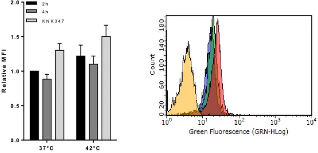
Surface Hsp72 detection by flow cytometry
Surface anti-Hsp72 cmHsp72.1 was specially produced and purchased from Dr G. Multhoff laboratories of Multimune, Munich, Germany. The use of the cmHsp72.1 antibody in this study insured that surface expression of Hsp72 is membrane embedded and not just receptor attached. Literature evidence showed by western blots that the TKD peptide antibody recognise specifically
23
the Hsp72 present in association with the membrane fraction of the cells [97]. This unique antibody specifically recognises the TKD peptide region, a 14-mer sequence, situated in the N-terminal region of the Hsp72 protein (aa 450–463; peptide sequence: TKDNNLLGRFELSG) and therefore detects protein which is embedded in the membrane.This has been demonstrated to be presented outside the cell membrane when the protein is embedded and is the target for natural killer (NK) cell anti-tumour responses [97]. Although the epitope for the Stressgen antibody overlaps the TKD region, it contains more amino acid residues (location between the amino acid residues 436 and 503), that become hidden in the membrane when the protein is embedded, making binding by the Stressgen antibody impossible. Cells were purified as described above, and concentration of cells was determined and adjusted to 1x10⁵cells*ml-1 prior to the labelling. The cell pellet was washed with wash buffer (5% FBS in PBS), prepared as described above, centrifuged at 500g for 5 min and supernatant was discarded. The pellet was then resuspended in antibody solution; samples were incubated for 40 min at 4°C in the dark. The incubation time was followed by a wash with 1 ml of wash buffer, centrifugation at 500g for 5 min followed by a discard of the supernatant. In order to dis ard i the a al sis a e roti ells, . μl of μg/ l propidiu iodide as added to the ell pellet a d left for i at °C. The ell pellet as the resuspe ded i μl of ash uffer a d analysed straight away to the flow cytometer. Cells negative to PI, therefore viable were the only gated, and the analysis of surface Hsp72 was restricted to them only.Intracellular Hsp72 detection by flowcytometry
Cells were purified as described above, and concentration of cells was determined and adjusted to 1x10⁶cells*ml-1 prior to the labelling. The cell pellet, was washed with wash buffer (5% FBS in PBS), prepared as described above, centrifuged at 500g for 5 min and supernatant was discarded. Cells were then fixed and permeabilised with Fix/Perm solution (BD biosciences) and incubated for 20 min at 25°C in the dark. Cells were washed by adding 1ml wash buffer, centrifuged 500g for 5 min and supernatant removed. Cells were then labelled for the intracellular Hsp72 antibody (santa scruz), mixed and incubated for 40 min at 25°C in the presence of dark. The incubation time was followed by a wash with 1 ml of wash buffer, centrifugation at 500g for 5 min followed by a discard of the supernatant. Cell pellet was resuspended in wash buffer and analysed at the flow cytometer.
CD25 surface detection in HUT78 cells
Cells were prepared as described above, and concentration of cells was determined and adjusted to 1x10⁶cells*ml-1 prior to the labelling. The cell pellet, was washed with wash buffer (5% FBS in PBS), centrifuged at 500g for 5 min and supernatant was discarded. Cells were then labelled for the surface CD25, mixed and incubated for 40 min at 25°C. Cells were the washed with 1 ml of wash
24
buffer, centrifuged at 500g for 5 min ad secondary antibody FITC labelled was added to the cell pellet and incubated for 30 min at 25°C in the dark. Cell pellet was washed and resuspended in wash buffer and analysed at the flow cytometer.Cell Cycle analysis by flowcytometry
Cell cycle analysis use the property of certain fluorescence molecules to bind the DNA in a stoichiometric manner, in proportion to the amount of DNA present in the cell. Therefore by looking difference fluorescence emission it is possible to distinguish between the S phase where DNA is more than cells in G1. The cells take up proportionally more dye and will fluoresce more brightly until they have doubled their DNA content. The cells in G2 are approximately twice as bright as cells in G1. The protocol used Propidium Iodide as DNA binding molecule. Briefly cells at concentration of 1x10⁶/ml were washed and resuspended in cold PBS, then fixed using 70% cold ethanol and incubated overnight at -20°C. Cell suspension was centrifuged and PI mastermix at 40µg/ml was added to the cell pellet and incubated at 37°C before the analysis. PI fluorescence was measured by flowcytometry.
2.12 Fluorescence Microscopy methods
Cells cultures were labelled with several antibodies in order to find co-localization of surface and intracellular Hsp72 with cellular and membrane markers. The fluorescence slides produced were analysed with the Nikon Eclipse TE2000-U fluorescence microscopy using IPLAB Suite Software.
Detection of surface Hsp72 and the lysosome marker LAMP-1
LAMP-1 protein is present in lysosome but it has been previously found to be present on the surface of cells in certain conditions; protocol was tested to check plasma membrane LAMP-1 or intracellular, therefore two protocols have been developed in order to check the presence in both locations. Cells at concentration of 1x10⁶cells*ml-1 were prepared, by washing with PBS, centrifuging at 500g 5min and removing the supernatant. Cell pellets were blocked with wash buffer (FBS 5% in PBS) for 30 min at 25°C, then washed by centrifuging at 500g for 5 min at 25°C, supernatant removed. Cell pellet was labelled with 100ul of LAMP-1 and Hsp72 mixed at appropriate concentrations diluted in buffer for 1 hour at 25°C in the presence of dark; cells were washed as described above and anti-mouse IgG-Cy3 conjugated was added at appropriate concentration for 1 hour at 25°C in the presence of dark. Unbound antibody was washed away with a washing step and cells were fixed with PFA 4% for 30 min. Cells were then washed and resuspe ded i μl of PBS; the hole ell solutio as added carefully on top of a polylysine-coated glass slides; slides were mounted by dropping anti-fade solution with DAPI on top of the
25
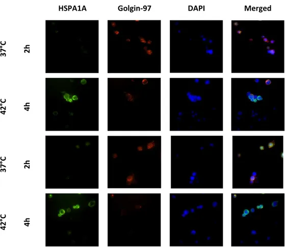
slides and by placing the cover slip on top taking care of not create any bubbles. Slides were left to recover overnight and fluorescence analysis was performed the following day.Detection of Hsp72 and Golgi apparatus marker, Golgin 97
Golgin 97 is an antibody that labels Golgi apparatus. The staining protocol was made in conjunction with Hsp72 in order to test intracellular co-localization of Golgi with Hsp72 protein. Cells at concentration of 1x10⁶cells*ml-1 were prepared, by washing with PBS, centrifuging at 500g 5min and removing the supernatant. Cell pellets were blocked with wash buffer (FBS 5% in PBS) for 30 min at 25°C, then washed by centrifuging at 500g for 5 min at 25°C, supernatant removed. The cell pellet was fi ed a d per ea ilised addi g μl of fi /per solutio BD Bios ie es for min at 25°C. Cells were washed as described above and 100ul of Golgin-97 and Hsp72 was added at appropriate concentrations diluted in buffer for 1 hour at 25°C in the presence of dark; cells were washed as described above and antimouse IgG-Cy3 conjugated was added at appropriate concentration for 1 hour at 25°C in the presence of dark. Unbound antibody was washed away with a washing step as previously described and cells resuspe ded i μl of PBS; the hole ell solutio was added carefully on top of a glass slide; the slide was mounted by dropping antifade solution with DAPI on top of the slides and by placing the cover slip on top taking care of not create any bubble. Slides were left to recover overnight and fluorescence analysis was performed the following day.
Detection of surface Hsp72 and the lipid raft marker GM-1
Lipid rafts are detergent, insoluble, shingolipid-and cholesterols-rich membrane microdomains that assemblies in the plasma membrane. Live cells were first labelled with, cholera toxin subunit B (CT-B)-alexa fluor 555 conjugated which binds to the pentasaccharide chain of plasma membrane ganglioside (GM-1) a component of lipid rafts. An antibody that specifically recognizes CT-B is then used crosslink the CT-B labelled lipid rafts into distinct patches on the plasma membrane, which were visualized by fluorescent microscopy. The lipid raft labelling was coupled with Hsp72 in order to obtain an analysis of co-localization of the protein with these membrane structures. Cells at concentration of 1x10⁶cells*ml-1 were prepared, by washing with PBS, centrifuging at 500g 5min and removing the supernatant. Cell pellets were blocked with wash buffer (FBS 5% in PBS) for 30 min at 25°C, then washed by centrifuging at 500g for 5 min at 25°C, supernatant removed. CT-B conjugate was added at appropriate concentrations for 30 min at 4°C. Cells were washed by centrifugation at 500 g for 5 min at 25°C and anti-CT-B together with Hsp72 antibody was added to the cell pellet at appropriate concentrations for 1 hour at 4°C in the presence of dark. Cells were ashed as des ri ed a o e a d fi ed ith μl PFA % for i at °C i the dark. The ell
26
suspe sio as ashed a d resuspe ded i μl of PBS; the hole ell solutio as added carefully on top of a glass slide; the slide was mounted by dropping antifade solution with DAPI on top of the slides and by placing the cover slip on top taking care of not create any bubble. Slides were left to recover overnight and fluorescence analysis was performed the following day.2.13 Exosome and microvesicles purification and characterization
Exosome and microvesicles purification
Exosome were purified adapting previously developed methods [98]. Briefly cell culture supernatant, from cell growing at 5*105 c/ml, was collected and centrifuged at 500xg for to remove cells; then the supernatant was centrifuged at 2000xg to remove apoptotic bodies and cellular debris; the supernatant was filtered in a 0.22µm filter, then transferred in a 100KDa Vivaspin tube (Sartorius); the sample was concentrated centrifuging the column at 1000xg; microvesicles were isolated centrifuging the resulting supernatant at 16000xg for 30 minutes, the pellet was recovered and the supernatant was isolated for exosome purification and transferred in a ultracentrifuge tube, at the bottom of the 1ml supernatant it was carefully inserted 300µl of a density gradient (20mM TRIS, 30% sucrose in D2O). Samples were ultracentrifuged in a Beckton Dickinson ultracentrifuge with TLA.100.3 rotor at 100,000xg a 4°C for 40 minutes. 350 µl of the exosome mix from the bottom of the tube was collected, diluted in 2.5ml of PBS and ultracentrifuged in TLA.100.3 rotor a 100,000xg at 4°C for 70 minutes. The pellet was collected at the bottom of the tube, resuspended in 200 µl of PBS and stored at -80°C for further use.
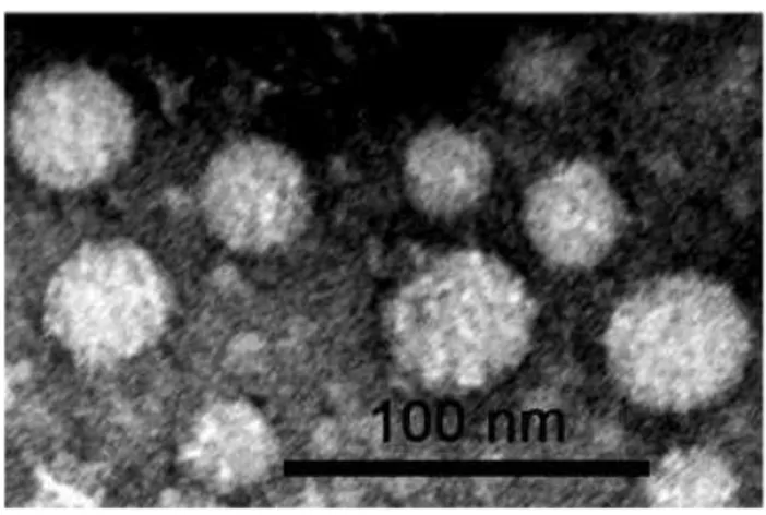
Transmission electron microscopy method for exosome visualization
Purified exosome were analysed by transmission electron microscopy: briefly exosomes in suspension were fixed with an equal volume of 2 % glutaraldehyde in 0.1 M cacodylate buffer (pH 7.4) and observed in transmission electron microscopy after negative staining conducted as follows: a drop (~20µl) of the sample was left to adsorb for 1 min on a 300 mesh nickel grid coated with formvar /carbon film. The sample was then contrasted with 4 steps of drops of uranyl acetate, 3% water for 30 seconds. The grids were air dried and samples were observed with CM10 electron microscope (Philips) at working voltage of 80 KV. The images of the samples were recorded by CCD camera Veleta 130,000X and the magnification of their diameters, expressed in nm, were measured by software iTEM (TEM Imaging Plaatform, Olympus).