1
Sapienza University of Rome
PhD in Clinical-Experimental Neuroscience and Psychiatry
Cycle XXXI
BRAIN-COMPUTER INTERFACE TECHNOLOGY
AND NEUROELECTRICAL IMAGING
TO IMPROVE MOTOR RECOVERY AFTER STROKE.
Neurophysiological studies based on high-resolution EEG and non-invasive brain stimulation.
PhD Candidate: Floriana Pichiorri MD (941105)
Tutor: Prof. Eleonora Palma
2
Table Of Contents
Preface ... 3
Brain-Computer Interface in neurologic rehabilitation practice ... 6
Rationale for BCI use in motor rehabilitation ... 7
Future perspectives ... 18
BCIs for rehabilitation of the upper limb: a multidisciplinary approach ... 21
Motor Imagery based prototype design and clinical validation ... 21
The Promotœr: BCI technology from laboratory to clinical practice ... 22
Hybrid EEG-EMG bci prototype: ongoing projects ... 25
An EEG Index of Sensorimotor Interhemispheric Coupling after unilateral stroke: clinical and neurophysiological study ... 27
Introduction ... 27
Methods ... 28
Results ... 31
Discussion ... 35
Overall Conclusions and Next Steps ... 38
3
Preface
Stroke is defined as a focal lesion in the brain caused by acute ischemia or hemorrhage. The events that characterize acute stroke as well as the spontaneous recovery process occurring in the subacute phase, demonstrate that the focal damage affects remote interconnected areas. On the other hand, interconnected areas largely contribute to reorganization of the central nervous system (CNS) along the recovery process (plasticity) throughout compensatory or restorative mechanisms which can also lead to unwanted effects (maladaptive plasticity). Such post-stroke brain reorganization occurring spontaneously or within a rehabilitation program, is the object of wide literature in the fields of neuroimaging and neurophysiology.
Brain-Computer Interfaces (BCIs) allow recognition, monitoring and reinforcement of specific brain activities as recorded eg. via electroencephalogram (EEG) and use such brain activity to control external devices via a computer. Sensorimotor rhythm (SMR) based BCIs exploit the modulation occurring in the EEG in response to motor imagery (MI) tasks: the subject is asked to perform MI of eg. left or right hand in order to control a cursor on a screen. In the context of post-stroke motor rehabilitation, such recruitment of brain activity within the motor system through MI can be used to harness brain reorganization towards a better functional outcome.
Since 2009 my research activity has been focused mainly on BCI applications for upper limb motor rehabilitation after stroke within national (Ministry of Health) and international (EU) projects. I conducted (or participated to) several basic and clinical studies involving both healthy subjects and stroke patients and employing a combination of neurophysiological techniques (EEG, transcranial magnetic stimulation – TMS) and BCI technology (De Vico Fallani et al., 2013; Kaiser et al., 2012; Morone et al., 2015; Pichiorri et al., 2011). Such studies culminated in a randomized controlled trial (RCT) conducted on subacute stroke patients in which we demonstrated that a one-month training with a BCI system, which was specifically designed to support upper limb rehabilitation after stroke, significantly improved functional outcome (upper limb motor function) in the target population. Moreover, we observed changes in brain activity and connectivity (from high-density EEG recordings) occurring in motor related
4 frequency ranges that significantly correlated to the functional outcome in the target group (Pichiorri et al., 2015).
Following these promising results, my activity proceeded along two main pathways during the PhD course.
On one hand, efforts were made ameliorate the prototypal BCI system used in (Pichiorri et al., 2015); the current system (called Promotœr) is an all-in-one BCI training station with several improvements in usability for both the patient and the therapist (it is easier to use, employs wireless EEG system with reduced number of electrodes) (Colamarino et al., 2017a,b). The Promotœr system is currently employed in add-on to standard rehabilitation therapy in patients admitted at Fondazione Santa Lucia. Preliminary results are available on chronic stroke patients, partially retracing those obtained in the subacute phase (Pichiorri et al., 2015) as well as explorative reports on patients with upper limb motor deficit of central origin other than stroke (eg. spinal cord injury at the cervical level). In the last year, I submitted research projects related to the Promotœr system to private and public institutions. These projects foresee i) the addition of a proprioceptive feedback to the current visual one by means of Functional Electrical Stimulation (FES) ii) online evaluation of residual voluntary movement as recorded via electromyography (EMG), and iii) improvements in the BCI control features to integrate concepts derived from recent advancements in brain connectivity. On these themes, I recently obtained a grant from a private Swedish foundation.
On the other hand, I conducted further analyses of data collected in the RCT (Pichiorri et al., 2015) to identify possible neurophysiological markers of good motor recovery. Specifically, I focused on interhemispheric connectivity (EEG derived) and its correlation with the integrity of the corticospinal tract (as assessed by TMS) and upper limb function (measured with clinical scales) in subacute stroke patients. The results of these analyses were recently published on an international peer-reviewed journal (Pichiorri et al., 2018).
In the first chapter of this thesis, I will provide an updated overview on BCI application in neurorehabilitation (according to the current state-of-the-art). The
5 content of this chapter is part of a wider book chapter, currently in press in Handbook of Clinical Neurology (Pichiorri and Mattia, in press).
In the second chapter, I will report on the status of BCI applications for motor rehabilitation of the upper limb according to the approach I developed along my research activity, including ongoing projects and prliminary findings.
In the third chapter I will present the results of a neurophysiological study on subacute stroke patients, exploring EEG derived interhemispheric connectivity as a possible neurophysiological correlate of corticospinal tract integrity and functional impairment of the upper limb.
Overall this work aims to outline the current and potential role of BCI technology and EEG based neuroimaging in post-stroke rehabilitation mainly in relation to upper limb motor function, nonetheless touching upon possible different applications and contexts in neighboring research fields.
6
Brain-Computer
Interface
in
neurologic
rehabilitation practice
It was about 10 years ago when the first exploratory studies appeared on the potential use of Brain-Computer Interface (BCI) technology for rehabilitation goals. Since then, this clinical application of BCIs has acquired a substantial scientific ground to delineate its transferability to clinical practice and to eventually provide a tool to promote the recovery of function after brain injury.
The evolution of BCI in rehabilitation has occurred in parallel with an emancipatory progress of neurologic rehabilitation that from a traditional approach mainly driven by teaching compensatory strategy to mitigate the dysfunction is turning into a medical discipline driven by evidence-based approaches to restore function.
The modern neurorehabilitation as a medical discipline has its foundations in the principles of neuroplasticity (Dimyan et al., 2008). Neuroplasticity encompasses all the modifications that occur in the components of the central nervous system (CNS) across the lifespan of an individual (Cramer et al., 2011). As such, it is involved in growth, normal aging, learning new skills and adaptation to the environment as well as in recovery after acquired brain injury. In the last few years, certain neurotechnologies have emerged that offer new routes of inquiry into the basis of plasticity occurring in the CNS (Alia et al., 2017; Dimyan and Cohen, 2011). Neuroplasticity has been widely demonstrated after acquired brain damage (Nudo, 2013) and is the fertile ground on which neurorehabilitation strategies intervene. The BCIs allow to act on the environment in absence of muscular activity, thus providing severely disabled people with an artificial channel to communicate and control. This ability resides into a technical pipeline by means of which BCIs directly measure brain activity, translate it into an action and provide feedback to the user in real time (Wolpaw et al., 2002). The ability to substitute or restore function (e.g. control of a neuroprosthesis) defines existing BCIs as assistive as compared to the rehabilitative BCIs which are meant to induce recovery of function (e.g. by modifying brain activity) (Wolpaw and Wolpaw, 2012).
The possibility to monitor and eventually modulate (by self-regulation) specific patterns of brain activity to induce (long-term) neuroplasticity, is the key element for
7 the design and application of BCIs for therapeutic purposes. Indeed, by re-training the brain to specific activities, an ultimate improvement of function is expected (Daly and Wolpaw, 2008; Riccio et al., 2016; Soekadar et al., 2015). Other features of BCI systems, such as the possibility to control external devices that assist movements (e.g. functional electrical stimulation - FES - or robots) further strengthen the BCI role among the repertoire of technologies to support neurorehabilitation (Ang et al., 2015a; Ramos-Murguialday et al., 2013; Varkuti et al., 2013).
RATIONALE FOR BCI USE IN MOTOR REHABILITATION
Stroke is a leading cause of long-term motor disability (Hankey, 2017). Despite the efforts of traditional rehabilitation approaches, more than two thirds of survivors are left with mild to severe paralysis of the upper/lower limb (Langhorne et al., 2011). For these reasons alternative approaches based on advanced technologies are being proposed: robot-assisted therapy, virtual reality, FES, non-invasive brain stimulation (NIBS) and among these, BCIs (Alia et al., 2017).
The current knowledge on the mechanisms underlying the activity-dependent brain plasticity (Dimyan et al., 2008; Nudo, 2011; Zeiler and Krakauer, 2013) and the brain reorganization that naturally occurs earlier after stroke in the motor areas (Cramer, 2008; Dimyan and Cohen, 2011; Ward, 2017) is instrumental for the use of BCIs to promote functional recovery. The majority of the current BCI applications for neurorehabilitation have been targeting upper limb motor function and their recovery after stroke (Remsik et al., 2016; Riccio et al., 2016; Soekadar et al., 2015).
Mechanisms underlying functional motor recovery after stroke
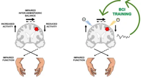
Motor training induces CNS plasticity (Dimyan and Cohen, 2011) and thus, much of neurorehabilitation rests on the assumption that motor learning contributes to motor recovery after injury (French et al., 2007; Zeiler and Krakauer, 2013). Further evidence from animal models and human studies suggest parallels between plasticity mechanisms in the developing nervous system and those taking place in the adult brain after stroke (Murphy and Corbett, 2009; Ward, 2017). Such innate plastic phenomena involve particularly the perilesional tissue in the injured hemisphere, but also the contralateral hemisphere, subcortical and spinal regions (Dancause and Nudo, 2011). There is now a general consensus that a stroke lesion in the motor cortical areas has widespread effects on the brain, involving the ipsilesional and the contralesional hemisphere as well as the balance between the two (Di Pino et al., 2014; Silasi and8 Murphy, 2014). These re-arrangements are conceptualized in a currently accepted theory that is schematized in Figure 1 (left panel). Several rehabilitative approaches (both traditional and novel) have been recently designed that aim at shaping the post-stroke motor system reorganization with manipulation of behavior and cortical activity to improve functional outcome (Dimyan and Cohen, 2011). The principles behind these strategies are schematized in figure 1 (right panel).
BCIs focus on brain activity and thus, offer a direct and effective way to induce plasticity resulting from a task-specific training (see below). Moreover, the BCI can recognize and reinforce motor related brain activity, resulting in an increase in the corticospinal excitability, as measured with Transcranial Magnetic Stimulation – TMS (Hallett, 2007), that is specific to the trained movement/limb (Kraus et al., 2016b; Pichiorri et al., 2011). For this ability to modify brain activity itself, BCI can been considered as a form of endogenous neuromodulation through which plastic re-modeling of brain activity can be induced (see Figure 1).
Figure 1: Schematic overview of the functional motor system reorganization after stroke and the neuromodulation strategies to promote recovery of (hand) function. Left panel: After the acute stroke event (red star over the left hemisphere), a reduction of activity is observed in the ipsilesional hemisphere as opposed to an overactive contralesional hemisphere (Dimyan and Cohen, 2011) (arrows aside the brain). Depending on the size of lesion (Kantak et al., 2012), the interhemispheric cross-talking becomes unbalanced (Perez and Cohen, 2009) (arrows above the brain). According to a currently accepted theory the overactivity of the contralesional hemisphere inhibits the restoration of activity in the ipsilesional with negative effects on functional recovery (Furlan et al., 2016). This abnormal inhibitory activity coming from the contralesional hemisphere towards the ipsilesional is one example of maladaptive plasticity. Right panel: Several rehabilitative approaches (both traditional and novel) have been recently designed according to this functional reorganization model, that aim at: i) stimulating the lesioned hemisphere such as facilitatory Non-Invasive Brain Stimulation (NIBS), distal upper limb training, ii) downregulating the contralesional hemisphere such as inhibitory NIBS, constraint induced motion therapy – CIMT (Kwakkel et al., 2015) in order to favor a re-establishment of the interhemispheric balance (e.g. bimanual training) (Furlan et al., 2016; Plow et al., 2009). The cartoon illustrates how NIBS and BCI can serve as neuromodulation strategies to manly contrast the abnormal unbalance between lesioned and non-lesioned cortical motor areas
9
and thus to improve motor function recovery. The facilitatory NIBS (yellow thunder) is applied to stimulate the ipsilesional hemisphere while inhibitory NIBS (blue thunder) is applied to inhibit the contralesional hemisphere. By enabling the manipulation of brain activity, Brain Computer Interfaces (BCIs) can be considered as a form of endogenous neuromodulation; in particular the BCI-mediated (by self-regulation) reinforcement of motor-related brain activity over the stroke hemisphere can counteract the abnormal interhemispheric unbalance thus, leading to functional recovery.
Different strategies have been put forward to exploit BCI as a tool to modulate brain reorganization towards improved motor function and against maladaptive changes (Daly and Wolpaw, 2008; Riccio et al., 2016). These strategies can be schematized as illustrated in Figure 2.
A first strategy (Figure 2, left panel; “brain-to-function” strategy) promotes the reinforcement of “close-to-normal” brain activity that in turn it would improve (motor) function (Daly and Wolpaw, 2008). The core of this strategy resides in using BCI closed loop not to control external devices (as for communication and control purpose), but exquisitely to reinforce those specific patterns of brain activity which underlies good recovery. The success of these BCIs requires to take into account physiologic motor learning principles to guide BCI “control” feature selection (Naros and Gharabaghi, 2015; Pichiorri et al., 2016).
One exemplary study of this strategy is from Pichiorri and colleagues (2015) wherein 28 subacute unilateral, first ever stroke patients were randomly assigned to receive (as adjunctive to conventional physiotherapy) either a 1month of motor imagery (MI) -based BCI training or the same MI training with no (contingent) feedback (ie, with no BCI). The aim was to establish the efficacy of the MI-based BCI training in promoting the clinical recovery of the paralyzed hand. Control features for BCI training were selected at Electroencephalografic - EEG relevant sensorimotor frequency (sensorimotor rhythms; SMRs) over the affected hemisphere also by ensuring a “correct” execution of kinaesthetic MI (Stinear et al., 2006) of the paralyzed hand movement (that is through the use of TMS). The practice of such MI rewarded by the contingent enriched visual feedback (see below) induced a significant increase of SMR oscillatory activity desynchronization (indicative of a volitional control of motor imagination/execution) only over the lesioned hemisphere that was associated with a clinically relevant improvement of the upper limb Fugl-Mayer scores (Gladstone et al., 2002). In the same study, EEG-derived resting state connectivity of the affected hemisphere positively correlated with the post-BCI training functional motor scores
10 and a re-enforcement of interhemispheric connectivity was observed, thus indicating a beneficial effect of BCI training on motor cortical plasticity.
The core of the second strategy (Figure 2, mid panel; “brain-to-limb” strategy) is that BCI controls external devices to assist limb movement execution such as FES (Daly et al., 2009; Young et al., 2016) or robotic devices (Ang et al., 2015a; Buch et al., 2008; Ramos-Murguialday et al., 2013). This approach is meant to close the sensorimotor loop which has been disrupted by the stroke event (Gomez-Rodriguez et al., 2011), and thus re-establish a connection between the CNS and the periphery. Representative of this strategy is a randomized controlled study by Ramos-Murguialday and colleagues (2013) wherein 32 chronic stroke patients underwent a training of attempted hand movement supported by a BCI-driven robotic orthosis. The 2 group interventions differed in the feedback delivery modality (ie, the orthosis actuation) that was either contingent (experimental intervention) or random (control intervention) with respect to the upregulation of the SMRs over the lesioned hemisphere. BCI training was administered as adjuvant to a physiotherapy program. Upper limb motor function was significantly increased in the “contingent feedback” BCI training with respect to control. Furthermore functional Magnetic Resonance Imaging – fMRI performed before and after the interventions revealed a shift of activation from the contralesional to the ipsilesional hemisphere only in the experimental group, thus indicating a beneficial effect of BCI in promoting brain plasticity and recovery of function.
A third strategy is represented by the possibility to combine BCIs with other neuromodulation strategies (Figure 2, right panel; “brain-to-brain” strategy). To boost neuroplasticity at the CNS level, BCIs have been employed in combination with NIBS techniques such as transcranial direct current stimulation (tDCS) (Ang et al., 2015b; Naros and Gharabaghi, 2017). In a randomized controlled trial with 19 chronic stroke patients, Ang and colleagues (2015b) tested the effects of facilitatory tDCS on the affected hemisphere preceding upper limb MI-based BCI training with robotic feedback delivered for 2 weeks. Although, no relevant clinical improvement was observed in both groups (tDCS+BCI versus BCI alone), the tDCS+BCI group showed a significantly higher SMR reinforcement over the affected hemisphere after training as compared to the BCI group. Similar results were obtained in a recent study with transcranial alternating current stimulation that was applied in chronic stroke patients
11 before or during BCI-training (specifically during resting inter-trial phases) (Naros and Gharabaghi, 2017). The Authors concluded that intermittent stimulation during training had a stronger effect on SMR desynchronization with respect to stimulation delivered before the session. These results highlight the importance of establishing the optimal stimulation modalities and parameters when aiming to boost the BCI effects on neuroplasticity. Learning from animal experiments (Jackson et al., 2006) this combined neuromodulation approach has been further developed, in such a way that the information on brain activity as derived from the BCI can be used to trigger the neuromodulation paradigm with optimal timing and parameters (brain state dependent stimulation) (Kraus et al., 2016a; Walter et al., 2012).
Regardless the strategy, the re-enforcement of the motor-related cortical activity has been sought mainly if not only, over the lesioned hemisphere. The contribution of the ipsilesional and contralesional hemisphere to post-stroke recovery in the subacute phase of stroke (that is when most of the neuroplasticity occurs), is however variable and strongly depends on the site and size of the lesion (Kantak et al., 2012). In the case of vast strokes where the recovery potential of the perilesional areas is limited, the contralesional hemisphere participates to motor recovery via uncrossing neural pathways, thus the contralesional activity might not be always detrimental for motor recovery (Buetefisch, 2015; Kantak et al., 2012; Perez and Cohen, 2009). A recent crossover study (Young et al., 2016) included 19 stroke patients who underwent training with a BCI-driven FES system for upper limb and were evaluated periodically with fMRI during both training and resting periods. No significant differences were found between the training and resting period as regards the ipsi- and contra- lesional corticospinal tract, the transcallosal motor fibers integrity (as assessed with Fractional Anysotropy – FA) (Sotak, 2002) and the behavioural measures. A further correlation analysis revealed that the contralesional corticospinal tract FA increase was correlated with poor upper limb functional scores thus, suggesting a possible maladaptive role of the contralesional motor system. This correlation was inverted during the BCI training and was also accompanied by increases in transcallosal FA. These preliminary findings suggest that BCI training could harness plasticity in the contralesional corticospinal tract from a possible detrimental role to a compensatory role.
12
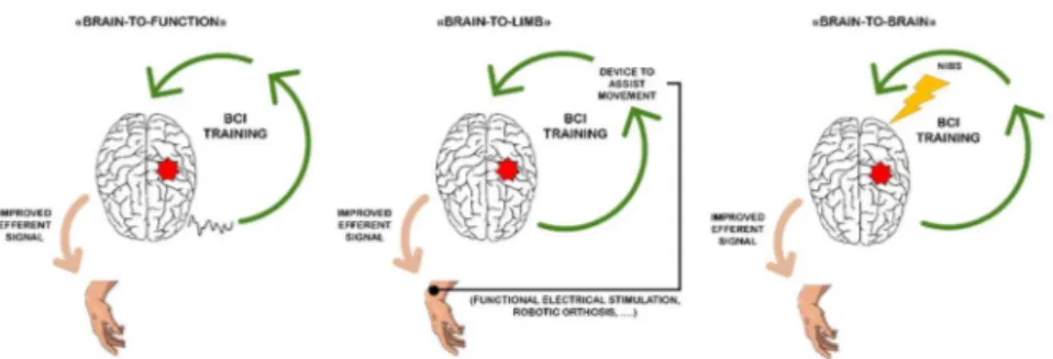
Figure 2: Overview of the Brain Computer Interface (BCI) applicability in poststroke rehabilitation. The figure illustrates 3 main strategies according to which most of the current BCIs for motor rehabilitation after stroke are designed. To note the 3 approaches are not mutually exclusive and can be combined to target specific aspects of rehabilitation. Left panel: in the "brain-to-function" strategy, the goal is to reinforce a close-to-normal brain activity to improve the efferent signal and thus, leading to better motor function. Mid Panel: in the "brain-to-limb" approach the BCI drives devices that assist movement and re-establish the connection between brain and periphery. Right panel: in the "brain-to-brain" strategy the information on brain activity derived from the BCI triggers/is combined with another neuromodulation paradigm (ie, non- invasive brain stimulaton - NIBS) to boost brain plasticity and improve motor function. Details are in the text. (Modified from Riccio et al., 2016).
As currently understood, the clinical and neurophysiologic consequences of a stroke are not only due to the focal lesion but also to the disruption of connections with other brain areas (Silasi and Murphy, 2014). Thus, several brain areas from the ipsi- and contra- lesional hemisphere contribute to the recovery process with positive as well as negative effects (Di Pino et al., 2014). Thanks to the advancement of theoretic/computational analyses, several signal processing techniques have been applied to elucidate the effects produced on distant brain areas by a focal stroke injury (Grefkes and Fink, 2014). This type of connectivity-based approach is related to the concept of connectome, which is defined by the connections between neurons (Sporns et al., 2004). Moreover, a number of studies have demonstrated that non-invasive connectivity measures can improve our ability to correlate behavioral (motor) deficits to clinical indices of dysfunction (ie, motor functional scales) (Carter et al., 2010, 2012; Cheng et al., 2015; De Vico Fallani et al., 2013). Such connectivity measures have been recently employed to assess the effects of rehabilitation interventions based on BCI (Pichiorri et al., 2015; Varkuti et al., 2013).
Recent studies mainly on healthy subjects are exploring the use of connectivity not only as a measure of BCI training induced effects but also as a BCI control feature (Billinger et al., 2013, 2015). Such approach by enabling a modulation of connectivity-based networks that best reflect neuroplasticity principles, could favor a close to-optimal brain reorganization e.g. in the motor system (Hamedi et al., 2016). In support of a connectivity-based as contrasted to activity-based control features (that is based on activation/deactivation of a specific brain area), recent neurofeedback literature has
13 shown that the former allows for a better control of the system as well as greater effects on brain reorganization (Kim et al., 2015).
Relevance of tasks in designing BCI for motor rehabilitation
Motor Imagery (MI) and voluntary movement attempt (MA) represent two main categories of tasks that can be exploited in BCIs designed for motor rehabilitation (Ang et al., 2015a; Mrachacz-Kersting et al., 2015; Pichiorri et al., 2015; Prasad et al., 2010). The MI has long been employed in motor rehabilitation as a strategy to access/engage the motor system when the execution even in the form of attempt, is not safe or feasible (e.g., high degree of spasticity; severe deficit leading to plegia) (Jeannerod, 1995; Malouin and Richards, 2010; Peters and Page, 2015). In stroke patients, MI can engage residual pathways that have been spared by the lesion that caused the motor deficit and also brain regions other than those with lesions (Sharma et al., 2006, 2009). The actual efficacy of MI as a therapeutic strategy is debated, as clinical trials have led to contrasting results (Ietswaart et al., 2011; Page et al., 2007). The causes of such uncertainty have been mainly attributed to the inhomogeneity of MI training protocols and the lack of objective measure of the actual adherence of patients to the required mental task. In this scenario, MI-based BCIs offer a tool to monitor and reinforce mental practice with motor content, provided that the task design endows the principles of task-specific training (ie, consistency between MI content and the rewarding feedback; goal-oriented action) which has been shown to induce motor cortical plasticity and translate the rehabilitation effects in the real world scenario (Peters and Page, 2015).
It can be argued that stroke might affect the ability of patients to perform MI, depending on the lesion side and site (Kemlin et al., 2016) and thus the brain reactivity to MI might be altered. Despite the proven ability of stroke patients to master MI-based BCIs (Buch et al., 2008; Pichiorri et al., 2015; Prasad et al., 2010), the question of which sub-group of stroke patients can benefit the most from MI-BCI-based interventions deserves further investigation. Buch et al. (Buch et al., 2012) have shown that the performance of hand grasping MI after chronic stroke relies on the integrity of the parieto-frontal network.
The ultimate goal of motor rehabilitation is the improvement of motor skills in everyday life situations. In stroke patients with residual motor abilities, the voluntary motor attempt (MA) is the most direct way to engage the motor system and it forms
14 the base of most of the traditional physical therapy (Nie and Yang, 2017). At the level of the brain, MA can be detected from the instants that precede and follow movement start (Shibasaki and Hallett, 2006). In BCI contexts, brain activity that is correlated with movement preparation and eventually execution can be detected and reinforced similarly as in MI-based BCIs for rehabilitation (Niazi et al., 2012). Furthermore, the muscular activation generated by MA can be included in the BCI paradigm via electromyographic (EMG) recording.
The so-called hybrid BCIs are characterized by the combination of brain signals with muscular activity as recorded by e.g. EMG (Wolpaw and Wolpaw, 2012). Hybrid EEG-EMG BCIs have been proposed in several BCI applications for communication or substitution where the signals can be fused as one input to the classifiers or used independently, to ultimately increase the accuracy of control (Choi et al., 2017; Millán et al., 2010; Müller-Putz et al., 2015; Riccio et al., 2015; Rohm et al., 2013).
In the case of rehabilitative BCIs, hybrid BCIs combine residual EMG activity with motor-related brain activation and provide a contingent reward which aims at re-establishing the link between the CNS and the periphery that is disrupted by the stroke (Chaudhary et al., 2016). It has been shown that even in severely paralyzed patients the residual EMG activity induced by MA can be reinforced via a MI-based BCI training and then reliably used as a control signal in a further stage of rehabilitation (Kawakami et al., 2016). Therefore, a modular approach including different biosignals (EEG only, EEG combined with EMG) according to the patients’ residual abilities and to the stage of recovery could be envisaged.
The integration of residual EMG activity in rehabilitative BCIs design requires some important clinical implications to be considered. Clear examples are the spasticity (ie, an abnormal increment of the physiologic muscular resistance to passive/active movements) and the abnormal muscular synergies (ie, abnormal functional recruitment of patterns of muscles) that are extremely common in post-stroke patients (Sunnerhagen, 2016). In this respect, the rehabilitative principle of promoting good plasticity and thus efficient recovery of function applies also to the muscular training/engagement possible operated by a hybrid BCI.
Although, a proof of concept of the hybrid approach in BCIs for post stroke rehabilitation has been given (Grimm et al., 2016) there is still the need for structured
15 trials with long-term comprehensive evaluation of the many clinical and neurophysiologic aspects reflecting the (ab)normal interplay between CNS and periphery. In particular, several different EMG features could be theoretically employed, eg. amplitude or frequency of EMG activity on the target muscle, ratios of amplitudes from different muscles; similarly, several ways of combining EMG activity with brain derived activity to drive the BCI system can be hypothesized including e.g. a measure of cortico-muscular coherence (von Carlowitz-Ghori et al., 2015). No consensus exists yet on these aspects and further studies are needed to define crucial aspects such as the “close-to-normal” EMG patterns to be reinforced or trained while discouraging those provoking spasticity and/or pathological synergies to eventually ensure a BCI mediated optimal re-establishment of brain-to-periphery connections.
Relevance of Feedback Modality in designing BCI for motor
rehabilitation
Within the BCI loop, the feedback is the means through which the subject receives real-time information about his own brain activity, and it is crucial for the instrumental learning occurring along the training (for a review see (Sitaram et al., 2017)). Feedback modality (e.g. visual, auditory, tactile) influences the subject’s engagement in the training and it has an effect on BCI performances (Kaufmann et al., 2013; McCreadie et al., 2013).
In rehabilitative BCIs aiming to re-establish the link between the brain and the periphery and to favor motor re-learning (Gomez-Rodriguez et al., 2011), the choice of the appropriate feedback is evident. In addition, participation and engagement of patients are factors of utmost importance in rehabilitation outcome (Paolucci et al., 2012).
Visual feedbacks such as arrows or cursor-target have been long employed in SMR-based BCI applications (Pfurtscheller and Neuper, 2001; Pichiorri et al., 2011; Schalk et al., 2004). Such abstract feedbacks might not be the best to facilitate patients’ engagement and even more importantly, to cope with the content of a BCI-mediated task-specific training for motor rehabilitation. The BCI feedback design in (Cincotti et al., 2012) and subsequently in (Pichiorri et al., 2015) was implemented as visual representation of the patients’ own hands characteristics and position, to optimize the adherence of patients to perform the MI task and to provide the best match between the content of MI task and the contingent visual feedback (Vargas et al., 2004).
16 Visually enriched feedback and virtual reality environment represent a valid instrument when other types of feedback (i.e. proprioceptive) may be hazardous, too bulky or too expensive. That is the case of e.g., walking rehabilitation for patients with severe lower limb motor deficit (Luu et al., 2016), or home-based rehabilitation programs with no direct assistance of professionals (Vourvopoulos and Bermúdezi Badia, 2016).
Proprioceptive feedback, i.e. feedback via activation of sensory terminations in the target limb, has the main objective to artificially link the brain activation related to motor intent with the peripheral activation of muscles and nerve terminations. Such objective may be obtained with several devices like robots or exoskeletons (passively moving the target limb), (Ang et al., 2015a; Buch et al., 2008; Ramos-Murguialday et al., 2013) or via electrical stimulation (FES) (Bhattacharyya et al., 2016). The effects of peripheral electrical stimulation on neuroplasticity have been attributed to the activation of afferent fibers inducing changes at the brain level (Quandt and Hummel, 2014).
In the so-called associative BCIs (Mrachacz-Kersting et al., 2012), the sensory afferent and the brain signals generated by an attempted movement are timely coupled, and thereby, the effects on brain plasticity subserving motor outcome are marked and occur quite rapidly. This BCI approach (Mrachacz-Kersting et al., 2012, 2015; Niazi et al., 2012; Xu et al., 2014) is based on the real-time detection of motor related cortical potential, MRCP (generated by the voluntary imagination/attempt of foot dorsiflexion) which triggers a peripheral nerve stimulation of the lower limb. This coupling of movement intention (MRCPs) with peripheral stimulation induced plasticity in the motor cortex (i.e. an increase in the corticospinal excitability as measured with TMS) that occurred after one training session (30-50 task repetitions) of healthy participants (Mrachacz-Kersting et al., 2012; Niazi et al., 2012; Xu et al., 2014). A RCT carried out with chronic stroke patients showed BCI-related functional improvements in walking speed and foot tapping on the paretic side after only one week of training (Mrachacz-Kersting et al., 2015).
BCI-based motor rehabilitation in SCI: beyond neuroprosthetics
control
Disability following SCI is virtually permanent and affects people of any age (Ackery et al., 2004) representing a major challenge in neurorehabilitation worldwide. The long
17 history of BCI research in SCIs have been substantially devoted to develop systems to control external devices with the purpose of restoring permanently lost hand function (Collinger et al., 2013; Hochberg et al., 2012; Kreilinger et al., 2013; Müller-Putz et al., 2006; Pfurtscheller et al., 2003) or mobility (Leeb et al., 2013).
SCI rehabilitation beyond neuroprosthetics is apparently at a turning point. Optimism is fostered by impressive research in animals combining pharmacological interventions with electrical stimulation delivered invasively via spinal implants (Capogrosso et al., 2016). Promising results are also coming from the field of regenerative medicine i.e. stem cell therapy (Anna et al., 2017). In this fervent scenario, recent findings in the non-invasive BCI field can potentially change the way BCI can be beneficial to SCI patients.
In a recent study (Donati et al., 2016), 8 chronic SCI patients participated In a long-term BCI-based gait rehabilitation training. Training protocol was based on lower limb MI and foresaw the use of different feedbacks with progressively increasing complexity: virtual reality, virtual reality during standing, robot-supported gait system on treadmill and overground walking with an exoskeleton. Enriched tactile feedback was also provided across all training stages. At the end of the 1-year protocol, all patients showed partial regain of sensation and of some motor function below the level of the lesion. The functional improvement was paralleled by changes in the EEG reactivity to MI suggesting a possible role of brain plasticity in the observed improvements. Although, all patients were clinically diagnosed as complete SCIs (ie no active movement below the level of the lesion), it can not ruled out that some surviving axon crossing the lesion might partially account for the reported clinical recovery.
In another pilot study, a BCI training in combination with FES of the upper limbs in tetraplegic SCI patients was compared with FES training alone. Following the treatment, functional improvements (increased muscle strength) and neurophysiologic changes were observed in the BCI-FES group only (Osuagwu et al., 2016).
These results, together with anecdotal reports on pain reduction in SCI patients after BCI training (Yoshida et al., 2016) are definitely changing the BCI approach to SCI rehabilitation from an exclusively long-term assistive device to a rehabilitation strategy. Further controlled studies with larger numbers are needed to explore such
18 potential especially in the subacute phase in which brain and spinal plasticity is at its climax.
FUTURE PERSPECTIVES
Along with the increasing knowledge of the mechanisms underlying neuroplasticity, neurorehabilitation is turning into an evidence-based discipline (Bernhardt and Cramer, 2013). Exemplary of this evolving scenario is the post-stroke neurorehabilitation. In this context, BCIs possess the essential properties to become not only a therapeutic intervention but also an instrument to complement the evaluation of recovery outcome over time (Pichiorri et al., 2016). As yet, the unanticipated results on lower limb recovery in SCI patients (Donati et al., 2016) are pushing further the rehabilitative expectations on these systems.
Despite the growing number of clinical studies providing evidence to advance the clinical transfer of BCI, a number of basic and clinical research issues still needs to be solved- if not even addressed for an exhaustive deployment of this technology to neurologic rehabilitation practice.
From a clinical standpoint, the current studies of BCIs in post-stroke neurorehabilitation have provided some encouraging evidence of their efficacy in promoting upper (Pichiorri et al., 2015; Ramos-Murguialday et al., 2013) and lower (Mrachacz-Kersting et al., 2015) limb functional motor recovery. Nevertheless, the issue of treatment efficacy requires new clinical trials to i) confirm the efficacy and safety (i.e. definition of intervention duration/frequency) of BCI on a larger scale; ii) identify patients who best benefit from BCI intervention (i.e., predictor of response; definition of relevant outcome measures); iii) verify the long-term maintenance of clinical benefit and effects on neuroplasticity (i.e., follow-up studies). Upon the success of clinical trials in demonstrating the effectiveness of BCI-based interventions, it will still be necessary to obtain regulatory approval.
This body of clinical research has to be paralleled by a better understanding of the neurophysiological mechanisms behind BCI use for motor recovery.
A peculiarity of BCI systems for neurorehabilitation is represented by the selection of the control features that should not be not simply to enable maximum accuracy (i.e. the best signals to discriminate classes as in control applications) but rather because of the relevance of a given signal in subserving motor (or cognitive) learning principles
19 and related function (Naros and Gharabaghi, 2015; Pichiorri et al., 2016). In this regards, a better understanding of the mechanisms underlying recovery and plasticity from a network perspective (Grefkes and Fink, 2014; Silasi and Murphy, 2014) might shape more effective control feature extraction for rehabilitation purpose. The design of connectivity-based BCIs (Billinger et al., 2015; Hamedi et al., 2016) appears a promising future research path to be undertaken.
As compared to other strategies aiming to favor CNS reorganization (e.g. NIBS) or to stimulate the periphery (e.g. FES or robotics devices), BCIs have the potential to combine these two aspects in a unique manner to serve neurorehabilitation purposes. The combination of endogenous neuromodulation (i.e., BCI) with peripheral activation mediated by FES or robotics effectors might be a key element in successful BCIs (Donati et al., 2016; Mrachacz-Kersting et al., 2015; Ramos-Murguialday et al., 2013). In this regard, hybrid EEG-EMG BCIs (Grimm et al., 2016; von Carlowitz-Ghori et al., 2015) are promising rehabilitative BCIs but further development in terms of optimal combination of brain and peripheral signals are needed.
In light of combined approaches to allow recovery of motor function, available findings are in favor of the integration of the BCI training in a comprehensive rehabilitative program i.e. with physical and occupational therapy (Donati et al., 2016; Ramos-Murguialday et al., 2013). Such integration could facilitate the generalization of the BCI induced improvements in motor function to daily life activities. Furthermore, BCI could act as a primer by posing the brain in an optimal state to boost the functional gains obtained with conventional physical therapy. The recent achievements in SCI patients (Donati et al., 2016) represent a further confirmation that a BCI training instantiated in a comprehensive rehabilitation program can boost CNS plasticity at the level of the motor system thus, leading to functional recovery far beyond the expected (i.e. in chronic SCI patients with stabilized sensorimotor impairment below the level of the lesion).
The likelihood that BCI systems will be adopted in clinical practice calls up for a reimbursement planning and thus, for a relevant role of policy makers such as insurance companies and/or health care systems (Roadmap BNCI Horizon 2020: The future in brain/neural-computer interaction, 2015). The pre-requisite step to address this factor in the BCI transferability is represented by the industrial take-up of research products. Technology-supported interventions such as BCI aim to reduce the costs of
20 rehabilitation by optimizing the ratio between a successful rehabilitation outcome and the required (human) resources to be involved. Home-based rehabilitation in this sense represents an attracting perspective of future BCI-based interventions.
To conclude, the successful clinical transfer of BCIs and the ultimate establishment of their value as neurorehabilitation tools is on its way. The accomplishment of such transfer relies on further multidisciplinary synergy between different actors in medicine, science and industry.
21
BCIs for rehabilitation of the upper limb: a
multidisciplinary approach
MOTOR IMAGERY BASED PROTOTYPE DESIGN AND CLINICAL
VALIDATION
Motor Imagery (MI) has been used extensively for rehabilitation in stroke patients that have little or no residual motor function (Braun et al., 2006), since kinaesthetic MI and actual movement performance activate common neural structures. However, divergent results have been reported as to the success of applying MI to stroke rehabilitation (Ietswaart et al., 2011; Zimmermann-Schlatter et al., 2008). There may be several reasons for this including differences in study design and methodology of the various RCTs, as well as a lack of adherence to the imagery task as the patient does not receive any immediate feedback on the purely mental exercise. To cope with this latter issue, as part of the EU project TOBI (www.tobi-project.org) and of a Ministry of Health project (RF-2010-2319611) we developed a non-invasive electroencephalographic (EEG) based BCI system to support MI training, by providing monitoring and feedback (Cincotti et al., 2012). The EEG data is continuously recorded while the patient imagines grasping or finger extension with their paretic hand. The SMR are extracted and classified in real time and used to trigger movements of a virtual image of the patient’s paretic arm and hand, thus providing for contingent and ecological visual feedback. In this way, the patient learns to associate their imagery with the corresponding movement of the virtual hand. Concomitantly the therapist is provided with BCI feedback of the patients MI performance on a separate screen. This allows the therapist to monitor the patient’s success in imagining the task and provide additional feedback via verbal instructions/encouragement, resembling the setting of a traditional motor rehabilitation session. The underlying hypothesis for the initial study was that by supporting MI training with the BCI, MI related activity in the affected motor cortex would be reinforced with the aim to improve motor function. Following the initial design of the prototype, a proof of principle study was conducted involving eight stroke patients that received the treatment as an add on to their conventional therapy across 12 sessions within four weeks, as well as 15 therapists (Morone et al., 2015). Visual analogue scales and the National Aeronautics and Space Administration Task load index (NASA-TLX)
22 were administered to monitor the satisfaction and motivation of the patients. Results revealed that all patients were able to tolerate and conduct the training and at completion, four patients had attained clinically significant improvements in the arm section of the Fugl-Meyer scale. Furthermore, BCI performance correlated significantly with interest and motivation. The therapists reported as one advantage that the system provided a quantitative measure of the patients’ adherence to the MI. However there were also concerns with regards to the technical skill required to operate the BCI, a need for more goal directed feedback (activities of daily living) and the possibility of an increase in arm spasticity during the MI. Overall, there was a high compliance and adherence to the training. The active involvement of the rehabilitation professionals in this type of BCI is a key component to its successful implementation in the clinic. The study further underlines the importance of feedback and thus positive reinforcement, which significantly influenced the patient satisfaction.
As a next step, we conducted a randomised controlled trial in 28 subacute patients (Pichiorri et al., 2015). Fourteen patients received the BCI supported MI training across four weeks, while 14 performed the MI without the BCI support. At completion of training, the BCI group had a significantly greater improvement in FMA scores that were clinically relevant. This was accompanied by a significant increase of EEG motor-related oscillatory activity over the lesioned hemisphere only in the target group. The correlation between functional improvements with changes in resting state brain network organization further supported the use of our BCI technology to promote early post-stroke functional motor recovery.
THE PROMOTŒR: BCI TECHNOLOGY FROM LABORATORY TO
CLINICAL PRACTICE
Following the successful RCT, a further translational effort at Fondazione Santa Lucia (FSL) (Morone et al., 2016) led to the implementation of an all-in-one BCI-supported MI training station, called the Promotœr, which is currently employed in add-on to standard therapy in patients admitted for rehabilitation with upper limb motor impairment due to central nervous system injury of different etiology. Approximately 70 patients have been treated until now, according to a protocol that was approved by the local ethics board
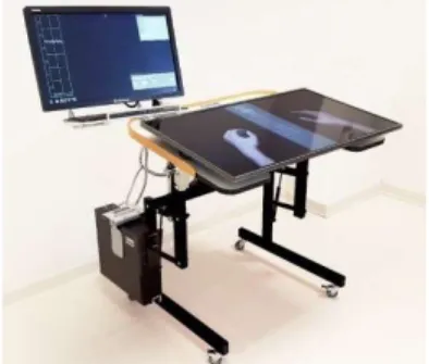
23 (Prot.CE/PROG.604). The Promotœr is an all-in-one training station that comprises a computer, a screen for the therapist feedback (EEG and EMG activity monitoring) and a screen for the ecological feedback to the patient (a virtual hand performing the imagined movement in successful trials); these components are mounted on a wheeled table (Figure 3). A commercial wireless EEG/EMG system is used to record brain and muscular activity from the patient: BCI control features are selected among electrodes placed above the affected sensorimotor area, EMG from forearm muscles is visualized online to ensure relaxation during MI training.
Figure 3: The Promotœr comprises a computer, a screen for the therapist feedback (EEG and EMG activity monitoring) and a screen for the ecological feedback to the patient (a virtual hand performing the imagined movement in successful trials). A commercial wireless EEG/EMG system (gTec, Austria) is used to record brain and muscular activity from the patient.
During training, the patient is seated on a chair (or wheelchair) with arms resting on a pillow. A visual representation of the forearms and hands is given on a dedicated screen, adjusted in size, shape and position as to resemble the patient’s own hands. The patient is asked to perform MI of affected hand (timing of exercise is provided via a spotlight on the screen enlightening the target hand and reinforced verbally by the therapist). During MI, the therapist is provided with continuous feedback of the patient’s brain activity on a dedicated screen; in brief, desynchronization occurring on electrodes placed above the affected sensorimotor area at sensorimotor relevant frequencies (BCI control features) is represented by a cursor moving towards a target (with speed proportional to the desynchronization). In successful trials (i.e. when the cursor reaches the target) the patient receives a positive reward represented by the visual representation of the affected hand moving accordingly with the imagined movement; otherwise, no visual feedback is represented on the patient’s screen. Along the whole session, the therapist is allowed to monitor the patient’s EEG and EMG activity
24 (recorded from forearm muscles) in order to ensure complete relaxation and to guide/encourage him/her during the exercise.
Preliminary results on chronic stroke patients (n=12) partially retrace those obtained in the RCT with subacute patients. Clinical improvements were observed (FMA) together with a significant decrease in muscle tone (as assessed by Modified Ashworth Scale, MAS) which is extremely relevant in chronic patients as spasticity represents a limiting factor in functional recovery during this phase (Pichiorri et al., 2019).
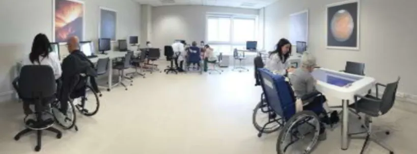
The Promotœr-guided MI training is now fully integrated in the specific rehabilitation program of each patient, involving therapists and doctors working at FSL (Figure 4). While the feedbacks from the patients have been enthusiastic as they are extremely motivated to complete the training with the Promotœr, this close interaction with both the clinical team and the patients has fostered important feedback as to possible novel applications of the device in the daily clinic. As an example, we are currently recruiting SCI patients with lesions at the cervical level, also with extremely positive feedback from patients and professionals.
Figure 4: A patient during a BCI session with the Promotœr (centre of the picture) in a room dedicated to technologically supported motor and cognitive rehabilitation at Fondazione Santa Lucia.
Alongside with the improvements described in the hardware and software components, advancements were made also regarding the process of BCI features selection.
EEG features to be used in BCIs for rehabilitation should be selected according to neurophysiological principles, thus specific expertise in the field is needed. We are currently implementing a procedure based on a semiautomatic method that combines physiological and machine learning approaches, with the twofold aim of reducing the operator variability and facilitate users without direct experience with BCI technology (usability). In a preliminary study we compared classification performances obtained using features selected manually by an
25 expert user and a semiautomatic procedure based on a priori neurophysiological principles; no significant differences were found between the two. The proposed physiologically-driven semi-automatic procedure could boost the transferability of BCI technology to support motor rehabilitation after stroke even in context without direct experience with BCIs, and altogether reduce the operator related variability putting the basis for a larger multicentric trial (Colamarino et al., 2017a,b, 2019).
HYBRID EEG-EMG BCI PROTOTYPE: ONGOING PROJECTS
In the context of stroke rehabilitation, MI practice offers a unique opportunity to access the motor system in severely affected patients; MI training, in our case BCI-supported via the Promotœr, is meant to improve actual motor recovery by boosting brain plasticity. Along the recovery process, therapists encourage and reinforce residual (or recovered) execution of the MI-trained movements. This practical approach is what we aim to incorporate in a second version of the Promotœr. For the development and preliminary testing of this updated prototype, the candidate recently received a grant from a private Swedish foundation (www.promobilia.se).
This enriched version of the Promotœr pursues a compensation of the lost physiological ability to perform a movement, by recognizing the motor attempt (mental pattern and related muscular activity) and producing an actual muscular contraction via Functional Electrical Stimulation (FES). The resulting device will be flexible and adaptable to different patients with variable degrees of impairment, as well as capable to follow each patient along the motor recovery process. We will provide the patient with a real proprioceptive feedback of his mental activity (EEG) related to motor attempt and reinforce the residual or recovered voluntary muscular activity (recorded via EMG) throughout the FES. The aim is to provide a proprioceptive experience as close as possible to the natural movement, artificially controlled by the same brain-muscle circuitry that controls the natural movement. To note, FES alone has been employed as a rehabilitative intervention after stroke for its capability to assist movement and has been shown to induce changes in the brain bearing witness of brain plasticity modulation (Quandt and Hummel, 2014).
26 The shift from MI to attempted movement and from visual feedback to FES feedback has several implications from the clinical point of view. Our approach will take into account solid rehabilitative principles, which partially overlap those already covered in the available version of the Promotœr. As for the brain patterns elicited by motor attempt (from EEG recordings) we will reinforce patterns collected from the affected hemisphere, with specific spatial and spectral characteristics (sensorimotor rhythms, resembling “physiological” activation in the healthy hemisphere during movement executed with the healthy upper limb). As for the residual muscular activity (from EMG recordings) and FES feedback, we will reinforce voluntary contraction reflecting correct muscle activation and discourage pathological synergies, co-contraction of antagonist muscles, leading to spasticity. Indeed, development of spasticity and abnormal synergies is a determinant factor along the recovery process of stroke patients. For these reasons, we will predominantly focus on finger and wrist extension, contrasting pathological flexion spasticity. Continuous EMG monitoring as well as extensive clinical evaluation and the constant presence of a therapist throughout the training will guarantee that possible unwanted effects are promptly recognized and corrected.
The system will be tested in a proof-of-concept framework in stroke patients already familiar with the previous version, with the assistance of rehabilitation professionals familiar with novel technologies for rehabilitation including BCI. This approach will provide a solid evaluation of feasibility and acceptability and put the basis for a subsequent clinical trial of efficacy. The updated version of the Promotœr will increase the currently available BCI-based opportunities for upper limb stroke rehabilitation in order to follow patients along the process of regaining motor abilities.
27
An EEG Index of Sensorimotor Interhemispheric
Coupling after unilateral stroke: clinical and
neurophysiological study
INTRODUCTION
In physiological conditions, activation of the primary motor cortex (M1) on one side induces inhibitory effects on the contralateral M1. Such phenomenon is known as interhemispheric inhibition and it has been extensively reported as altered after unilateral stroke (Perez and Cohen, 2009). The neuroanatomical substrate of interhemispheric inhibition resides in the corpus callosum. This is the largest white matter structure of the brain whose topographical organization has been widely demonstrated (Huang et al., 2005) to reflect the respective interconnected cortical areas. Structural and functional changes in interhemispheric coupling after stroke have been related to the extent of damage in the corticospinal tract (CST), and thus, to the extent of the motor impairment (Li et al., 2015).
The clinical consequences of a stroke are not only due to the effect of the focal lesion, but also to the disruption of connections with other areas (Silasi and Murphy, 2014). On the other hand, interconnected areas contribute to the recovery process, with positive as well as detrimental effects. For these reasons, brain connectivity seems a particularly apt instrument to provide insights on post-stroke recovery mechanisms (Grefkes and Fink, 2014), as well as to eventually assess the extent of damage and the effects of rehabilitation interventions.
In a recent study, we have provided evidence that interhemispheric connectivity (IHC) in subacute stroke patients is modulated by different rehabilitation interventions: specifically a Brain-Computer Interface (BCI)-based motor imagery training of the paretic upper limb in which ipsilesional electroencephalograpic (EEG) sensorimotor rhythms were reinforced, lead to an increase in IHC at rest, specific to the EEG frequency ranges engaged in the training (Pichiorri et al., 2015).
In this study, we aim at defining an EEG-derived index of IHC correlated with CST integrity and severity of clinical impairment. The Motor Evoked Potential
28 (MEP) as assessed by Transcranial Magnetic Stimulation (TMS) will be used as indicator of CST integrity and excitability, as an already established predictor of clinical outcome after stroke (Bembenek et al., 2012). To corroborate the possible clinical relevance of the identified index, we will seek for correlations between such index and the degree of motor impairment in a population of subacute stroke patients undergoing rehabilitation.
METHODS
Patients and Corticospinal Tract Integrity Assessment
Thirty stroke patients (of which 19 already participated in a previous study (Pichiorri et al., 2015)) were consecutively enrolled among those admitted for rehabilitation in our rehabilitation hospital at Fondazione Santa Lucia, IRCCS. The study was approved by the local ethics board (Prot.CE/AG4-PROG.244-105), and written informed consent was obtained from each patient. Inclusion criteria were: age between 50 and 80 years; first ever unilateral stroke (cortical, subcortical or mixed) occurred no longer than 6 months before enrollment and causing hemiparesis or hemiplegia. Upon enrollment, patients were evaluated by means of the European Stroke Scale (ESS) (Hantson et al., 1994) and the upper limb section of the Fugl-Meyer Assessment (FMA) (Gladstone et al., 2002). The ESS is a 14-item scale to assess neurological deficit derived from stroke (including motor and non-motor functions), ranging from 0 (most affected) to 100 (least affected) (Lyden and Hantson, 1998). The FMA is a functional scale to assess limb motor function; the upper limb section (motor domain) is often used separately and it ranges from 0 (most affected) to 66 (least affected) (Gladstone et al., 2002).
Corticospinal tract integrity and excitability was determined bilaterally by means of TMS as follows. Single-pulse magnetic stimuli were delivered through a round coil that was connected to a Magstim 200 (Magstim Company, Whitland, UK) over the motor cortex in the optimal position to elicit MEPs in the First Dorsal Interosseus (FDI) muscle. The electromyographic (EMG) activity from FDI was recorded through Ag/AgCl surface electrodes in a belly-tendon montage (Galileo-NT; Italy). The amplified and bandpass-filtered (0.1 Hz to 2 kHz) raw EMG signal was digitized at a 20-kHz sampling rate and stored for offline analysis.
29 Corticospinal tract integrity was tested at 100% of output stimulator intensity. The presence/absence of a MEP of at least 50 μV was determined by averaging 10 EMG traces. If the MEP was not inducible at rest, patients were instructed to attempt voluntary contraction. The presence or absence of MEP on affected side allowed us to divide the sample into two groups which will be labeled YES/NO, respectively. The motor threshold at rest (RMT) was defined as the lowest intensity that produced MEPs greater than 50 μV in at least 5 of 10 consecutive trials in the FDI muscle and was determined bilaterally (affected side, AS and unaffected side, US) in the YES patients and on the US only in NO patients.
EEG Data Acquisition and Analysis
EEG data was acquired in a separate session (within one week from clinical evaluation and TMS assessment). During the EEG data acquisition patients were comfortably seated in an armchair in a dimly lit room with their upper limbs resting on a desk. Scalp EEG potentials were collected from 61 positions, assembled on an electrode cap (according to an extension of the 10-20 International System; bilateral earlobe reference), band pass-filtered between 0.1 and 70 Hz, digitized at 200 Hz, and amplified by a commercial EEG system (BrainAmp, Brainproducts GmbH, Germany). Five minutes of EEG recordings at rest (relaxed, eyes closed) were acquired. EEG data were down-sampled at 100 Hz (with anti-aliasing low-pass filter) and band pass-filtered (1-45 Hz). Artifact rejection was performed using a semiautomatic procedure, based on the definition of a voltage threshold (± 80μV). EEG traces were then segmented in 1s-epochs and, after relation to the average reference (Common Average Reference, CAR), spectral analysis was performed for all the 61 electrodes: at least 100 artifact-free trials were used for each subject for fast Fourier transformation and power spectral analysis (PSD) within the 5 frequency bands of interest. These latter were individually defined according to the Individual Alpha Frequency (IAF) (Klimesch, 1999) as determined by means of the Fast Fourier Transform spectra over posterior leads (parietal, parieto-occipital, and occipital). The band filtering parameters were: theta 6/IAF-2), alpha (IAF-2/IAF+2), lower beta (IAF+2/IAF+11), upper beta (IAF+11/IAF+20) and gamma (IAF+20/IAF+35). IAF range in our patients was 9.45±0.54 Hz.
30 To investigate the brain network properties under resting conditions, we estimated the statistical dependencies between EEG data (preprocessed as described above, except for CAR), applying methods that have been detailed elsewhere (Pichiorri et al., 2015).
In brief, the full multivariate spectral measure Partial Directed Coherence (PDC) (Astolfi et al., 2006; Baccalá and Sameshima, 2001; Toppi et al., 2016) was used to estimate causality in the statistical sense (expressed as PDC matrices) in each patient. The patterns significance against the null case was assessed by means of an asymptotic statistic method (Takahashi et al., 2007) and the obtained estimations were averaged within the 5 frequency bands as described above. The index called normalized Inter-Hemispheric Strength (nIHS) was defined as the sum of weights of inter-hemispheric links (ihwi) normalized by the total weight of the network (wTOT):
𝑛𝐼𝐻𝑆 = ∑ 𝑖ℎ𝑤
𝑤 (1)
with:
𝑤 = 𝑤 (2)
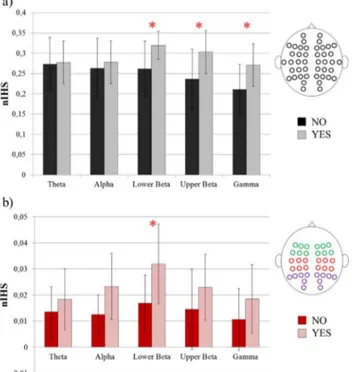
where NIHC in equation (1) is the number of non null inter-hemispheric links, wi is the weight of i-th connection and N the total number of connections in the network. For each patient, nIHS was computed and extracted considering the inter-hemispheric connections in the whole scalp area (global nIHS – 44 electrodes) and in three different scalp subareas (12 electrodes each): central zone (corresponding to sensorimotor cortices) occipital and frontal zones (see Figure 5 for the complete list of electrodes).
This subdivision was performed in order to investigate the possibility to isolate sensorimotor interhemispheric coupling from anterior and posterior areas on the scalp; i.e. to investigate the macroscopic topographical specificity of the proposed index.
Statistical Analysis
31 Statistical comparisons were performed to evaluate differences between YES/NO groups in demographical and clinical characteristics (age, time from event, ESS, FMA), RMT on US (unpaired-two tailed Welch test). Moreover, differences in RMT between AS and US were tested in YES patients (paired t-test).
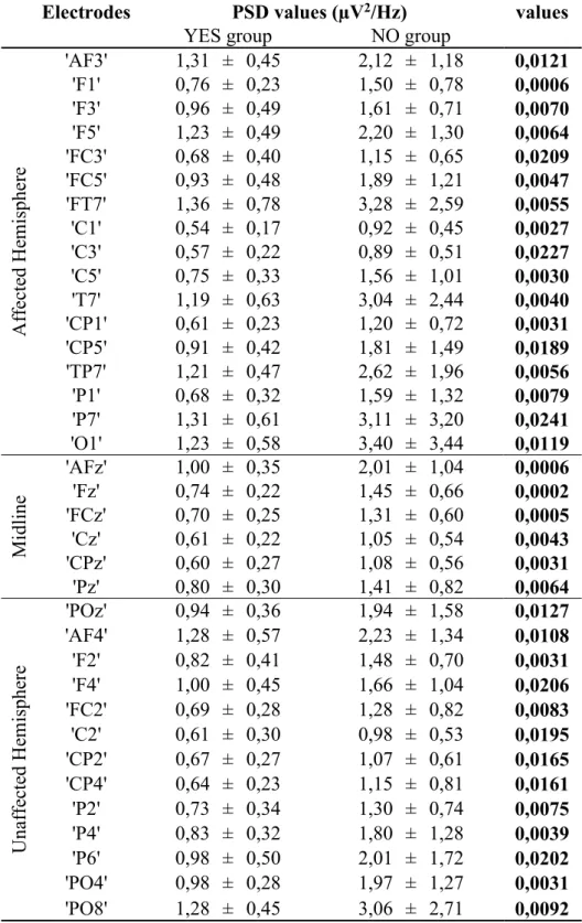
To evaluate between-group (YES/NO) differences in the spectral activity at rest, we performed unpaired two-tailed Welch t-tests between PSD values for each channel (electrode) and frequency band. For this group analysis we flipped the functional (EEG time series) and anatomical (scalp electrode positions) data of patients with right-sided lesions along the midsagittal plane, so that the ipsilesional side was common to all patients (De Vico Fallani et al., 2013; Pichiorri et al., 2015).
For brain network characteristics, statistical analysis was performed (unpaired two-tailed Welch t-tests) to identify differences between groups for the whole scalp ("global"), each scalp area ("sensorimotor", "frontal" and "occipital") and EEG frequency band.
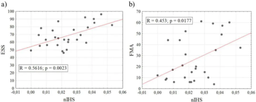
A correlation analysis (two tailed Spearman correlation) between nIHS and clinical scales (ESS and FMA) was performed to further investigate the potential of the proposed effective connectivity index as a neurophysiological descriptor of stroke derived impairment.
Significance level was set at p<0.05. False Discovery Rate correction for multiple comparisons was applied to avoid the occurrence of type I errors.
RESULTS
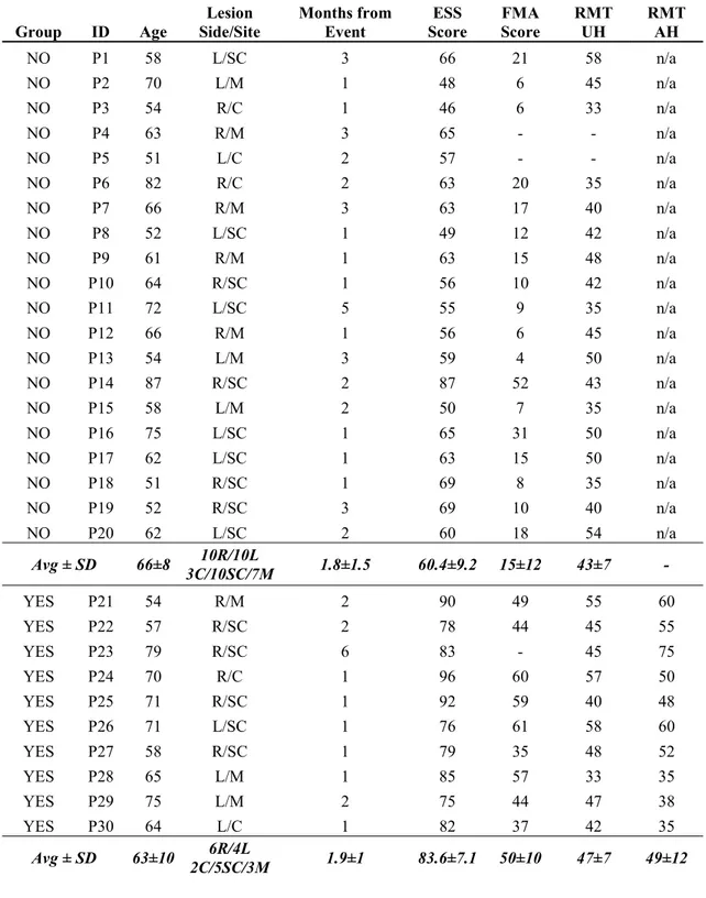
Table 1 shows demographics, clinical characteristics and RMT values of patients, classified as YES or NO groups. Ten patients had inducible MEPs on the affected FDI muscle (YES).
No significant between-group differences were observed for age and time from the stroke event (p > .05). As expected, the YES group had significantly higher ESS and FMA scores than the NO group (p < 10-6 for both ESS and FMA).
32
Table 1: Demographical, Clinical and TMS parameters of patients upon enrollment. L/R (Left/Right). C/SC/M (Cortical/Subcortical/Mixed). ESS: European Stroke Scale; FMA: Fugl-Meyer Assessment; RMT: resting motor threshold; UH: unaffected hemisphere; AH: Affected Hemisphere. FMA data is missing for P4,5 and 23; TMS data is missing for P4 and 5.
Group ID Age Side/Site Lesion Months from Event Score ESS FMA Score RMT UH RMT AH NO P1 58 L/SC 3 66 21 58 n/a NO P2 70 L/M 1 48 6 45 n/a NO P3 54 R/C 1 46 6 33 n/a NO P4 63 R/M 3 65 - - n/a NO P5 51 L/C 2 57 - - n/a NO P6 82 R/C 2 63 20 35 n/a NO P7 66 R/M 3 63 17 40 n/a NO P8 52 L/SC 1 49 12 42 n/a NO P9 61 R/M 1 63 15 48 n/a NO P10 64 R/SC 1 56 10 42 n/a NO P11 72 L/SC 5 55 9 35 n/a NO P12 66 R/M 1 56 6 45 n/a NO P13 54 L/M 3 59 4 50 n/a NO P14 87 R/SC 2 87 52 43 n/a NO P15 58 L/M 2 50 7 35 n/a NO P16 75 L/SC 1 65 31 50 n/a NO P17 62 L/SC 1 63 15 50 n/a NO P18 51 R/SC 1 69 8 35 n/a NO P19 52 R/SC 3 69 10 40 n/a NO P20 62 L/SC 2 60 18 54 n/a Avg ± SD 66±8 3C/10SC/7M 10R/10L 1.8±1.5 60.4±9.2 15±12 43±7 - YES P21 54 R/M 2 90 49 55 60 YES P22 57 R/SC 2 78 44 45 55 YES P23 79 R/SC 6 83 - 45 75 YES P24 70 R/C 1 96 60 57 50 YES P25 71 R/SC 1 92 59 40 48 YES P26 71 L/SC 1 76 61 58 60 YES P27 58 R/SC 1 79 35 48 52 YES P28 65 L/M 1 85 57 33 35 YES P29 75 L/M 2 75 44 47 38 YES P30 64 L/C 1 82 37 42 35 Avg ± SD 63±10 2C/5SC/3M 6R/4L 1.9±1 83.6±7.1 50±10 47±7 49±12
As for the TMS data analysis, no significant differences in RMT values were observed between groups on US, neither between AS and US in the YES group (p > .05).