1 Vincenzo Frusciante MD, 2 Cristina Ferrari MD, 1 Manuela Totaro MD, 1 Guido Valle MD, 3 Claudio Carmine Guida MD,
3 Filippo Aucella MD, 2 Paola Caputo MD, 2 Giuseppe Rubini MD, PhD
1. Nuclear Medicine, IRCCS “Casa Sollievo della Sofferenza”, San Giovanni Rotondo, Foggia, Italy
2. Nuclear Medicine, D.I.M., University of Bari “Aldo Moro”, Bari, Italy
3. Nephrology, IRCCS “Casa Sollievo della Sofferenza”, San Giovanni Rotondo, Foggia, Italy
Keywords: Congenital erythropoietic porphyria -Brain perfusion defect -Single photon emission tomography
-Magnetic resonance imaging
Corresponding author: Giuseppe Rubini, Professor MD, PhD
Piazza G. Cesare, 11, 70124 Bari, Italy Phone number: +39 080 5592913 Fax number: +39 080 5593250 [email protected] Rece ved: 23 March 2018 Accepted revised : 10 April 2018
Brain perfusion defects by SPET/CT and neurostat
semi-quantitative analysis in two patients with congenital
erythropoietic porphyria
Abstract
Background: Congenital erythropoietic porphyria (CEP) is a rare autosomal recessively inherited disorder with chronic and relatively stable presentation. Till now brain blood ow derangements have been descri-bed only in acute hepatic porphyrias. We describe the rst ndings of brain perfusion defects, studied by single photon emission tomography/computed tomography (SPET/CT), in two patients affected by CEP, by using a semi-quanti cation anatomic-standardized voxel-based program compared with magnetic reso-nance imaging (MRI) results. Subjects and Methods: Two Pakistanis brothers were investigated for CEP con rmed by a genetic test. The disease was severe with: skin burning, mood depression and haemolytic anemia. Considering depression, patients underwent brain SPET/CT and MRI. Single photon emission to-mography/CT images were processed by neurostat semi-quantitative software. Data obtained were com-pared to a normal database and z-score images were generated. Results: In both patients we found several perfusion defects evident in transaxial slices and in z-score images obtained by neurostat processing. Mag-netic resonance imaging was negative in both patients. Biochemical mechanisms inducing localized brain hypoperfusion are uncertain. However, mismatch between SPET/CT data and MRI was probably due to ab-sence of necrosis. Conclusion: In our opinion, SPET/CT could have a key role in this setting of patients due to its high sensitivity and reliability in mild-to-moderate brain perfusion defects detection. Moreover, the quantitative analysis by using neurostat may allow to recognize even mild brain perfusion alterations, diffi-cult to detect only visually.
Hell J Nucl Med 2018; 21( ): 43-47 1 Published online: 5 2 April 2018
Introduction
C
ongenital erythropoietic porphyria (CEP), or Gunther's disease, is an autosomal re-cessively inherited disorder that affects heme, involving one of the eighth enzy-matic steps which starts and ends in the mitochondria with intermediate steps ta-king place in the cytosol.Congenital erythropoietic porphyria belongs to the non-acute porphyrias group and it is caused by de ciency of uroporphyrinogen III synthase (UROS), the fourth cytoplasma-tic enzyme of the heme biosynthecytoplasma-tic pathway, which catalyzes the conversion of the hyd-roxymethylbilane into uroporphyrinogen III [1].
Heme is an essential component of several hemoprotein such as hemoglobin, cytoc-hrome enzymes and myoglobin. Hemoglobin synthesis in erythroid precursor cells acco-unts for about 85% of heme synthesis, while synthesis of cytochrome P450 enzymes in hepatocytes accounts for most of the other heme synthesis [2].
Forty ve different CEP-causing mutations have been identi ed throughout the UROS gene on chromosome 10q25.2-26.3, with impact on disease severity that can ranges from fetal hydrops to adult onset with mild cutaneous photosensitivity [3].
Typically, CEP begins with red urine shortly after birth. Bullous cutaneous photosensi-tivity and skin fragility starts in early infancy, leading to scarring with photomutilation. Other manifestations include hypertrichosis, erythrodontia, chronic hemolytic anemia, osteoporosis, corneal ulceration and scarring [4, 5].
No neurological and/or psychiatric symptoms were described to date neither in CEP patients nor in other non-acute porphyrias. Conversely, literature reports several neuro-logical manifestations for acute porphyrias such as motor weakness, sensory loss, psychi-atric problems, seizures, and mental and behavioral abnormalities, even if
pathophysi-ology is not yet fully understood [ ]. 6
Several authors reported the presence of cerebral ische-mia in patients with acute intermittent porphyria (AIP). Tota-ro et al. (2013) [7] described the possible Tota-role of single pho-ton emission tomography (SPET) in the early detection of mild brain perfusion defects in a series of 15 AIP patients, be-fore any morphological changes were evident on MRI. Com-parable SPET perfusion pattern was observed in patients with hereditary coproporphyria (HCP) [8, 9].
We describe in two patients affected by CEP, to the best of our knowledge, the rst ndings in literature of brain SPET/ CT perfusion defects by using a quanti cation anatomic-standardized voxel-based analysis.
Patients' description
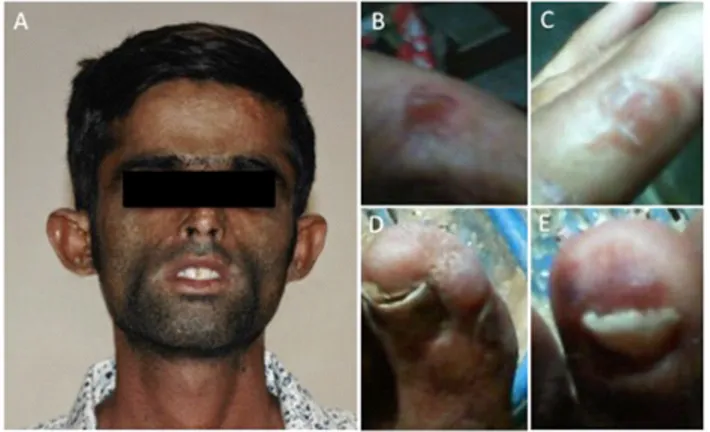
Two Pakistanis brothers, of 27 and 23 years of age respecti-vely, were admitted at our hospital presenting red to yellow urine, mild yellow teeth coloration, hypertrichosis, skin fri-able with red bullae and blisters prone to rupture and infec-tion, skin thickening, focal hypopigmentation and hyper-pigmentation, skin burning, light sensitivity and symptoms like paresthesia, worsening fatigue and depression (Figure 1).
Figure 1. One of the two Pakistanis brothers with signs of CEP (patient 1). A. Evi-dence of yellow teeth coloration and hypertrichosis; B, C Fingers with red bullae and blisters prone to rupture and; D, E. Toes with ulcerations and infection.
Hemolytic anemia was demonstrated and osteopenia re-sulted at dual-energy X-ray absorptiometry (DEXA) exami-nation. The disease was investigated by genetic testing and con rmed by biomolecular analysis.
Considering patients' depression and taking into account that in other porphyrias, even if acute, brain perfusion ab-normalities have been already demonstrated [7-10], the pre-sence of a possible brain vascular damage was assessed by performing brain perfusion SPET/CT and MRI.
Clinical conditions that could interfere with the brain per-fusion like hypertension or family history of stroke or of ot-her cardiovascular diseases were denied. None of the two patients was tobacco smoker or drug abuser [11]. A written informed consent was obtained from both patients.
Imaging Methods
SPET/CT examinationsPatients received an intravenous injection of 740MBq of 99m
Tc-hexamethylpropyleneamine oxime (HMPAO) when ly-ing down in the supine position with eyes closed in a dimly lit, quiet room. Thirty minutes after the injection of the ra-diopharmaceutical, brain SPET/CT was performed using a double-head rotating gamma-camera Symbia T16 (Sie-mens, Erlangen Germany) equipped with high resolution low-energy collimators. Data were acquired with 128x128 matrix in step & shoot mode, 60 steps for each camera at 30 seconds per angle; data were acquired with an auto contour orbit; scatter correction was performed with two windows method. Spiral CT data were acquired with 130kV and 130 mAs. The SPET data were reconstructed with Flash 3D itera-tive algorithm method (Siemens) and corrected for attenu-ation by CT maps.
The qualitative assessment was performed analyzing transahxial, sagittal and coronal images. Any area of decre-ased radiopharmaceutical uptake was considered as hypo-perfused and then pathologic. According with the usual practice, the evaluation of the severity of the perfusional ab-normalities has been done on the basis of a personal evalu-ation expressed by the consensus of two nuclear physicians with at least 5 years of experience [12].
Moreover, the semi-quanti cation of defects' severity was performed by processing the brain perfusion reconstructed images with an anatomic-standardized voxel-based pro-gram named neurostat software installed on a windows 7 64-bit computer (Microsoft, Redmond USA) [13, 14]. An indi-vidual brain image set was aligned to the midsagittal plane. The AC-PC line (a line passing through the anterior and pos-terior commissure) was estimated by an iterative matching between the individual image set and a standard atlas tem-plate. Differences in size between the individual brain and the standard template were removed by linear scaling. Then a nonlinear warping along the major neuronal ber bundles was performed to adjust the individual brain shape to the stereotactic atlas of Talairach and Tournoux [15]. At the end of the process neurostat produced a standardized set of 60 slices 128x128 matrix, voxel size of 2.25mm.
Then, neurostat compared the obtained patients' data to a normal HMPAO SPET database resulting in a set of z-score surface images normalized to cerebellar activity: right la-teral, left lala-teral, right medial, left medial (Figure 2A). On the two lateral z-score images 9 standardized regions of interest (ROI) were overlaid: frontal, pre-central, post-central, supe-rior parietal, infesupe-rior parietal, antesupe-rior temporal, postesupe-rior temporal, occipital, cerebellar (Figure 2 B, C); while, on the two medial z-score images 1 standardized ROI was posi-tioned on posterior cingulate.
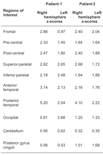
Areas with a z-score >2 were considered as hypoperfused. Moreover, hand-made polygonal ROI of the same pixel-si-ze were drawn on the two more severely hypoperfused regi-ons on transaxial Talairach standardized images and the ra-tio of counts in this areas to counts of contralateral specular areas was calculated.
MRI examinations
Magnetic resonance imaging examinations were perfor-med using a Philips Ingenia 3 T scanner (Philips Healthcare, Best, The Netherlands). Each MRI study included: SE T1, on
transversal and sagittal planes, TR 450ms, TE 8.90ms, FA 90, Thickness 5mm, Gap 5; BLADE T2, on transversal and coro-nal planes, TR 3800ms, TE 99ms, FA 141, Thickness 5mm, Gap 5mm; 3D uid-attenuated inversion recovery (FLAIR) (voxel
3
1.2×1.2×1.2mm , TR/TE/TI=4,800/350/1,600 milliseconds, 3
ip angle 90°, 250×250×180mm FOV, NEX=1,163 slices,), susceptibility weighted imaging (SWI) on transversal plane,
3
(0.7×0.7×0.7mm , TR 15, TE 0 milliseconds, ip angle 15°, 3
250×250×180mm FOV, NEX=1,200 slices), Diffusion Weig-hted Imaging (DWI) TR 7200ms, TE 80ms, FA 90, Thickness 5mm, Gap 5mm, b values 0 and 1000; post-contrast T1-MPR-AGE TR 1900, TE 3.02, FA 15, Thickness 1mm, Gap 0mm. All the sequences where acquired using the same Matrix (320\320) and 1 NEX, except for DWI in which 2 NEX were used.
Figure 2. Brain perfusion SPET z-score surface images produced by neurostat (A), with the 9 standardized regions of interest (ROI) overlaid on right-lateral (B) and left-lateral (C) images, in one of the two Pakistan brothers (patient 1).
Results
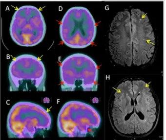
In patient 1 qualitative analysis demonstrated moderate brain perfusion defects in the parietal cortex bilaterally es-pecially in the right upper gyrus and the bilateral temporal cortex with high hypoperfusion in the right posterior gyrus, and mild hypoperfusion in left occipital cortex, as showed in Figure 3.
The neurostat analysis demonstrated wide hypoperfused areas located in: the frontal, pre-central, post-central, supe-rior and infesupe-rior parietal, antesupe-rior and postesupe-rior temporal standardized regions of the right hemisphere and in the su-perior and inferior parietal, anterior and posterior temporal and the occipital regions of the left hemisphere, by quanti-fying the defect severity, as reported in Table 1.
Considering the most severely hypoperfused areas, we drew two specular ROI on the right and left medium tempo-ral gyri and two specular ROI on right and left post-centtempo-ral gyri; The ratios of the counts from the right to left regions for the temporal gyri and the post-central gyri were: 0.26 and 0.29, respectively.
In patient 2 qualitative analysis demonstrated mild to mo-derate brain perfusion defects in: frontal cortex bilaterally,
particularly in the right inferior frontal area, and the anterior and posterior temporal cortex bilaterally, as showed in Figu-re 4 (A-F).
Figure 3. Brain perfusion SPET/CT of patient 1, axial (A-E) and coronal (B-F) slices. Mild to moderate hypoperfusion is detectable in: parietal cortex bilaterally espe-cially in the right upper gyrus (A, B), bilateral temporal cortex (C, D) and the left oc-cipital cortex (E, F), with the qualitative analysis.
Table 1. Right and left lateral z-score of patients 1 and 2,
respecti-vely. Areas w ith a z -score >2 w ere c onsidered a s h ypoperfused.
Regions of interest Patient 1 Right Left hemisphere z-scores Patient 2 Right Left hemisphere z-scores Frontal 2.86 0.97 2.40 2.04 Pre-central 2.33 1.45 1.64 1.64 Post-central 2.47 1.80 2.40 1.89 Superior parietal 2.82 2.65 2.66 1.72 Inferior parietal 2.18 2.48 1.64 1.88 Anterior temporal 3.14 2.13 2.16 1.76 Posterior temporal 5.20 2.04 4.10 2.22 Occipital 0.81 2.68 1.20 1.33 Cerebellum 0.56 0.62 0.32 0.35 Posterior gyrus cinguli 0.06 0.53 1.01 1.68
Figure 4. Brain perfusion SPET/CT (A-F) and related MRI imaging ndings (G-H) of patient 2, axial (A, D, G, H), coronal (B, E) and sagittal (C, F) slices. Moderate with the qualitative analysis hypoperfusion is detectable in the: frontal cortex bilate-rally especially in the inferior right frontal area (A-C) and the anterior and posterior temporal cortex bilaterally (D-F). Some hyperintense lesions are located in the periventricular white matter and the subcortical frontal white matter on MRI-FLA-IR sequences (G-H).
The neurostat analysis demonstrated similar but wider perfusion defects located in: the frontal, post-central, supe-rior parietal, antesupe-rior and postesupe-rior temporal regions of the right hemisphere and in the frontal and posterior temporal regions of the left hemisphere, by quantifying the defect se-verity, as reported in Table 1.
Then, considering the more severely hypoperfused regi-ons, we drew two specular ROI on the right and left tempo-ral gyri and another two specular ROI on the right and left post-central gyri; the ratios of right to left regions 0.31 and 0.30 for the temporal gyri and the post-central gyri respec-tively.
The MRI studies did not show any signi cant pathological brain ndings, related to SPET/CT abnormalities for both pa-tients; only some micro-focal FLAIR and T2-w hyperin-tensities were present in patient 2, related to chronic micro-vascular injury (Figure 4 G-H).
Discussion
Congenital erythropoietic porphyria is extremely rare, with a prevalence in Europe of 1 in 1,000,000 or less [16, 17]. It re-sults from the markedly de cient, but not absent, activity of URO-synthase, leading to overproduction and accumula-tion of porphyrins, heme precursors. The major clinical ma-nifestations of CEP are either erythropoietic, with hemolytic anemia, or cutaneous, resulting from phototoxicity, with a chronic, relatively stable, presentation. Excess porphyrins are also deposited in teeth and bones.
Differently from other acute porphyrias, in which the ex-cess of porphyrins can cause toxicity with
neuro-pathic visceral (abdominal) pains and mental status change effects [2], these complications, to the best of our knowled-ge, have never before been described in CEP. The main mec-hanism implicated seems to be the hepatic production of a neurotoxic substance, presumably ALA (a γ-aminobutyric acid analog) and/or PBG, that may interact with γ-aminobu-tyric acid or glutamate receptors, resulting in uptake re-duction and accumulation of catecholamines, with hyper-tension and tachycardia commonly observed during the acute porphyria attack [18].
Nuclear medicine imaging methods have always been distinguished, as they are able to highlight functional altera-tions before the evidence of morphological changes [19]. In particular, the role of brain perfusion SPET/CT with semi-qu-antitative analysis has already been tested in several other brain diseases such as encephalitis dementia and/or mild cognitive impairment [20, 21].
The young age of our patients, the absence of other vascu-lar risk factors (diabetes, hypertension, dyslipidemia, high
brinogenemia, pro-coagulative disorders) and of other en-dangerment conditions, like tobacco smoke and drug ad-diction, leave little doubt that the observed hypoperfusion phenomena at SPET/CT scan are depending from CEP.
The observation as above, in reference to acute porphy-rias, is consistent with the presence of regional hypoperfu-sion also in subjects with CEP. Likewise it is difficult to make a guess on the biochemical/functional mechanisms inducing localized brain hypoperfusion. The molecules thought to be able to induce vascular derangements in porphyrias are ALA and PBG that reduce the re-uptake of catecholamines [18]. However, in our two cases these molecules were within the normal range and therefore their causative role appears the-refore unlikely. On the contrary, serum urobilinogen and coprobilinogen were very elevated but in the extremely li-mited medical literature on this issue there is no evidence that these molecules might induce arterial spasm or that might trigger derangements of blood supply.
However, since heme is required for a variety of hemopro-teins, including hemoglobin, myoglobin, respiratory cyto-chromes and the cytochrome P450 enzymes we can specu-late that other different mechanisms can be implicated in brain perfusion defects, in “non acute” porphyrias line CEP. First of all, the severe hemolytic anemia which is characteristic of CEP could be the basis of chronic hypoxia that, as a mild but constant factor affecting microcirculation, could explain the hypoperfusion defects detected on brain SPET/CT. In fact, SPET/CT is able to detect even mild functional alterations in contrary to morphological imaging methods like MRI, for which brain hypoxia was not sufficient to determine neither necrosis nor cytotoxic oedema. A mismatch between the SPET (positive for ischemia) and MRI (normal) patterns has been observed in our CEP patients, similar to what is observed in other porphyrias like AIP and HCP [7, 8, 10].
It could also be hypothesized, as has already been specu-lated, that porphyric patients may have a disorder of heme enzymes, like nitric oxide synthase and prostacyclin syn-thase implicated in vascular tone regulation [10, 22]. A
dys-function of this latter heme-enzyme, that catalyzes the syn-thesis of PGI2 from prostaglandin H2, may induce hyper-tension, and cerebral and myocardial infarction [23]. Also, the heme-citochrome CYP4F2 haplotype has been repor-ted to be associarepor-ted to cerebral infarction [24]. Other causes however cannot be excluded and the genesis of vascular dysfunction in CEP remains to be ascertained.
Probably, all these conditions together, incurred by a ch-ronic stimulus, contribute to the onset of cerebral vascular damage even at early age, thus explaining the neurological symptoms and brain damage, which probably could incre-ase and get worse with age.
In conclusion, to the best of our knowledge, this is the rst
brain SPET/CT perfusion study in congenital erythropoietic porphyria and therefore the rst SPET/CT study demonstra-ting a brain vascular damage in these patients. In our opini-on, SPET/CT could have a key role in this setting of patients due to its high sensitivity and reliability in the detection of mild-to-moderate brain perfusion defects and to follow the disease course. Moreover, the semi-quantitative analysis by using neurostat may allow to recognize even mild brain per-fusion changes, difficult to detect by only visual assessment.
The authors declare that they have no con icts of interest.
Bibliography
1. Das D, Murad A, Saad A. Erythropoietic Porphyria-Role of Curative He-matopoietic Stem Cell Transplantation. J Hematol Transfus 2013; 1(1): 1005.
2. Balwani M, Desnick RJ. The porphyrias: advances in diagnosis and tre-atment. Blood 2012; 120(23): 4496-504.
3. Warner CA, Yoo HW, Roberts AG, Desnick RJ. Congenital erythropoietic porphyria: identi cation and expression of exonic mutations in the uroporphyrinogen III synthase gene. J Clin Invest 1992; 89: 693-700. 4. Poh-Fitzpatrick MB. The erythropoietic porphyrias. Dermatol Clin 1986;
4: 291-6.
5. Bishop DF, Johansson A, Phelps R et al. Uroporphyrinogen III synthase knock-in mice have the human congenital erythropoietic porphyria phenotype, including the characteristic light-induced cutaneous lesi-ons. Am J Hum Genet 2006; 78: 645-58.
6. Solinas C, Vajda F. Neurological complications of porphyria. J Clin Neu-rosci 2008; 15: 263-8.
99m
7. Totaro M, Guida CC, Frusciante V et al. Perfusional brain Tc-Bicisate
(Neurolite) Single Photon Emission Computed Tomography (SPECT) in Acute Intermittent Porphyria (AIP). Clin Transl Imaging Rev Nucl Med Mol Imaging 2013; 1 (Suppl 1), S1: S88.
8. Guida CC, Totaro M, Aucella F et al. Brain Perfusion in Acute Intermittent
99m
Porphyria (AIP) and in Hereditary Coproporphyria (HCP): Tc-Bicisate (Neurolite) Single Photon Emission Computed Tomography (SPECT) studies. Clin Chem Lab Med 2013; 51(5): eA7.
9. Mullin S, Platts A, Randhawa K, Watts P. Cerebral vasospasm and anterior circulation stroke secondary to an exacerbation of hereditary copro-porphyria. Pract Neurol 2012; 12: 384-7.
10. Valle G, Guida CC, Nasuto M et al. Cerebral Hypoperfusion in Hereditary Coproporphyria (HCP): a Single Photon Emission Computed Tomogra-phy (SPECT) study. Endocr Metab Immune Disord Drug Targets 2016; 16 (1): 39-46.
11. Gigante AF, Defazio G, Niccoli Asabella A et al. Smoking in Patients with Parkinson's Disease: preliminary striatal DaT-SPECT ndings. Acta Ne-urol Scand 2016; 134(4): 265-70.
12. Rana KM, Narwal V, Chauhan L et al. Structural and perfusion abnor-malities of brain on MRI and technetium-99m-ECD SPECT in children with cerebral palsy: a comparative study. J Child Neurol 2015; 31: 3-4. 13. Minoshima S, Koeppe RA, Frey KA et al. Anatomic Standardization:
line-ar scaling and nonlineline-ar wline-arping of functional brain images. J Nucl Med 1995; 35: 1528-37.
14. Nishimya M, Matsuda H, Imabayashi E et al. Comparison of SPM and NEUROSTAT in voxelwise statistical analysis of brain SPECT and MRI at the early stage of Alzhiemer's disease. Ann Nucl Med 2008; 22: 921-7. 15. Talairach J, Tournoux P. Co-planar stereotaxic atlas of the human brain.
New York: Thieme; 1988.
16. Deybach JC, Badminton M, Puy H et al. European porphyria initiative (EPI): a platform to develop a common approach to the management of porphyrias and to promote research in the eld. Physiol Res 2006; 55 (Suppl 2): S67-73.
17. Elder GH, Harper P, Badminton M et al. The incidence of inherited por-phyrias in Europe. J Inherit Metab Dis 2013; 36: 849-57.
18. Beal MF, Atuk NO, Westfall TC, Turner SM. Catecholamine uptake, accu-mulation, and release in acute porphyria. J Clin Invest 1979; 60: 1141-8.
18
19. Altini C, Niccoli Asabella A, Ferrari C et al. F-FDG PET/CT contribution to diagnosis and treatment response of rhino-orbital-cerebral mucor-mycosis. Hell J Nucl Med 2015; 18(1): 68-70.
20. Barai S, Sanjay G, Shankar PD, Manish O. Sequential brain perfusion ab-normalities in various stages of Japanese encephalitis. Hell J Nucl Med 2006; 9(3): 163-6.
21. Tranfaglia C, Palumbo B, Siepi D et al. Semi-quantitative analysis of perfusion of Brodmann areas in the differential diagnosis of cognitive impairment in Alzheimer's disease, fronto-temporal dementia and mild cognitive impairment. Hell J Nucl Med 2009;12(2): 110-4. 22. Kupferschmidt H, Bont A, Schnorf H et al. Transient cortical blindness
and bioccipital brain lesions in two patients with acute intermittent porphyria. Ann Int Med 1995; 123: 598-600.
23. Nakayama T. Genetic polymorphisms of prostacycline synthase gene and cardiovascular disease. Int Angiol 2010; 29: 33-42.
24. Fu Z, Nakayama T, Sato N et al. Haplotype of the CYP4F2 gene is associ-ated with cerebral infarction in Japanes men. Am J Hypertens 2008; 21: 1216-23.