Dottorato di ricerca
in Scienze Farmaceutiche
Ciclo XIV- Anno di discussione 2016Coordinatore: Chiar.mo Prof. Gianluca Sbardella
DESIGN AND SYNTHESIS OF
MODULATORS OF APOPTOTIC
ACTIVITY
settore scientifico disciplinare di afferenza: CHIM/08
Dottorando Tutore
‘The cancer and p53 “guardian of the genome”’
1.1 Introduction: Carcinogenesis, an overview 2
1.2 Cell cycle regulation 10
1.2.1 The cancer and the cell cycle
1.3 Apoptosis 15
1.3.1 Connecting apoptosis and proliferation in cancer
1.4 Molecular targeting of cell proliferation and apoptosis 21
1.5 p53: molecular target 23
1.5.1 The role of p53 in normal cells 1.5.2 The role of p53 in cancer cells
1.6 p53 network: co-activators and associated proteins 29
1.6.1 p53 and the DNA repair 1.6.2 p53 and the growth arrest 1.6.3 p53 and the apoptosis
1.6.3.1 p53 role in trascription dependent apoptosis
CHAPTER II:
‘Search setting’
37
2.1 Aim of the study 38
CHAPTER III:
‘Design, synthesis, results and discussion of potential p53
modulators (SERIES 1 AND SERIES 2)’
Index
3.1 Background and design 44
3.2 Chemistry 46
3.2.1 Chemistry of 1st series
3.2.2 Chemistry of 2nd series
3.3 Biological effects 52
3.3.1 Antiproliferative activity
3.3.2 Modulating p53-MDM2 interaction in vitro 3.3.3 Modulating p53-MDM2 interaction in cell 3.3.4 Cell cycle progression
3.3.5 Apoptotic cell death
3.4 Molecular modeling studies 63
CHAPTER IV:
‘Experimental section for series 1 and 2’
65
4.1 Chemistry 66
4.2 Biology 79
4.3 Molecular modeling methods 83
CHAPTER V:
‘Design, synthesis, results and discussion of potential p53 modulators (SERIES 3 AND SERIES 4)’
85
5.1 Background and design 86
5.2 Chemistry 87
5.2.1 Chemistry of 3rd series 5.2.2 Chemistry of 4th series
Chapter VI:
‘Experimental section for series 2 and 3’
93
6.1 Chemistry 94
6.2 Biology 101
CHAPTER VII:
‘Biological bases for SM13 tumour growth inhibition’
103
7.1 Background 104
7.2 Effects of SM13 on KAT-4 cell proliferation in vitro 104 7.3 Effects of SM13 on FRO cell proliferation in vitro 107 7.4 Effects of SM13 on KAT-4 cell proliferation in vivo 108
7.4.1 Evaluation of side effects after treatment with SM13
7.5 Discussion 111
Chapter VIII:
‘Experimental section’
115
CHAPTER IX:
‘Design and synthesis of thiazolidine and diketopiperazine as potential p53 modulators’
119
9.1 Background and design 120
Index
9.2.1 Chemistry of thiazolidine based derivatives 9.2.2 Chemistry of diketopiperazine based derivatives
9.3 Biological effects 125
CHAPTER X:
‘Experimental section for thiazolidine and diketopiperazine derivatives’
127
10.1 Chemistry 128
CHAPTER XI:
‘Dihydrithieno [2,3-b]naphto-4,9-dione analogues as anticancer agents: synthesis and in cell pharmacological studies’
135
11.1 Introduction 136
11.2 Background and design 136
11.3 Chemistry 138 11.3.1 Chemistry of series F 11.3.2 Chemistry of series G 11.4 Biological effects 141 11.4.1 In vitro cytotoxicity 11.4.2 Cardiotoxicity
11.4.3 Biological effects of compound 14 on LN299 cells
CHAPTER XII:
‘Experimental section for series F and G’
149
CHAPTER XIII:
Conclusions
161
REFERENCES 165
Abstract
p53 is a transcription factor with tumour suppressor properties, which is able to induce mitochondrial apoptosis independently of its transcriptional activity. Analogues of the spiro[imidazo[1,5c] thiazole3,3′indoline] 2′,5,7(6H,7aH) -trione, previously synthesized from my research group, as p53 modulators were synthesized during my PhD, aiming to explore new structural requirements at the thiazolidine domain to increase the antiproliferative activity and improve p53 modulation. Derivative 5-bromo-3′- (cyclohexane carbonyl) -1-methyl-2oxospiro[indoline-3,2′-thiazolidine] (SM13) emerged as the most potent compound of all series, inhibiting, in vitro, 30% of p53−MDM2 interaction at 5 μM and the cell growth of different human tumor cells at nanomolar concentrations. Docking studies confirmed the interactions of SM13 with the well-known Trp23 and Phe19 clefts, explaining the reasons for its binding affinity to MDM2. SM13 at 50 nM is capable of inducing the accumulation of p53 protein, inducing significant apoptotic cell death without affecting the cell cycle progression. Comparative studies using nutlin in the same cellular system confirmed the potential of SM13 as a tool for increasing understanding of the process involved in the nontranscriptional proapoptotic activities of p53. Thus, the effectiveness of this compound in tumors carrying a mutated form of the p53 gene without transcriptional activity was verified.The effectiveness of SM13 in cancer cell lines carrying WT, mutated and null p53 gene were evaluatedi vitro. At the same time, in vivo studies were performed in BALB/c nude mice and the signal-dependent mitochondrial apoptosis was evaluated by western stain. SM13 reduced cell proliferation and induced apoptosis in the in vitro studies, suggesting that its effect is independent of p53 transcriptional activity. On the contrary, SM13 had no effect in a null p53 cell line. In vivo, SM13 induced tumor cell death in a dose dependent manner through the activation of death mitochondrial-dependent signaling in cells mutated p53. Overall these studies highlights the efficacy of SM13 as anticancer cancer to be used for the treatment of p53-dependent tumors, even in the absence of transcriptional activity of p53.
(piperidin-1-yl) acetamido)-2,3,4,9-tetrahydronaphtho[2,3-b]thiophene-3- carboxamide (compound 14), showed a reduced cardiotoxicity, inducing, at the ame time, cell differentiation and was distributed mainly in the cytoplasm in the human glioblastoma LN229 cell line. Moreover, compound 14 reduced both cellular glucose uptake and serine/threonine kinase AKT expression, and triggered cell apoptosis. These findings suggest that highly functionalized DTNQ-based derivatives are promising pharmacological tools for the study of human solid tumours.
- 1 -
CHAPTER I:
THE CANCER AND P53 “GUARDIAN OF THE
Chapter I: The cancer and p53 “guardian of the genome”
- 2 -
1.1 Introduction: Carcinogenesis, an overview
Cancer is the name given to a collection of related diseases, which have a common feature: their cells grow without stopping and spread into surrounding tissues. Cancer originates in our own cells, but several intrinsic and external factors can add to the cancer risk. Infections, diet, toxins, smoking, alcohol, obesity, environmental chemical pollutants, industrial effluents and some therapeutic drugs can act as a stimulus to induce and promote cancer development. Cancer research has generated a rich and complex body of knowledge crystallizing, so far, in two drastically different approaches to understanding the driving forces behind cancer onset and proliferation. These are the somatic mutation theory (SMT)1 and the tissue organization field theory (TOFT).2 The essence of SMT is that cancer is derived from a single somatic cell which has successively accumulated multiple DNA mutations, and that those mutations occur on gene controlling cell proliferation and cell cycle. Thus, according to SMT the neoplastic lesions that destroy normal tissue architecture are the results of DNA-level events. Conversely, according to TOFT, carcinogenesis is primarily a problem of tissue organization: carcinogenic agents (eg, environmental chemicals, inflammation, viruses) destroy the normal tissue architecture thus disrupting cell-to-cell signaling and compromising genomic integrity. Hence, in TOFT the DNA mutations are the effect, and not the cause, of the tissue-level events. However, a large grey zone of biological facts and clinical cases exists which poses the questions that are difficult to resolve from either of these view-points.3 Essentially, for a long time the theories conceptually close to SMT and TOFT were considered as two different facets (along with many others) of the complex phenomenon of carcinogenesis. Indeed, the cornerstone of SMT is the notion that carcinogenesis is triggered by a single aberrant cell which happened to acquire multiple DNA mutations, and that these mutations predominantly damage the genes responsible for the cell cycle and apoptosis. Cancer cells develop the ability to defy the mechanisms of
- 3 -
Figure 1. Intracellular Signaling Networks Regulate the Operations of the Cancer
Cell: SMT theory. An elaborate integrated circuit operates within normal cells and is reprogrammed to regulate hallmark capabilities within cancer cells. Separate subcircuits, depicted here in differently colored fields, are specialized to orchestrate the various capabilities. At one level, this depiction is simplistic, as there is
considerable crosstalk between such subcircuits. In addition, because each cancer cell is exposed to a complex mixture of signals from its microenvironment, each of these subcircuits is connected with signals originating from other cells in the tumor microenvironment
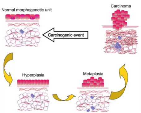
In direct opposition to these views, TOFT depicts carcinogenesis as general deterioration of the tissue microenvironment due to extracellular causes. This deterioration hinders normal cell-to-cell signaling thus making normal functioning of the intracellular machinery impossible and eventually leading it to the breaking point. In such a scenario, the deleterious mutations should be scattered all over the genome randomly and incoherently with little chance for clonal homogeneity. (Figure 2)
Chapter I: The cancer and p53 “guardian of the genome”
- 4 -
Figure 2. Carcinogenesis according to the TOFT. A single or multiple carcinogenic
exposure acts disturbing the reciprocal biophysical and biomechanical
communication between the parenchyma and the mesenchyme/stroma in a given morphogenic field. This results in miscues that manifest morphologically in both the stroma and the epithelium. The proliferation and motility restraints imposed by normal tissue architecture loosen and as a consequence, hyperplasia of the
epithelium may occur. Further alteration of the reciprocal interactions between tissue compartments will induce metaplasia, dysplasia, and carcinoma. The stroma also may show alterations (desmoplasia, inflammatory cells)
According to SMT, cancer progression is a unidirectional and mostly irreversible process; the disease cannot be cured unless the entire tumor mass is surgically removed, radiationally killed, or deprived of its aggressive nature by carefully targeted chemotherapeutic agent. Paramount importance of TOFT for cancer biology, for the practice of clinical oncology and for cancer prevention, dwells in the fact that according to this view carcinogenesis is not a unidirectional process; rather, it is curable and reversible.4
Recently a new approach outlines a plausible scenery, in an attempted reconciling the two previous theories, in which a single event, insignificant on its own, may trigger a system-wide catastrophic restructuring. Such a scenery
- 5 -
requires participation of a large number of transcription factors (TFs) which essentially are the proteins expressed by other genes. In turn, these supporting proteins cannot come into existence unless their parent genes have complete teams of their own TFs coming from yet another set of genes. This tight interdependence of genes (gene-to-gene interactions) creates the situation when each gene may be expressed only with the support of many other genes, essentially of the entire network. Ideally, the system can only work in a perfectly synchronized manner, with each of thousands of parts being produced and delivered where needed in a timely manner. If, however, at least one part fails to arrive in time to its destination, the corresponding assembly line then comes to a stop, thus triggering the domino effect of secondary failures and ultimately driving the entire system to a complete halt. Comparatively simple and universal forces driving the living systems towards critical conditions are always present behind the scenes in all SOC phenomena. In very general terms, a hallmark of living entities is their ability to replicate and proliferate themselves. In a community of such entities, unstoppable proliferation will inevitably drive the community towards exhaustion of common resources, whatever these resources are, thus bringing the populations to the verge of extinction. SOC plays an important role in DNA damage and transition of cellular machinery into chaotic state. The first cellular factor to sustain the damage is the RNA Polymerase II during the transcription of an active gene; this damage leads to a stalled transcription fork. The stalled fork triggers DNA repair mechanisms by attracting a large number of proteins which, in turn, allosterically modify binding affinities of many other proteins. It may happen that the damage occurs in the so called “hub” proteins (such as p53 protein) which are capable of
Chapter I: The cancer and p53 “guardian of the genome”
- 6 -
modifying a large number of vital cellular functions simultaneously.6 A subtle balance always exists between the rates of damage and repair. Up to a certain level of mutagenic load, the repair mechanisms are capable of containing damage, thus maintaining a generally healthy cell population. However, the last straw effect may also occur when the cell, after an insult, remains unrepaired yet undestroyed, thus giving rise to a genetically aberrant sub-population. This last straw event is analogous to the last grain of sand in the sandpile avalanche because it fires up multiple, very complex, and mostly irreversible pathways. Such a massive complex response to a seemingly minor event is a hallmark of SOC. It would be an obvious misjudgment to regard any particular minor event as a cause of the system’s collapse. Rather, one may expect that a mutationally overloaded system would collapse anyway, whatever a minor event actually happens to be the trigger. In this context both the self-organized criticality and the somatic mutation theories match. (Figure 3) In particular, as observed by Nowell,7 cytogenetic studies have demonstrated that in many primary tumors all cells show the same abnormal karyotype; the immunoglobulin produced by plasma cell tumors has in almost every case the homogeneity characteristic of a single clone. As an ultimate manifestation of this paradigm, direct evidence of a single catastrophic event triggering carcinogenesis has been presented by Stephens et al.8 The authors explain that the overwhelming majority of rearrangements leading the distinctive genomic structures, present in the different cancer types, occurs in a single catastrophic event. In this scenario, the chromosome or chromosomal region shatters into tens to hundreds of pieces, some (but not all) of which are then stitched together by the DNA repair machinery in a mosaic patchwork of genomic fragments. A cell suffering tens to hundreds of DNA breaks in a single cataclysmic event would be expected to undergo apoptosis.
- 7 -
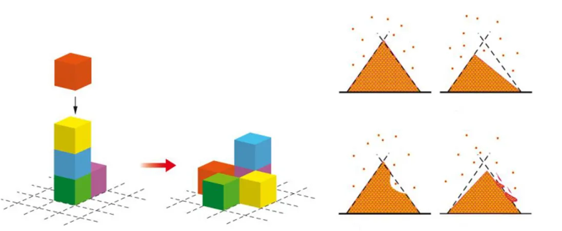
Figure 3. Different representations of self-organized criticality theory. Sandpile
paradigm: if additional sand grains are randomly added to a sand pile then inevitably an instance will occur when local steepness of the slope surpasses a certain critical threshold thus causing local failure of structural stability. The excess of material will cascade into adjacent areas of the pile causing their failure as well
That a cell can survive such an insult and progress to become cancerous suggests that the extensive remodeling of the genome may confer significant selective advantage to that clone. Self-organized criticality confirms also one basilar point of the tissue organization field theory on the evolution of tissue, from a healthy state to a precancerous state and further to tumorigenesis, using some concepts of the somatic mutation theory. In particular, chronic systemic inflammation has been widely recognized to be among the leading factors in progression of healthy tissue towards precancerous and cancerous lesions. The specific mechanisms of such progression include sustained cell proliferation in an environment rich in inflammatory cells and molecular agents causing DNA damage.9 Excessive and pathologic inflammation causes DNA damage, genomic instability, epigenetic dysregulation, and alteration of intracellular signaling, all of which are involved in neoplastic transformation.10 It is important to realize that inflammation-triggered carcinogenesis cannot be reduced to just cell proliferation and conquering new tissue territories. A number of complex molecular mediators facilitate proliferation of genomic
Chapter I: The cancer and p53 “guardian of the genome”
- 8 -
damage, among which an important role belongs to inflammasomes, i.e., the multi-protein complexes that mediate immune response.11
Another fondamental aspect of carcinogenesis is the DNA methylation. In normal tissue, gene methylation is mostly localized in the coding region whereas the promoter region remains mostly unmethylated. A different pattern is observed in neoplasia: the genome-wide hypomethylation is accompanied by localized hypermethylation. Evidence suggests that methylation is an important factor in carcinogenesis since genome-wide hypomethylation can trigger the chromosome instability and increase the mutation rates, playing an key role in different cancer types and probably at different stages of oncogenesis.12, 13 Generally, abnormal patterns of methylation signify elevated cancer risk due to heightened susceptibility to cancer cell proliferation. According to Vendramini-Costa and Carvalho,14 tumor initiation involves irreversible changes in DNA through activation of oncogenes or inactivation of tumor suppressor genes. Further development leads mutated cells to expansion through increased proliferation and suppression of cell death. In the process of invasion of adjacent tissues cancer cells may accumulate other mutations, thus exacerbating their phenotype. Again, the process is quite similar to the forest fire propagation, which accumulates additional strength while invading new territories. Last, the disruption of cell-to-cell communication is an important aspect characterizing precancerous tissue, and it is a central component of a bigger process of tissue disorganization.15 The viewpoint is that a community of cells is not simply a collection of units dwelling within certain architectural structures. With the destruction of signaling pathways, not only the normal regulation of individual cellular processes is damaged, but also a blow is dealt, so to speak, to the mental capabilities of the community as a whole. Its collective memory is wiped out or distorted, customary division of labor between subpopulations is shifted towards aberrant modalities, and community-wide self-defense mechanisms are weakened or broken. These processes in turn cause a shift in expression profiles
- 9 -
may be seen as catastrophic on the level of individual cell would necessarily lead to carcinogenesis. Vast majority of those events would fade and disappear without traces. This is because the immune system remains on guard of tissue homeostasis. When tissue homeostasis is perturbed, sentinel macrophages and mast cells release cytokines, chemokines, reactive oxygen species (ROS), and other bioactive mediators that induce mobilization of additional leukocytes.16 This means that the mutant cell capable of starting the domino-effect of subsequent failures should be able to overcome the tissue’s natural defenses; this may happen only if the tissue is already preconditioned for failure and resides on the verge of systemic collapse. All these findings provide just a glimpse of extremely complex and tangled transition of healthy tissue towards precancerous state. Obviously, even a complete knowledge of each and every process contributing to this transition does not automatically lead to understanding the process as a whole. Resorting to the sandpile analogy, it would be as difficult as understanding the phenomenon of avalanche from observations of each sand grain trajectory. This is why systemic approaches are not simply helpful, they are absolutely necessary and unavoidable for synthesizing existing biomolecular knowledge into a coherent picture of carcinogenesis. Thus, recognition of the widespread applicability of concepts, outlined here, will increasingly affect the development of new strategies to treat human cancer.
Chapter I: The cancer and p53 “guardian of the genome”
- 10 -
1.2 Cell cycle regulation
With advancements on the basic mechanisms of oncogenesis, we have gained a better understanding of the role that the cell cycle regulation plays in malignant transformation and in the development of resistance to chemotherapy, laying the bases for development of a new class of anticancer therapeutics in clinical development.17
The fondamental task of the cell cycle is to ensure that DNA is faithfully replicated once during S phase and that identical chromosomal copies are distributed equally to two daughter cells during M phase.18 The machinery for DNA replication and chromosome segregation is insulated from interruption by extracellular signals, and its essential and autonomous nature implies that damage to the pivotal components would be highly debilitating, if not fatal, to cells. Therefore, genes commanding these processes should not be frequent targets of mutation, deletion, or amplification in cancer. Oncogenic processes exert their greatest effect by targeting particular regulators of GI phase progression.19 During the G, phase, cells respond to extracellular signals by either advancing toward another division or withdrawing from the cycle into a resting state (Go).20 Unlike transit through the S, G2, and M phases, G progression normally relies on stimulation by mitogens and can be blocked by antiproliferative cytokines. Cancer cells abandon these controls and tend to remain in cycle, and because cell cycle exit can facilitate maturation and terminal differentiation, these processes are subverted as well.21 The decision to divide occurs as cells pass a restriction point late in GI, after which they become refractory to extracellular growth regulatory signals and instead commit to the autonomous program that carries them through to division. An appreciation of restriction point control is central to our understanding of how and why cancer cells continuously cycle. The cell cycle is a critical regulator of the processes of cell proliferation and growth as well as of cell division after DNA damage. It governs the transition from quiescence (G0) to cell proliferation, and through
- 11 -
through the cell cycle is promoted by a number of CDKs which, when complexed with specific regulatory proteins called cyclins, drive the cell forward through the cell cycle. There exist corresponding cell cycle inhibitory proteins (CDK inhibitors [CDKIs]) that serve as negative regulators of the cell cycle and stop the cell from proceeding to the next phase of the cell cycle (Figure 3). The INK4 (for inhibitor of cdk4) class of CDKIs, notably p16lnk4a, p15lnk4b, p18lnk4c, and p191nk4 days, bind and inhibit cyclin D–associated kinases (CDK2, -4, and -6). The kinase inhibitor protein (KIP) group of CDK inhibitors, p21waf1, p27kip1, and p57kip2, negatively regulate cyclin E/CDK2 and cyclin A/CDK2 complexes.22
Chapter I: The cancer and p53 “guardian of the genome”
- 12 -
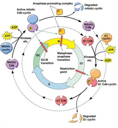
Figure 4. The cell cycle
The pattern of cyclin expression varies with a cell’s progression through the cell cycle, and this specific cyclin expression pattern defines the relative position of the cell within the cell cycle.23 At least nine structurally related CDKs (CDK1- CDK9) have been identified, though not all have clearly defined cell cycle regulatory roles. A considerable number of cyclins have been identified to date (cyclin A–cyclin T). CDK/cyclin complexes themselves become activated by phosphorylation at specific sites on the CDKby cdk7/cyclin H, also referred to as CDK-activating kinase (CAK).24 Cyclin D isoforms (cyclin D1-D3) interact with CDK2, -4, and -6 and drive a cell’s progression through G1. The association of cyclin E with CDK2 is active at the G1/S transition and directs entry into S phase. S phase progression is directed by the cyclin A/CDK2 complex, and the complex of cyclin A with CDK1 (also known as cdc2) is important in G2. CDK1/cyclin B is necessary for mitosis to occur. The cell
- 13 -
‘restriction point’ (R point); it is a central event in normal cellular proliferation control. It has been demonstrated that pRb is the molecular device that serves as the R point switch. pRb is hypophosphorylated in resting G0 cells, is increasingly phosphorylated during progression through G1 and is maintained in a hyperphosphorylated state until late mitosis. pRb phosphorylation seems to be related to mitogenic signals, which converge on the cell cycle machinery, represented by the cyclin D1/cdk4 (cdk6) complex in the early and mid-G1, and composed of cyclin E/cdk2 in late G1.
1.2.1 The cancer and the cell cycle
In physiological conditions, activation of CDK/Cyclin kinases is tightly controlled both spatially and temporally. However, CDK/Cyclins are dysregulated in several human cancers, which wreaks havoc in the coordinated cycle of cell growth and proliferation and contributes to the uncontrolled proliferation characteristic of cancer cells.25 In fact, together with mutations in proto-oncogenes, mutations leading to hyperactivation of CDK activity have been reportedly found in human cancer genomes, and confer selective growth advantage to cells, whilst mutations that inactivate checkpoint regulators, tumour suppressor genes or CKIs result in loss of cell cycle inhibition.26 CDK/Cyclin hyperactivation may result from one of several causes, including gene amplification and protein overexpression of either the CDK or cyclin subunit, alternative splicing and expression of truncated cyclin variants, untimely expression and mislocalization, or constitutive activation of CDK/Cyclins by preventing their inactivation through binding to INK or KIP/CIP inhibitors.27 A representative panel of mutations which occur in CDKs
Chapter I: The cancer and p53 “guardian of the genome”
- 14 -
and Cyclins may be found in the catalogue of Cosmic Mutations in Cancer which integrates all mutations identified through sequencing of human cancer tissue samples. (Figure 5)
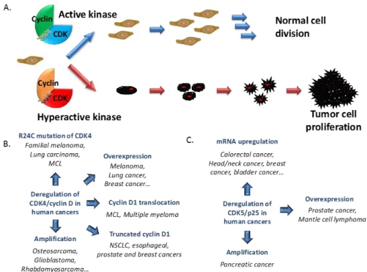
Figure 5. CDK/Cyclins and Cancer. (A) Schematic representation of normal cell
growth and division regulated by cyclin-dependent kinases. Hyperactivation of these kinases contributes to development of cancer cell proliferation; (B) CDK4/cyclin D in cancers: Among all the CDK/cyclins, the complex CDK4/cyclin D is the one which presents most aberrations in cancers. Hyperactive CDK4/cyclin D is found in several human cancers associated with the R24C mutation of CDK4 which prevents the fixation of the endogenous inhibitor p16INK4A, mutation of p16INK4a itself or CDK4 or cyclin D amplification; (C) CDK5/p25 in cancers.
A second class of growth-deregulating mutations comprises those that target the principal late-G1 cell-cycle checkpoint regulated by pRB. Loss of pocket protein functions may induce cell cycle deregulation and lead to a malignant phenotype. Defects in this pathway, which may be universal in human cancers, include deletion of the RBgene itself and deregulation of the CDKs that
- 15 -
mitogen availability in normal cells, but it is usually expressed in a deregulated or elevated manner in tumor cells.30,31 Myc seems to be a strategic controller of cell proliferation that acts pleiotropically to coordinate both cell growth and concomitant progression through the cell cycle.32 The presence in individual tumors of multiple mutations that affect each of the pathways discussed above suggests that each pathway contributes a discrete type of proliferative function to the neoplastic phenotype. But precisely what such functions are and how and why they interact, remains unknown. In addition to driving aberrant cell division, mutations in the various proliferative control pathways have a profound impact on other cell functions. For example, many of the proliferative lesions in tumor cells also contribute to the inhibition of differentiation, thereby preventing the elimination of progeny cells from the proliferative compartment of many types of tissue. pRB, for example, is essential in differentiation of several tissue types through interactions with factors such as the helix–loop– helix proteins MyoD26 33 and Id2. Loss or inhibition of pRB function prevents normal differentiation, a contribution to tumor development distinct from the direct deregulation of cell-cycle progression. Deregulated Myc expression also inhibits differentiation, in part by activation of Id2 expression.34
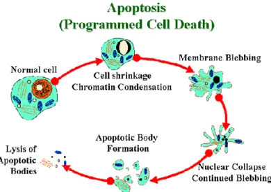
1.3 Apoptosis
Apoptosis, or programmed cell death (Figure 6), is a normal component of the development and health of multicellular organisms. Apoptosis occurs during the normal development of multicellular organisms and continues throughout adult life. The combination of apoptosis and cell proliferation is responsible for shaping tissues and organs in developing embryos. Cells die in response to a
Chapter I: The cancer and p53 “guardian of the genome”
- 16 -
variety of stimuli and during apoptosis they do so in a controlled, regulated fashion.
There are a number of mechanisms through which apoptosis can be induced in cells. The sensitivity of cells to any of these stimuli can vary depending on a number of factors such as the expression of pro- and anti-apoptotic proteins (eg. the Bcl-2 proteins or the Inhibitor of Apoptosis Proteins), the severity of the stimulus and the stage of the cell cycle. Some of the major stimuli that can induce apoptosis include virus infection, cell stress and DNA damage. In some cases the apoptotic stimuli comprise extrinsic signals such as the binding of death inducing ligands to cell surface receptors called death receptors. These ligands can either be soluble factors or can be expressed on the surface of cells such as cytotoxic T lymphocytes. The latter occurs when T-cells recognize damaged or virus infected cells and initiate apoptosis in order to prevent damaged cells from becoming neoplastic (cancerous) or virus-infected cells from spreading the infection. Apoptosis can also be induced by cytotoxic T-lymphocytes using the enzyme granzyme. In other cases apoptosis can be initiated following intrinsic signals that are produced following cellular stress. Cellular stress may occur from exposure to radiation or chemicals or to viral infection. It might also be a consequence of growth factor deprivation or oxidative stress caused by free radicals. In general intrinsic signals initiate apoptosis via the involvement of the mitochondria. The relative ratios of the various bcl-2 proteins can often determine how much cellular stress is necessary to induce apoptosis.
Upon receiving specific signals instructing the cells to undergo apoptosis a number of distinctive changes occur in the cell. A family of proteins known as caspases is typically activated in the early stages of apoptosis. These proteins breakdown or cleave key cellular components that are required for normal cellular function including structural proteins in the cytoskeleton and nuclear proteins such as DNA repair enzymes. The caspases can also activate other
- 17 -
is a process in which cells play an active role in their own death (which is why apoptosis is often referred to as cell suicide).
Figure 6. Apoptosis
1.3.1 Connecting apoptosis and proliferation in cancer
The central engines of apoptosis are the caspases, cascades of cysteine aspartyl proteases that implement cell death by cleaving a variety of intracellular substrates that trigger cell dissolution. Caspases are synthesized as latent zymogens that are activated by proteolytic cleavage: typically through the action of upstream apical caspases. An activation pathway (extrinsic pathway) is mediated by transmembrane death receptors of the CD95 (Apo-1 or Fas)/TRAIL/tumor-necrosis factor (TNF) receptor 1 family, whose ligation triggers recruitment and assembly of multiprotein complexes that activate apical caspase 8.35 The other principal apoptotic death-signaling pathway
Chapter I: The cancer and p53 “guardian of the genome”
- 18 -
involves the mitochondrion (intrinsic pathway), which acts as an integrating sensor of multiple death insults by releasing cytochrome c into the cytosol where it triggers caspase activation. The mitochondrial pathway is thought to be the principal target of survival signaling pathways, which act by stabilizing mitochondrial function and integrity and suppressing release of cytochrome c.36 Once cytochrome c has been released from the mitochondrion, it orchestrates assembly of an intracellular apoptosome complex that recruits apical caspase 9 via the adaptor protein Apaf-1.37 The anti-apoptotic oncoproteins 2 and Bcl-xL, which exert their principal effects through stabilization of the mitochondrion, are overexpressed in several tumor types and recent analyses have indicated that loss of Apaf-1 is a relatively frequent event in malignant melanoma that presumably confers resistance to apoptosis.38 A particularly potent driving force for the suppression of apoptosis in tumor cells is the coupled relationship between cell proliferation and cell death, a phenomenon exemplified by the Myc protein. In addition to its well documented growth-promoting property, Myc was found to be a powerful inducer of apoptosis, especially under conditions of stress, genotoxic damage or depleted survival factors.39 Consideration of such observations led to the proposal that the innate apoptotic potential of Myc serves as an inbuilt foil to its oncogenic capacity (Figure 7).
- 19 -
Figure 7. Activation of growth-deregulating lesions triggers ‘sentinel’
In this example, the oncoprotein Myc is shown activating a p53 damage sentinel through the ARF/MDM-2 pathway, thereby sensitizing the cell to any DNA damage. Myc also promotes release of holocytochrome c from the mitochondrion into the cytosol where it triggers apoptosis. Release of holocytochrome c is inhibited by paracrine ‘survival’ signals that are typically restricted both in supply and location. Clonal outgrowth driven by relentless Myc expression outstrips survival factor availability, triggering the ‘trophic sentinel’ to kill the cell. Another common pathway through which a wide variety of proliferative signals influence the apoptotic program is through induction of ARF, an alternate product of the INK4a locus, one of whose functions is to trigger upregulation of p53 through its inhibitory action on MDM-2.40 Another potent selective pressure in cancers to suppress apoptosis arises from the fact that programmed cell death is the typical response of somatic cells to many forms of stress and damage; in particular damage to cell DNA (a fact exploited by most classical cancer therapeutics). Stress-associated signals that activate apoptosis include many of those encountered by the incipient tumor cell,
Chapter I: The cancer and p53 “guardian of the genome”
- 20 -
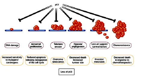
including hypoxia and nutrient deprivation, as well as DNA damage arising from telomere erosion, defective repair, oncogene deregulation and therapy. The p53 protein is important in transducing such diverse signals into tumor-suppressive apoptotic or growth-arresting responses, which implies that there is strong selection for tumor cells to loose p53 function.41 Importantly, differing p53-activating stresses tend to arise at different stages of carcinogenic progression. For example, oncogene deregulation occurs early, as it is a prerequisite for clonal expansion, whereas hypoxia is significant only after the tumor reaches macroscopic size. Consequently, p53 exerts a tumor-suppressive role at multiple stages of carcinogenic progression, (Figure 8) offering an explanation for why loss of p53 has such a profound effect on tumor development.
Figure 8. Many stress signals encountered during tumor progression activate p53,
resulting in apoptosis or growth arrest
Loss either of the ability to activate p53 or of p53 function itself has considerable impact on the ‘success’ of the carcinogenic process, as it increases the chances of a tumor cell surviving progressively adverse conditions. Inability to activate p53 in response to stress signals encountered early during tumor development, such as deregulated proliferation, may to be sufficient to allow
- 21 -
pathways that activate or respond to p53, or loss of p53 by direct mutation of the gene itself, may be selected during progression to more malignant cancers.
1.4 Molecular targeting of cell proliferation and apoptosis
Because deregulated proliferation and inhibition of apoptosis lie at the heart of all tumor development, they present two obvious targets for therapeutic intervention in all cancers. Clearly there are numerous mechanisms through which these two defects can occur, and the success of targeted therapy will depend to a large part on the molecular fingerprinting of individual tumours.42 Although most existing cancer drugs are anti-mitotic, they act not by targeting the specific lesions responsible for deregulated tumor growth, but by crudely interfering with the basic machinery of DNA synthesis and cell division. Moreover, we now know that the surprising selectivity of such crude agents results largely from the increased sensitivity to apoptosis afforded to tumor cells by their oncogenic lesions. Drugs designed to specifically inhibit growth-deregulating lesions are currently being tested in clinical trials, and include inhibitors of RTKs, Ras, downstream signaling kinases such as the mitogen-activate protein kinase and Akt pathway, and CDKs. At first glance, targeted inhibition of growth-deregulating lesions in cancer would be seem to have limited therapeutic efficacy, as they would at best be cytostatic. However, unexpected therapeutic bonuses may emerge from such an approach because growth deregulation induces a plethora of downstream activities in affected cells and their adjacent tissues. Therapeutic inhibition of the offending oncoprotein in tumors arising from cell lineages where terminal differentiation has been blocked could be sufficient to trigger a resumption of that differentiation
Chapter I: The cancer and p53 “guardian of the genome”
- 22 -
program, permanently expelling the tumor cell from the proliferating compartment.43
The second obvious strategy for cancer therapy is to target the lesions that suppress apoptosis in tumor cells. The potent proapoptotic effects of growth-deregulating mutations mean that tumors are peculiarly dependent upon their particular suite of antiapoptotic mutations for continued survival. Thus, although apoptosis in tumor cells is sufficiently suppressed to below a critical threshold to enable them to survive, they remain acutely sensitized to apoptosis. In most, if not all, cancer, this ability to survive results in part from inhibition of the p53 pathway, either by inactivating mutations in p53 itself, perturbation of the signaling pathways that allow activation of p53 in response to stress, or defects in the downstream mediators of p53-induced apoptosis. Reintroduction of p53 function is sufficient to induce apoptosis in many tumor cells, and several mechanisms to reactivate p53 are being considered as therapeutic strategies. These include introduction of wild-type p53 into tumors expressing a mutant protein, or inhibition of negative regulators of p53, such as MDM-2, in those tumors that retain wild-type p53.41 Regardless of efficiency in cell killing, the success of repairing the apoptotic response in tumor cells depends on the extent to which such therapies confine death to the cancer cells, and allow survival of normal tissue. Many conventional chemotherapies induce significant toxicity, particularly in tissues that normally maintain a proliferative compartment, such as gut epithelium and the hematopoietic system. This DNA damage-induced toxicity is mediated in part through p53, leading to the suggestion that inhibition of p53 in these normal tissues may protect against drug-induced toxicity, thereby improving the tolerance of conventional cancer therapies. However, implicit in the development of drugs that target specific lesions responsible for tumor cell growth is the prediction that these approaches will show significantly more specificity for tumor cell killing than conventional therapies. Although activation of apoptotic pathways can lead to the death of untransformed cells, a
- 23 -
requisite survival signals. Repair or replacement of a single apoptotic signal, be it reactivation of p53 or removal of a survival signal, could well prove too much for a tumor cell already burdened with a heavy apoptotic load. By contrast, the same perturbation may scarcely ruffle the equilibrium of a normal cell, safely buffered in its appropriate soma and enjoying the full gamut of trophic support that ensures normal cell survival.
1.5 p53: molecular target
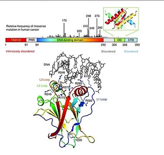
p53, (53KD) also known as tumor protein 53 (TP53),is a transcription factor that regulates the cell cycle and apoptosis, in case of cellular insults, and hence functions as a tumor suppressor. p53 contains a natively unfolded amino-terminal transactivation domain (TAD), which can be further subdivided into the subdomains TAD1 and TAD2, followed by a proline-rich region (PRR). The structured DNA-binding and tetramerization domains (OD) are connected through a flexible linker region. (Figure 9) Similarly to the TAD region, the regulatory domain at the extreme carboxyl terminus (CTD) is also intrinsically disordered.44 The vertical bars, shown in Figure 9, indicate the relative missense-mutation frequency in human cancer for each residue based on the TP53 Mutation Database of the International Agency for Research on Cancer45, showing that most cancer mutations are located in the DNA-binding domain. The structure of the DNA-binding domain (PDB code 1TSR) is shown (Figure 9) as a ribbon representation and colored with a rainbow gradient from the amino terminus (blue) to the carboxyl terminus (red).
Chapter I: The cancer and p53 “guardian of the genome”
- 24 -
Figure 9. Domain structure of p53
p53 has been described as "the guardian of the genome", "the guardian angel gene", or the "master watchman", referring to its role in conserving stability by preventing genome mutation. The transcription factor p53 responds to diverse cellular stresses to regulate target genes that induce cell cycle arrest, apoptosis, senescence, DNA repair, or changes in metabolism.46 In addition, p53 appears to induce apoptosis through nontranscriptional cytoplasmic processes.47 In unstressed cells, p53 is kept inactive essentially through the actions of the ubiquitin ligase MDM2, which ftinhibits p53 transcriptional activity and ubiquitinates p53 to promote its degradation.47 Numerous posttranslational modifications modulate p53 activity, most notably phosphorylation and acetylation. Several less abundant p53 isoforms also modulate p53 activity.
- 25 -
Activation of p53 can result in a number of cellular responses, and it is possible that different responses are induced by different stress signals. There is evidence that p53 can play a part in determining which response is induced through differential activation of target-gene expression. Although the importance of these responses to tumor suppression is clear, previously unanticipated contributions of these responses to other aspects of human health and disease are being uncovered. The role of p53 in tumor suppression, development and ageing is likely to depend on which cellular response is activated and on the context in which the activation occurs. p53 is an intensively studied protein, its fame stemming mainly from its clear role as a tumor suppressor in humans and other mammals.48 Loss or mutation of p53 is strongly associated with an increased susceptibility to cancer, and most functions of p53 have been considered in the light of how p53 might protect from malignant progression.49 Some p53-null mice can develop normally 50 an observation that has been taken to rule out major functions for p53 in normal physiology. But recent studies are questioning whether p53 is truly such a single-minded protein, and other functions of p53 that might be profoundly important during normal life are being uncovered. These include roles for p53 in regulating longevity and ageing, glycolytic pathways that might determine endurance and overall fitness, and apoptotic responses during ischemic and other types of stress. Evidence for genetic variations in the activity of the p53 pathway in humans gives these ideas extra relevance.51 One of the major mechanisms by which p53 functions is as a transcription factor that both positively and negatively regulates the expression of a large and disparate group of responsive genes (Figure 7).52 Although some of these p53- responsive genes have an important role in mediating cell-cycle
Chapter I: The cancer and p53 “guardian of the genome”
- 26 -
arrest, senescence and apoptosis (the best understood activities of p53), it is now evident that the ability of p53 to influence gene expression has wider reaching effects. Numerous studies have identified p53-regulated genes that could have a role in a number of different and sometimes unexpected responses.53 Although some of these still need to be fully validated, there is now clear evidence for a role of p53 in the regulation of glycolysis,54 and autophagy,55 the repair of genotoxic damage,56 cell survival and regulation of oxidative stress,57 invasion and motility,58 cellular senescence,59 angiogenesis,60 differentiation,61 and bone remodeling.62 The cellular pathways in which p53 is involved, are schematically represented in Figure 10. In these aspects, its worthy to analyze there is any cancer cells are expressing wild type p53, and if they are expressing, its role in cancer cells has to be studied before clinical use of p53 mediated gene therapy as an anticancer therapy.
Figure 10. Activation and functions of p53
1.5.2 The role of p53 in cancers cells
In the two decades since its original discovery, p53 has found a singularly prominent place in our understanding of human cancer. Although the biochemistry of p53 has been worked out in some detail, our knowledge of the biologic consequences of p53 dysfunction is still quite rudimentary.
- 27 -
unwanted activities such as a gain-of-function or be dominant negative inhibitors of wt p53 activity. In this regard, it will be important to determine how best to harness the complex properties of p53’s ability to induce cellular growth arrest and cell death to generate novel, effective approaches to cancer therapy. Furthermore, a clearer appreciation of the direct interaction epigenetic factors with p53 will lead to development of strategies to inhibit tumor initiation and progression. DNA damage was the first type of stress found to activate p53 and, based on this, p53 has been widely regarded as “the guardian of the genome”.63 Extensive characterization of the signaling routes that connect DNA damage with p53 have identified a cascade of Ser/Thr kinases that includes ATM, ATR, Chk1 and Chk2, which phosphorylate p53.64 This signaling cascade is permanently activated in human cancer, suggesting that the cancerous state is intrinsically associated to the generation of DNA damage .65,66 The constitutive DNA damage present in cancer cells is thought to emanate primarily from the strong generation of reactive oxygen species,67 as well as, from the aberrant firing of DNA replication origins.68 Recent characterization of mice genetically manipulated with a knocked-in p53 that cannot be phosphorylated at two of the main residues targeted by ATM/ATR/Chk1/Chk2, namely, Ser18 and Ser23 (Ser15 and Ser20 in human p53), indicates an important role of these phosphorylation sites in some, but not all, the DNA damage induced and p53-dependent responses.69 In agreement with this, mice carrying p53 S18A/S23A alleles are tumor prone,70 although this phenotype is considerably milder than in the case of p53-null mice.71 These data suggest that the activation of p53 in response to DNA damage occurs through multiple pathways, which in addition to the well-established kinase cascade of
Chapter I: The cancer and p53 “guardian of the genome”
- 28 -
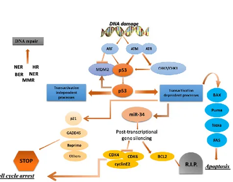
ATM/ATR/Chk1/Chk2, probably include other kinases such as p38, JNK/SAPK and c-Abl (Figure 11).72 Regarding human cancer, the available information gathered from the analysis of epigenetic aberrations indicates that the aforementioned DNA damage signaling kinases are not, in general, significant targets of genetic and epi-genetic inactivation.73 The only exception to this is found in hematological malignancies, which present a high incidence of mutations in ATM (13–40% depending on the particular type of malignancy).74 In line with this, a recent large-scale sequencing effort of 210 diverse human cancers has identified ATM among the three most frequently mutated kinases (5% incidence).75 Based on the above genetic evidence, it can be concluded that DNA damage is conveyed to p53 through multiple redundant pathways in which many transducers participate, but none of them plays a critical role and, therefore, alteration of a single component does not have a significant impact on p53 function.
Figure 11. p53 is at the center of a complex network of biological interactions that
- 29 -
interaction with p53.76 In non-stressed conditions these proteins bind p53, ubiquitylate it and target it for degradation by the proteasome. In stressed conditions the function of the MDM2–MDM4 complex is blocked by phosphorylation, protein-binding events and/or enhanced degradation. Hence, phosphorylation of MDM4 is essential for the p53 response to ionizing radiation, and the response to oncogene activation depends on the binding of ARF to MDM2. Many p53-activating small molecules function by causing the release of ribosomal proteins from the nucleolus to the nucleoplasm, where they bind to MDM2 and MDM4 and inhibit their function. Molecules that activate wild-type p53 in tumors by disrupting MDM2 activity can compensate for any missing upstream components of the p53 pathway. However, defective downstream p53 signaling might substantially decrease their effectiveness. Therefore, the ability to identify tumors in which downstream p53 signaling is unaffected is important. The development of strategies to ensure that the desired p53 response is initiated when it is reactivated might be necessary and could require the judicious use of drug combinations.
1.6 p53 network: co-activators and associated proteins
The interaction between p53 and transcriptional co-activators also influences its affinity for promoters. It is therefore plausible that the specific co-factors expressed in a particular cellular context determine the repertoire of p53-target genes induced, and consequently whether the cell undergoes growth arrest or apoptosis, or even a particular apoptotic pathway, may be subject to the availability of co-activators.76 Once the p53 protein is activated, it initiates a transcriptional program that reflects the nature of the stress signal, the protein
Chapter I: The cancer and p53 “guardian of the genome”
- 30 -
modifications and proteins associated with the p53 protein. The p53 protein binds to a specific DNA sequence, termed the p53- responsive element (RE),77 and induces the expression of downstream genes. An algorithm that identifies p53-responsive genes in the human and mouse genome has been utilized to detect a number of new genes regulated by the p53 protein.78 The genes in this p53 network mainly initiate one of three programs that result in cell cycle arrest, DNA repair or apoptosis. The exact criteria that influence p53 to stimulate cell cycle arrest or apoptosis are only partially understood and are the subject of intense study. Several general factors that influence this decision include p53 expression levels, the type of stress signal, the cell type and the cellular context at the time of exposure to stress. Several intriguing observations have recently provided insight into the apparent intricacies of such cell fate determination. The examples described below involve the binding of p53 to its canonical binding sequence in target genes. Note, however, that p53 can also activate target genes through a non-canonical sequence. The first such example is in the p53-induced gene 3 (PIG3), which has been implicated in the accumulation of reactive oxygen species and apoptosis induction. PIG3 can be induced by p53 through a microsatellite sequence within its untranslated region. Another recently described example is the gene encoding the pro-apoptotic phosphatase PAC1, which is induced through binding of p53 to a novel palindromic binding site. This might represent a new mechanism for transcriptional regulation of apoptotic genes by p53, which differs from that already described (see below). Exacting discrimination between p53 arrest and apoptotic functions has been critical to the identification of the importance ofthe latter in tumor suppression.
1.6.1 p53 and the DNA repair
Soon after having established TP53 as the most frequently altered gene in human tumors in the 1990s,79 p53 was understood as a major component of the DNA damage response pathway. After the introduction of DNA injuries the
- 31 -
protein (Cbp)/p300, and by the poly (ADPribose) polymerase 1 (Parp-1), which prevent proteolysis via the Arf-mouse double minute 2 (MDM2) pathway and/or enhance binding of p53 to consensus sequences within the genome.80 Initially, investigations on a direct participation of p53 in DNA repair were spurred by a number of biochemical observations. Thus, the C-terminal 30 amino acids of p53 were shown to recognize several DNA damage-related structures, such as DNA ends, gaps, and insertion/deletion mismatches. p53 was also demonstrated to catalyze reannealing of short stretches of single- and double-stranded DNA and to promote strand exchange between them . Further, p53 binds to three-stranded heteroduplex joints and four-three-stranded Holliday junction DNA structures with localization specifically at the junction, suggesting that p53 directly participates in recombinational repair.81 Moreover, several groups demonstrated a Mg-dependent 3’–5’exonuclease activity intrinsic to p53. Noticeably, the same central region within p53, where tumorigenic mutations are clustered, recognizes DNA sequence specifically, is required for junction specific binding of heteroduplex joints and is necessary and sufficient for the 3’–5’ exonuclease activity on DNA.82 In addition to p53’s biochemical activities, numerous reports on physical and functional protein interactions further strengthened the proposal of a direct role of p53 in nucleotide excision repair (NER), base excision repair (BER), and double-strand break (DSB) repair.83
1.6.2 p53 and the growth arrest
p21WAF1/CIP1 is known to be a p53-downstream gene, and has been suggested to mediate p53-induced growth arrest triggered by DNA damage. The p21
Chapter I: The cancer and p53 “guardian of the genome”
- 32 -
protein is a cyclin-dependent kinase inhibitor that associates with a class of CDKs and inhibits their kinase activities. This will facilitate the accumulation of hypophosphorylated form of pRB that in turn associates with E2F inhibiting its transcriptional activity, leading to cell cycle arrest. As long as pRb is bound to E2F, the cell is prevented from entering into S phase. This G1 arrest affords the cell time to repair the DNA damage. Should repair be unsuccessful, P53 levels drop and CDK-cyclin protein kinase activity resumes, leading to entry into S phase. In the event that the DNA is not repair, p53 triggers apoptosis.84
1.6.3 p53 and the apoptosis
Pivotal to the tumor-suppressor activity of p53 is its ability to activate apoptosis via multiple different pathways. Since the most-studied function of p53 is its role as a transcription factor that can activate transcription of an ever-increasing number of target genes, its transcriptional activation of pro-apoptotic genes, as well as its transcriptional repression of anti-apoptotic genes, has been widely analyzed.85 However, although a large number of genes regulated by p53 during induction of apoptosis are known no single target gene has been identified whose altered expression alone can sufficiently explain p53 mediated transcription dependent apoptosis, and whose genetic deficiency phenocopies p53 deficiency in vivo. As an additional mode of p53’s pro-apoptotic activity, recent studies have placed non transcriptional pro- apoptotic activities of p53 at the center of an active debate that aims to establish a comprehensive understanding of p53- mediated apoptosis.86
1.6.3.1 p53 role in transcription dependent apoptosis
The past twenty-five years have seen intensive and varied investigations to better understand the functions that p53 uses to mediate apoptosis. The first indication of the role of p53 in apoptosis was obtained using the M1 mouse myeloid leukemia cell line lacking endogenous p53. Using M1 cells stably
- 33 -
been implicated in p53-mediated apoptosis. One is p53 activation to up-regulation of pro-apoptotic Bax and down-up-regulation of pro-survival Bcl-2.88 More recently its determined that p53-mediated apoptosis of M1 cells involves rapid activation of the pro-apoptotic Fas/CD95 death pathway-via up-regulation of membrane bound Fas and the intrinsic mitochondrial pathway, which results in activation of caspases 8, 9 and 10. (Figure 12) Either Fas blocking antibody or inhibition of the apical caspases 8 and 10, were each almost as effective as IL-6 in abrogating p53 mediated apoptosis. These observations argue that p53 regulation of the bcl-2 members Bax and BcI-2, associated with the intrinsic mitochondrial apoptotic pathway, is ancillary to the extrinsic Fas/CD95 apoptotic pathway in mediating p53 induced apoptosis of M1 myeloid leukemia cells.89
Chapter I: The cancer and p53 “guardian of the genome”
- 34 -
Figure 12. p53 mediated apoptosis
In other cell types up-regulation IGF-BP3 90 which sequesters the cell survival factor insulin-like growth factor-1 has been associated with p53 mediated apoptosis. The gene encoding for the cathepsin-D protease, PAG-608 which encodes a nuclear zinc finger protein and the human homolog of the Drosophilasina gene have also been implicated as mediators of p53 induced apoptosis in various cell types. Furthermore, a series of p53-induced genes (PIG genes) were documented to encode proteins that respond to oxidative stress, suggesting that p53-mediated apoptosis involves activation of redox- controlling targets followed by increase in ROS, oxidative damage to mitochondria and caspase activation. Along this research line it was recently observed that p53 suppresses Nrf2-dependent transcription of antioxidant response genes, presumably to prevent the generation of antioxidants that could hinder induction of apoptosis.91 Clearly established is p53’s role as a nuclear
- 35 -
respectively, in stressed cells. Puma and Noxa are thought to indirectly induce mitochondrial outer membrane permeabilization (MOMP), known to be induced by the activation of Bax and Bak, via interfering with Bax and Bac interaction with prosurvival Bcl-2 family members. Interestingly, it was observed that Puma and Noxa differentially contribute to the regulation of p53-mediated apoptotic pathways. In normal cells, Puma was found to induce mitochondrial outer membrane permeabilization via an ER-dependent pathway; however, upon E1A oncoprotein expression, cells also became susceptible to mitochondrial outer membrane permeabilization induction by Noxa via an ER-independent pathway. In several instances, transcriptional activation by p53 was observed to be dispensable for p53-dependent apoptosis, since mutants p53 which fail to activate transcription could still induce apoptosis.92 In addition, p53-dependent apoptosis could occur in the presence of inhibitors of transcription and translation.93 In recent years it has become clear that p53 also harbors a direct proapoptotic function at the mitochondria via engaging in protein-protein interactions with anti- and pro-apoptotic Bcl2 family members, including BclXL and Bak. 94 It has been reported, certain transcriptionally inactive mutants of p53 can still induce apoptosis when over expressed in tumor cells. Also, in response to some stresses, such as hypoxia, p53 induces apoptosis but does not function as a transactivator. Intriguingly has been demonstrated that during p53-dependent apoptosis a fraction cellular p53 protein localizes to mitochondria and induces cytochrome c release; however, this is not observed during p53-mediated cell cycle arrest.95 Additional support for the concept that p53 has a cytoplasmic role in apoptosis induction resulted from functional analysis of polymorphic variants of p53 (within exon 4 of the p53 gene, a
Chapter I: The cancer and p53 “guardian of the genome”
- 36 -
common single-nucleotide polymorphism (SNP) at codon 72 leads to the incorporation of either an arginine (R72) or a proline (P72) at this position of the protein. When explored the potential mechanisms underlying the observed functional difference between the two p53 variants, made the initially surprising discovery that the greater apoptotic potential of the R72 form correlated with its much better ability to traffic to mitochondria. Based on these data, therefore concluded that the enhanced apoptosis-inducing activity of the R72 protein related, at least in part, to its greater mitochondrial localization. An analysis of whole cell or mitochondrial extracts by immune precipitation-western blot analysis, demonstrated the R72 form of p53 binds better to the mitochondrial death-effectors protein BAK than does the P72 variant, correlating with the difference in apoptotic potential of the two p53 variants. In healthy cells, Bak resides at mitochondria as an inactive monomer. In response to various death stimuli, it undergoes an activating allosteric conformational change that promotes homo-oligomerization. This leads to formation of a pore in the outer mitochondrial membrane, and allows the release of cytochrome c and other caspase cascade (Figure 4). Recently, like BAK, the BCL2 family members BAX and BCL-XL have also been implicated in mitochondrial apoptosis induction by p53 (Figure 12).96
- 37 -
CHAPTER II
SEARCH SETTING
Chapter I1: Search setting
- 38 -
2.1 Aim of the study
The ability of p53 to respond to stress signals by triggering cell-cycle arrest and cell death by apoptosis is crucial to inhibit tumor development and for the response to anticancer therapy.49,97,98 Inactivation of p53 by mutation occurs in about half of all human tumors. Tumors that retain wild-type p53 often acquire an alternative mechanism for its inactivation, largely through deregulation of MDM2 (murin double minute-2) protein. Negative regulation of p53 activity and stability is enhanced in many human tumors and effectively impairs the activities of the p53 pathway. Therefore, recovery of p53 activity in cancer cells by antagonizing MDM2 has been proposed as a novel approach for treating cancer and validated in vitro by macromolecular studies. MDM2 and p53 are part of an auto-regulatory feedback loop (Figure 13).99,100 MDM2 is transcriptionally activated by p53 and MDM2, in turn, inhibits p53 activity in several ways. MDM2 binds to the p53 transactivation domain and there by inhibits p53-mediated transactivation MDM2 also contains a signal sequence that is similar to the and, after binding to p53, it induces its nuclear export nuclear export signal of various viral proteins. As p53 is a transcription factor, it needs to be in the nucleus to be able to access the DNA; its transport to the cytoplasm by MDM2 prevents this. Finally, MDM2 is an ubiquitin ligase, so is able to target p53 for degradation by the proteasome. In normal cellular conditions, p53 is constantly degraded by MDM2, and is therefore present at low levels.101