Adhesion and biofilm formation by oral streptococci on different commercial brackets.
Claudio Passariello1*, Pierangelo Gigola2
1
Department of Public Health and Infectious Diseases, " Sapienza" University of Rome, Rome Italy. 2
Department of Surgical Specialities, Radiologic and Medico-Forensic Sciences University of Brescia, Brescia Italy.
Key Words: Bacterial adhesion, biofilm formation, brackets
Corresponding author: Dr. Claudio Passariello, Dipartimento di Sanità Pubblica e Malattie Infettive, Università “La Sapienza”, Piazzale Aldo Moro 5, 00185, Rome, Italy. Email address
Abstract Objective.
To compare early bacterial adhesion and biofilm formation in vitro by different oral streptococci on a variety of commercial brackets
Materials and Methods.
Adhesion and biofilm formation in vitro of 6 Streptococcus spp. on 15 different commercial brackets, in standard culture medium and in human saliva was evaluated by the MTT reduction assay.
Results.
Significant differences were evidenced in both early adhesion and biofilm formation among the studied brackets and between the two conditions of growth. Gold brackets resulted less prone to colonization, while composites brackets were the most prone ones. The rates of growth of the different tested species on the different tested materials were significantly different.
Conclusions.
The adopted experimental plan, dissecting the two phases of plaque formation on different brackets in different conditions, showed that composite brackets are more susceptible to adhesion and
colonization by streptococci, while the remaining tested brackets do not show differences that could be clinically relevant. Data suggest that different personal behaviors affecting the oral environment could significantly affect colonization of brackets by cariogenic bacteria.
Introduction
In recent decades orthodontic treatment has undergone a great increase in its diffusion, mostly due to patients seeking orthodontic treatment to improve their dentofacial esthetics, while only a minority require treatment for medical or dental reasons [Shaw et al., 1991; Harris, 2011]. Such a situation, in turn, greatly improved the necessity for orthodontic treatment to be safe and free from collateral effects. Among short- and long-term complications of fixed orthodontic treatment, those related to increased formation of dental plaque are of great relevance [van Gastel et al., 2007]. The insertion of fixed orthodontic appliances induces significant ecological changes affecting the composition, metabolic activity, and pathogenicity of the oral microbiota, thus favoring the
incidence of periodontal inflammation and incipient carious lesions [Atack et al., 1996; Naranjo et al., 2006; Ahn et al., 2007].
While ecological alterations and periodontal inflammation are considered to be largely reversible in children and adolescents [Ristic et al., 2007], incipient caries results at least in the formation of persisting white spot lesions [Øgaard, 1989; Sallum et al., 2004] that contrast with aesthetic requirements of patients and their parents. Enamel decalcification and caries formation, due to increased prevalence of Streptococcus mutans and Lactobacillus species in dental plaque have been extensively studied as a complication of fixed orthodontic treatment in children [Forsberg et al., 1991; Rosenbloom and Tinanoff, 1991]. Although once believed to be essentially a consequence of quantitative alteration of the dental microbiota [Boyd, 1983], this side-effect is also caused by the specific properties of bracket materials, affecting the quality of dental microbiota [Fournier et al., 1998; Anhoury et al., 2002; Brusca et al., 2007].
These evidences prompted researchers to try to develop materials that were less susceptible to bacterial colonization thus minimizing plaque accumulation around brackets and other fixed appliances [van Gastel et al., 2007; Ahn et al., 2007; Papaioannou et al., 2007; Faltermeier et al., 2008; van Gastel et al., 2009].
With few exceptions, these studies addressed the problem of adhesion of cariogenic bacteria to bracket materials, while less attention was dedicated to other oral streptococci. This study, in consequence, was aimed to compare early bacterial adhesion and biofilm formation in vitro by different oral streptococci on a variety of different commercial brackets.
Materials and Methods
Brackets. Fifteen commercially available brackets, made of different materials, were used (Table 1). All brackets were maxillary premolar brackets, with the Roth prescription and a 0.022-inch slot Twelve brackets for each bacterial strain were tested.
Bacterial strains and cultures. Six reference strains of different species of oral streptococci were used: Streptococcus salivarius DSM20560, Streptococcus gordonii DSM6777, Streptococcus sanguinis DSM20567, Streptococcus oralis DSM20627, Streptococcus mutans DSM20523 and Streptococcus sobrinus DSM20742. All strains were maintained in stock cultures freezed at -80°C in Tryptic Soy broth (TSB) containing glycerol (20% v/v). For adhesion assays, isolated colonies of each strain were inoculated in TSB and incubated at 37°C with mild shaking till the mid logarithmic phase of growth. Bacterial cells were then collected by centrifugation and suspended in fresh sterile 0.5xTSB (i.e TSB diluted 1/2 in PBS pH 7.2) or sterile 0.5xSaliva (i.e. human pooled saliva diluted 1/2 in TSB) at an OD600nm = 0.1. Saliva was obtained by paraffin stimulation from 15 healthy volunteers (having refrained from eating and drinking in the previous 2 hours) and checked for pH being in the range 7.0 to 7.3. Saliva samples were subjected to sonication (1 minute at 30W with refrigeration), filtered through a 70µm filter (Cell Strainer, Becton Dickinson Italia, Buccinasco, Italy) and centrifuged at 22,000 x g for 60 minutes at 4°C. Supernatants were pooled, sterilized by sequential filtration through 0.45 µm and 0.2 µm filters, stored at 4°C and used within the next 48 hours.
Adhesion assays. In order to perform standardized adhesion assays, brackets were mounted on 0.6 x 0.6 cm polished clear acrylic blocks (K-Mac Plastics Wyoming, MI, USA) sticked to the cover of a 24 wells polystyrene plate. All the mounting process was performed by a single operator inside a sterile class II biohazard cabinet. The central region of each block, in the exact position were a bracket had to be fixed, was roughened with a diamond coated burr in such a manner that these areas were completely covered by the bracket bases The brackets were then bonded with Transbond Plus color change adhesive (3M Unitek, Monrovia, CA, USA). Excesses of adhesive were removed
carefully and the composite was light-cured for 30 seconds from both sides. Brackets mounted this way were completely immersed when each well was filled with 1.1 ml of bacterial suspension. Before contact with the bacterial cultures, brackets were placed in 24 well plates containing either sterile 0.5xTSB or sterile 0.5xSaliva and incubated at 37°C for 1 hour.
Pre-conditioned brackets were then transferred to a new plate with wells filled with the bacterial suspension in the same medium, and incubated for 4 and 48 hours at 37°C on a orbital shaker at 60 RPM.
Following contact with the different bacterial suspensions, the brackets were removed with a sterile pliers and transferred into an adequately coded well of a flat bottom 96 well plate containing 0.1 ml of sterile PBS. Brackets were then washed five times with sterile PBS and further processed for the enumeration of adherent bacteria by the MTT reduction assay.
Quantitation of adherent bacteria by the MTT reduction assay.
The amount of bacteria adherent to each bracket was determined by the MTT-reduction assay [Kairo et al., 1999; Walencka et al., 2006]. Briefly, each bracket-containing well was carefully emptied of any residul liquid, and 0.15 ml of PBS were added, followed by 0.05 ml of MTT (Sigma Chemical Co. USA) (0.3% v/v in PBS). Samples were then incubated 2 hours at 37°C and MTT was replaced with 0.15 ml of dimethyl sulfoxide and 0.025 ml of glycine buffer (0.1 M, pH 10.2) for 15 minutes at room temperature. In this assay, bacteria with an active electron transport system reduce the pale yellow tetrazolium salt to water soluble purple formazan. The amount of formazan produced by each reaction was determined using a BioRad model 680 microplate reader at A550. Quantitative analysis was performed following construction of species specific standard curves for each tested strain.
Statistics
Statistic evaluation of the significance of differences among results of adhesion assays was performed by the Student T test available in the Microsoft Excel software. Differences yielding
values of P in the range >0.01 to ≤ 0.05 were considered significant while differences yielding values of P ≤ 0.01 were considered very significant.
Results
Mean values of adherent streptococci detected at the surface of different brackets in adhesion and biofilm formation assays performed in TSB or saliva are reported in Figure 1. The 15 tested
brackets were divided in 6 groups depending on material they were made of: i) ceramic (brackets A, B, C and D), stainless steel (brackets E, F and G), gold (brackets H and I), composites (brackets J, K and L), titanium (bracket M) and monocrystalline sapphire (brackets N and P). Results showed that streptococci are significantly different in their adhesiveness to the studied materials.
Streptococcus mutans and Streptococcus sobrinus constantly yielded among the highest values with all tested materials and conditions, while Streptococcus gordonii and Streptococcus sanguinis showed to be less adhesive. Adhesion assays performed in the presence of human saliva yielded significantly higher values for all tested species and materials, although the influence of saliva was different depending on both bacteria and materials. In fact, comparison of cumulative results of adhesion assays for the different materials showed values of P (obtained by the Student T test) for the comparison TSB vs Saliva ranging 0.01 to 7.91x10⁻⁷ (ceramic P= 7.91x10⁻⁷; stainless steel P= 3.25x10⁻³; gold P= 7.70x10⁻⁶; composites P= 1.07x10⁻⁵; titanium P= 0.01; monocrystalline sapphire P= 2.62x10⁻³).
Biofilm formation, assessed by counting adherent bacteria after 48h of growth in TSB or saliva, showed that the studied bacteria have different capacities to form biofilms in the studied conditions and that results obtained after growth in TSB are significantly different from those obtained in saliva (Figure 1).
When results obtained at 4h and 48h were analyzed cumulatively for each group of brackets, differences in the possibility of the different materials to favor biofilm growth appeared evident in both TSB and saliva(Figure 2). In both media composites resulted as the worst performing brackets, while gold brackets showed the best performances.
Analysis of ratios (saliva/TSB) of adherent bacteria detected at the surface of the studied brackets showed that ratios differed significantly between 4h and 48h for all studied species (Figure 3a), but
not for all tested brackets (Figure 3b). In fact, biofilm formation at 48h was significantly greater on gold, stainless steel and ceramic brackets, but not on the remaining ones (Figure 3b). Values of standard deviation reported in figure 3 (panels a and b) show that biofilm formation at the surface of brackets is strongly influenced by both the material and the industrial process. These differences are not evident from adhesion assays at 4h.
Discussion
The present study was aimed to evaluate the susceptibility of 15 different brackets to adhesion by 6 different species of oral streptococci and to evaluate the ability of these brackets, made of 6 classes of materials, to support biofilm formation by the studied bacteria, in different in vitro conditions. The rationale of this study is to support furhter informations on differences existing among commercially available brackets as to their capacity to favor formation of streptococcal biofilms that can give rise to initial caries.
In fact, data of the international literature show that fixed orthodontic appliances induce increased plaque accumulation [Balenseifen and Madonia, 1970; Årtun and Brobakken, 1986] and favor colonization by the cariogenic mutans streptococci and lactobacilli [Forsberg et al., 1991;
Rosenbloom and Tinanoff, 1991], thus increasing the risk of decalcification, which can involve up to 50% of patients, and lead to the development of caries.
Brackets are per se an obstacle to correct oral hygiene, but constructive material is believed to play a relevant role in determining the amount and quality of bacterial plaque accumulating around teeth of patients undergoing fixed orthodontic treatment. Colonization of a site depends on two distinct phases, early adhesion, mostly due mostly to electrostatic and hydrophobic interactions
[Papaioannou et al., 2007] and subsequent biofilm formation, that is influenced by a variety of environmental and surface properties [Anhoury et al., 2002; Demling et al., 2010].
Surface free energy is believed to play a relevant role in bacterial adhesion to surfaces and materials with high surface free energies, as stainless steel [Eliades et al., 1995] should favor adhesion of mutans streptococci more than other materials [Ahn et al., 2007; Faltermeier et al., 2008; An et al., 1998]. Our data, nevertheless, indicate that although stainless steel effectively favors initial
bacterial adhesion, as compared to other materials, with the exception of composites, it
subsequently does not support biofilm formation to the same extent, so that final values of bacterial accumulation are substantially comparable to those obtained with other materials, including
mutans group. These discrepancies were already evident from data of other studies [Fournier et al., 1998; Ahn et al., 2002], and could possibly depend on the role played in biofilm formation by the acquired pellicle and on material dependent differences in its composition [Ahn et al., 2002; 2003] as demonstrated in our experiments by different rates of adhesion and biofilm formation obtained in TSB as compared to saliva containing medium. Although significant discrepancies can be found between our data and data by other authors concerning relative performances of the different materials [Fournier et al., 1998; Ahn et al., 2002; 2003], this is a consequence of the different experimental protocols that have been adopted.
Overall, our experimental data show that bracket material is relevant for plaque formation around teeth treated with fixed orthodontic appliances. Although statistical analyses suggest that significant differences exist among results obtained with almost all tested materials, an overview to obtained results suggests clearly that composite brackets are characterized by a higher susceptibility to colonization by the different tested streptococci and particularly by those of the mutans group while all the other tested brackets have better performances. Although the non-composite brackets yielded significantly different results in the adhesion and bofilm formation assays, these differences are not so relevant to be considered clinically important. Gold brackets appear to be the better performing brackets in all tests while a cumulative evaluation of the remaining groups suggests that although less prone to initial colonization, titanium and monocrystalline sapphire brackets, must overall be considered comparable to ceramic and stainless steel brackets.
Data evaluating the influence of the medium on adhesion and biofilm formation suggest that in vivo the bacterial colonization of brackets, and consequently the formation of incipient caries could be influenced by food and other personal behaviors.
Our experimental plan allowed us to dissect the two basic moments of bacterial colonization of orthodontic appliances and could constitute the basis for further competitive colonization studies. Acknowledgements
This work was supported by a grant from MIUR (Project PRIN 2007LXNYS7_003 granted to CP) and by funds of the University of Brescia granted to PG.
References
Ahn S-J, Kho H-S, Lee S-W, Nahm D-S. Roles of salivary proteins in the adherence of oral streptococci to various orthodontic brackets. J Dent Res. 2002;81(6):411–415.
Ahn S-J, Kho H-S, Kim K-K, Nahm D-S. Adhesion of oral streptococci to experimental bracket pellicles from glandular saliva. Am J Orthod Dentofacial Orthop. 2003;124: 198-205.
Ahn S-J, Lee S-J, Lim B-S, Nahm D-S. Quantitative determination of adhesion patterns of cariogenic streptococci to various orthodontic brackets. Am J Orthod Dentofacial Orthop. 2007;132:815-821.
An YH, Friedman RJ. Concise review of mechanisms of bacterial adhesion to biomaterial surfaces. J Biomed Mater Res. 1998;43:338-348.
Anhoury P, Nathanson D, Hughes C V, Socransky S, Feres M, Chou L L. Microbial profile on metallic and ceramic bracket materials. Angle Orthod. 2002; 72: 338-343.
Årtun J, Brobakken B. Prevalence of caries and white spots after orthodontic treatment with multibonded appliances. Eur J Orthod. 1986; 8:229-234.
Atack N E, Sandy J R, Addy M. Periodontal and microbiological changes associated with the placement of orthodontic appliances. A review. J Periodontol. 1996; 67: 78–85.
Balenseifen JW, Madonia JV. Study of dental plaque in orthodontic patients. J Dent Res. 1970; 49:320–324.
Boyd R L. Longitudinal evaluation of a system for self-monitoring plaque control effectiveness in orthodontic patients. J Clin Periodontol. 1983; 10: 380-388.
Brusca MI, Chara O, Sterin-Borda L, Rosa AC. Influence of different orthodontic brackets on adherence of microorganisms in vitro. Angle Orthod. 2007; 77:331-336.
Demling A, Elter C, Heidenblut T, Bach Fr-W, Hahn A, Schwestka-Polly R, Stiesch M, Heuer W. Reduction of biofilm on orthodontic brackets with the use of a polytetrafluoroethylene coating. Eur
Eliades T, Eliades G, Brantley W A. Microbial attachment on orthodontic appliances: I. Wettability and early pellicle formation on bracket materials. Am J Orthodont Dentofac Orthop. 1995; 108: 351–360.
Faltermeier A, Bürgers R, Rosentrittc M. Bacterial adhesion of Streptococcus mutans to esthetic bracket materials. Am J Orthod Dentofacial Orthop. 2008;133:S99-103
Forsberg CM, Brattstrom V, Malmberg E, Nord CE. Ligature wires and elastomeric rings: two methods of ligation, and their association with microbial colonization of Streptococcus mutans and lactobacilli. Eur J Orthod. 1991;13:416–420.
Fournier A, Payant L, Bouclin R. Adherence of Streptococcus mutans to orthodontic brackets. Am J Orthod Dentofacial Orthop. 1998;114:414-417.
Harris EF. Sex differences in esthetic treatment needs in American black and white adolescent orthodontic patients. Angle Orthod. 2011; 81: 743-749.
Kairo SK, Bedwell J, Tyler PC, Carter A, Corbel MJ. Development of a tetrazolium salt assay for rapid determination of viability of BCG vaccines. Vaccine 1999;17:2423-2428.
Naranjo A A, Trivino M L, Jaramillo A, Betancourth M, Botero J E. Changes in the subgingival microbiota and periodontal parameters before and 3 months after bracket placement. Am J Orthod Dentofacial Orthop. 2006; 130: 275.e17–22.
Øgaard B. Prevalence of white spot lesions in 19-year-olds: a study on untreated and
orthodontically treated persons 5 years after treatment. Am J Orthod Dentofacial Orthop. 1989; 96: 423–427.
Papaioannou W; Gizani S; Nassika M; Kontou E; Nakou M. Adhesion of Streptococcus mutans to different types of brackets. Angle Orthod. 2007; 77: 1090-1095.
Ristic M, Vlahovic Svabic M, Sasic M, Zelic O. Clinical and microbiological effects of fixed orthodontic appliances on periodontal tissues in adolescents. Orthod Craniofacial Res. 2007; 10: 187–195.
Rosenbloom RG, Tinanoff N. Salivary Streptococcus mutans levels in patients before, during, and after orthodontic treatment. Am J Orthod Dentofacial Orthop. 1991; 100:35-37.
Sallum EJ, Nouer DF, Klein MI, Gonçalves RB, Machion L, Wilson Sallum A, Sallum EA Clinical and microbiologic changes after removal of orthodontic appliances. Am J Orthod Dentofacial Orthop. 2004; 126: 363–366.
Shaw WC, Richmond S, O’Brien KD, Brook P, Stephens CD. Quality control in orthodontics: indices of treatment need and treatment standards. Br Dent J. 1991; 170:107–112.
van Gastel J, Quirynen M, Teughels W, Coucke W, Carels C. Influence of bracket design on microbial and periodontal parameters in vivo. J Clin Periodontol. 2007; 34:423–431.
van Gastel J, Quirynen M, Teughels W, Pauwels M, Coucke W, Carels C. Microbial adhesion on different bracket types in vitro. Angle Orthod. 2009; 79:915–921.
Walencka E, Sadowska B, Rózalska S, Hryniewicz W, Rózalska B. Staphylococcus aureus biofilm as a target for single or repeated doses of oxacillin, vancomycin, linezolid and/or lysostaphin. Folia Microbiol (Praha) 2006; 51:381-386.
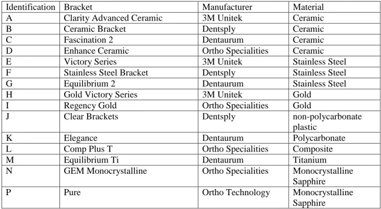
Table 1. List of brackets used during the study, their identification keys in the text and results section, manufacturer and construction material.
Identification Bracket Manufacturer Material
A Clarity Advanced Ceramic 3M Unitek Ceramic
B Ceramic Bracket Dentsply Ceramic
C Fascination 2 Dentaurum Ceramic
D Enhance Ceramic Ortho Specialities Ceramic
E Victory Series 3M Unitek Stainless Steel
F Stainless Steel Bracket Dentsply Stainless Steel
G Equilibrium 2 Dentaurum Stainless Steel
H Gold Victory Series 3M Unitek Gold
I Regency Gold Ortho Specialities Gold
J Clear Brackets Dentsply non-polycarbonate
plastic
K Elegance Dentaurum Polycarbonate
L Comp Plus T Ortho Specialities Composite
M Equilibrium Ti Dentaurum Titanium
N GEM Monocrystalline Ortho Specialities Monocrystalline
Sapphire
P Pure Ortho Technology Monocrystalline
Figure legends
Figure 1. Adherent bacteria detected at the surface of different brackets in adhesion (4h) and biofilm formation (48h) assays performed in Tryptic Soy broth (TSB) or in human saliva. Results are
reported as means for brackets grouped according to construction material. Individual standard deviations are reported.
Figure 2. Ratios of adherent bacteria detected at 4h and 48h of incubation at the surface of the studied brackets after incubation in human saliva as compared to results obtained in Tryptic Soy broth (TSB). Results are grouped according to tested strain (panel a) and bracket material (panels b). Individual standard deviations are reported. * indicates significant differences corresponding to values of P in the range >0.01 to ≤ 0.05; ** indicates very significant differences corresponding to values of P≤ 0.01.
Figure 3. Mean biofilm growth curves obtained at the surface of different types of brackets with the tested bacteria grown in Tryptic Soy broth (TSB) or human saliva.