Abatacept Reduces Levels of Switched Memory
B Cells, Autoantibodies, and Immunoglobulins in
Patients with Rheumatoid Arthritis
Mirko Scarsi, Lucia Paolini, Doris Ricotta, Antonio Pedrini, Silvia Piantoni, Luigi Caimi,
Angela Tincani, and Paolo Airò
ABSTRACT. Objective. Abatacept (ABA) is a chimeric molecule, able to block the CD28-mediated costimulatory pathway. To evaluate the hypothesis that, through this mechanism of action, ABA may down-modulate the immune responses of B lymphocytes in rheumatoid arthritis (RA), we investi-gated the serum levels of immunoglobulins (Ig), free light chains (FLC), anticitrullinated protein antibodies (ACPA), and rheumatoid factor (RF), as well as the number of B lymphocytes differen-tiated into post-switch memory cells in patients treated with ABA.
Methods. The serum levels of Ig, FLC, different ACPA, RF isotypes, and the B cell phenotype were longitudinally evaluated in 30 patients with RA treated with ABA.
Results. At baseline, the proportion of total and post-switch memory B cells was lower in RA than in healthy individuals. After 6 months of ABA treatment we observed significant reductions of serum levels of IgG, IgA, and IgM, as well as FLC, with a normalization in many patients who had initially abnormal values. A significant reduction of the titers of IgG- and IgA-ACPA, as well as of IgM-, IgA-, and IgG-RF was also observed. A decrease of autoantibodies below the upper limits of normal values was found in 2 of 26 patients (8%) initially seropositive for IgG-ACPA, 1 of 14 (7%) for IgA-ACPA, 5 of 22 (23%) for IgM-RF, 7 of 22 (30%) for IgA-RF, and 5 of 16 (31%) for IgG-RF. After treatment, the proportion of circulating post-switch memory B cells was also further signifi-cantly decreased.
Conclusion. ABA treatment in patients with RA can reduce signs of polyclonal B cell activation, inducing a trend toward normalization of serum levels of different classes of Ig and of FLC, decreasing titers of ACPA and RF, and percentages of post-switch memory B cells. (First Release March 1 2014; J Rheumatol 2014;41:666–72; doi:10.3899/jrheum.130905)
Key Indexing Terms:
ABATACEPT RHEUMATOID FACTOR FREE LIGHT CHAINS ANTICITRULLINATED PROTEIN ANTIBODIES B LYMPHOCYTES
From the Rheumatology Unit and Clinical Immunology, Spedali Civili, Brescia; and Clinical Biochemistry, Department of Molecular and Translational Medicine, University of Brescia, Brescia, Italy.
Supported by Bristol-Myers-Squibb Italy. Inova Diagnostic Inc. Italy and The Binding Site Italy provided diagnostic kits for the study.
M. Scarsi, MD, Rheumatology Unit and Clinical Immunology, Spedali Civili and University of Brescia; L. Paolini, PhD; D. Ricotta, MD, Clinical Biochemistry, Department of Molecular and Translational Medicine, University of Brescia; A. Pedrini, Med. Sci.; S. Piantoni, MD, Rheumatology Unit and Clinical Immunology, Spedali Civili and University of Brescia; L. Caimi, MD, Clinical Biochemistry, Department of Molecular and Translational Medicine, University of Brescia; A. Tincani, MD; P. Airò, MD, Rheumatology Unit and Clinical Immunology, Spedali Civili and University of Brescia, Italy. Address correspondence to Dr. P. Airò, Rheumatology and Clinical Immunology Unit, Spedali Civili di Brescia, Piazzale Spedali Civili 1, 25123 Brescia, Italy. E-mail: [email protected]
Accepted for publication December 19, 2013.
B cells play a central role in the pathophysiology of
rheumatoid arthritis (RA)
1,2, and their hyperactivation is
demonstrated by hypergammaglobulinemia and increased
levels of serum free light chains (FLC)
3. B cells can also
produce rheumatoid factor (RF) and other autoantibodies,
including anticitrullinated protein antibodies (ACPA), whose
presence is associated with a more severe disease
1. The
ability to produce different classes of immunoglobulins (Ig),
including autoantibodies, is acquired after antigen
presen-tation in lymphoid tissue with the aid of T lymphocytes, by a
fraction of B cells that have undergone somatic
hypermu-tation. These cells can generally be identified phenotypically
as post-switch memory cells (CD19+CD27+IgD–)
4,
although a smaller CD27– population of memory B cells
with mutated Ig genes has also been described
5. Post-switch
memory B cells have been shown to accumulate in the
synovial compartments of patients with RA
6,7, and this
underlines their relevance in the autoimmune/inflammatory
process of RA.
B cells expressing the costimulatory molecules CD80
and CD86 can also act as antigen-presenting cells and
therefore activate T cells providing signals to the CD28
receptor
1,2. The relevance of this pathway as a therapeutic
target in RA has been demonstrated by clinical results
obtained with abatacept (ABA), a cytotoxic T
lympho-cyte-associated antigen 4 immunoglobulin (CTLA-4-Ig)
fusion protein
8. Through its CTLA4 portion, this agent can
bind to CD80 and CD86 on B cells, thereby inhibiting CD28
costimulation
9. CD28-mediated signals are relevant in the
upregulation of CD154 (the ligand for CD40) on T cell
surface, a key process in the acquisition of the T cell
“helper” function
10. The engagement of CD40
(constitu-tively present on B cell membrane) by CD154 plays a
crucial role in the process of isotype switching and B cell
maturation
10. It can therefore be hypothesized that, blocking
this pathway, ABA may downmodulate the immune
responses of B lymphocytes and the production of
autoanti-bodies
9. However, not much information is currently
available on the effect of ABA therapy on B cells and
autoantibody levels in patients with RA
9.
The aim of our study was to evaluate whether the
blockade of costimulation performed by ABA may reduce
the ability of B lymphocytes to differentiate into post-switch
memory B cells and to produce ACPA and RF. The levels of
total serum Ig and of FLC were also evaluated.
MATERIAL AND METHODS
Patients. Thirty consecutive patients with RA treated for at least 6
consec-utive months with ABA were enrolled in our study. Their main clinical and demographic characteristics are shown in Table 1. Four patients had been previously treated with the anti-B cell agent rituximab (RTX). Median time from the last RTX infusion at the moment in which ABA was started was 14.5 months (range 8–68). No patients had renal failure or monoclonal gammopathies. The clinical disease activity and the response to the treatment were evaluated respectively with the DAS28 (based on CRP) and the European League Against Rheumatism (EULAR) criteria of response to the treatment11.
The local ethics committee approved our study, and all patients provided informed consent.
Twenty-four blood donors [18 women (75%); median age 39 yrs (25th–75th percentile: 34–46)] served as healthy controls (HC).
Serum analysis. Serum samples were collected and stored at –80°C
immediately before the first administration of ABA (T0) and then after 6 months (T6). In 16 patients, a further sample was collected after 12 months (T12). Testing for the different assays was carried out simultaneously on all serum samples at the end of the study.
Serum IgG, IgA, and IgM levels were measured by a nephelometric immunoassay method (Siemens Healthcare Diagnostics Products GmbH) using a Dimension Vista 500 (Siemens). Reference ranges were provided by the package insert of the commercial kit and were derived by a consensus of a group of professional societies and diagnostic companies based on the standardization against the calibrated reference material 47012.
Serum FLC levels were measured by a latex-enhanced immunoassay (Freelite, The Binding Site) with use of the turbidimetric platform SPA-PLUS analyzer (The Binding Site). The diagnostic ranges had been previously established by the manufacturer to include 100% of a reference population of 282 serum samples13.
IgG-ACPA and IgA-ACPA were tested using a commercially available third-generation indirect solid-phase ELISA kit (Quanta-Lite CCP 3.1; Inova Diagnostics). The upper limit of normal (ULN; 20 U/ml) was set in accordance with the manufacturer’s recommendations. Serum samples showing high concentration (> 250 U/ml) were evaluated after further dilutions (1/4 and, when necessary, 1/16) and then corrected for these additional dilution factors. The different RF isotypes (IgM, IgA, and IgG) were assessed using ELISA kits (Quanta-Lite RF; Inova Diagnostics). According to the manufacturer’s recommendations, the test ULN was 6 U/ml. Only high titer IgM-RF (> 100 U/ml) were evaluated after further 1/20 dilution.
Flow cytometry. B cell counts were determined by flow cytometry
(Cytomics FC-500, Beckman Coulter Inc.). Briefly, 100 µl of fresh whole blood were stained for 20 min at 4°C with a mixture of PC5-CD19, PE-CD27, and FITC-IgD (from Beckman Coulter, or R&D Systems Inc.), to identify naive (CD19+CD27–IgD+), memory (CD19+CD27+), or post-switch memory (CD19+CD27+IgD–) populations4. Absolute cell count was determined by single-platform analysis using Flow-Count beads (Beckman Coulter).
Statistical analysis. Data are expressed as the median (25th–75th
percentile). The comparison between quantitative variables among different groups was performed by Mann-Whitney U test, while Wilcoxon signed-rank test was applied to assess variation within paired quantitative variables. The association between nominal variables was assessed with chi-square test with Yates’ correction or Fisher’s exact test. The correlation between quantitative variable was evaluated with the linear simple regression.
RESULTS
Evidence of B cell hyperactivation before ABA treatment.
Before starting treatment with ABA (T0), patients with RA
had higher serum levels of IgM, IgA, and FLC than those
observed in HC (Table 2). In comparison with reference
ranges, raised levels of serum IgG, IgA, or IgM were
observed in 17%, 37%, and 20% of patients, respectively,
whereas no one had hypogammaglobulinemia.
Measure-ment of FLC demonstrated raised levels of κ chains in 18
out of 29 evaluated patients (68%) and of λ chains in 5/29
(17%). The κ:λ ratio was above normal levels in 8 of 29
patients (27%), and normal in 21/29.
As far as autoantibodies (Table 3), at T0, 87% and 47%
of patients tested positive (> 20 IU/ml) for IgG- and
IgA-ACPA, respectively, whereas 73%, 73%, and 53%
showed the presence (> 6 U/ml) of IgM-, IgA-, and IgG-RF.
Table 1. Demographic features of patients with rheumatoid arthritis (n =
30). Data are expressed as the median (25th–75th percentile) and range unless otherwise indicated.
Sex (male/female), n 4/26 Age, yrs 53 (44–60) Disease duration, yrs 6.5 (2.25–11.75) Smokers, n (%) 13 (43) No. previous DMARD 3 (1–5) No. previous biological agents 2 (0–3)
TNF-a blocking agents 24
Rituximab, no. patients 4
Tocilizumab, no. patients 4
Anakinra, no. patients 6
ABA as firstline biological treatment 5 Concomitant use of methotrexate, n (%) 24 (80) Median dosage of methotrexate at baseline 12.5 (5.62–15) DAS28-CRP at baseline 5.12 (4.71–5.93) Serum creatinine (mg/dl) 0.70 (0.64–0.72)
DMARD: disease-modifying antirheumatic drug; TNF: tumor necrosis factor; ABA: abatacept; DAS28-CRP: 28-joint Disease Activity Score based on C-reactive protein.
Patients previously treated with RTX (minimum interval
was 8 mos) did not differ from the other patients at T0 in Ig
and FLC serum levels, but in 2 cases ABA was started
before complete B cell reconstitution. These patients were
therefore excluded from B cell analysis.
At T0, the proportions of circulating total memory B
cells (CD19+CD27+) and of not-switched memory B cells
(CD19+CD27+IgD+) were lower in patients with RA than
in HC (p = 0.019 and p = 0.05, respectively; Figure 1; Table
4), whereas there was no difference in post-switch memory
B cells (CD19+CD27+IgD–). Moreover, setting a cutoff at
the 25th percentile of HC range, 28% and 71% of patients
with RA at baseline had a low absolute number of
circu-lating naive or memory B cells, respectively.
ABA treatment modulates the B cell compartment. After 6
months of ABA treatment (T6), 70% and 53% of patients
achieved the EULAR good clinical response and clinical
remission, respectively.
At this time, we observed a significant reduction of
serum levels of IgG, IgA, and IgM (Table 2). Baseline
abnormal values of IgG, IgA, and IgM normalized at 6
months in 2 of 5 (40%), 3 of 11 (27%), and 4 of 6 (67%)
patients, respectively. Analogously, both serum free κ and λ
chains decreased significantly after 6 months of therapy
(Table 2), normalizing respectively in 6 of 18 (33%) and 4
of 5 (80%) patients with raised levels at T0. Despite this
decrease in both types of light chains, the κ:λ ratio was also
significantly reduced after therapy (Table 2), normalizing in
4 of 8 patients (50%) with raised ratio at T0.
We observed also a significant reduction of the titers of
IgG- and IgA-ACPA, as well as of IgM-, IgA-, and IgG-RF
(Table 3). A decrease of autoantibodies below the ULN
values was observed in 2 of 26 patients (8%) initially
seropositive for IgG-ACPA, 1 of 14 (7%) for IgA-ACPA, 5
of 22 (23%) for IgM-RF, 7 of 22 (30%) for IgA-RF, and 5
of 16 (31%) IgG-RF, whereas only 1 patient initially
seronegative showed a weak (8.3 IU/ml) new positivity for
IgG-RF at T6.
Finally, the proportion of post-switch memory B cells
were found to be significantly decreased following ABA
treatment (p = 0.03; Figure 1; Table 4).
In 16 patients, serum samples were obtained after 12
months of therapy (T12). There was a further significant
decrease of total serum IgA, and of IgA- and IgM-RF,
compared to values observed at T6 in these individuals
(Tables 2 and 3). One patient who still had raised levels of
serum IgA and 1 who still had positive IgA-RF at T6
normalized at T12.
All the variations here described were statistically
signifi-cant even if patients who had received RTX prior to ABA
therapy (n = 4) were excluded from the analysis. The only
exception was the reduction of the titer of IgG-RF, which
after exclusion of these patients, was significant only at T12
and not at T6.
Analysis of patients with low numbers of circulating B
memory cells. As described in RA
6,14, a large proportion of
our patients had low numbers of circulating memory B cells.
Comparing these patients with the others, no difference was
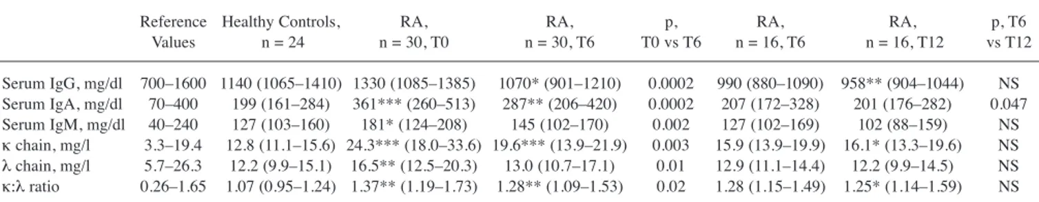
Table 2. Variations of Ig and FLC after therapy with ABA and comparison with healthy controls. Data are expressed as the median (25th–75th percentile).
Reference Healthy Controls, RA, RA, p, RA, RA, p, T6 Values n = 24 n = 30, T0 n = 30, T6 T0 vs T6 n = 16, T6 n = 16, T12 vs T12 Serum IgG, mg/dl 700–1600 1140 (1065–1410) 1330 (1085–1385) 1070* (901–1210) 0.0002 990 (880–1090) 958** (904–1044) NS Serum IgA, mg/dl 70–400 199 (161–284) 361*** (260–513) 287** (206–420) 0.0002 207 (172–328) 201 (176–282) 0.047 Serum IgM, mg/dl 40–240 127 (103–160) 181* (124–208) 145 (102–170) 0.002 127 (102–169) 102 (88–159) NS κchain, mg/l 3.3–19.4 12.8 (11.1–15.6) 24.3*** (18.0–33.6) 19.6*** (13.9–21.9) 0.003 15.9 (13.9–19.9) 16.1* (13.3–19.6) NS λchain, mg/l 5.7–26.3 12.2 (9.9–15.1) 16.5** (12.5–20.3) 13.0 (10.7–17.1) 0.01 12.9 (11.1–14.4) 12.2 (9.9–14.5) NS κ:λratio 0.26–1.65 1.07 (0.95–1.24) 1.37** (1.19–1.73) 1.28** (1.09–1.53) 0.02 1.28 (1.15–1.49) 1.25* (1.14–1.59) NS * p < 0.05 of controls; ** p < 0.01 of controls; *** p < 0.001 of controls. NS: not significant; Ig: immunoglobulin; FLC: free light chains; ABA: abatacept; RA: rheumatoid arthritis; T0: first administration of ABA; T6: after 6 months of ABA treatment; T12: after 12 months of ABA treatment.
Table 3. Variations of ACPA and RF after therapy with ABA. Data are expressed as the median (25th–75th percentile), except for reference values and p values.
Ig (IU/ml) Reference
Values RA, n = 30, T0 RA, n = 30, T6 p, T0 vs T6 RA, n = 16, T6 RA, n = 16, T12 p, T6 vs T12 IgG-ACPA < 20 283 (165–1497) 184 (68–1178) 0.05 140 (63–235) 130 (49–229) NS IgA-ACPA < 20 20 (6–258) 13 (4–224) 0.01 7 (3–39) 10 (3–35) NS IgM-RF < 6 87 (9–150) 31 (3–101) 0.03 31 (2–99) 24 (2–81) < 0.01 IgA-RF < 6 23 (10– > 100) 16 (4–95) 0.01 14 (2–29) 8 (3–27) 0.02 IgG-RF < 6 7 (2–13) 3 (0.3–14) 0.03 3 (2–10) 3 (0.2–8) NS
NS: not significant; ACPA: anticitrullinated protein antibodies; Ig: immunoglobulin; ABA: abatacept; RA: rheumatoid arthritis; T0: first administration of ABA; T6: after 6 months of ABA treatment; T12: after 12 months of ABA treatment; RF: rheumatoid factor.
observed in Ig, FLC, and autoantibody levels. The absolute
number of circulating memory B cells at T0 was inversely
correlated with the level of disease activity by Disease
Activity Score 28 (DAS28) at T0 (r –0.47, p = 0.02), but
treatment-induced change in levels of circulating memory B
cells did not correlate with changes in DAS28 score.
However, the reduction of Ig, FLC, and autoantibodies
after ABA therapy was significant only in patients with low
numbers of circulating memory B cells and not in the others
(with the exception of the decrease of IgM-RF (data not
shown).
Comparison between patients who achieved clinical
remission and those who did not. The decrease of FLC,
ACPA, and RF was significant only in patients with clinical
remission at T6, but not in those without it, whereas serum
IgG and IgA levels decreased in both groups (Table 5).
Moreover, the reductions of free λ chains and IgM-RF were
significantly correlated with the reduction of
DAS28-C-reac-tive protein (CRP) (r 0.47, p = 0.012; and r 0.46, p = 0.03,
respectively).
Evaluating B cell markers as predictors of response to
ABA, baseline free λ chain serum levels were lower in
patients achieving clinical remission at T6 [14.7 mg/l
(12.1–17.0) vs 18.5 (14.1–27.2); p = 0.045]. The differences
of the other measurements did not reach significance,
although they often approached it [in particular, IgA-ACPA
titers in patients achieving remission were 6.7 IU/ml
(4.8–55.1) vs 197.4 IU/ml (9.6–370.1); p = 0.058].
DISCUSSION
Our current study shows that ABA costimulation blockade
in patients with RA induces a trend toward normalization of
serum levels of total Ig of different classes and of FLC,
variables that were above normal values in 17–68% of our
patients before starting ABA. FLC are produced by B cells,
plasma blasts, and plasma cells
15and raised levels have
been shown to correlate with disease activity in RA
3,15,16.
Interestingly, the reduction of FLC was significant in
patients achieving clinical remission after ABA therapy, but
not in those who did not. Similar results were observed in
patients with RA who were treated with rituximab, but not
with tumor necrosis factor (TNF)-blocking agents
15.
Moreover, in the present series, the decrease of λ serum free
chains was correlated with clinical improvement, and their
baseline levels appeared to be the best B cell marker
associated with clinical response to ABA. This is at variance
with what was observed in patients treated with RTX, in
which the presence of autoantibodies and the levels of
serum IgG were described as the better predictors of
response
17. However, our results derive from the
obser-vation of a small cohort, and further studies are needed to
clarify whether they really reflect drug-specific differences
(possibly related to different mechanisms of action). In the
French nationwide registry, response to ABA was associated
with ACPA positivity, but FLC were not evaluated
18.
We have observed a significant decrease of the serum
titers of IgG- and IgA-ACPA, as well as of IgM-, IgA-, and
IgG-RF. The probability of a seroreversion within normal
Figure 1. Flow cytometry evaluation of B cell subsets in a representative
patient with rheumatoid arthritis before therapy with abatacept (ABA; T=0) and after 6 months (T=6), and in a representative healthy donor (HD). Dot-plot analysis is of CD19+ gated lymphoid cells stained with CD27 and antiimmunoglobulin D (IgD). PSM: post-switch memory cells; NSM: not-switched memory cells; DNM: double-negative memory cells.
values after 6 months of ABA therapy appeared to be more
frequent for RF (23–31% of patients initially seropositive,
according to the different isotypes) than for ACPA (7–8%).
These results are in agreement with those of several studies
in patients with RA receiving treatments other than ABA.
Data from a cohort of 143 seropositive patients with RA
demonstrated that, after 6 months of therapy, RF and ACPA
titers decrease significantly, but the median changes
were –36% of the baseline value and –15%, respectively
19.
Conflicting data have been reported on the effect of
TNF-blocking agents: most studies describe a decrease in
RF, whereas ACPA are generally reported to decrease
significantly only in responders
20,21,22,23,24. The effect on
autoantibodies is more evident in patients treated with
RTX
25, but even then the reduction of RF titer is more rapid
and pronounced than in ACPA
26. These data prompted the
suggestion that RF is preferentially produced by short-lived
plasma cells, whereas ACPA is predominantly produced by
rather longer-lived plasma cells. We suggest therefore that
the reductions of IgM-RF and of FLC λ serum levels after
ABA therapy were directly correlated with clinical
improvement, whereas the modifications of ACPA were not,
because FLC and RF levels may better reflect the activation
state of the B cells, while ACPA may be more associated to
the immunological memory.
At T0, the proportions of circulating total memory B
cells and of not-switched memory B cells were lower in
patients with RA than in HC. This is in accordance with
results that have shown that this effect is present from the
early phase of RA
14. It has been hypothesized that in
patients with RA, circulating not-switched memory B cells
are recruited to the synovial membrane or the secondary
lymphoid organs
6,14. The observation that reductions of Ig,
FLC, and autoantibody levels after ABA therapy were more
evident in patients with low absolute numbers of memory B
cells might suggest that in these patients (in which more B
cells might be present in the synovial and lymphoid tissue),
ABA might better control signs of B cell hyperactivation.
Finally, we observed that ABA therapy can limit the
differentiation of B lymphocytes to the effector population
of post-switch memory cells. Post-switch memory cells are
also reported to accumulate in the synovium of patients with
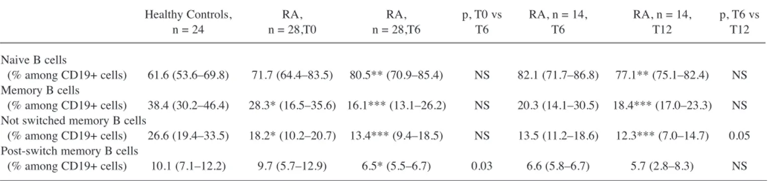
Table 4. Variations of B cell subset proportions after therapy with abatacept (ABA), and comparison with healthy controls. Except for p values, data are
expressed as median (25th–75th percentile).
Healthy Controls, RA, RA, p, T0 vs RA, n = 14, RA, n = 14, p, T6 vs n = 24 n = 28,T0 n = 28,T6 T6 T6 T12 T12 Naive B cells
(% among CD19+ cells) 61.6 (53.6–69.8) 71.7 (64.4–83.5) 80.5** (70.9–85.4) NS 82.1 (71.7–86.8) 77.1** (75.1–82.4) NS Memory B cells
(% among CD19+ cells) 38.4 (30.2–46.4) 28.3* (16.5–35.6) 16.1*** (13.1–26.2) NS 20.3 (14.1–30.5) 18.4*** (17.0–23.3) NS Not switched memory B cells
(% among CD19+ cells) 26.6 (19.4–33.5) 18.2* (10.2–20.7) 13.4*** (9.4–18.5) NS 13.5 (11.2–18.6) 12.3*** (7.0–14.7) 0.05 Post-switch memory B cells
(% among CD19+ cells) 10.1 (7.1–12.2) 9.7 (5.7–12.9) 6.5* (5.5–6.7) 0.03 6.6 (5.8–6.7) 5.7 (2.8–8.3) NS * p < 0.05 of controls; ** p < 0.01 of controls; *** p < 0.001 of controls. NS: not significant; RA: rheumatoid arthritis; T0: first administration of ABA; T6: after 6 months of ABA treatment; T12: after 12 months of ABA treatment.
Table 5. Comparisons of variations of Ig, FLC, ACPA, and RF in patients with and without disease remission after 6 months of abatacept therapy. Data are
expressed as the median (25th–75th percentile).
Remission, n = 16 Not Remission, n = 14
T0 T6 p T0 T6 p Serum IgA, mg/dl 322 (248–363) 242 (182–343) 0.016 463 (360–694) 353 (269–474) 0.007 Serum IgM, mg/dl 175 (149–210) 163 (112–185) NS 189 (108–195) 127 (74–152) 0.013 Serum IgG, mg/dl 1295 (1014–1370) 1075 (885–1112) 0.006 1340 (1140–1390) 1070 (929–1230) 0.009 κchain, mg/l 23.0 (16.4–28.9) 17.3 (13.6–20.3) 0.019 35.5 (19.1–36.1) 20.3 (14.7–26.2) NS λchain, mg/l 14.7 (12.1–17.0) 11.7 (10.7–14.5) 0.016 18.9 (15.4–27.8) 14.1 (12.9–23.1) NS IgA ACPA, IU/ml 7.5 (5.2–80.9) 7.4 (3.1–39.5) 0.011 203.2 (13.3–378.4) 159.8 (10–268.4) NS IgG ACPA, IU/ml 242 (174–425) 140 (83–273) 0.022 1004.8 (31.4–1884.8) 606.4 (18–1272) NS IgA RF, IU/ml 15.3 (10.4–41.5) 10.2 (3–23.4) 0.014 35.4 (12.6– > 100) 23.4 (5–> 100) NS IgM RF, IU/ml 101 (34.3–129.4) 40.5 (4.5–97.7) 0.008 31 (4.3–455) 4.15 (1.0–289) NS IgG RF, IU/ml 7.3 (3.75–10.8) 3 (2.1–9.9) NS 4.2 (1.6–12.8) 1.5 (0.2–15.4) NS NS: not significant; T0: first administration of ABA; T6: after 6 months of ABA treatment; ACPA: anticitrullinated protein antibodies; RF: rheumatoid factor; FLC: free light chains; Ig: immunoglobulin.
established RA
6,7. Indeed, depletion of this B cell
population can be induced also by therapy with RTX and
was associated with good clinical response
27. Taken
together, all these data suggest that the blockade of
costim-ulation can reduce B cell ability to differentiate into
post-switch memory B cells and produce autoantibodies.
Accordingly with our hypothesis, data available from
immunohistological analysis of synovial tissue from
patients with RA treated with ABA also provide evidence
for a modest but significant reduction observed in mature B
cells
28,29.
The effect of ABA therapy on the B cell compartment of
patients with RA shown in our results was not previously
described, but was observed in patients receiving other
drugs. This kind of observation holds true for many effects
of the biological and nonbiological therapies on other cell
targets, and has led to the hypothesis of a possible common
final pathogenic pathway leading to RA, which may be
targeted by various and differently acting therapeutic
agents
30. Nevertheless, the specific effect of ABA on the
costimulation blockade provides a clear possible mechanism
of action accounting for the here-described effects of the
drug on B cells
9. In murine models, ABA administration
blocked antigen-specific T cells in the lymph nodes to
acquire a phenotype associated with migration to B cell
follicles. This led to reduced specific antibody responses,
despite normal B cell clonal expansion
31. In accordance
with the hypothesis of a direct effect of ABA in lymphoid
organs, data from a human RA synovium/severe combined
immunodeficiency mouse model suggested that ABA does
not act directly on synovial T cells, but more likely prevents
T cell activation at a systemic level of the immune system
32.
The data here presented, and the results of previous studies
on T cells
33,34, are in agreement with a model in which ABA
plays a “central effect,” modulating T and B cell
differenti-ation after antigenic presentdifferenti-ation and their trafficking (also
in human settings), thereby modifying the pathophysiology
of RA.
The effects of ABA on B cells may have clinical
implica-tions, in particular for the response to vaccination and
longterm humoral memory. In fact, it has been shown that
ABA (in combination with methotrexate) significantly
reduced the humoral response to the 2009 pandemic
influenza A/H1N1 vaccine in patients with RA compared to
patients with RA treated with methotrexate only
35. Because
others have observed similar findings
36, even though they
need to be confirmed, clinicians may consider vaccinating
patients against pathogens before starting ABA therapy.
Despite these data, which suggest a possible impairment of
the adaptive immune response, longterm safety of therapy
with ABA in the clinical setting is confirmed by reassuring
results showing no unexpected events and low incidence
rates of serious infections and malignancies
37.
These data provide new insight on the effects of CD28
costimulation blockade in patients with RA, demonstrating
a reduction of polyclonal B cell activation.
REFERENCES
1. McInnes IB, Schett G. The pathogenesis of rheumatoid arthritis. N Engl J Med 2011;365:2205-19.
2. Nakken B, Munthe LA, Konttinen YT, Sandberg AK, Szekanecz Z, Alex P, et al. B-cells and their targeting in rheumatoid arthritis — current concepts and future perspectives. Autoimmun Rev 2011;11:28-34.
3. Gottenberg JE, Aucouturier F, Goetz J, Sordet C, Jahn I, Busson M, et al. Serum immunoglobulin free light chain assessment in rheumatoid arthritis and primary Sjogren’s syndrome. Ann Rheum Dis 2007;66:23-7.
4. Klein U, Rajewsky K, Kuppers R. Human immunoglobulin (Ig)M+IgD+ peripheral blood B cells expressing the CD27 cell surface antigen carry somatically mutated variable region genes: CD27 as a general marker for somatically mutated (memory) B cells. J Exp Med 1998;188:1679-89.
5. Wei C, Anolik J, Cappione A, Zheng B, Pugh-Bernard A, Brooks J, et al. A new population of cells lacking expression of CD27 represents a notable component of the B cell memory compartment in systemic lupus erythematosus. J Immunol 2007;178:6624-33. 6. Souto-Carneiro MM, Mahadevan V, Takada K, Fritsch-Stork R,
Nanki T, Brown M, et al. Alterations in peripheral blood memory B cells in patients with active rheumatoid arthritis are dependent on the action of tumour necrosis factor. Arthritis Res Ther
2009;11:R84.
7. Michelutti A, Gremese E, Morassi F, Petricca L, Arena V, Tolusso B, et al. B-cell subsets in the joint compartments of seropositive and seronegative rheumatoid arthritis (RA) and no-RA arthritides express memory markers and ZAP70 and characterize the aggregate pattern irrespectively of the autoantibody status. Mol Med 2011;17:901-9.
8. Maxwell LJ, Singh JA. Abatacept for rheumatoid arthritis: a Cochrane systematic review. J Rheumatol 2010;37:234-45. 9. Cutolo M, Nadler SG. Advances in CTLA-4-Ig-mediated
modulation of inflammatory cell and immune response activation in rheumatoid arthritis. Autoimmun Rev 2013;12:758-67.
10. van Kooten C, Banchereau J. CD40-CD40 ligand. J Leuk Biol 2000;67:2-17.
11. Wells G, Becker J-C, Teng J, Dougados M, Schiff M, Smolen J, et al. Validation of the 28-joint Disease Activity Score (DAS28) and European League Against Rheumatism response criteria based on C-reactive protein against disease progression in patients with rheumatoid arthritis, and comparison with the DAS28 based on erythrocyte sedimentation rate. Ann Rheum Dis 2009;68:954-60. 12. Dati F, Schumann G, Thomas L, Aguzzi F, Baudner S, Bienvenu J,
et al. Consensus of a group of professional societies and diagnostic companies on guidelines for interim reference ranges for 14 proteins in serum based on the standardization against the IFCC/BCR/CAP reference material (CRM 470). Eur J Clin Chem Clin Biochem 1996;34:517-20.
13. Katzmann JA, Clark RJ, Abraham RS, Bryant S, Lymp JF, Bradwell AR, et al. Serum reference intervals and diagnostic ranges for free kappa and free lambda immunoglobulin light chains: relative sensitivity for detection of monoclonal light chains. Clin Chem 2002;48:1437-44.
14. Moura RA, Weinmann P, Pereira PA, Caetano-Lopes J, Canhão H, Sousa E, et al. Alterations on peripheral blood B-cell
subpopulations in very early arthritis patients. Rheumatology 2010;49:1082-92.
15. Kormelink TG, Tekstra J, Thurlings RM, Boumans MH, Vos K, Tak PP, et al. Decrease in immunoglobulin free light chains in patients
with rheumatoid arthritis upon rituximab (anti-CD20) treatment correlates with decrease in disease activity. Ann Rheum Dis 2010;69:2137-44.
16. Gottenberg JE, Miceli-Richard C, Ducot B, Goupille P, Combe B, Mariette X. Markers of B-lymphocyte activation are elevated in patients with early rheumatoid arthritis and correlated with disease activity in the ESPOIR cohort. Arthritis Res Ther 2009;11:R114. 17. Sellam J, Hendel-Chavez H, Rouanet S, Abbed K, Combe B, Le
Loët X, et al. B Cell activation biomarkers as predictive factors for the response to rituximab in rheumatoid arthritis. Arthritis Rheum 2011;63:933–8.
18. Gottenberg JE, Rayaud P, Cantagrel A, Combe B, Flipo RM, Schaeverbeke T, et al. Positivity for anti-cyclic citrullinated peptide is associated with a better response to abatacept: data from the ‘Orencia and Rheumatoid Arthritis’ registry. Ann Rheum Dis 2012;71:1815-9.
19. Bohler C, Radner H, Smolen JS, Aletaha D. Serological changes in the course of traditional and biological disease modifying therapy of rheumatoid arthritis. Ann Rheum Dis 2013;72:241-4.
20. Alessandri C, Bombardieri M, Del Papa N, Cinquini M, Magrini L, Tincani A, et al. Decrease of anti-cyclic citrullinated peptide antibodies and rheumatoid factor following anti-TNFa therapy (infliximab) in rheumatoid arthritis is associated with clinical improvement. Ann Rheum Dis 2004;63:1218–21.
21. Bobbio-Pallavicini F, Caporali R, Alpini C, Avalle S, Epis OM, Klersy C, et al. High IgA rheumatoid factor levels are associated with poor clinical response to tumour necrosis factor a inhibitors in rheumatoid arthritis. Ann Rheum Dis 2007;66:302–7.
22. Atzeni F, Sarzi-Puttini P, Dell’Acqua D, de Portu S, Cecchini G, Cruini C, et al. Adalimumab clinical efficacy is associated with rheumatoid factor and anti-cyclic citrullinated peptide antibody titer reduction: a one-year prospective study. Arthritis Res Ther 2006;8:R3.
23. Chen HA, Lin KC, Chen CH, Liao HT, Wang HP, Chang HN, et al. The effect of etanercept on anti-cyclic citrullinated peptide antibodies and rheumatoid factor in patients with rheumatoid arthritis. Ann Rheum Dis 2006;65:35-9.
24. De Rycke L, Verhelst X, Kruithof E, Van den Bosch F, Hoffman IE, Veys EM, et al. Rheumatoid factor, but not anti-cyclic citrullinated peptide antibodies, is modulated by infliximab treatment in rheumatoid arthritis. Ann Rheum Dis 2005;64:299-302.
25. Cambridge G, Leandro MJ, Edwards JC, Ehrenstein MR, Salden M, Bodman-Smith M, et al. Serologic changes following B
lymphocyte depletion therapy for rheumatoid arthritis. Arthritis Rheum 2003;48:2146-54.
26. Thurlings RM, Vos K, Wijbrandts CA, Zwinderman AH, Gerlag DM, Tak PP. Synovial tissue response to rituximab: mechanism of action and identification of biomarkers of response. Ann Rheum Dis 2008;67:917-25.
27. Moller B, Aeberli D, Eggli S, Fuhrer M, Vajtai I, Vögelin E, et al. Class-switched B cells display response to therapeutic B-cell depletion in rheumatoid arthritis. Arthritis Res Ther 2009;11:R62. 28. Buch MH, Boyle DL, Rosengren S, Saleem B, Reece RJ, Rhodes
LA, et al. Mode of action of abatacept in rheumatoid arthritis patients having failed tumour necrosis factor blockade: a histological, gene expression and dynamic magnetic resonance imaging pilot study. Ann Rheum Dis 2009;68:1220-7.
29. Kanbe K, Chiba J, Nakamura A. Immunohistological analysis of synovium treated with abatacept in rheumatoid arthritis. Rheumatol Int 2013;33:1883-7.
30. Smolen JS, Aletaha D. Forget personalized medicine and focus on abating disease activity. Ann Rheum Dis 2013;72:3-6.
31. Platt AM, Gibson VB, Patakas A, Benson RA, Nadler SG, Brewer JM, et al. Abatacept limits breach of self-tolerance in a murine model of arthritis via effects on the generation of T follicular helper cells. J Immunol 2010;185:1558–67.
32. Koenders MI, Marijnissen RJ, Joosten LA, Abdollahi-Roodsaz S, Di Padova FE, van de Loo FA, et al. T cell lessons from the rheumatoid arthritis synovium SCID mouse model: CD3-rich synovium lacks response to CTLA-4Ig but is successfully treated by interleukin-17 neutralization. Arthritis Rheum 2012;64:1762-70. 33. Scarsi M, Ziglioli T, Airò P. Decreased circulating CD28-negative T
cells in patients with rheumatoid arthritis treated with abatacept are correlated with clinical response. J Rheumatol 2010;37:911-6. 34. Airò P, Scarsi M. Targeting CD4+CD28- T cells by blocking CD28
co-stimulation. Trends Mol Med 2013;19:1-2.
35. Ribeiro AC, Laurindo IM, Guedes LK, Saad CG, Moraes JC, Silva CA, et al. Abatacept and reduced immune response to pandemic 2009 influenza A/H1N1 vaccination in patients with rheumatoid arthritis. Arthritis Care Res 2013;65:476-80.
36. Adler S, Krivine A, Weix J, Rozenberg F, Launay O, Huesler J, et al. Protective effect of A/H1N1 vaccination in immune-mediated disease—a prospectively controlled vaccination study.
Rheumatology 2012;51:695-700.
37. Weinblatt ME, Moreland LW, Westhovens R, Cohen RB, Kelly SM, Khan N, et al. Safety of abatacept administered intravenously in treatment of rheumatoid arthritis: integrated analyses of up to 8 years of treatment from the abatacept clinical trial program. J Rheumatol 2013;40:787-97.