UNIVERSITÁ DEGLI STUDI ROMA TRE
Facoltà di Scienze Matematiche, Fisiche e Naturali Dipartimento di Biologia
Scuola Dottorale in Biologia
Sezione di Biologia Applicata alla Salute
dell’Uomo (BASU) – XXIII ciclo
THE ENDOCRINE DISRUPTORS: EFFECTS AND
ACTION MECHANISMS ON ESTROGEN-INDUCED
CELL FUNCTIONS
GLI INTERFERENTI ENDOCRINI: EFFETTI E
MECCANISMI D’AZIONE NELLE FUNZIONI
CELLULARI ESTROGENO-DIPENDENTI
Ph.D. Student: Dr. Pamela Bulzomi A.A. 2010/2011
Supervisor: Prof. Maria Marino
To my family, for its never ending support and patience
To Alessandro, For standing by me, always encouraging me “through thick and thin fate” To Maria, For patiently and constantly guiding me, conveying her passion for research to me
INDEX
SUMMARY………...I RIASSUNTO...IV
1.BACKGROUND………...1
1.1 Endocrine disruptors……….……….….……..1
1.1.1 Flavonoids, a particular class of EDs ……..………..4
1.1.1.1 Flavonoid bioavalability……….7
1.2Estrogen…..………10
1.2.1 Estrogen Receptors………..10
1.2.2 ER distribution………….………...……….12
1.2.3ER mechanisms of action………..12
1.3 Flavonoid –dependent modulation of ER activities..…………18
2.AIM………...21
3. FLAVONOID EFFECTS ON E2-INDUCED REGULATION OF CELL PROLIFERATION.……..……….22
3.1 Introduction………..22
3.2 Results………...………....24
3.2.1 Quercetin effect on HeLa cell growth……….24
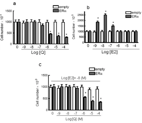
3.2.1.1Quercetin decreases cell number of ERα-transfected HeLa cells………..24
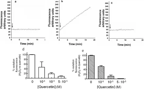
3.2.1.2 Quercetin as pro-oxidant/antioxidant……….25
3.2.1.3 Effect of quercetin as kinase inhibitor……….25
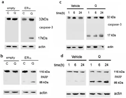
3.2.1.4 Quercetin as pro-apoptotic agent………....…….27
3.2.1.5 Quercetin decreases ERβ-transfected HeLa cell number...30
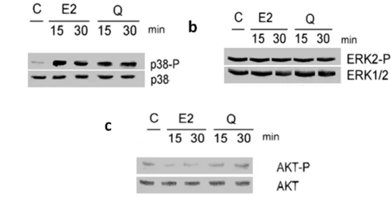
3.2.1.6 Quercetin effect on kinase activation in ERβ-transfected HeLa cells………..31
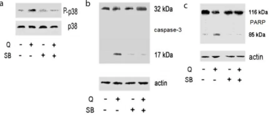
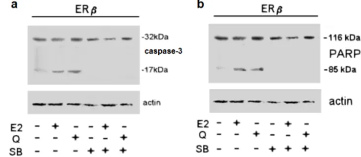
3.2.1.7 Quercetin:ERβ complex induced pro-apoptotic cascade activation via p38 pathway………....33
3.2.1.8 Quercetin as modulator of ERα and ERβ transcriptional activity……….34
3.2.2.Effect of Naringenin and E2 coadministration…………..…36
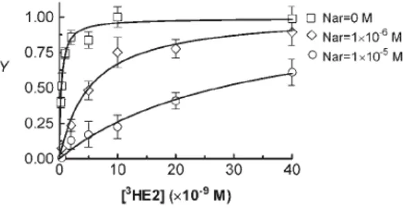
3.2.2.1 E2 and Nar Binding to ERα………...……….….33
3.2.2.2 ERα Transcriptional Activities………..…..37
3.2.2.3 ERα-Dependent Rapid Signals………38
3.2.2.4 ERα-Dependent E2-Induced Cell Proliferation………...41
4. ANTIPROLIFERATIVE FLAVONOID EFFECTS IN MIXTURE
WITH OTHER EDs…………...49
4.1 Introduction………..………49
4.2 Results………51
4.2.1 BPA and Nar binding to ERα………..………51
4.2.2 BPA and Nar mixture effect on breast cancer cells survival and proliferation………..51
4.2.3 BPA and Nar mixture effect on ERα-activated extranuclear signals.54 4.2.4 BPA binding to ERβ………...57
4.2.5 Effects of E2 and BPA on DLD-1 cell survival and propoapototic cascade activation……….58
………..88
4.2.6 BPA effect on ERβ genomic and extra-nuclear activities…………...60
4.2.7 BPA effect on ERβ molecular interactions with caveolin-1 and p38.60 4.2.8 Effect of BPA and Nar mixture on DLD-1cancer cells growth……..62
4.2.9 Effect of BPA and Nar mixture on ERβ levels………...…....65
4.3 Discussion………..66
5. FLAVONOID EFFECT ON E2 INDUCED SKELETAL MUSCLE PROTECTION………...………...69
5.1 Introduction………..69
5.2 Results………72
5.2.1 ER expression in differentiation-induced L6 cells………..72
5.2.2 Naringenin effect on L6 cell differentiation ………..………...73
5.2.3 Nar specifically impairs E2- induced differentiation in L6 cells……73
5.2.4 Narigenin effect on L6 cell growth ………78
5.2.5 Nar effect in C2C12 cells………...……….79
5.2.6 Nar effect on ROS production in L6 myoblasts……….82
5.3 Discussion………..84
6. CONCLUSION………...87
7. REFERENCE……….91
ACKNOWLEDGEMENTS ... 116 APPENDIX... Material and Methods available on CD-ROM
I
SUMMARY
Besides E2, ERs bind a wide variety of compounds with remarkable structural and chemical diversity (Ascenzi et al., 2006) collected into the class of Endocrine Disruptor (EDs). EDs are defined as “exogenous substances that cause adverse health effects in an intact organism or in its progeny, consequent to changes in endocrine function”. Even if present in minute amounts (part per trillion) EDs could interfere with the synthesis, secretion, transport, metabolism, binding, action, or elimination of natural hormones responsible for homeostasis maintenance, reproduction, and developmental processes (Colborn etal.,1993). Animals, including humans, are especially sensitive to EDs at the early stages of development but some effects exerted at these stages may be expressed only in the adult life or even in subsequent generations (Rhind, 2009). Among EDs, several synthetic chemicals have been described to induce several degenerative disease. For example Bisphenol A (BPA) has been demonstrated to promote the development of endometriosis (Signorile et al., 2010) and of various cancer type (e.g. breast, endometrial and prostate cancer) because of its estrogen mimetic activity (Bolli et al., 2008; Ricupito et al., 2009). On the contrary, natural compounds such as flavonoids show a protective effect against various degenerative phenomena (i.e. cardiovascular disease, osteoporosis, several cancer type) (Cassidy et al., 2000; Dang and Lowik, 2005; Keinan- Boker et al., 2004). To date, it’s not clear if the discrepant effects between synthetic and natural compounds depend on ED interaction with ERs, or on other mechanisms whose occur independently from ED binding to ERs. Furthermore, since the most of the studies on EDs, particularly flavonoids beneficial effects, were conducted in the absence of E2, the physiological relevance of these findings is not clear. Moreover, since human beings intake daily about 500 g of different chemicals, which exhibit endocrine effects in vivo and in vitro (Marino and Galluzzo, 2006), mammalians are exposed to several EDs, resulting in systemic circulation of ED and flavonoid mixtures in the body. Although in the last years, some evidence has become available to show the combined effects of EDs (Kortenkamp et al., 2008), the research on the effects of “dietary” and synthetic mixtures, at relevant human levels, remain inconclusive.
Aim of this project is to assess the effects of natural chemicals (i.e. flavonoids) alone or in mixture with the endogenous hormone (E2) or with other EDs (i.e. BPA) on E2-dependent cell functions (i.e. cell proliferation/apoptosis balance, cellular differentiation and oxidative stress)
II
evaluating their effects, action mechanisms, and the putative involvement of ERα and ERβ.
Our data demonstrated that the well known growth inhibition and cell death effects of the flavonol quercetin, one of the most frequently studied and ubiquitous bioactive flavonoid, are not related to its high-concentration requiring (5×10⁻⁵M) antioxidant activity but to quercetin ability to activate,
in a very small amount (i.e., 10-6M), a pro-apoptotic cascade mainly
modulating both ER activities. Particularly quercetin is able to bind both ERs, and in turn to act as an E2-mimetic, leading cancer cell to apoptotic death, in tissue expressing ERβ, such as colon (Galluzzo et al., 2007), or to antagonize E2 proliferative effect in tissue expressing ERα, such as breast cancer (Bulzomi et al., 2010). Quercetin underlying action mechanism requires ER activity modulation: in the ERβ presence quercetin activates the same E2 pathways (both genomic and extranuclear signal), whereas in presence of ERα, quercetin only allows ERα direct transcriptional activity, impairing ERα-mediated rapid signals important for ERα-induced cell proliferation. Thus, at nutritionally relevant concentration, quercetin antiproliferative activities depends on ER activity modulation, rather than its antioxidant activity. Our data also demonstrate that flavonoids, such as the flavanone Naringenin, preserve their ability to induce apoptosis in ERα-expressing cancer cells also in the presence of E2. Nar is one of the best absorbed flavonoids in the human gastrointestinal tract, and the peak of plasma aglycone Naringenin ranges from 0.7 to 14.8×10⁻⁶ M (Erlund et al.,
2001; Bugianesi et al., 2002; Manach et al., 2004). According to the reported plasma concentration the obtained data demonstrate that 10-6 M
Nar, although didn’t affect E2-induced direct transcriptional activity of ERα, reverts the proliferative effect of E2 impairing ERα-mediated rapid signals and inducing different proapoptotic signal transduction pathways in ERα-expressing cancer cells. As a whole, the assays with nutritionally relevant concentration of Nar, against a background of physiological level of E2 allowed us to elucidate Naringenin disrupting action mechanism giving a physiological meaning to the antiproliferative activity of this compound. The importance of flavonoid chemoprotective properties is strongly supported also by the data obtained on flavonoid and man-made ED mixtures, whose indicate that the small amount of Nar recovered in human plasma (Manach et al., 2004) is sufficient to counteract BPA cancer promoting effect. Our data indicate that BPA is, like Nar, a double sided action mechanism compound, which promotes tumor incidence in breast and other target organs that predominantly express ERα but inhibits the E2
III
protective effects in the ERβ-expressing colon cell. These two divergent aspects could act synergistically by increasing the E2-disrupting potential of this widespread environmental polluter. Our data, demonstrating Nar ability to revert BPA estrogenic activity in ERα expressing cancer cells and to preserve its antiproliferative activity in ERβ-expressing cancer cells, strongly support the theory of the cell fate as the resulting balance of the ED-activated pathway, highlighting the importance of investigating the chemopreventive effect of flavonoids. Since E2 effects go beyond cell proliferation, and ERα and ERβ are coexpressed in several tissue, we assessed the impact of the Nar on E2 protective effect in non cancerous cells when both receptor isoforms are present. The data obtained on E2 and Nar mixture in skeletal muscle myoblasts, expressing both ERs, allow us to affirm that ERα and ERβ mediate different E2 effect in skeletal muscle. ERα-activated rapid signals are essential for E2-induced skeletal muscle differentiation, while ERβ-activated pathways are the only involved in the E2-protective effect from ROS-induced oxidative stress. Nar ability to specifically affect only E2-induced differentiation raise the existence of a gender-related susceptibilities to flavonoids in the different physiological stages of life.
As a whole, these data enlarge our knowledge of the mechanisms underlying the (anti)estrogenicity of dietary compounds pointing to rapid mechanisms as the most susceptible target of endocrine disruptors. In fact nor Naringenin or Quercetin modify direct ERE-containing promoter transcription, but decoupling ERα from rapid signals, drive cells to different destiny, highlighting the importance of investigating flavonoid effect in different stages of life that could be characterized by a different level and role of this receptor isoform.
Since E2 effects depend on the balance of the relative expression of ER isoform and on the balance of the signals originated by each isoform, ED actions are more complex than originally considered, since different ligands induce ERs to assume different conformations responsible for specific signaling pathway activation.
IV
RIASSUNTO
Oltre ad E2, i recettori degli estrogeni (ER) legano una vasta gamma di molecole, con spiccate differenze chimiche e strutturali (Ascenzi et al., 2006), raggruppate nella classe degli Interferenti Endocrini (IE). IE sono definiti come “sostanze esogene che causano effetti avversi alla salute negli interi organismi o nella progenie, in seguito ad alterazioni delle funzioni endocrine”. Sebbene presenti in parti per trilione, IE possono interferire con la sintesi, la secrezione, il trasporto, il metabolismo, il legame, l’azione o l’eliminazione degli ormoni responsabili del mantenimento dell’omeostasi, della riproduzione e dei processi di sviluppo (Colborn et al.,1993). Gli animali sono particolarmente sensibili agli IE nelle prime fasi dello sviluppo, sebbene alcuni effetti possono manifestarsi nella vita adulta o nelle generazioni successive (Rhind, 2009). Tra gli IE, diverse molecole di origine sintetica sono state dimostrati essere in grado di indurre l’insorgenza di diverse patologie. Il Bisfenolo A (BPA), ad esempio, è stato dimostrato promuovere l’insorgenza dell’endometriosi (Signorile et al., 2010) e di diversi tipi di cancro (e.g. cancro al seno, alla prostata e all’endometrio) a causa della sua attività estrogeno mimetica (Bolli et al., 2008; Ricupito et al., 2009). D’altro canto, composti di origine naturale, quali i flavonoidi, hanno mostrato avere un effetto protettivo contro diversi fenomeni degenerativi (i.e. patologie cardiovascolari, osteoporosi, cancro) (Cassidy et al., 2000; Dang and Lowik, 2005; Keinan- Boker et al., 2004). Ad oggi, non è chiaro se le discrepanze tra gli effetti descritti per i composti di origine sintetica e naturale dipendano dall’interazione degli IE con gli ER, o da meccanismi indipendenti da questo. Inoltre, poiché la maggior parte degli studi sugli IE, in particolare sugli effetti benefici dei flavonoidi, sono stati condotti in assenza di E2, il significato fisiologico di queste scoperte non è chiaro. L’uomo assume quotidianamente circa 500 g di diversi composti dimostrati avere attività ormone-simile sia in vitro che in vivo (Marino e Galluzzo, 2006), risultando esposto a più IE contemporaneamente e conseguentemente a miscele di composti naturali e sintetici. Sebbene siano stati recentemente evidenziati alcuni degli effetti combinati di diversi IE, gli effetti di miscele costituite da molecole di origine alimentare e sintetica sono tuttora ignoti. Scopo di questo progetto è quello di valutare gli effetti di composti naturali (i.e. flavonoidi), da soli o in miscela con l’ormone endogeno (E2) o con altri IE (BPA), sulle funzioni cellulari regolate da E2 (i.e. bilancio tra proliferazione e morte cellulare,
V
differenziamento e stress ossidativo) evidenziando gli effetti, i meccanismi d’azione, e l’eventuale coinvolgimento di ERα e ERβ.
I nostri dati dimostrano che il noto effetto di inibizione della crescita e induzione dell’apoptosi del flavonolo quercetina, uno dei flavonoidi bioattivi più comune e più studiato, non è dovuto alla sua attività antiossidante, che si estrinseca ad elevate concentrazioni (5×10⁻⁵M), ma alla
capacità della quercetina di attivare a basse concentrazioni (i.e., 10-6M) una
cascata apoptotica modulando l’attività di entrambi gli ER. In particolare, la quercetina è in grado di legare entrambi gli ER e di agire come un estrogeno-mimetico, portando le cellule ad apoptosi, nei tessuti che esprimono ERβ, quale il colon (Galluzzo et al., 2007), o di antagonizzare l’effetto proliferativo di E2 nei tessuti esprimenti ERα, quale il seno (Bulzomi et al., 2010). Il meccanismo di azione della quercetina prevede la modulazione delle attività degli ER: in presenza di ERβ, la quercetina attiva le stesse vie di segnale di E2 (sia genomiche che extranucleari), mentre in presenza di ERα, la quercetina preserva la sola capacità trascrizionale di ERα, bloccando i segnali rapidi attivati da questo recettore, importanti per la proliferazione cellulare._Ne consegue che a concentrazioni nutrizionalmente rilevanti, l’effetto antiproliferativo della quercetina dipende dalla modulazione delle attività degli ER piuttosto che dalle sue proprietà antiossidanti. I nostri dati hanno inoltre dimostrato che i flavonoidi, quale il flavanone Naringenina, mantengono la capacità di attivare l’apoptosi in cellule di cancro esprimenti ERα anche in presenza di E2. La Naringenina è uno dei flavonoidi meglio assorbiti nel tratto gastrointestinale umano, e il picco plasmatico di Naringenina aglicone va da 0.7 a 14.8 × 10⁻⁶ M (Erlund et al., 2001; Bugianesi et al., 2002; Manach et
al., 2004). In accordo con le concentrazioni plasmatiche riportate, i dati ottenuti in cellule esprimenti ERα mostrano che 10-6 M Nar, sebbene non
influenzi l’attività trascrizionale diretta di ERα, reverte l’effetto proliferativo di E2 bloccando l’attivazione dei segnali rapidi mediati da questo recettore e attivando vie di segnale coinvolte nell’apoptosi. Nel complesso, gli esperimenti condotti in presenza di concentrazioni fisiologiche di E2 e concentrazioni nutrizionalmente rilevanti di Nar ci hanno permesso di elucidare il meccanismo di interferenza della Nar dando un significato fisiologico alla proprietà antiproliferativa di questo flavonoide. L’importanza delle proprietà chemoprotettive dei flavonoidi sono supportate dai dati ottenuti utilizzando miscele di flavonoidi e IE di origine sintetica, i quali indicano come le piccole quantità di Nar ritrovate nel plasma (Manach et al., 2004) siano sufficienti per contrastare l’effetto
VI
cancerogenico del BPA. I nostri dati infatti mostrano come il BPA sia, al pari di Nar, un composto dalla duplice azione, in grado di promuovere l’insorgenza di cancro al seno e in altri organi bersaglio che esprimono prevalentemente ERα e di bloccare l’effetto protettivo di E2 nei tessuti esprimenti ERβ, quali il colon. Questi aspetti divergenti possono agire in maniera sinergica incrementando il potenziale effetto di interferente di questo contaminante ambientale. I nostri dati, dimostrando che Nar reverte l’attività estrogenica del BPA nelle cellule esperimenti ERα ma che mantiene la sua attività antiproliferativa nelle cellule esprimenti ERβ anche in presenza di BPA, supportano fortemente la teoria del destino cellulare come risultato del bilanciamento delle vie di segnale attivate dai diversi IE, supportando l’importanza degli studi sugli effetti antiproliferativi dei flavonoidi. Poiché l’effetto di E2 va ben oltre la regolazione della proliferazione cellulare, e gli ER sono co-espressi in diversi tessuti, abbiamo valutato l’impatto della Nar in cellule non cancerose esperimenti entrambe le isoforme di ER. I dati ottenuti stimolando mioblasti di ratto, esperimenti entrambi gli ER, ci ha permesso di individuare distinti ruoli per ERα e ERβ nel muscolo scheletrico. Mentre le vie di segnale rapide attivate da ERα sono fondamentali per l’induzione del differenziamento mediato da E2, i segnali rapidi attivati da ERβ sono gli unici coinvolti nell’effetto protettivo di E2 dallo stress ossidativo indotto dai ROS. La capacità della Nar di interferire specificatamente esclusivamente con il differenziamento indotto da E2 pone l’attenzione sulla possibile esistenza di una suscettibilità correlata al genere ai flavonoidi nei diversi stati fisiologici della vita. Nel complesso, i dati ottenuti ampliano la nostra conoscenza sui meccanismi alla base dell’ (anti)estrogenicità dei composti di origine alimentare evidenziando come i meccanismi di azione rapidi siano i bersagli più suscettibili agli interferenti endocrini. Infatti né la Naringenina né la quercetina modificano la trascrizione diretta di promotori contenenti la sequenza ERE, ma, disaccoppiando le attività rapide di ERα da quella trascrizionale diretta, porta le cellule verso destini diversi. Ciò evidenzia l’importanza di studiare gli effetti dei flavonoidi nei diversi stati della vita i quali possono essere caratterizzati da differenti livelli proteici e distinti ruoli di questa isoforma di recettore. Poiché gli effetti di E2 dipendono dal rapporto tra i livelli di espressioni degli ER e dal bilanciamento dei segnali attivati da ciascuna isoforma, gli effetti degli IE risultano molto più complessi di quelli originariamente considerati, dal momento che diversi leganti inducono gli ER ad assumere conformazioni diverse, responsabili dell’attivazione di specifiche vie di segnale.
1
1. BACKGROUND
1.1 Endocrine disruptors
Global concerns have been raised in recent years over the potential adverse effects that may result from exposure to chemicals that have the potential to interfere with the endocrine system (WHO, 2002). Discovered in the early 1900s, when pig farmers in the USA complained of fertility problems in swine herds fed on mouldy grain (McNutt et al.,1928), and picked out in the 1940s by reports of infertility in sheep grazing on certain clovers in Western Australia (Marino and Mita, 2007) it has become evident that many chemicals present in the environment can interfere with both human and wildlife health mimicking, antagonizing or altering the physiological actions of endogenous hormones, mainly, sex steroid hormones. These substances present in the environment, are now classified as endocrine disruptors (EDs), defined as “exogenous substances that cause adverse health effects in an intact organism or in its progeny, consequent to changes in endocrine function” (European commission DGXII, 1996). Although present in minute amounts (part per trillion), EDs could interfere with the synthesis, secretion, transport, metabolism, binding, action, or elimination of natural hormones responsible for homeostasis maintenance, reproduction, and developmental processes (Diamanti-Kandarakis et al., 2009).
Currently more than 100 chemicals have been identified as EDs. About half of these compounds are substituted with halogen groups, mostly chlorine and bromine, and within this heterogeneous group of molecules we find: (a) synthetic chemicals used in industry, agriculture, and consumer products (e.g. polychlorinated biphenyls (PCBs), polybrominated biphenyls (PBBs), dioxins, plasticizers as phthalates bisphenol A (BPA), pesticides as methoxychlor, chlorpyrifos, dichlorodiphenyltrichloroethane (DDT) and fungicides, like vinclozolin (b) synthetic chemicals used as pharmaceutical drugs (e.g. diethylstilbestrol, DES), and (c) natural chemicals found in human and animal food (e.g. flavonoids) (Marino and Mita, 2007, Diamanti-Kandarakis et al., 2009). EDs have long environmental half-life resulting in a continue increase of their global concentration in the environment and can be detected and may concentrate at great distances from where they are produced, used or released (Mita and Marino, 2007). As a consequence, some EDs are detectable in so-called “pristine” environments at remote distances from the site they were produced, used, or released due to water and air currents and via migratory animals that spend part of their life in a contaminated area, to become incorporated into the
food chain in an otherwise uncontaminated region. As these substances do not decay easily, they may not be metabolized, or they may be metabolized or broken down into more toxic compounds than the parent molecule; even substances that were banned decades ago remain in high levels in the environment, and they can be detected as part of the body burden of virtually every tested individual animal or human (Porte et al., 2006; Calafat and Needham, 2007). On the contrary, other EDs may not be as persistent but are so widespread in their use that there is prevalent human exposure (Diamanti-Kandarakis et al., 2009). The sources of exposure to EDs are diverse and vary widely around the world. Humans and animals can be exposed involuntarily to EDs by drinking contaminated polluted water, breathing contaminated air, ingesting food, or, contacting contaminated soil or even in the workplace (Diamanti-Kandarakis et al., 2009). Exposure can be the result of uptake through skin, gills or lungs, through food and drink, and through maternal blood (eg. foetus) or milk (eg. neonate) (Rhind, 2009). In general, persistent endocrine disruptors have low water solubility and extremely high lipid solubility, leading to their bioaccumulation in adipose tissue (Rhind, 2009).
Figure 1.1: Chemical structure of common EDs. Chemical structure of synthetic chemicals used in industry, agriculture, and consumer products (BPA, DDT, PCB), as pharmaceutical drugs (DES) and basic structure of natural compounds (flavonoids). DES: diethylstilbestrol, DDT: dichlorodiphenyltrichloroethane, BPA: bisphenol A , PCBs: polychlorinated biphenyls.
EDs can exert both short and long term effect in many physiological states. The precise nature of these effects will depend on the extent to which they are taken up by the organisms and then on the extent to which they are
2
3
degraded, excreted or metabolized; each of these factors in turn, will depend on species, sex, age and compound class (Rhind, 2009). Animals, including humans, are especially sensitive to EDs at the early stages of development but some effects exerted at these stages may be expressed only in the adult life or even in subsequent generations (Rhind, 2009). Since the 1930s an increase in the frequency of development abnormalities of the male reproductive tract, particularly cryptorchidism and hypospadias, as well as a decline of sperm quality, have been reported (WHO, 2002). Developmental exposure to pesticides or to PCBs produce alterations in the dopaminergic system and has been linked to neurodegenerative disorders, including Parkinson's disease (Jones and Miller, 2008). Several concerns have also been raised about the influence of EDs on the timing of puberty and on onset of several pathologies. For example Bisphenol A (BPA) has been demonstrated to promote the development of endometriosis (Signorile et al., 2010) and of various cancer type (e.g. breast, endometrial and prostate cancer) because of its estrogen mimetic activity (Bolli et al., 2008; Ricupito et al., 2009).From a physiological perspective, an endocrine-disrupting substance is a compound, either natural or synthetic, which, through environmental or during inappropriate developmental exposures, alters the hormonal and homeostatic systems that enable the organism to communicate with and to respond to its environment (Diamanti-Kandarakis et al., 2009). Several historical examples of toxic pills or contamination show a direct causal relationship between an unique chemical and the manifestation of an endocrine or reproductive dysfunction, due to the alteration of the milieu interieur (le Maire, et al., 2010). However, these types of single exposure are not representative of more common persistent exposures to a broad mix of chemicals and contaminants (le Maire, et al., 2010). The basic tenet of toxicology from Ames and Gold (2000) that ‘‘dose alone determines the poison’’ is too limited for EDs because both the timing of exposure and the dose can dictate not only the effect, but also whether the effects are adverse versus beneficial, or permanent versus transient (Hotchkiss et al., 2008). The so called “genotropic” effects of EDs appear at concentrations well below that at which they are toxic in the conventional sense (Wetherill et al., 2007). In fact, EDs may exert nontraditional dose-response curves, such as inverted-U or U-shaped curves (vom Saal, 2007) typical of hormone actions. As a consequence, EDs can induce cellular and molecular alterations of endocrine function at low dosage levels producing a cascade of effects that could be more potent effects than higher doses. This concept have been known for hormone and neurotransmitter actions, but only in the past decade it has begun to be appreciated for EDs.The properties of these
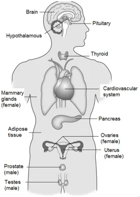
substances are particularly well suited for study by endocrinologists because they so often activate or antagonize hormone receptors. There is no endocrine system that is immune to EDs (Fig.1.2). However, because of the shared structures and properties of the chemicals and the similarities of the receptors (Thornton, 2001) and enzymes involved in the synthesis, release, and degradation of hormones, the most caught hormones are the sex steroid hormones (Diamanti-Kandarakis et al., 2009).
.
Figure 1.2: Endocrine systems targeted by EDs. Model of hormone-sensitive physiological systems vulnerable to EDs (Diamanti-Kandarakis et al., 2009).
1.1.1 Flavonoids, a particular class of EDs
A particular class of EDs is composed by natural polyphenols. The biological activity of these compounds, particularly flavonoids, drew the attention of many researchers for their ability to prevent several degenerative disease in humans. Flavonoids have a long history in science.
5
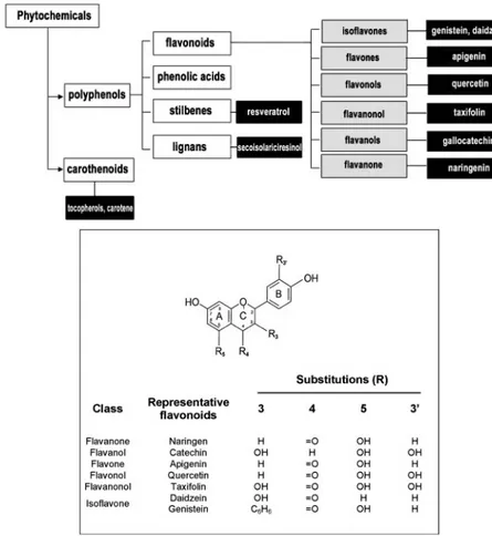
Often referred to as weak estrogens, they were chemically synthesized before the ring structure of the mammalian steroids was determined in the 1920’s and 1930’s (Barnes, 2004). They re-emerged from obscurity in the 1940’s as the anti-estrogenic principle in red clover that caused infertility in sheep in Western Australia (Bennetts et al., 1946). This adverse effect of flavonoids, caused by interfering in some way with sex hormone actions, placed these substances in the class of EDs (Jacobs and Lewis, 2002). Primarily recognized as the pigments responsible for the many shades of yellow, orange, and red in flowers (Timberlake and Henry, 1986; , Brouillard and Cheminat, 1988) and as part of plant defense mechanism against stresses of different origins (Birt et al., 2001) at the present more than 4000 flavonoids have been identified in edible plants (Timberlake and Henry, 1986; Manach et al., 2004) and are consumed regularly with the human diet (Timberlake and Henry, 1986).These low molecular weight substances are phenylbenzo-pyrones (phenylchromones), possessing an assortment of structures based on a common three-ring nucleus (Middleton et al., 2000) in which primary substituents (eg, hydroxyl, methoxyl, or glycosyl groups) can be further substituted (e.g., additionally glycosylated or acylated) sometimes yielding highly complex structures (Cheynier, 2005) (Fig. 1.3). Flavonoids, have been categorized into 6 subclasses as a function of the type of heterocycle involved: flavonols, flavones, flavanols, flavanonols, flavanones, and isoflavones (Fig 1.3) (Birt et al., 2001; Manach et al., 2004). The role played by flavonoids as EDs have more recently been confirmed in vivo. Numerous effects in both male and female rats exposed to genistein from gestational day 7 into adulthood through placental transfer, lactational exposure and ingestion were observed including hyperplasia of mammary glands in both sexes, aberrant or delayed spermatogenesis, histological changes in the vagina and ovary, mineralization of renal tubules in males, modulation of natural killer cell activity, myelotoxicity, neuroendocrine changes associated with behavioural outcomes, and sexually dimorphic brain development (Flynn et al., 2000; Delclos et al., 2001; Guo et al., 2005; Doerge et al., 2006). However, for the past 10–15 years scientific evidence has indicated that adult human diets rich in flavonoids lead to significantly decreased serum concentrations of total cholesterol, low-density lipoproteins (LDL) and triglycerides (Kirk et al., 1998; Ricketts et al., 2005), as well as a reduced incidence of cardiovascular diseases (Hertog et al., 1997; Cassidy et al., 2000), and osteoporosis (Dang and Lowik, 2005). These effects, recognized as estrogen-mimetic effects, are currently being explored to prevent osteoporosis (Mikkola and Clarkson, 2002; Dang and Lowik, 2005), the risk of coronary artery disease (Middleton et al. 2000;
Kris- Etherton et al., 2002), and the vasomotor flushing related to estrogen deficiency in women during menopause (Mikkola and Clarkson, 2002; Fitzpatrick, 2003).
Figure 1.3: Schematic model and chemical structure of flavonoids. Subdivision of bioactive compounds from plants present in foods (top panel). The white, grey, and black boxes are representative of phytochemical families, flavonoid classes, and demonstrative compounds, respectively. General structure and numbering pattern for common food flavonoids (bottom panel) (Marino and Galluzzo, 2008).
7
The need to develop new estrogen-mimicking agents derives from the necessity of producing their desired beneficial effects without the accompanying adverse side effects of estrogen treatment (Fitzpatrick, 2003). In fact, estrogens are tumor promoting agents known to increase the risk of breast and uterine cancer in women taking estrogen replacement therapy (Castagnetta et al., 2004; Yager and Davidson, 2006). On the contrary, Asian women (large isoflanoid consumers) and vegetarians have a lower than average breast cancer risk (Limer and Speirs, 2004). In addition, flavonoids have been shown to induce responses consistent with the protective effects of fruit and vegetable rich diets against cancer in both in vitro test systems and small animal models (Hollman et al., 1996; Gamet-Payrastre et al., 1999; Birt et al., 2001; Brownson et al., 2002; Keinan- Boker et al., 2004). The anticancer effect of nutritional flavonoids could represent other anti-estrogenic effects ascribed to these compounds. As a result of all these potentially beneficial effects, a huge number of preparations are now commercially available on the market as health food products. As dietary supplements they are obtainable as plant extracts or mixtures, containing varying amounts of isolated or concentrate flavonoids in bakery, dairy, infant formulas (Tomar and Shiao, 2008). The commercial success of these supplements is evident and the consumption of these compounds in Western countries is increasing even though their mechanisms of action is not well understood.Several mechanisms, such as inhibition or modulation of different kinases or antioxidant activities, have been proposed for flavonoids actions (Marino and Bulzomi, 2009). However, these pathways require high flavonoid concentration (>50 μM) (Marino and Bulzomi, 2009). At concentrations more physiologically achievable in the plasma (from 10-7 M
to 10-5M) after the consumption of meals rich in flavonoids (Manach et al.,
2004), these compounds are thought to function by regulating estrogen receptor (ERs) activity (Birt et al., 2001; Totta et al., 2004) leading to estrogenic or antiestrogenic effects (Totta et al., 2004; Galluzzo and Marino 2006; Galluzzo et al., 2008; Marino and Bulzomi, 2009). At present the relative importance of each of these pathways at physiological level and their putative cross-talk, as well as the correlation among the proposed mechanism and clinical significance in nutritionally relevant flavonoid concentration remain to be established.
1.1.1.1 Flavonoid bioavalability
Plant flavonoid metabolism and composition are highly variable both qualitatively and quantitatively; some of the compounds are ubiquitous, whereas others are restricted to specific families or species (e.g., isoflavones
8
in legumes). In most cases, foods contain complex mixtures of polyphenols, which are often poorly characterized. Several factors affect the flavonoid content of plants, including ripeness at the time of harvest, environmental factors, processing, and storage (Manach et al., 2004; Cheynier, 2005). It is quite well established that once eaten, flavonoids enter a complex pathway of bio-transformation so that, the molecular forms reaching the peripheral circulation and tissues to be excreted are usually different from those present in foods (Manach et al., 2004). Ring scission occurs under the influence of intestinal microorganisms, which also account for the subsequent demethylation and dehydroxylation of the resulting phenolic acids (cinnamic acid derivatives and simple phenols). Intestinal bacteria also possess glycosidases capable of cleaving sugar residues from flavonoid glycosides. Such glycosidases do not appear to exist in mammalian tissues. Flavonoids can undergo oxidation and reduction reactions, as well as methylation, glucuronidation, and sulfation in animal species (Middleton et al., 2000). During the course of absorption, polyphenols are conjugated in the small intestine and later in the liver (Hollman and Katan, 1999; Birt et al., 2001; Manach et al., 2004). These glucuronide and sulphate conjugates are more readily transported in the blood and excreted in bile or urine than are the parent aglycones. The spectrum of conjugation products may be species- and gender-dependent and these metabolites are not necessarily biologically inert (Manach et al., 2004). The solubility, the metabolic fate of compounds, due to endogenous and exogenous biotransformation, and their interaction with other dietary components determine flavonoid bioavailability (Hendrich et al., 1998) and effects. Bioavailability differs greatly from one polyphenol to another, so that the most abundant polyphenols in our diet are not necessarily those leading to the highest concentrations of active metabolites in target tissues. The metabolites present in blood, resulting from digestive and hepatic activity usually differ from the native compounds. The plasma concentrations of total flavonoid metabolites ranged from 0 to 4×10⁻⁶M with an intake of 50 mg aglyconeequivalents, and the relative urinary excretion ranged from 0.3% to 43% of the ingested dose, depending on the polyphenol. Gallic acid and isoflavones are the most well-absorbed polyphenols, followed by catechins, flavanones, and quercetin glucosides. The least well-absorbed polyphenols are the proanthocyanidins, the galloylated tea catechins, and the anthocyanins (Manach et al., 2004). As a consequence, the maximum concentration of flavonoids reached in the circulation ranged from 10-7 M to 10-5M (Manach
et al., 2004).
Among the huge number of flavonoids, the most studied and well characterized are the flavonol quercetin and the soy isoflavones, such as
9
genistein or daidzein. Flavonols, especially quercetin, have been extensively studied, mainly because they are widely distributed in dietary plants and because of its excellent antioxidant activity (Hanasaki et al., 1994; Haenen and Bast, 1999; Terao, 2009). However, quercetin content in the diet is generally quite low (Justesen et al., 1997; Hertog et al., 1993; Pietta et al., 1996; Sampson et al., 2002; Manach et al., 2004) and it is not present in plants as an aglycone thus occurs only in conjugated forms (i.e. glycosides). As a consequence, baseline quercetin aglycon concentrations, were generally from 5×10⁻⁸ M to 8×10⁻⁸M and values were even lower when alow-polyphenol diet was given to the volunteers before a test meal (Noorozi et al., 2000; Erlund et al., 2002). Quercetin aglycon maximum plasma concentrations reached to 0.6 ×10⁻⁶M-1.5 10-6M after 28 days of
supplementation with high doses of quercetin (from 0.8 to 1 g/d) (Conquer et al., 1998; Moon et al., 2000). Thus, quercetin plasma concentration appear to be too slow to exert any of the in vitro described effects .
Isoflavones are the most well-absorbed polyphenols (Manach et al., 2004), but these compounds are provided only by soybean-derived products, thus are typical of an Asiatic diet. Furthermore, in some cases isoflavone metabolite (i.e. equol) has been shown to be more active than its precursor (i.e. daidzein) in many in vitro studies and in animal models (Setchell et al., 2002). Since a great interindividual variability in the capacity to produce equol exist and only 30-40% of the Western population are “equol producer” (Manach et al., 2004),only “equol producers” seems to be susceptible to this compound action. The biological activity of flavonoid metabolites received scarce attention and only few paper reported the effects of such compounds (Totta et al., 2005). Furthermore, to date, no clear correlations between dietary habits or microflora composition and the capacity to produce flavonoid metabolite have been reported (Manach et al., 2004). Intriguingly, flavanones, representing a small group of compounds present in plants mainly in glycoside form, have been demonstrated to be more rapidly absorbed as aglycones. In an elegant paper Bugianesi et al. showed that peak plasma concentrations (Cmax, 10-7M) of naringenin
aglycone (Nar), a 4’,5,7-trihydroxyflavanone widely present in citrus fruits and skin tomato, was reached as early as 2 h after the ingestion of tomato paste (Bugianesi et al., 2002). Furthermore Nar Cmax of 0.6×10⁻⁶M and
6×10⁻⁶M, were reached in the plasma of volunteers after the ingestion of
orange juice and grapefruit juice, respectively (Erlund et al., 2001). Even though Nar represents one of the most widely present flavonoid in Mediterranean diet, regularly consumed in the meal, it is one of the less studied flavonoids. Since at concentration achievable in the plasma, Naringenin has been demonstrated to exert cholesterol-lowering properties
10
by inhibiting cholesteryl ester synthesis (Borradaile et al., 1999), to act as a modulator of immune system (Nahmias et al., 2008) and to exhibit anti-estrogenic activity (Jacob and Kaul, 1973; Miksicek, R. J. 1993; Ruh et al., 1995; Totta et al., 2004; Virgili et al., 2004; Galluzzo et al., 2008), that may be responsible for the decreased incidence of breast cancer in western women consuming a large amount of phytoestrogens (Adlercreutz et al., 1992) major studies are necessary in order to investigate the chemopreventive and protective effect of this compound.Thus, in thesis, we focused our attention on Naringenin underlying action mechanisms in modulating E2-dependent cellular effect.
1.2 Estrogens
Estrogens and in particular 17β-estradiol (E2) the most potent estrogen in humans, regulate a widespread of physiological functions (Ascenzi et al., 2006). Just to mention some of them E2 regulates the development of the secondary sexual features, bone turnover inhibition, vasodilatation and relaxation of vascular smooth muscle (Mendelsohn, 2000) white adipose metabolism and location in females. In addition, protective effect of E2 against colon cancer growth (Galluzzo et al., 2007), neurodegenerative diseases (Deroo and Korak, 2006), atherosclerosis (Ascenzi et al., 2006), as well as in skeletal muscle mass maintenance (Galluzzo et al., 2009) have been reported. Furthermore, estrogens, derived from testosterone conversion to estradiol by aromatase, are also fundamental for the masculinization of developing male brain, for prostate growth, for male bone mineralization, and for male fertility (Gorski, 1985; Revelli et a., 1998; Simerly, 1998; Simerly, 2002; Christian et al., 2000; Clarke and Khosla, 2009; Ulubaev et al., 2009).
1.2.1 Estrogen Receptors
The biological actions of E2 are mediated by two estrogen receptor isoforms (ERα and ERβ) (Ascenzi et al., 2006). ERα and ERβ (NR3A1 and NR3A2, respectively) are the products of separate genes (ESR1 and ESR2, respectively) present on distinct chromosomes (locus 6q25.1 and locus 14q23-24.1, respectively) (Gosden et al., 1986; Enmark et al., 1997; Luisi et al., 2006; Zhou et al., 2006) functioning as ligand-activated transcription factors (O’Malley, 2005). ERs, like all the members of the nuclear receptor super-family, are modular proteins sharing common regions, named A/B, C, D, and E/F, as well as a high sequence homology (Fig. 1.4). These regions participate in the formation of independent but interacting functional domains. The N-terminal domain (A/B region) is involved in both inter-molecular and intra-inter-molecular interactions as well as in the activation of
gene transcription. The DNA binding domain (DBD, C region) allows ER to dimerize and to bind to the specific estrogen response element (ERE) sequence on DNA through its two “zinc finger” structures. The hinge domain (D region) has a role in receptor dimerization and in binding to chaperone heat-shock proteins (Hsp). The ligand binding domain (LBD, E/F region, C-terminal) comprises the estrogen-binding domain and works, synergistically with the N-terminal domain in the regulation of gene transcription (Mosselman et al., 1996; Nilsson et al., 2001; Claessens and Gewirth, 2004; Kumar et al., 2004, Ascenzi et al., 2006). ERs contain two regions called activation functions (AFs) important for ligand-dependent transcriptional activity (Fig. 1.4) (Mosselman et al., 1996; Nilsson et al., 2001; Claessens and Gewirth, 2004; Kumar et al., 2004). AF-1 and AF-2 regions of ERs, interacting with a number of trancription co-activators, can independently activate transcription but in most cases, they synergize with one another in a promoter- and cell-context specific manner (McEwan, 2004). AF-1 could be activated even in a ligand-independent manner, depending on the phosphorylation status of ERs. In particular, the Ser118 residue in the AF-1 region of ERα, as well as residues Ser106 and Ser124 in the AF-1 region of ERβ, are the phosphorylation sites essential for the ligand-independent activation of ERs through the Ras-mitogen activated protein kinase (MAPK) signaling cascade (Ortì et al., 1992; Lannigan, 2003; Ascenzi et al., 2006).
Figure 1.4: A schematic structural comparison of human ERα and ERβ functional domains. Receptor domains are illustrated with different colored boxes, and the approximate size of each domain is indicated. The A/B domain contains the ligand-independent transcriptional-activation function AF-1, the C domain represents the DNA-binding-domain (DBD), the D domain corresponds to the hinge region, and the E/F domain contains the hormone-binding domain (LBD) and the hormone-dependent transcriptional-activation function AF-2, the dimerization domain, and part of the nuclear localization region.The number inside each box of ERβ refers to the percentage of amino acid identity.
12
Recent progress in studies on genomic and cDNA sequences has accelerated the identification of gene splice variants in the NR super-family. Numerous mRNA splice variants exist for both ERs and the best-characterized splice variants are ERα46 and ERβcx, which are frequently co-expressed with their wild-type counterparts. The exact function and potential role of these and other ERs splice variants in physiology and human disease remain to be elucidated (Herynk and Fuqua, 2004; Marino et al., 2006; Ascenzi et al., 2006).1.2.2 ER distribution
Both ERs are widely distributed throughout the body, displaying distinct or overlapping expression patterns in a variety of tissues (Couse and Korach, 1999; Pettersson and Gustafsson, 2001). In particular, ERα mRNA is highly expressed in epididymis, testis, ovary, kidney, and adrenal. Moderate amounts of ERα are also present in the prostate gland, bladder, liver, and thymus. The highest amounts of ERβ mRNA were detected in the prostate gland, brain, ovary, gastrointestinal tract and bladder, hematopoietic and central nervous systems. ERα and ERβ are, however, coexpressed in a number of tissues including the mammary gland, epididymis, thyroid, adrenal, bone, and certain regions of the brain. Although both ER subtypes may be expressed in the same tissue, they might not be expressed in the same cell type. In the rat ovary, ERβ is the predominant ER in the granulosa cells, whereas ERα is largely present in the thecal and interstitial cells (Hiroi et al., 1999; Sar and Welsch, 1999; Nilsson et al., 2001). Furthermore, a switch in ER expressions during development has been reported (Brandenberger et al., 1997; Nishihara et al., 2000; Deroo and Korach, 2006; Ascenzi et al., 2006). Nonetheless, ERα and ERβ proteins have been simultaneously detected in many cell types including neurons and thymocytes (Greco et al., 2001; Mor et al., 2001), and these as well as other cell types that coexpress both ER subtypes are targets for potential interplay between the two receptors. However it has been demonstrated that when coexpressed with ERα, ERβ appears to act as a dominant negative regulator of estrogen signaling causing a concentration dependent reduction in ERα-mediated transcriptional and rapid activities (Pettersson et al., 2000; Liu et al., 2002; Matthews and Gustafsson, 2003) even if other underlying mechanisms cannot be excluded.
1.2.3 ER mechanisms of action
The mechanisms underlying ERα and ERβ action are complex pathways that involve two distinct types of signaling which lead to protein kinase activation (rapid membrane-initiated mechanism) and direct or
indirect transcription of target genes (nuclear mechanism) (Fig. 1.5). All these pathways synergize each other to determine the overall effects of E2.
Figure 1.5: Schematic rappresentation of E2:ER complex mechanisms of action. Upon E2 binding, activated ER can dimerize and bind to ERE sequence on DNA, and/or activate signaling cascade important also for ER indirect transcriptional activity, depending on ER interactions with Sp1 and AP-1 factors. E2: 17β-estradiol; ER: Estrogen receptor; AP-1: activating factor-1; Sp1: stimulating factor-1.
In the nuclear mechanism of action, estrogens diffuse into the cell membrane and bind to ERs causing ERs to dissociate from heat shock proteins, dimerize and traslocate into the nucleus. The nuclear ERα- or ERβ-E2 complex directly binds DNA through the ERE (estrogen responsive element) sequences or indirectly through protein-protein interactions with activator protein-1 (AP-1) or stimulating protein (Sp1), resulting in recruitment of coregulatory proteins (coactivators or corepressors) to the promoter, increased or decreased mRNA levels, protein synthesis, and physiological responses (Ascenzi et al, 2006; Deroo and Korach, 2006) (Fig.1.5). A large subset of coregulatory proteins (e.g., steroid receptor coactivator-1, 2, and 3) helps the hormone-receptor complex in the
14
recruitment of histone acetyltransferases and methyltransferases which, in turn, possess chromatin-remodeling ability and tether activated receptors to the basal transcriptional machinery (Smith and O’Malley, 2004). Both ERα and ERβ regulate gene transcription through this classical mechanism involving ERE, even if ERβ seems to be a weaker transactivator (Cowley and Parker, 1999). AF-1 activity of ERβ is weak compared with that of ERα on ERE, whereas their AF-2 activities are similar (Cowley and Parker, 1999). Consequently, when both AF-1 and AF-2 functions are active in a particular cell and/or on a particular promoter, the activity of ERα greatly exceeds that of ERβ, whereas ERα and ERβ activities are similar when only AF-2 is required (McInerney et al, 1998; Cowley and Parker, 1999; Ascenzi et al, 2006). It has been postulated that differences in the ERα and ERβ activities are due to differences in the ability of the receptors to interact with coregulatory proteins, because of the low amino acid identity in A/B domain of ERs (Fig. 1.4) (Smith and O’Malley, 2004; Ascenzi et al, 2006).Only a fraction of the known mammalian EREs reflects the consensus palindromic element ERE (GGTCAnnnTGACC), initially described based on the ERE in the Xenopus laevis vitellogenin A2 promoter (Klein-Hitpass et al., 1986; Ponglikitmongkol et al., 1990). Thus, many target genes contain response elements that bear little similarity to consensus EREs and affects the affinity that a given receptor isoform has for binding DNA (Loven et al., 2001). Even if ERα and ERβ have similar effects on ERE-mediated gene transcription, only the complex E2:ERα activates promoters lacking any ERE-like sequences and requiring a second DNA-binding transcription factor (e.g., Sp1 and AP-1) to mediate ER association with the DNA (O’Lone et al, 2004). E2 binding to ERβ does not result in the formation of a transcriptionally active complex at a promoter containing Sp1 elements (Saville et al, 2000) and inhibits AP-1-mediated promoter activity (Paech et al, 1997). As an example ERα and ERβ, in the presence of E2, oppose each other’s function in the regulation of the cyclin D1 promoter (Liu et al., 2002). Deletion of AP-1 and Sp1 responsive element motifs in the cyclin D1 gene promoter resulted in attenuation of promoter responsiveness to E2 (Marino et al, 2002, 2003). Unlike ERα, E2-bound ERβ did not activate cyclin D1 expression (Acconcia et al, 2005a), important for the progression of cells through the G1 phase of the cell cycle, and blocks ERα-E2-mediated induction when both receptor isoforms are present (Matthew and Gustafsson, 2003). Consequently, these differences in transcriptional activity between the ERα and ERβ may account for the major differences in their tissue specific biological actions.
The ‘genomic action’ of steroid hormones occurs after a time-lag of at least 2 hours after E2 stimulation and explains some hormone functions in
15
physiological and pathological situations (Farach-Carson and Davis, 2003; Marino et al., 2005). A physiological dose of E2 was reported to increase the uterine cAMP level in ovariectomized rats within 15 seconds (Szego and Davis, 1967), and only seconds are requested for an E2-induced increase of intracellular calcium level in granulose cells (Morley et al., 1992) and to increase inositol trisphosphate (IP3) production in the liverand in liver derived HepG2 cells (Marino et al., 1998; Marino et al., 2001a). These effects are too rapid to be accounted for genomic action(s). It is interesting to note that the cell membrane impermeable E2-bovine serum albumin conjugate mimics the E2 effects in activating rapid signal transduction pathways (Marino et al., 2002; Levin, 2005). Furthermore these events are insensitive to inhibitors of transcription (e.g., actinomycin D) and translation (e.g., cycloheximide) (Losel et al., 2003), and due to the short time required for the activation they have been termed “rapid or non-genomic”. Actually the term “non-genomic” is not adequate when referring to rapid changes that may also initiate new gene transcription (Farach-Carson and Davis, 2003; Kampa and Castanas, 2006) and the term extranuclear is now referred. These E2-induced rapid effects have been attributed in most cells to a population of ERs present on the plasma membranes. Debate continues over whether structural changes target nuclear ERs in separate pools localizing them to the membrane (Chambliss, et al., 2000; Acconcia and Kumar, 2005; Marino et al., 2005; Kampa and Castanas, 2006), or whether membrane ER represents a novel receptor (Ahola et al., 2002; Filardo et al., 2002; Ropero et al., 2002; Toran-Allerand et al., 2002; Thomas et al., 2005; Vivacqua et al., 2006). Besides these data, much evidence favors the idea that the membrane-localized ER is the same protein as the nuclear-localized receptor (Pappas et al., 1995; Norfleet et al., 1999; Razandi et al., 1999; Marino et al., 2002, 2003) and that ERα and ERβ must be considered a population of protein(s) which localization in the cell is able to dynamically change, shuttling from membrane to cytosol and to the nucleus, depending on ligand binding (Razandi et al., 1999; Dan et al., 2003; Marino et al., 2005; Leclercq et al., 2006). Current evidence indicates that a small population of ERα and ERβ localize at the plasma membrane exists within caveolar rafts. It is at the plasma membrane that E2-liganded ER associates with the scaffolding protein caveolin-1 and a variety of signal transduction cascade activation occurs. ERs do not contain a trans-membrane domain (Björnström and Sjöberg, 2005; Ascenzi et al., 2006), thus the ability of ERα and ERβ to associate with the plasma membrane could be due to its association with membrane proteins and/or by post-translational addition of lipids to ERα (Acconcia et al., 2005b; Levin, 2005). Recently it has been demonstrated that ERα undergoes to
S-16
palmitoylation on a cysteine residue (Cys447) present in the LBD which allows receptor anchoring to plasma membrane, association to caveolin-1, and which accounts for the ability of E2 to activate different signaling pathways (Acconcia et al., 2005a). The Cys399 residue present in the LBD of ERβ is also subjected to S-palmitoylation (Galluzzo et al., 2007) indicating that a similar mechanism also works for ERβ localization to the plasma membrane and association to caveolin-1 (Marino and Ascenzi, 2008). E2-induced reversible S-palmitoylation of ERα and ERβ could account for the coexistence of both membrane-bound and soluble isoforms of ERα and ERβ (Marino and Ascenzi, 2006; Galluzzo et al., 2007). S-palmitoylation is necessary for E2-induced rapid events as demonstrated by the loss of signaling cascade activation in human cancer cells treated with physiological concentration of E2 in presence of the palmitoyl-acyl-transferase inhibitor or transfected with the ERα Cys447Ala mutant (Acconcia et al., 2005b, Pietras et al., 2005; Pedram et al., 2007). Various signaling pathways are activated upon E2 binding to membrane ERs. These rapid events may be classified into four main signaling cascade: phospholipase C (PLC)/protein kinase C (PKCs) (Morley et al., 1992; Marino et al., 1998, 2001a, 2001b; Picotto et al., 1999; Perret et al., 2001; Incerpi et al., 2003), Ras/Raf/MAPK (Marino et al., 2002; Watter et al., 1997; Russel et al., 2000; Dos Santos et al., 2002; Migliaccio et al., 2002; Tanaka et al., 2003; Klinge et al., 2005; Woo et al., 2005), phosphatidyl inositol 3 kinase (PI3K)/AKT (Castoria et al., 1999, 2001; Simoncini et al., 2000; Marino et al., 2003; Björnström and Sjöberg , 2005; Levin, 2005; Acconcia et al., 2005a; Marino et al., 2005; Chambliss et al., 2005), and cAMP/protein kinase A (PKA) (Gu and Moss, 1996; Farhat et al., 1996; Picotto et al., 1996; Chen et al., 1998; Malyala et al., 2005). These pathways present numerous interactions with several other pathways. The ERα:E2 complex interacts with the IGF-1 receptor, leading to IGF-1 receptor activation and hence to MAPK signaling pathway activation (Kahlert et al., 2000). In addition, the ERα:E2 complex activates the EGF receptor by a mechanism that involves activation of guanine nucleotide exchange proteins (G-proteins), Src, and matrix metalloproteinases, leading to an increase in extracellular regulated kinases (ERK) and PI3K/AKT activities (Dos Santos et al., 2002; Driggers and Segars, 2002; Improta-Brears et al., 1999; Razandi et al., 2003; Zhang et al., 2004; Kupzig et al., 2005). AKT and PKC could modulate the MAPK pathway through Raf phosphorylation (Chambliss et al., 2000, 2005; Marino et al., 2005; Kim and Bender, 2005). It has been demonstrated that a sub-population of ERβ transfected into Chinese Hamster ovary cells is capable of stimulating IP3 production,17
Geraldes and coworkers reported that E2 reduces ERK activity through ERβ stimulation in porcine smooth muscle cells (Geraldes et al, 2003). Recently, E2:ERβ complex has been demonstrated to rapidly induces a persistent membrane-initiated activation of p38/MAPK in ERβ-trasnfected cells and DLD-1 colon cancer cells, endogenously expressing a great amount of ERβ (Acconcia et al., 2005; Galluzzo et al., 2007; Caiazza eta l., 2007). Also E2:ERα complex increased p38/MAPK phosphorylation, however E2:ERα dependent p38 activation is transient (Acconcia et al., 2005). In fact E2:ERα complex, activating ERK and AKT pathways, suppress the activity of the apoptosis signal regulating kinase 1(ASK1), one of the upstream activators of p38. Particularly, E2 induces ASK1 phosphorylation at Ser83 via ERα-AKT cascade (Kim et al., 2001; Yuan et al., 2003; Du et al., 2004; Mabuchi et al. 2004). Thus, the ability of the ERα-E2 complex to activate rapidly ERK and AKT avoids the persistent p38 activation.The physiological significance of these ER-dependent rapid pathways is quite clarified, at least for some E2 target tissues. E2 actions on proliferation have been assumed to be exclusively mediated by ERα-induced rapid membrane-starting actions (e.g., PI3K/AKT and ERK/MAPK pathway) (Marino et al., 2005; Ascenzi et al., 2006). E2 treatment of mammary-derived MCF-7 cells triggers the association of ERα with Src and p85α leading to DNA synthesis (Castoria et al., 2001). In HepG2 cells multiple and parallel membrane starting pathways are rapidly activated by the ERα-E2 complex (Marino et al., 1998, 2002, 2003) and the blockade of PLC/PKC, ERK, and PI3K/AKT pathways completely prevents the E2-induced DNA synthesis (Marino et al., 2002, 2003). ERK/MAPK and PI3K/AKT pathways, rapidly activated by the ERα-E2 complex, also have a critical role in E2 action as a survival agent. In fact, these pathways enhance the expression of the anti-apoptotic protein Bcl-2, block the activation of the p38/MAPK, reduce the pro-apoptotic caspase-3 activation, and promote G1-to-S phase transition via the enhancement of the cyclin D1 expression (Marino et al., 2002, 2003; Acconcia et al., 2005a). E2 affects neural functions, both in male and in female brain, in part by inducing such rapid responses (Farach-Carson and Davis, 2003; Losel et al., 2003). In the skeleton, ERα-dependent Src/Shc/ERK pathway transmits survival signals and prolongs the life span of osteoblasts (Kousteni et al., 2003). At the same time, E2 delivers a pro-apoptotic signal to bone-resorbing osteoclasts, shortening their life span (Kousteni et al., 2002; Manolagas et al., 2002; Kousteni et al., 2003). In the liver, rapid E2-induced signals (i.e., PLC/PKC) are strongly linked to the increased expression of the LDL receptor which leads to a decreased level of LDL-cholesterol in the plasma (Marino et al., 2001b; Distefano et al., 2002). E2-activated PI3K/AKT
18
pathway is responsible for E2-induced survival signals (Acconcia et al., 2005b) and for activation of endothelial nitric oxide synthase (eNOS), which is at the root of E2 vascular protection in ischemia/reperfusion injury in vivo (Simoncini et al., 2000; Chambliss and Shaul, 2002). ERα-dependent PI3K/AKT activation is also essential for E2-induced skeletal myoblast differentiation (Galluzzo et al., 2009).Collectively these evidences demonstrate that the integration of the E2:ER “genomic action” together with the ability of membrane starting pathways to signal through multiple cascades are at the root of estrogen pleiotropic effects.
1.3 Flavonoid-dependent modulation of ER activities.
Given the wide spectrum of function regulated by E2:ERs and the reported flavonoid estrogen-like or estrogen antagonistic activities it is necessary to understand the mechanism underlying flavonoid-dependent modulation of ER action. A plethora of papers, supported by epidemiological and experimental data, indicates the ability of flavonoids to bind to ER isoforms leading to estrogen mimetic or anti-estrogenic effects (Kuiper et al., 1998; Bolli et al., 2008; Marino and Bulzomi, 2009). From a biochemical point of view, all effective ER ligands require at least one E2 A ring-like phenolic hydroxyl group and a second E2 D ring-like hydroxyl group separated by a rigid hydrophobic linker region (Pike et al., 1999, 2000, 2001; Kumar et al., 2004; McDonnell, 2004; Ascenzi et al., 2006). Each molecule of this wide spectrum of compounds, binds to ERs with different affinity inducing the repositioning of LBD of ERα, which result in different ER conformations that may favor or impair co-activators recruitment and, in turn, receptor transcriptional activity (Kuiper et al., 1997, 1998; Ascenzi et al., 2006). Several studies indicate the ability of flavonoids to bind both ER isoforms maintaining the ER gene transcriptional ability (Kuiper et al., 1997,Totta et al., 2004, Virgili et al., 2004). Nevertheless several epidemiological and experimental data show that flavonoid effects can be both estrogen mimetic and antiestrogenic. Several groups have demonstrated that flavonoid affinity to ERs is lower than E2 (Kuiper et al., 1997). Competition binding studies confirm that nutritional molecules (e.g., genistein, coumestrol, daidzein, and equol) show a distinct preference for ERβ (Kuiper et al., 1997; Mueller et al., 2004; Escande et al., 2006), although the prenylated chalcone occurring in hops, 8-prenylnaringenin, has been found to be a potent ERα agonist, but a weak agonist of ERβ in E2 competition assays (Stevens and Page, 2004). Phytochemicals as the isoflavonoids daidzein and genistein, the flavanone naringenin, and the flavonol quercetin increase the activity of
ERE-19
luciferase reporter gene construct in cells expressing ERα or ERβ (Mueller, 2002; Totta et al., 2004; Virgili et al., 2004; Totta et al., 2005), but impair ERα interaction with Sp1 and AP-1 (Paech et al., 1997; Liu et al., 2002; Virgili et al., 2004). Cluster analysis of DNA microarray in MCF-7 cells show a very similar profiles between estrogen responding genes and 10 μM genistein (Terasaka et al., 2004) while the expression of only five genes is affected by daidzein with respect to E2 in TM4 Sertoli cells. These five genes were related to cell signaling, cell proliferation, and apoptosis, suggesting a possible correlation with the inhibition of cell viability reported after treatment with daidzein (Adachi et al., 2005). As a whole, even though E2 effects are the final outcome of the integration of genomic and rapid signals, studies on flavonoid ability to act as an estrogen mimetic or an antiestrogen have been mainly focused on their ability to activate the transcription of ERE promoter containing genes (Routledge et al., 2000; Bramlett et al., 2001; Mueller, 2002; Mueller et al., 2004; Virgili et al., 2004; Totta et al., 2004; Totta et al., 2005).As far we know, a little number of studies reported flavonoid effects due to a flavonoid-dependent modulation of ER rapid action mechanisms (Totta et al., 2004; Virgili et al., 2004; Watson et al., 2007a, 2007b). The capability of flavonoids to influence E2 rapid actions in both reproductive and non-reproductive E2-target tissues and how such effects may impact the normal development and physiological properties of cells largely have not been tackled until very recently (Somjen, 2005; Watson, 2005). In fact, scarce information is available on the extranuclear signal transduction pathways activated after the formation of flavonoids:ERα and flavonoids:ERβ complexes. Since ERs do not posses intrinsic effector domains with outcome functions such as chromatin (i.e.,, histone) -remodelling, acetyltransferase, or kinase activity, nor it is able to directly interact with and trigger the activation of the basal transcriptional machinery (Ascenzi et al., 2006), the molecular outcomes of ligands-bound ERs depend on the coupling between ligand recognition and the recruitment of partner macromolecules. Thus, it is possible that flavonoids could induce different conformational changes of ERs, also precluding the activation of rapid signaling cascades (Galluzzo et al., 2008). As support of this hypothesis our group have recently demonstrated that both quercetin and Nar hamper ERα-mediated rapid activation of signaling kinases (i.e., ERK/ MAPK and PI3K/AKT) and cyclin D1 transcription only when HeLa cells, devoid of any ER isoforms, were transiently transfected with a human ERα expression vector (Virgili et al., 2004). In particular, Nar, inducing conformational changes in ER, provokes ERα depalmitoylation faster than E2, which results in receptor rapid dissociation from caveolin-1, impairing
20
ERα binding to molecular adaptor and signaling proteins (e.g., modulator of non genomic actions of the ER, c-Src) involved in the activation of the mitogenic signaling cascades (i.e., ERK/MAPK and PI3K/AKT) (Galluzzo et al., 2008). Moreover, Nar induces the ERα-dependent, but palmitoylation-independent, activation of p38/MAPK, which in turn is responsible for naringenin-mediated antiproliferative effects in cancer cells. Naringenin, decoupling ERα action mechanisms, prevents the activation ERK/MAPK and PI3K/AKT signal transduction pathways thus, drives cells to apoptosis (Galluzzo et al., 2008). On the other hand, Nar does not impair the ERα-mediated transcriptional activity of an ERE-containing promoter (Totta et al., 2004; Virgili et al., 2004). As a whole, this flavanone modulates specific ERα mechanisms and can be considered as ‘mechanism-specific ligands of ER’ (Totta et al., 2004).21
2. AIM
As extensively reported before, besides E2, ERs bind a wide variety of compounds with remarkable structural and chemical diversity (Ascenzi et al., 2006) collected into the class of Endocrine Disruptor (EDs). Among EDs, several synthetic chemicals have been described to induce several degenerative disease (i.e. BPA) whereas natural compounds such as flavonoids show a protective effect against various degenerative phenomena. To date, it’s not clear if these discrepant effects depend on ED interaction with ERs, or on other mechanisms whose occur independently from ED binding to ER. Furthermore, since the most of the studies on EDs, particularly flavonoids beneficial effects, were conducted in the absence of E2, the physiological relevance of these findings is not clear. In fact, flavonoid ability to maintain their protective effects against cancer growth even in the presence of E2 is completely unknown. This latter point is particular intriguing in that the final outcome of the exposure to a single or a mixture of these compounds, is strictly dependent on the interaction of the flavonoid-activated and hormone-activated signals (Bulzomi and Marino, 2010). Moreover, human beings intake daily about 500 g of different chemicals, which exhibit endocrine effects in vivo and in vitro (Marino and Galluzzo, 2007). As a consequence, mammalians are exposed to several EDs, rather than a single compounds resulting in systemic circulation of ED and flavonoid mixtures in the body. As a consequence, although in the last years, some evidence has become available to show the combined effects of EDs (Kortenkamp et al., 2008), the research on the effects of “dietary” and synthetic mixtures, at relevant human levels, remain inconclusive..
Aim of this project is to assess the effects of natural chemicals (i.e. flavonoids) alone or in mixture with the endogenous hormone (E2) or with other EDs (i.e. bisphenol A, BPA) on E2-dependent cell functions (i.e. cell proliferation/apoptosis balance, cellular differentiation and oxidative stress) evaluating their effects, action mechanisms, and the putative involvement of ERα and ERβ.