Università degli Studi di Urbino Carlo Bo
Dipartimento di Scienze Biomolecolari (DiSB)
Corso di Dottorato di Ricerca in:
Scienze della Vita, Salute e Biotecnologie
CICLO XXIX
Developmental characterization of Group 1
Innate Lymphoid Cells: from mouse to human
Settore Scientifico Disciplinare (SSD): BIO-16
Coordinatore:
Chiar.ma Prof.ssa Elisabetta Falcieri
Relatore:
Chiar.mo Prof. Loris Zamai
Dottorando:
Dott.ssa Sara Gabrielli
Abstract ... I
Introduction ... 1
1. Innate Lymphoid Cells ... 1
1.1 Group 1 Innate Lymphoid Cells: NK and ILC1 cells ... 2
1.2 ILC1 and NK cells homeostasis ... 3
2. Transcriptional regulation of innate lymphoid cell fate and generation of different ILC1 and NK cell subsets ... 6
2.1 The Common Lymphoid progenitor (CLP) ... 7
2.2 Differentiation of NK cells and ILC1 ... 9
3. Natural Killer cells ... 11
3.1 Natural Killer cell markers and receptors ... 12
4. Natural Killer cell functions ... 31
4.1 Cytotoxic activity ... 31
4.2 Cytokines production ... 35
5. Natural Killer cell subsets ... 37
5.1 Mouse NK cells subsets ... 37
5.2 Human NK cell subsets ... 39
Aim of the work ... 43
Materials and Methods ... 44
1. Mouse model ... 44 2. Human model ... 47 Results ... 52 4.1 Mouse model ... 52 4.2 Human model ... 61 Discussion ... 67 References... 72
I
Abstract
Natural Killer (NK) cells are a heterogeneous population of cytotoxic cells that can be grouped in phenotypically and functionally different subsets. Among them, human CD56bright and CD56dim NK cells show important differences in their cytotoxic activity, cytokine production, and responses to cytokine activation. Moreover, CD56bright NK cells differ from CD56dim ones for the phenotypic expression of CD117, CD16 and the HLA class I inhibitory receptors (CD94/NKG2A and KIRs). CD56bright NK cells have been proposed to represent either a mature NK cell subpopulation or an immature stage of the CD56dim NK subset. Considered that CD56bright/CD16dim/neg NK cells are virtually all licensed by CD94-NKG2A expression, it is not clear which subset represents the real immature stage of the licensed CD56dimCD16bright NK cells.
Human CD56bright NK cells are thought to be the counterpart of mouse thymic NK (tNK) cells, because they share some characteristics like the requirement for GATA3 and the dependence on IL-7, but it is not completely clear weather they are NK cells or a different subset of Group 1 Innate Lymphoid Cells (Group 1 ILCs); in fact tNK cells have been described with hybrid features of immature NK cells and ILC1.
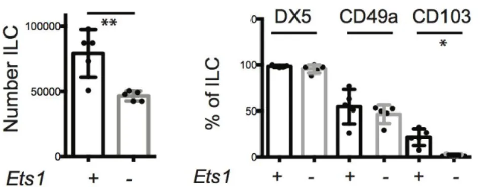
We have investigated the mechanisms governing tNK cell functions, demonstrating that tNK cells express the transcription factor EOMES and that they developed independent of the essential ILC1 factor TBET, confirming their placement within the NK lineage. Moreover, tNK cells developed independent of the E protein transcription factor inhibitor ID2 and their numbers were only mildly affected by the loss of ETS1.
Our data revealed that in the thymus of mice there is an absence of ILC1, setting the stage for deeper studies of the relationship between murine tNK cells and human CD56bright NK cells.
In the first part of this project, using culture systems capable of generating CD56bright and CD56dim NK cells from the human hematopoietic progenitors CD34+ circulating in the peripheral blood through the administration of appropriate cytokine combinations, we have been able to characterize the differentiating NK cells. Thus, we indicate that CD56dim and CD56bright NK cells, would originate from distinct progenitors, which, along with their differentiation into mature cells, would generate two distinct cell NK subsets with convergent phenotypes and functions. Moreover, during their development CD56dim and CD56bright NK cells would exploit different mechanisms to prevent cytotoxicity against healthy cells.
1
Introduction
1.
Innate Lymphoid Cells
NK cells represent a homogeneous group of IFN-γ producing cells, which express characteristic markers (NK1.1 in mouse and CD56 in human, and NKp46 in both species) and depend on the transcription factor T-bet for their development. In 2008 a second subset of cells, involved in the mechanisms of the innate immunity, has been discovered. These cells share many attributes with conventional NK (cNK) cells, but they differ in several aspects like their transcriptional requirement and their localization, suggesting that they differ for some aspect in their role in immune response (Diefenbach et al., 2014). All of these cells, unlike adaptive immune cells, develop in the absence of recombination-activating gene 1 (Rag1) or 2 (Rag2), they require few hours after their activation to respond, and they all develop from the common lymphoid progenitor (CLP); moreover, they all have a lymphoid morphology and they lack myeloid and dendritic antigens (Spits & Cupedo 2012). These cells, called ILCs (Innate-like Lymphoid Cells), have been discovered and divided into three main groups (Annunziato et al., 2015) based on their cytokine and transcription factor expression (Figure 1): The ILC1 family is composed of the T-bet expressing cells and includes NK cells and ILC1
cells; ILC2 are Gata-3-expressing cells (also known nuocytes) originally discovered in lung, skin, or fat tissue (Price et al., 2010; Neill et al., 2010); and ILC3 that produce IL-17 and/or IL-22 and express the transcription factor Rorγt. All of the three populations are made up of several subsets, but among them Group 1 ILC is the most heterogeneous and confused subset. In fact, NK cells have been considered the prototype of Group 1 ILCs for several years, but more recently it has become evident that distinct populations within this group exist and their diversity seems to be very important for immune protection.
2
1.1
Group 1 Innate Lymphoid Cells: NK and ILC1 cells
NK cells where identified for their ability to spontaneously lyse tumor cell lines in vitro, but more recently other recombinant activating gene (RAG)-independent ILC populations have been discovered. The ILC1 family, better known as Group 1 ILCs, is mainly composed of T-bet expressing cells: NK cells and ILC1 cells. Group 1 ILC is a group of non-cytotoxic lineage negative (Lin-) cells which can produce IFN-γ and TNF-α and are involved in immunity to intracellular bacteria and parasites (Fuchs et al., 2013; Klose et al., 2014). To date the ILC1 lineage is not well defined but it’s known that it requires TBET for lineage specification and function and produce “type 1” cytokines such as IFN- (Spits et al., 2013). NK cells have been considered the prototype member of this group, but they are not the only one. Another subset of group 1 ILCs that produces IFN-γ but not Th2 cell- or Th17 cell-associated cytokines, and that is distinct from NK cells, has been identified in mice and humans (Vonarbourg et al., 2010). In humans, the ILC1 subset lacks expression of c-kit (also known as CD117) and expresses high levels of TBET and low levels of RORγt. ILC1 have been identified in a variety of different tissue locations resulting in the identification of distinct populations. In the adult liver NK cells coexist with ILC1. These ILC1 are distinct from cNK cells because they do not circulate throughout the body and they remain at their steady state only in the liver (Peng et al., 2013). Liver ILC1 are CD49a+Trail+ (Takeda et al., 2005), and Trail seems to be a marker to distinguish these two different lineages. Moreover, these two populations have different transcription factor requirement and a distinct gene profile (Daussy et al., 2014). Liver ILC1 share the expression of some antigens like NKp46, CD122 and NK1.1 with cNK cells, but they also express a pool of antigens not common to that expressed by cNK cells like some chemokine receptor and adhesion molecules (CXCR6, CXCR3), cytokines and cytokine receptors. A functional profile of liver NK cells has been outlined, and it correspond to that of a cell population that surprisingly kill target cells with mechanisms that differ from NK cells, and that could be involved in regulatory roles either directly or indirectly via interactions with T cells. In the intestine it has been identified at least two populations of ILC1. Fuchs and colleagues characterized a human ILC1 subset that produces INF-γ in response to IL-12 and IL-15 and which has a unique phenotypic profile. This population of cells have been found in human tonsils and express markers such as CD160, CD49a, CXCR6 and CD39 (Fuchs et al., 2013); it has been proposed that because of sharing some features with tissue-resident memory CD8+ T cells intraepithelial ILC1 may be their innate counterparts; with an activated-memory phenotype and the secretion of INF-γ and other lytic mediators, these subsets of ILC1 could have a pro-inflammatory function. The murine counterpart of human intraepithelial ILC1 has been identified in the CD160+ NKp46+ NK1.1+ INF-γ producing cells in response to stimulation with IL-12 and IL-15
3
(Fuchs et al., 2013) and they are largely independent of IL-15R, corroborating that these ILCs are distinct from conventional NK cells, which require IL-15R for development.
Another study revealed that in inflamed intestine from individuals with Crohn’s disease there is an accumulation of INF-γ producing ILC1 (Bernink et al., 2013), and this subset of cells is the most represented ILC subset. Even if at least a portion of these cells differentiated from ILC3 cells under the influence of IL-12, ILC1 develop after colonization of the gut with commensals and this population may be involved in the early innate immune response against certain bacteria. Moreover, the adoptive transfer of human fetal hematopoietic stem cells (HSC) into transgenic mice lacking lymphocytes, NK cells, and ILCs, demonstrated that human ILC could reconstitute the intestinal ILC compartment in mice (Bernink et al., 2013).
In mouse salivary glands (SG) another population of ILC1 has recently been identified. This population of cells express both TBET and EOMES, but are poor producers of IFN-γ and opposite to cNK cells they do not depend on NFIL3 to develop (Cortez et al. 2014). How this ILC1 group fits into the innate landscape is not completely clear because these SG ILC1 have some confusing characteristics: they have a unique integrin pattern of expression (e.g.: CD103, VLA1) similar to that of intestinal intraepithelial ILC1s, which however are largely NFIL3 dependent and produce IFN-γ. Moreover, SG ILC1 express EOMES, while liver VLA1+ NK cells do not.
1.2 ILC1 and NK cells homeostasis
The immune system has evolved the ability to have a broad reactivity but also a high specificity to protein antigens thanks to the continuous expression or recombination activation genes (Rag1/2) by B and T cells; few selected B and T cells can recognize an antigen and clonally expand to produce a long-lived memory effector. In contrast, a heterogeneous pool of short-lived NK cells mediates the inflammatory response by secreting pro-inflammatory cytokines and cytotoxic granules. The current understanding of lymphocyte homeostasis is that when a system experiences a deficiency in a cell type due to infection/chemotherapy/irradiation, the biological system induces a replacement of these cells from progenitors or residual cells. For that reason, the homeostasis and the activation of NK cells is tightly regulated in an antigen-independent manner, both with extrinsic and intrinsic factors.
Intrinsic factors
Among the intrinsic factors involved in NK and ILC1 homeostasis, an important role is made by the protein tyrosine phosphatase CD45 (encoded by Ptprc), which is a key
4
negative regulator of both NK cell and liver ILC1 homeostasis (Huntington et al., 2005); in fact, hepatic ILC1 and cNK cell numbers are significantly elevated, correlating with an increased proliferation. Nevertheless, ILC1 and NK cells develop differently and they also have different transcription factor requirements, so the exact mechanism of how CD45 negatively regulates NK cell and ILC1 homeostasis is not clear.
In the differentiation of peripheral NK cell subsets and their functional diversification, an important role is acted by the Ikaros family of zinc finger proteins Aiolos (encoded by Ikzf3). It is early expressed during haematopoiesis and it regulates several aspects of lymphoid lineage development (Morgan et al., 1997), and mice lacking this gene display an unusual arrest in NK cell differentiation (Holmes et al., 2014). This Ikzf3−/− NK cells produce less IFN-γ even though they show a higher in vivo killing potential, and they still have normally expressed transcription factors known to regulate NK cell development. The arrested NK cell differentiation phenotype of Ikzf3−/− mice resembles that of B-lymphocyte-induced maturation protein-1 (Blimp-1, encoded by Prdm1) deficiency (Holmes et al. 2014). Even though Blimp-1 plays a key role in the terminal B- and T- cells differentiation, its expression increases during NK cell differentiation and is rapidly up regulated upon IL-12 and IL-21 stimulation.
Also Forkhead box protein O1 (Foxo1) is a transcription factor that has recently been identified as a negative regulator of NK cell differentiation. The homeostatic cytokines IL-2 and IL-15 induced the phosphorylation of Foxo1, preventing it from binding to its target gene, like TBET, which is essential for NK cell differentiation and Tbx21−/− mice do not develop mature NK cells and have significantly fewer total NK cells (Daussy et al., 2014; Gordon et al,. 2012). The expression of TBET and Foxo1 during NK cell ontogeny is inversed with TBET increasing and Foxo1 decreasing during differentiation. Foxo1 was found to bind Tbx21 at the proximal promoter region and Tbx21 expression was significantly elevated in Foxo1-null NK cells indicating that Foxo1 acts as a repressor of TBET expression to limit NK cell differentiation in vivo (Deng et al., 2015).
Extrinsic factors
The principle factor known to regulate NK cell homeostasis is IL-15 (Huntington 2014) but IL-15 is also necessary for the development and maintenance of other lymphocyte subsets. Also, IL-15 is important for driving NK cell maturation, in particular it is responsible of the up regulation of KLRG1 (Huntington et al., 2007). IL-15 is critical for the enhanced homeostatic proliferation and accumulation of KLRG1+ NK cells in
Rag1−/− mice: in fact there is a dose-dependent reduction of KLRG1+ NK cells with the deletion of one or two copies of IL15. Also commensal bacteria could have a role in the
5
homeostatic expansion of NK cells, probably linked to IL-15 production via myeloid and non-hematopoietic cells as a result of NOD signalling. This competition between NK cells and T cells for IL-15 and commensal bacteria have important consequences for immune responses. This competition between NK cells and T cells for IL-15 and commensal bacteria have important consequences for immune responses. In response to MCMV, KLRG1− NK cells are functionally superior to KLRG1+ NK cells and experience a significantly greater Ly49H-m157 expansion 7 days of post-infection (Kamimura & Lanier, 2015).
IL-2 is another member of common γ cytokine family, like IL-15, but even if they have sometimes overlapping pathways and overlapping functions, IL-2-/- and IL-15-/- mice present very different phenotypes suggesting unique roles for these cytokines in NK cell homeostasis (Ring et al., 2012); in fact both cytokines require the heterodimeric IL-2R β/γ complex for their signalling, whereas IL-15Rα is required to trans-present IL-15 to IL-2Rβ/γ expressing cells but does not intrinsically alter IL-15 signalling (Lodolce et al. 1998).
Transforming growth factor beta (TGF-β) is another potent immune-regulatory cytokines (Laouar et al., 2005); and it has been proposed that dendritic cells are a possible source of TGF- β1 for NK cells during immune response, and they can suppress and/or alter NK cell activity by altering TGF-β1 and IL-12 levels (Sarhan et al., 2015).
6
2.
Transcriptional regulation of innate lymphoid cell fate
and generation of different ILC1 and NK cell subsets
In the hematopoietic system, the hematopoietic stem cell (HSC) is the multipotent and self-renewing cell which gives rise to the generation of all the hematopoietic lineages. Haematopoiesis is a multistep process, during which HSC progressively lose the cell-fate potential, and the main step in lymphopoiesis is the generation of the common lymphoid progenitor (CLP).
NK cell is considered the founding member of the ILC family, one of the three lineages of lymphocytes to originate from the CLP, along with T and B cells, and several transcription factors and growth factors are known to be involved in CLP development into the downstream precursor. In these years, rare Lineage negative cells have been identified in the fetal liver, fetal gut and adult bone marrow; these cells express CD127 and the 7 integrin, and have also lost B and T cell potential even if they can still generate NK cells, dendritic cells and LTi cells (Yoshida et al., 2001). Recently, a committed ILC precursor (ILCp) has been identified within the IL-7Rα+α4β7+ population in bone marrow and fetal liver (Constantinides et al., 2014). This precursor expresses high levels of the transcription factor pro-myelocytic leukemia zinc finger protein (PLZF, encoded by Zbtb16) and it is required for ILC development. Several transcription factors have been identified to drive the generation of the ILCs subsets, which act in different ways and in the different lineages (Figure 2).
7
2.1
The Common Lymphoid progenitor (CLP)
ID2
ID2 is a member of the ID family of transcriptional repressor; proteins of this family lack the DNA-binding domain and share the highly conserved helix-loop-helix domain (Sun et al., 1991). ID2 regulates the differentiation and the development of a lot of lineages, and its ablation has dramatic effects on the differentiation of myeloid and Figure 2: General model for (mouse) ILC development. Innate lymphoid cells (ILCs) differentiate from
haematopoietic stem cells via a common lymphoid progenitor (CLP). Interleukin-7 (IL-7) and the transcription factors ID2 and NFIL3 promote the differentiation of potential common precursors of ILC-restricted progenitors. Immature NK (iNK) cells appear after expression of EOMES. ILC1s may derive from NKPs and/or CHILPs in response to an IL-15-induced transcriptional programme that involves transcription factors such as TBET, EOMES, GATA3 and/or NFIL3. I [From: Serafini et al., 2015]
8
lymphoid lineages (Verykokakis et al., 2014). Members of this family bind and functionally inactivate a set of transcription factors known as E-proteins, like E2A, E2-2 and HEB within the hematopoietic system.
ID2 deficient mice have been reported to have a selective loss of NK cells and lymphoid tissue, whereas B- and T-cell development is substantially the same (Boos et al., 2007). Moreover, overexpression of ID proteins inhibits B cell and T cell development, while it strongly promotes ILC generation, in particular NK cells (Heemskerk et al., 1997). All CLPs express little or no ID2, and high levels of ID2 would essentially restrict the lymphoid precursor to the ILC lineage (Klose et al., 2014). Moreover, some ILCs can develop in the absence of ID2 if also E protein are ablated (Boos et al., 2007), indicating how ILCs development might represent a default pathway for CLPs: ID2 induction appears to be one of the first molecular steps in the induction of the ILC lineage; however, the mechanism of Id2 up-regulation in ILC progenitors remains unclear.
NFIL3
NFIL3 (nuclear factor interleukin-3; also known as E4-binding protein 4, or E4BP4) has been first described as a critical transcriptional regulator for NK cell development affecting mature NK cells (Gascoyne et al., 2009; Kamizono et al., 2009) and thymic NK cells (Seillet et al., 2014a). It is broadly expressed in different tissue and it is involved in several developmental and biological processes. Within lymphocytes, NFIL3 ablation has a dramatic effect on NK cell development in particular at the pre-NKP stage, but it seems to be not so fundamental in mature NKp4+ cells (Gascoyne et al., 2009). NFIL3-deficient mice also display a broad loss of ILC populations including ILC1, 2, and 3 together with LTi cells. This loss appears to stem from inhibition of the development of the bone marrow α4β7+
lymphoid progenitors and ILCPs (Seillet et al., 2014b) and thus the development of a common innate lymphoid progenitor, prior to PLZF up-regulation.
CLPs express little or any NFIL3, but it can be regulated by cytokines and IL-7 can strongly induces NFIL3 expression in CLP (Ikushima et al., 1997). NFIL3 is not required for all ILC and discrimination between different peripheral subsets has been linked to the induction of EOMES. Indeed, all EOMES-expressing NK cells, including conventional medullary and thymic NK cells, are absent in absence of NFIL3, whereas TRAIL+ NK cells that do not express EOMES appear unaffected by its loss (Seillet et al., 2014a). Even if the strength importance for NFIL3 in the NK cell development, how its expression is regulated is not completely clear.
9
GATA3
The transcription factor GATA3 has many roles in T cell and ILC2 differentiation (Tindemans et al., 2014). In 2006 for the first time a role for GATA3 has been appreciated for thymic NK (tNK) cells, a subset of NK cell different from that of cNK cells (Vosshenrich et al., 2006); since then it has been shown that deletion of GATA3 in all hematopoietic cells impaired the development of all IL-7Rα+ ILC subsets but did not interfere with the development of cNK cells (Yagi et al., 2014), showing that in the absence of GATA3, Lin- CD127+ cells fail to develop in the fetal liver and adult bone marrow.
In general, GATA3 has a broad role in ILCs development, and it has been proposed that it is involved in the segregation of “helper” from “killer” ILC lineages, because the development of splenic CD127- EOMES+ NK cells still occur also in its absence (Samson et al., 2003).
PLZF
The transcription factor PLZF has a very important role in the differentiation of T cell subsets, but only recently its role in the differentiation of ILCs has been demonstrated. Fetal liver and bone marrow PLZF+ precursor include committed ILC precursor; however, PLZF deficient mice have only partial defect in ILC1 and ILC2 development, thus, even if its molecular targets are not totally known, it seems to be important for the generation of CHILP from CLP. Fate mapping experiments in mice have shown that PLZF expression promotes commitment to the ILC lineages, but not LTI or NK cells (Constantinides et al., 2015).
2.2
Differentiation of NK cells and ILC1
Transcription factors govern the development of NK cells, starting from the earliest progenitor. Although many of these DNA-binding and chromatin-modifying proteins are shared with other cells of the immune system or even with non-hematopoietic cells, some are unique to the NK cell lineage. Already mentioned transcription factors ID2, E proteins, PLZF and Nfil3 are among proteins known to drive early stages of NK cell development. Additional transcription factors, including TBET, EOMES, and ETS1 play specific roles at distinct stages of NK cell development and maturation.
TBET and EOMES
TBET is the transcription factor encoded by Tbx21, and it is the signature transcription factor for mature Group 1 ILC that produce IFN-γ; it binds to the Ifng locus to activate
10
it via chromatin remodelling that allows other binding of factors that improve Ifng expression (Djuretic et al., 2007). Both ILC1 and NK cells express TBET, but they depend on it in a different manner, while only NK cells express EOMES so it can be used to discriminate between these two subsets.
In NK cells, TBET is required altogether with EOMES to promote their maturation and function (Gordon et al., 2012). These two transcription factors act in a sequential manner, but TBET directs the development of iNK cells and stabilizes the immature phenotype while EOMES induces the expression of a diverse repertoire of Ly49 receptors in NK cells and acts to maintain the mature phenotype (Gordon et al. 2012). EOMES seems to be a downstream target of NFIL3 in NK precursor (NKP) (Male et al., 2011) which drives early stage differentiation, and this is an explanation for the severe NK cell deficiency in NFIL3 deficient mice. Indeed, these mice have other severe defects also in the other ILCs lineages, suggesting that EOMES is not the only downstream target of NFIL3 (Seillet et al., 2014a). Moreover, Analyses of EOMES reporter mice showed that at steady-state TBET+ EOMES− cells appear to be a stable population that do not subsequently give rise to TBET+ EOMES+ cells, suggesting that these cells are not immature, but represent a distinct population (Daussy et al., 2014). Daussy and colleagues also demonstrated that TBET expression is repressed in the bone marrow allowing the development of EOMES+ NK cells, and EOMES- NK cells are not precursors of EOMES+ NK cells in homeostatic conditions and rather correspond to a distinct lineage of ILCs (Daussy et al., 2014). It was also demonstrated that TBET and EOMES cooperate to induce high expression of CD122, the β chain that binds IL-15 (Intlekofer et al., 2005)
ETS1
ETS1 is a member of the large family of ETS transcription factors, it is a proto-oncogene expressed in all lymphoid lineages, with widespread roles in development (Sharrocks, 2001). ETS1 is required for the optimal development of mature NK cells (Barton et al., 1998), and NKP are significantly reduced in the absence of ETS1, in fact transcriptional analysis have revealed that ETS1 sustains ID2 and TBET expression in this progenitors (Ramirez et al., 2012).
11
3.
Natural Killer cells
Natural Killer (NK) cells were first discovered in the mouse at the beginning of 1970s as a population of cells able to spontaneously kill cancer cells (Kiessling et al., 1975), and few years later they were found also in human (Pross & Jondal, 1975; Jondal & Pross, 1975). This name is due to their capacity of spontaneous cytotoxicity (natural killing) that was different to that one mediated by cytotoxic T lymphocytes; in fact T cell cytotoxicity is dependent on the expression of Major Histocompatibility Complex (MHC), while NK cells were able to kill also tumor cells without the expression of MHC antigens on their surface (Trinchieri & Santoli, 1978). Even if they were first considered as a “background noise” in T cells cytolytic assays, about ten years later NK cells were characterized as a distinct type of mononuclear cells in the peripheral blood (Perussia et al., 1983; Lanier et al., 1986b); they were described as Large Granular Lymphocytes (LGL) because of their homogeneous morphology with granules inside the cytoplasm (Grossi et al., 1982), and they were present in both lymphoid organs and non-lymphoid peripheral tissues. Nevertheless, because NK cells share the LGL morphology with some other subsets of T cells and dendritic cells, they can’t be described only on the basis of their morphology.
NK cells are included as part of the innate immune system, with monocytes and granulocytes (Scott & Trinchieri, 1995), because they rapidly respond to infectious agents without prior sensitization, and they are highly cytotoxic versus infected cells and tumor cells when these cells do not express class I MHC molecules (MHC-I) (Zamai et al., 2007). NK cells represent about the 10-15% of all the circulating cells in the peripheral blood, but they can be found also in other lymphoid tissues, spleen, lymph nodes and bone marrow (Ferlazzo et al., 2004), and in case of necessity they can also migrate towards sites of inflammation.
Even if T and NK cells share some functional and phonotypical characteristics they have complementary action, because T cells have the requirement that MHC-I is expressed on the surface of pathogen-infected cells in order to recognize them, while NK cells can lyse target cells without prior sensitization resulting powerful effector when B and T cells are not enough and for that reason they have been considered among the main components of the innate immunity. Moreover, as opposed to T cell and B cell responses that are dictated by unique, somatically recombined, and clonally distributed antigen receptors, NK cell responses are controlled by a more limited repertoire of germ line-encoded receptors (Vivier et al., 2011). The main feature that distinguishes NK cells from other lymphoid populations is the lack of specific receptors for foreign antigens, in particular the absence of the surface receptor CD3, the T Cell receptor
12
(TCR) typical of T lymphocytes or surface Immunoglobulins (sIg or B Cell Receptor, BCR) characteristics of B lymphocytes (Ritz et al., 1985; Lanier et al., 1986a).
NK cells play a pivotal role in defending our body, with an intense cytolytic activity against tumor cells, cells infected by viruses or bacteria, and allogeneic cells (Moretta, et al., 2001) being able to lyse them through the activation of cytolytic pathway in the absence of a previous stimulation, and being active against cells that do not express MHC class I molecules (Lanier et al., 1985).
The most important aspect in defining this population able to mediate the spontaneous cytotoxicity has been to unequivocally identify it through the presence of surface markers in the absence of CD3/TCR or sIg. Although NK cells do not express rearranged gene products TCR, adult NK cells expressing the CD3 ζ chain; moreover, NK fetal constitutively present in the cytoplasm the γ, δ, and ε chains of CD3 molecule (Lanier et al., 1992). Activated NK cells in the peripheral blood of adult individuals express the ε chain of the CD3 intracytoplasmic (cytCD3ε), but not the other chains of this receptor with the exception of the ζ chain (that is bound to CD16). Some evidence suggests that the initial expression of cytCD3ε can identify a common T/NK progenitor present in the fetal thymus (Miller et al., 1994; Spits et al., 1995) but no functional role has been found in NK cells.
The lack of CD3ε chain in other hematopoietic lines makes this molecule potentially important in discriminating NK cells and immature T cells, when these lack other markers or phenotypic functional characteristics. However, human NK cells exhibit numerous other proteins on the cytoplasmic membrane that can be used as phenotypic markers exclusive for easily distinguish them from other lymphocyte populations. NK cells can be divided into functionally distinct populations, which seem to be different in humans and mice: human NK cells can be mainly classified based on the expression levels of CD56 and CD16, while mouse NK cells share many characteristics with human NK cells, but the lack of CD56 expression and other surface markers makes it difficult to identify functionally comparable NK cell subsets in mice.
3.1
Natural Killer cell markers and receptors
CD56
CD56 is a transmembrane glycoprotein that belongs to the immunoglobulin superfamily (Cunningham et al., 1987) and it has a molecular weight between 175 and 220 kDa, it is characterized by the presence of an immunoglobulin domain in the extra-cellular portion. The CD56 molecule is homologous to the neural cell adhesion molecule N-CAM, but it is not able to transduce an activator signal and it is not directly involved in
13
the activation of NK cytolytic machinery. CD56 is involved into an increased adhesion between the NK cell and its target (Lanier et al., 1991), and through homophilic binding it plays an important role in the process of target cell killing by an apoptosis mechanism (Takasaki et al., 2000). NCAM in the peripheral blood is prematurely expressed on NK cell surface and it is considered an important marker for the identification of NK cell subsets in the human.
CD16
One of the most studied activating receptor expressed on the NK cell surface, is the low affinity receptor for the crystallisable fragment (Fc) of IgG, CD16 (FcγRIIIa).
CD16 is a transmembrane glycoprotein of 50-80 kDa and it is expressed by most human NK cells, activated monocytes and a subpopulation of T lymphocytes (Lanier et al., 1985). This receptor is important because it is a signal-transducing molecule that after the binding with its ligand, it induces (also in combination with other stimuli) the transcription of genes encoding for other surface activation molecules or cytokines important in enhancing NK cells activation (Anegòn et al., 1988).
CD16 has low affinity for the crystallisable fragment (Fc) of G Immunoglobulins (Ravetch & Perussia, 1989), it binds opsonized targets cells and it activates the antibody-dependent cell cytotoxicity (antibody dependent cell-mediated cytotoxicity, ADCC) mechanism, through the association with γ chain of the high affinity receptor for IgE (FcεRI) and the ζ chain of the CD3 that contains an activator immune-receptor (Immune Tyrosine-based activating Motif, ITAM) (O 'Shea, et al., 1991).
The cross-linking of CD16 with its ligand leads to a rapid activation of the tyrosine-kinases protein (PTK), which follows the phosphorylation of a tyrosine residue of the ITAM sequence of the intracytoplasmic tails of FcεRIγ and CD3ζ chains. Subsequently, they are recruited on this phosphorylated PTK site belonging to the Syk/ZAP70 family (Brumbaugh et al., 1997) and the Src family (Azzoni et al., 1992) and they are able to phosphorylate both the adapter protein LAT (Jevremovic et al., 1999) and the phosphatidylinositol-3-kinase (PI3K) (Weiss & Littman, 1993). At this point distinct metabolic events can occur: on one side there are activated phospholipase γ1 and C-γ2 which, in turn, lead to the activation of protein kinase C (PKC), by the production of diacylglycerol (DAG) as a second messenger, and to a transient increase in the intracellular concentration of Ca2 +, due to the inositol 1,4,5-triphosphate (IP3) (Einspahr et al., 1991; Ting et al., 1992). Another way involves the Vav-Rac1 protein (Galandrini, et al., 1999), while a different pathway involves the activation of phosphatidylinositol-3-kinase (PI3K), which plays a major role only in the ADCC mechanism. The phosphorylation of the ITAM sequences allows, besides the generation of second messengers, also the translocation into the nucleus of transcription factors such as NFATp and NFATc.
14
After stimulation by CD16, NK cells mediate antibody-dependent cell-mediated cytotoxicity (ADCC), and they secrete a variety of cytokines such as IFN-γ, tumor necrosis factor (TNF)-α, and some colony stimulating factors such as IL-3 and granulocyte monocyte colony stimulating factor, GM-CSF.
Because of the low affinity for immunoglobulin G (IgG1 and IgG3 specifically recognizes) CD16 binds only to IgG coated cells or immune complexes and not to free circulating IgG in the blood; so that the free IgG in the plasma do not interfere with the recognition by the CD16 cell covered by Ig, allowing the accomplishment of the cytotoxic action versus target cells. In this process the IgG have the dual role of specific recognition structures of the target cell that needs to be eliminated and mediator of NK cell binding of the target cell, ensuring the specificity effector.
The CD16 antigen is not an exclusive molecule of NK cells, it is also present on the surface of granulocytes, where, however, it does not induce the ADCC mechanism, in fact the main function of CD16 on granulocytes is to enable the anchoring of the latter to opsonized cell, thus allowing the process of phagocytosis. This is due to the fact that CD16 on granulocytes is not an integral membrane protein, and, unlike the NK cells, it has no got the transmembrane and intracytoplasmic domains able to transduce the signal inside the cell, being CD16 simply anchored to the surface cell by means of a phosphatidylinositol.
CD27
CD27 is a member of the tumor necrosis factor receptor superfamily, known to play a very important role in cell growth and differentiation, as well as apoptosis or programmed cell death (Prasad et al., 1997). It binds to ligand CD70, and it is one of the most important markers for the characterization on NK cells in mice: altogether with the gradual decrease of CD11b expression, there is the gradual up-regulation of CD27, which became a marker of the latest stages of differentiation of NK cells (Chiossone et al., 2009).
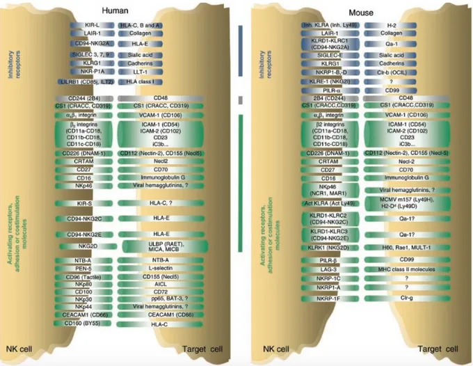
NK inhibitory and activating receptors
NK cells mediate spontaneous immune response against a variety of cells including, in some conditions, the autologous cells. The study of the regulatory mechanisms of this response has involved many researchers and has led to the definition of a new mechanism of lymphocyte regulation mediated by surface receptors, both activators and inhibitors, expressed by NK cells (Moretta &Moretta, 2004).
A key role in the recognition of appropriate target cells is played by the ubiquitously expressed major histocompatibility complex (MHC) class I molecule, a ligand for which NK cells generally have multiple receptors. NK receptor gene complexes are
15
intimately associated from a genetic and functional point of view with MHC recognition, and the interaction of various combinations of NK cells receptors (NKRs) and MHC class I molecules may have contribute also to human survival in the presence of epidemic infections (Parham, 2005). In the absence of inhibitory signals, the activating receptors induce biological responses and this is the reason why NK cells spontaneously lyse tumor cell lines which do not express HLA class I antigens (Moretta et al., 2001). Healthy cells are protected from lysis mediated by NK cells by the expression of the major histocompatibility complex class I (MHC-I) complex that acts as a ligand for the inhibitory receptors of NK cells (Lanier, 2005).
NK cell receptors should be distinguished according to their function. Most of the molecules expressed on the NK cell membrane is expressed by other hematopoietic cells, such as T-lymphocytes, myeloid cells and monocytes. Moreover, many of these receptors are not expressed by all NK cells, indicating the existence of phenotypic and functional heterogeneity within this cell population. Receptors involved in inhibitory functions (or those whose activation induces the inhibition of the cytotoxic activity and cytokine production), are important because they represent a sort of safety-check in preventing attacks by NK cells against normal autologous cells and they are represented by killer immunoglobulin receptors (Killer Immunoglobulin (Ig) -like receptor, KIR), which recognize different allelic groups of HLA-A -B, -C molecules; by the complex CD94 / NKG2A that recognizes HLA-E molecules, and to a lesser extent by the LIR. A common feature of inhibitory receptors is the presence in their intracytoplasmic chain of a tyrosine immune-receptor with inhibitory function (immune-receptor Tyrosine-based Inhibitory Motif, ITIM) which, following the binding with the appropriate MHC, activates the phosphatase SHP-1 and SHP-2. These phosphatases in turn inhibit the cascade of signals induced by activating receptors. There are also several receptors involved in the activation of NK cells, like the natural cytotoxicity receptors (Natural Cytotoxicity Receptors, NCRs) NKp46, NKp44, NKp30. These receptors are very important not only for their functions but also because they are expressed almost exclusively by NK cells, so they can be very important for their identification. In the NK immune response there are also numerous surface molecules that perform specific functions such as co-activators (CD2, 2B4, DNAM- 1) and adhesion molecules (such as CD56 and LFA-1).
Inhibitory NK receptors
Inhibitory receptors are randomly distributed on NK cells surface and they distinguish different subsets NK. In the early '90s, basing on some experimental evidences showing that NK cells killed preferentially cancer cells, which don’t express MHC-I molecules, the hypothesis of an immune surveillance mechanism which eliminate cells with deterioration in the expression of MHC molecules was formulated. In particular, the
16
cytolysis is inhibited when the appropriate molecules HLA (the major histocompatibility system of the human) of class I are expressed by target cells, but it is implemented when these molecules, which identify its own cells as "self", are missing, and there is the situation known as "missing self" (Ljunggren & Kärre, 1990). The “missing self” hypothesis suggests that NK cell could attack only target cells with a reduced or aberrant MHC or HLA-I (like cancer cells or virus-infected cells), become unable to send inhibitory signals to NK cells and, therefore, they become susceptible to NK lysis. Thus, when MHC-I is expressed (normally by healthy tissues) on target cells, the activation of NK cells in inhibited (Kärre, 2008). Almost all NK cells express at least one specific inhibitory receptor for MHC I "self-molecules" and NK cells that do not express them are hypo-functional (Anfossi et al., 2006), in this way the autologous cells that express MHC I antigens are protected from NK lysis. This inhibition is essential for the role of NK cells: because of the abundant expression of MHC-I on many cells, NK cells remain non-responsive to healthy tissue (Figure 3).
In the last decade there was the molecular characterization of human NK inhibitory receptors, which allowed their division in different classes, among these there are structurally distinct families of receptors that are sensitive to the expression of MHC class I molecules I: the family of the KIRs (Killer immunoglobulin-like receptors) and LIRs (Leukocyte immunoglobulin-like receptor-1) belonging to the immunoglobulin superfamily and the receptors belonging to the family of C-type lectins (CD94/NKG2A-C) (Moretta et al, 1996). The expression of KIR and CD94/NKG2 receptors is not restricted to NK cells, because they are expressed also by a subpopulation of T lymphocytes, which inhibits both the cytotoxicity and the production of cytokines induced by TCR. Inhibitory receptors in NK cells determine the inhibition of signals derived from activating receptors, including cytotoxicity signals and the production of cytokines (Augugliaro et al., 2003).
Humans and primates have evolved different families of receptors when compared to mice; but even if they have different developmental origins and structures there are some evidences of their convergent evolution, such as the ITIM (immunoreceptor Tyrosine-based Inhibitory Motifs) and DAP-12 ITAM (immunoreceptor tyrosine-based activating motifs) signalling, the recognition of MHC-I and the presence of both inhibitory and activating receptors (Barten et al., 2001)
17
KIRs
KIR (Killer Immunoglobulin-like Receptors) are integral membrane glycoproteins expressed on the surface of human NK cells and also T cell, in which an extracellular, a transmembrane and an intracytoplasmic portion can be distinguished; they have evolved from the Ig-superfamily and they have two or three Ig-like domains with short or long cytoplasmic tail that are able to specifically recognize different types of HLA (Colonna & Samaridis, 1995; Wagtmann et al., 1995). Some members of the KIR family specifically bind certain alleles of HLA class I, and the decision of which KIRs are expressed on each NK cells is randomly regulated by the methylation of KIR gene loci (Chan et al., 2003).
KIR receptors specifically bind HLA-A, -B and –C molecules, but each individual has a different KIR haplotype, that is the expression of a particular repertoire of KIR genes; but three KIR genes are common to all haplotypes: KIR3DL3, KIR2DL4, KIR3DL2. Based on the number of Ig domains present in the extracellular portion there are two subfamilies KIR: KIR2D (which has two Ig domains: D1 and D2) and KIR3D (with three Ig domains: D0, D1, D2). The members of each members of KIR subfamily differ also for the length of the intracytoplasmic chain, those long-chain (long, L), which has two ITIM motifs, induce a signal type inhibitory (KIR2DL and KIR3DL); in contrast, KIR with short tail (KIR2DS and KIR3DS) have the ITAMs sequences that generate activation signals upon interaction with their respective ligands.
KIRs inhibitory receptors are:
KIR2DL: it is also called p58 (Moretta et al., 1993) and it has two extracellular Ig domains (D1 and D2) and an intracytoplasmic chain with 76-84 amino acidic Figure 3: Schematic representation of the “missing-self” hypothesis. a. NK cell interacting with a
normal autologous target cell which expresses the appropriate MHC-I so that inhibitory signals block the lysis. b. NK cell is activated by the missing of expression of MHC-I on the surface of the target cell; it does not receive inhibitory signals and therefore lyses the target cell [Adapted from: Kumar & McNerney, 2005].
18
residues. The recognition of KIR2D depends mainly on the nature of the MHC-I amino acid present at position 80; in this family we can find two main subsets: - KIR2DL1, also called CD158a, which is a protein of 58kDa that binds
HLA-C2 molecules and it induces an inhibitory signal after the contact of NK cells with their target (Melero & Salmeròn, 1994).
- KIR2DL2, also called CD158b, which is a glycoprotein of 58kDa that binds HLA-C1 molecules, and it leads to the inhibition of NK cell-mediated cytotoxicity.
KIR3DL: it is also called p70 (Litwin et al., 1994) and it has three extracellular Ig domains (D0, D1, D2) and an intracytoplasmic chain with 84-95 amino acidic residues. Also this family have different members, but the main can be considered KIR3DL1 (also called CD158e1 or NKB1). It is a glycoprotein of 70kDa and it binds some HLA-A and HLA-B alleles of the serologic group Bw4.
The biochemical mechanisms that mediate this inhibition are known only in part and it was only recently discovered that inhibition of NK function involves recruitment and activation of the tyrosine phosphatase SHP-1. The signal transduction cascade of KIR inhibitory receptor is triggered at the level of the ITIM amino acid sequence, present on the cytoplasmic tail of these receptors. When this structural motif containing tyrosine residues recognizes its specific ligand, there is the phosphorylation of a tyrosine residue, the recruitment and activation of a phosphatase (SHP-1) thus determining the inhibition of activating signal which, otherwise, would lead to the lysis of the target cell (Burshtyn, et al., 1996).
There are also KIR activators which have an extracellular portion similar to that of inhibitors, but a short intracytoplasmic portion, in particular:
KIR2DS, also called p50 (Bottino, et al., 2000), has two extracellular Ig domains (D1 and D2) and an intracytoplasmic chain with 39 amino acid residues. In this family, it is possible to distinguish subgroups:
- KIR2DS1 (also called CD158h) is a glycoprotein of 50 kDa which upon interaction with a group of HLA-C alleles HLA-denominated C2, it produces a activating signal.
- KIR2DS2 (also called CD158j) is a 50 kDa glycoprotein which results in a signal that activates the cytotoxicity mediated by NK cells, but its ligand is still not well known.
KIR3DS1 (also called CD158e2) has three immunoglobulin extracellular domains (D0, D1, D2) and it is able to transduce activating signals when it encounters its ligand. It seems that it can bind the HLA-alleles Bw4, but in a manner dependent on the assembled peptide.
19
The KIR2DL4 is actually a receptor with activating function, despite the long cytoplasmic tails, and that recognizes the HLA-G molecule expressed exclusively in the fetus.
The cytotoxic effect of NK cells toward target cells is a balance between inhibitory KIR and stimulating KIR: the former can inhibit the cytotoxic activity mediated by KIR activators if the target or antigen presenting cell also expresses the ligand for the inhibitory KIR (Moretta &Moretta, 2004). While recognizing the same HLA-I molecules, the latest evidence suggests that the KIR activators have greater affinity for HLA-I associated peptides of viral antigens; while the KIR inhibitors have greater affinity for HLA-I associated peptides self-antigens.
KIRs have a role in the induction of NK cell tolerance of self-tissue, preventing the activation of NK cells against normal healthy tissues. It’s important to notice that KIR and HLA segregate independently and the expression of KIRs is not driven by HLA (Gumperz et al., 1996).
Ly49 receptors
Instead of having polygenic and polymorphic KIRs, rodents have expanded their Ly49 genes, resulting in a remarkable diversity across different inbred mouse strains (Kirkham & Carlyle 2014). This complex in the mouse comprises about 20 genes and pseudo genes similar to KIRs in humans from a functional point of view, because both receptors families have inhibiting and activating members.
Ly49 receptors are type II integral membrane proteins that form disulphide-linked homodimers on the cell surface, and they composed of a carboxy-terminal lectin domain, also known as NK domain (NKD), which gives specificity for distinct allotypic groups of MHC-I and MHC-I like molecules (Karlhofer et al., 1992). The NKD is bind to the cell membrane by an extended stalk region, about 70 amino acidic residues in length. Within this lectin domain, like in the KIRs family, there is genetic variation both in which Ly49 genes are present and in the sequence of individual genes across different mouse strains (Carlyle et al., 2008).
More than 20 Ly49 genes have been identified, the majority of which encode for inhibitory receptors, whose prototypes are Ly49A and Ly49C. Both of them have the ITIM motif in their cytoplasmic regions that, after the binding of the molecule in the target cell, recruit and activate phosphatases such as SHP-1, to inhibit NK cell activation (Nakamura et al., 1997). Within the Ly49 family, there are some members that have been evolved more recently, like Ly49D and Ly49H, that have gained the capacity to interact with the small disulphide bonded homodimers such as DAP12 or DAP10 (Smith et al., 1998).
The pathway of MHC-I recognition by Ly49 receptors requires the presence of a peptide bound in the furrow of the MHC molecule, and each Ly49 receptor has a
20
different specificity for this peptide binding, because they exhibited different binding proprieties (Hanke et al., 1999). It has been found that Ly49 molecules, such as Ly49A, not only bind its ligand on potential target cells (trans), but also it is constitutively associate on the same cell (cis); thus lowering the threshold at which NK cell activation exceeds NK cell inhibition, cis interaction allows optimal discrimination of normal and abnormal host cells (Doucey et al., 2004). Moreover, the nature of Ly49 receptors (cis or trans) can affect the signalling outcome.
C-type lectin family of receptors
The receptors CD94/NKG2 are heterodimers composed of a common chain, CD94, associated with one of the products of genes NKG2. The most representative of the inhibitory receptor superfamily of C-type lectins consists of the CD94 glycoprotein subunits and NKG2A tied in a heterodimeric complex. These receptors react with a non-classical MHC-I on the surface of target cells, and they seem to be crucial for the prevention of inappropriate NK cell activation (Borrego et al., 1998); in particular their ligand is the product of the HLA-E gene, which expression depends on the presence of MHC-I molecules and it is expressed in most normal autologous cells (Braud et al., 1998). The interaction of the receptor with the HLA-E therefore allows to prevent "self-reactivity" against normal cells. In humans the expression of these receptors seems to be related to KIR gene expression, as suggested in an important study where they demonstrate that NK cell clones lacking the expression of an inhibitory KIR, expressed an inhibitory CD94/NKG2 heterodimer (Valiante et al., 1997).
The CD94 is the invariable component of the receptor, and it is a type II integral membrane protein, encoded by a single gene with no apparent polymorphism (Chang, et al., 1995), and which has a very short intracytoplasmic chain that alone it is not capable of inducing a signal transduction after the bounding with the specific ligand; for this reason to be functional, CD94 need to be bounded through a disulphide bond with a member of the NKG2 family receptors.
NKG2 is a multigene family expressed either on the cell surface of NK cells on CD8+ T lymphocytes (Ykoyama & Seaman, 1993), consisting of five different proteins (NKG2A, NKG2B, NKG2C, NKG2D/F, NKG2E). Like KIRs, some members have inhibitory function (NKG2A and B) and other have activating functions (NKG2C, NKG2D/F, NKG2E).
Despite the structural heterogeneity of the various inhibitory receptors that interact with MHC I molecules, the mechanism responsible for their inhibitory activity is common. In fact in the intracytoplasmic tail of an inhibitory receptor, both KIR of the CD94/NKG2 family (-A / -B), there are one or two ITIM sequences, containing tyrosine residues, which upon interaction with the ligand MHC I are phosphorylated. The phosphorylated ITIM domain is therefore responsible for the recruitment of tyrosine
21
phosphatases, and in particular SHP-1, responsible for the propagation of the negative signal and then by blocking the activation of NK cell (LeDrean, et al., 1998). The other members of the NKG2 family instead transduce activating signals, because they are associated with trans-membrane proteins such as DAP10 and DAP12 that contain ITAM sequences. It is important to underline the fact that each inhibitory receptor bound can only inhibit the activators of receptors signals that are around it (Kaplan, et al., 2011).
Another member belonging to the superfamily of C-type lectins is the NKR-P1 receptor (or CD161), expressed in dimeric form on most NK cells and on a subpopulation of T lymphocytes (Lanier et al., 1994). The genes coding for NKR-P1 were identified both in humans and in rodents and are located in a chromosomal region called "NK complex". Unlike the mouse, in humans there is only one family member, NKR-P1A, while in the mouse NKR-P1 receptors can be both activating and inhibitory, and five different receptors have been identified (Plougastel et al., 2001). The physiological ligand of NKR-P1 seems to be the lectin-like transcript-1, LLT1. The interaction between NKRP1A on NK and LLT1 on target cells inhibits both the cytotoxic activity that the secretion of cytokines (Rosen et al., 2008). In humans, some evidence suggests that it can work as a receptor both activating and inhibitory, depending on the cell type.
LIRs
The family of the LIRs (Leukocyte Inhibitory Receptors) is quite wide and also distributed in other cell types, including B cells, dendritic cells and certain T lymphocytes (Fanger et al., 1999). These receptors such as KIRs, are inhibitory receptors able to recognize a wide variety of MHC-I molecules presenting a very similar structure characterized by the inhibitory ITIM sequences and thereby acting with the same mechanisms.
The function of LIR in the regulation of NK cell activation is not completely clear, but it has been found a specific LIR which binds UL18, a protein encoded by human cytomegalovirus, with greater affinity than for HLA-I (Chapman et al., 1999).
NK activating receptors
According to the hypothesis of the missing-self, in the absence of inhibitory signals due to a failure or reduced expression of HLA class I on the target cell membrane, the activating receptors lead to the activation of NK cell that is now free to perform its cytotoxic activity and to produce cytokines (Moretta et al., 2001). NK cells have many activating and co-activating receptors. In addition to activators members of KIRs and NKG2 family (especially NKG2D), among the activating receptors members of the
22
NCR (Natural Cytotoxicity Receptors) family are very important, and they are represented by NKp46, NKp44 and NKp30.
NCRs
NK cells express three different receptors that mediate directly the natural cytotoxicity, called NCR, which are NKp30, NKp44 and NKp46; they were discovered in the late 1990s as being express on human NK cells. Even if they share some functional skills, the do not share many similarities either in their amino-acid sequence or in their structure (Joyce & Sun, 2011).
The NCRs belong to the Ig superfamily (Ig-SF), and they are integral membrane proteins in which an extracellular portion with one or two Ig domains, a transmembrane portion and an intracellular portion can be identified. The intracellular portion is associated through a disulphide bridge to molecules presenting ITAM sequences and that transduce activation signals of the natural cytotoxicity mediated by NK cells when they are engaged with their specific ligands (Figure 4).
The NKp30 receptor is a glycoprotein of 30 kDa that is expressed in all mature NK cells including those immature that are produced in vitro from CD34+ cells and it cooperates with NKp46 and NKp44 in the induction of cytotoxicity against several tumor targets (Pende et al., 1999), moreover it can be expressed also on other type of cells, such as cord blood T cells after IL-15 exposure (Tang et al., 2008). NKp30 is a Figure 4: Schematic representation of the NCRs molecules. All NCRs have a positively charged
amino acid in the transmembrane domain but NKp30 and NKp44 possess only one Ig-like domain, whereas NKp46 has two. [From: Kruse et al., 2014 ].
23
molecule composed of one extracellular Ig domain, the transmembrane domain has a charged arginine associated with the negatively charged present in the transmembrane portion of the CD3ζ molecule, and the intracellular domain has no additional signalling domain. It is associated to the ζ chain of the CD3 complex and both the CD3ζ homodimers and the CD3ζ/FcRg heterodimers can bind to the charged arginine in the transmembrane domain. There are six different splicing variants of NKp30 known to be expressed on the cell surface: three of them encoding for a molecule with an extracellular V-type domain, and the other three encoding for a C-type Ig domain (Neville & Campbell, 1999). NKp30 plays an important role in the selection (editing) of dendritic cells induced by NK cells, the latter are in fact able to recognize and eliminate immature dendritic cells that would not do properly the work of antigen presentation (Della Chiesa et al., 2003). The cross-linking mediated by the specific monoclonal antibody (mAb) on NKp30, induces cellular responses identical to those induced by NKp46: flow of Ca2+ ions, cytotoxicity, and production of cytokines. Recently it was shown that TGF-β1 (Transforming Growth Factor β1) influences the expression of NKp30 and in part also that of NKG2D to the cell surface. TGF-β1, which is issued by various tumor such as melanoma, neuroblastoma, cancer and leukemia, is proficient in inducing a negative regulation of the expression of the receptor-inducing cytotoxic activity, as if cancer cells have found a mechanism escape surveillance by NK cells (Romero et al., 2001). Among the cellular ligands that NKp30 is able to recognize we find B7-H6 (Pogge von Strandmann, et al., 2007), BAT3 (Brandt, et al., 2009), the pp65 protein of HCMV (Arnon, et al., 2005), and heparan sulphate (HS) as a co-ligand (Hecht, et al., 2009).
The NKp44 receptor is another member of the NCR family, with a molecular weight of 44KDa, which induces NK-mediated cytotoxicity as a result of cross-linking with specific antibodies. There is no coding gene for NKp44 in the mouse, but the gene has been found in other primates; in particular in humans it is expressed on the surface of activated cells (Vitale et al., 1998). NKp44 is composed of a single extracellular V-type Ig domain, a single transmembrane domain rich in lysine amino-acidic residues, and a short cytoplasmic domain containing a sequence with a not functional ITIM motif (Campbell et al., 2004); only recently it has been demonstrated that it can be functional depending on the ligand (Rosental et al., 2011). The transmembrane portion is responsible for associating this molecule with the KARAP/DAP12 complex, which in turn has only one ITAM region (Lanier et al., 1998). Although the NCR ligands are still poorly understood, it has been recently seen that the protein E of the flavivirus can be linked to the NKp44 receptor (Hershkovitz, et al., 2009).
24
NKp46 was the first NCR to be identified; it is the only NCR conserved in human and
mice and it is expressed on NK cells regardless of whether they are resting or activated (Sivori et al., 1997). It is a transmembrane glycoprotein of 46 kDa characterized by two extracellular domains of the Ig-like C2 type, followed by a sequence of amino-acidic residues that connects them with the transmembrane and cytoplasmic portion (Pessino et al., 1998). The intracytoplasmic portion does not contain the ITAM domains necessary to promote activating signals, but the signal mediated by NKp46 depends on its association with the adapting molecules CD3ζ and FCεRIγ, containing the ITAM sequences that, after their phosphorylation at the level of the tyrosine residue, transduce the activation signal. It is seen that the polypeptides CD3ζ and FcεRIγ are also involved in other intracellular signal transduction, for example via CD16. Following the assignment of this receptor with its ligand there is a mobilization of Ca2+ ions from intracellular stores and the release of lytic granules, which determine the lysis of target cells and release of cytokines. NKp46 also has an important role in the regulation of NK cell function, in fact in a mutant mouse in which NKp46 was not stably expressed at the cell surface, NK cells were hyper responsive due to an overexpression of the Helios transcription factor (Narni-Mancinelli et al., 2012). A possible ligand of NKp46 is represented by the hemagglutinin (HA) of influenza virus (Mandelboim et al., 2001). The interaction between the HA protein and the receptor provides a mechanism by which NK cells can specifically recognize and eliminate virus-infected cells. In vitro studies have shown that after an initial up-regulation of NKp46 in response to the virus, occurs a subsequent down-modulation of the receptor probably induced by chronic stimulation produced virus (Jost et al., 2011). A recent study by Jaron-Mendelson has demonstrated that a dimerization (between two molecules NKp46) needs to happen to let NKp46 perform its task because it influences the binding of the receptor to the target cells via an allosteric effect (Jaron-Mendelson et al., 2012). It is unclear whether the dimerization is contingent upon ligand binding and allosteric change produces the signal transduction or whether the dimerization affects ligand binding.
NKG2D
Recently, some studies have shown that clones of NCRbright NK cells could kill some tumor cell lines through an NCR-dependent mechanism, while the killing of other cellular targets requires coordinated action by both the NCR and NKG2D. Thus, NKG2D plays an important role in activating NK cells, and its activity is complementary to that of NCR (Pende et al., 2001); in particular, this receptor has been shown to be important in the NK cell-mediated control of some cancers (Guerra et al., 2008).
NKG2D is only related to the NKG2 family, and it does not form a heterodimer with CD94, but it is expressed as a homodimers, and its signalling works by recruiting
DAP-25
10 or DAP-12 molecules; in fact, to perform its cytolytic function, it needs the association with adapter proteins that transduce the signal such as DAP10 and DAP12. These proteins contain ITAM motifs in their intracytoplasmic portion and are able to activate the enzyme PI 3-Kinase (phosphatidyl-inositol-3 kinase), after the phosphorylation of a tyrosine residue present in the intracytoplasmic chain ITAM (Wu et al., 1999). In the mouse there are two isoforms of the NKG2D molecule, a longer isoform and a shorter one, and after their stimulation they signal through DAP-12 resulting in both cytokine secretion and cytotoxicity, and through DAP-10 they stimulate a strong cytotoxic response (Gilfillan et al., 2002). In human, NK cells only express the long isoform of NKG2D, which associates with DAP-10 to induce both cytotoxic and cytokine-mediated response.
NKG2D recognizes different ligands, including MHC-I related proteins whose expression is regulated by a DNA damage and heat shock response pathways. In humans these ligands are represented by surface molecules induced by stress such as MHC-like protein, and MIC-B MIC-A (MHC class I-related chain A and B) and UL16-binding proteins called proteins, ULBPs (Groh et al., 1999; Sutherland et al., 2001). The MIC-A and MIC-B molecules are transmembrane molecules normally expressed gastro-intestinal epithelium but also on other epithelia such as the lung, breast, kidney, ovary, prostate and some pathologies such as colon cancer and melanoma. The expression of these molecules is increased in response to cellular stress and following the infection by pathogens. Recently it has been discovered another ligand for NKG2D known as ULBP-16, produced by the human cytomegalovirus. In the mouse, NKG2D binds to retinoic acid early transcript-I molecules ( and ), as well as mouse UL16-binding-like transcript-I and H60 molecules (Carayannopoulos et al., 2002).
NKG2D has been shown to have a role in the immune response to certain immunogenic tumors, which have been reported to secrete NKG2D ligands, such as MIC-A, which can serve as a decoy to NK cells (Groh et al., 2002). Tumor cells use different strategies to escape NKG2D mechanism, like the secretion of transforming growth factor-1, which can lead to down regulation of expression of NKG2D on NK cells (Castriconi et al., 2003).
Co-activating receptors
Other surface molecules are expressed on NK cells (but also on other lymphoid cells), and they seem to have a co-receptor function. In fact, their ability to activate NK cells depends on the simultaneous activation of other activating receptors (Moretta et al., 2001) and they are able to amplify the cytotoxic effect when co-stimulated with activating receptors (NCR and NKG2D CD16), acting as co-activators. Some of these proteins are: 2B4, CD2 and DNAM-1 (Bryceson et al., 2006)
![Figure 1: Main subsets of the Innate Lymphoid Cell family. [From: Artis & Spits, 2015 ]](https://thumb-eu.123doks.com/thumbv2/123dokorg/4776290.48149/5.892.131.765.657.894/figure-main-subsets-innate-lymphoid-family-artis-spits.webp)