Accepted Manuscript
Title: Differential TBXA2 receptor transcript stability is dependent on the C924T polymorphism
Authors: Vincenzo De Iuliis, Sebastiano Ursi, Alfonso Pennelli, Marika Caruso, Angela Nunziata, Antonio Marino, Vincenzo Flati, Francesco Cipollone, Maria Adele
Giamberardino, Gianfranco Vitullo, Elena Toniato, Pio Conti, Stefano Martinotti
PII: S1098-8823(17)30006-0
DOI: http://dx.doi.org/doi:10.1016/j.prostaglandins.2017.07.001
Reference: PRO 6232
To appear in: Prostaglandins and Other Lipid Mediators Received date: 9-1-2017
Revised date: 26-5-2017 Accepted date: 3-7-2017
Please cite this article as: Iuliis Vincenzo De, Ursi Sebastiano, Pennelli Alfonso, Caruso Marika, Nunziata Angela, Marino Antonio, Flati Vincenzo, Cipollone Francesco, Giamberardino Maria Adele, Vitullo Gianfranco, Toniato Elena, Conti Pio, Martinotti Stefano.Differential TBXA2 receptor transcript stability is dependent on the C924T polymorphism.Prostaglandins and Other Lipid Mediators http://dx.doi.org/10.1016/j.prostaglandins.2017.07.001
This is a PDF file of an unedited manuscript that has been accepted for publication. As a service to our customers we are providing this early version of the manuscript. The manuscript will undergo copyediting, typesetting, and review of the resulting proof before it is published in its final form. Please note that during the production process errors may be discovered which could affect the content, and all legal disclaimers that apply to the journal pertain.
Differential TBXA2 receptor transcript stability is dependent on the C924T
polymorphism
Vincenzo De Iuliis1,Sebastiano Ursi1, Alfonso Pennelli2, Marika Caruso2, Angela Nunziata2, Antonio
Marino1, Vincenzo Flati3,Francesco Cipollone4, Maria Adele Giamberardino4, Gianfranco Vitullo1,
Elena Toniato2,*, Pio Conti5, Stefano Martinotti1,2
1
SS Annunziata University Hospital, Unit of Clinical Molecular Biology and Predictive Medicine,
University of Chieti, ASL Lanciano-Vasto-Chieti, Chieti, Italy
2
Department of Medical, Oral and Biotechnological Sciences, Via dei Vestini 31, University of
Chieti, Italy
3
Department of Biotechnological and Applied Clinical Sciences, University of L’Aquila, L’Aquila,
Italy
4
Department of Medicine and Science of aging, Via dei Vestini 31, University of Chieti, Italy
5
Immunology Division, Postgraduate Medical School, University of Chieti, Viale Unità d’Italia 73, Chieti, Italy
*Corresponding Author
Elena Toniato
Department of Medical, Oral and Biotechnological Sciences, University of Chieti, Italy
Via dei Vestini 31, Chieti, Italy
Telephone: +3908713554161
Fax: +390871357340
Highlights
- role of the C924T polymorphism of the TBXA2R gene
- effect on platelet aggregation linked to C924T polymorphism
- protective role for the TBXA2R TT genotype against atherothrombosis
Abstract
Background. In order to better characterize the molecular mechanisms involved in processing
mutated transcripts, we investigated the post-transcriptional role of the C924T polymorphism (rs4523)
located in the 3’ region of the TBXA2R gene.
Methods and Results. Experiments of dose response with Actinomycin D on MEG-01 human cell
line showed a significant decrease on cell viability that was more evident on cells treated for 24 h. In
addition, we showed that treatments with 5-10 M, 15 M and 20 M of actinomycin D reduced cell viability by 44%, 72% and 75%, respectively, compared to the control group. Conversely, the samples
treated with 1 M of actinomycin D did not show significant difference on cell viability as compared to the control group. Analysis of the steady state mRNA level of TBXA2R by qRT-PCR evidenced an
increase in mRNA stability for the wild type (C) compared to the mutant (T) allele. Furthermore, the
expression levels of TBXA2R on wild type (CC) and mutant type (TT) patients, based on C924T
polymorphism, were analyzed. The wild type showed a higher expression of TBXA2 receptor also
with two different degrees of glycosylation (55 and 64 kDa), when compared to the mutant. These
observations correlated with platelet aggregation, which was reduced in TT, independently of the
platelet aggregation stimuli.
Conclusions. The instability of the TBXA2R transcript and the lack of effect on platelet aggregation
might suggest a protective role for the TBXA2R TT genotype against atherothrombosis and its
Key Words: gene expression ● platelet aggregation ● single nucleotide polymorphism
Introduction
Interactions between the vascular system and inflammatory cells play a major role in the pathogenesis
and development of atherosclerosis [1, 2]. Several studies have shown the involvement of several
pro-inflammatory mediators in plaque formation and lesion progression [3]. As widely debated,
Thromboxane A2 (TXA2) biosynthesis (detected as Thromboxane B2) is increased during
atherosclerosis, and the initial progression of this process is promoted by TXA2. This regulator exerts
its physiological and pathophysiological responses by binding to the TXA2 receptor (TBXA2R),
inducing activation of platelet cells, controlling endothelial integrity and leukocyte-endothelial cell
interaction [4]. As a matter of fact, several studies demonstrated an increased expression of TXA2 and
thromboxane A2 receptor (TBXA2R) during the atherogenesis progression, indicating that the
TBXA2R signaling pathway plays a key role in advanced atherosclerosis [5]. Interestingly, in the
ApoE knockout mouse model, the pharmacological inhibition of TBXA2R was effective in reducing
the development of atherosclerotic plaques rather than the systemic depletion of TXA2 levels [6].
Moreover, clinical and experimental data pointed out a critical role of the TBXA2R in ischemia and
myocardial infarction. Inhibition of TBXA2R by receptor antagonists prevented the extension of
ischemic damage in myocardial ischemia and improved early survival following permanent coronary
artery ligation [7].
Based on several clinical and physiological observations, TXA2 is a potent mediator of the vascular
TXA2 is a member of the eicosanoids, synthesizedthrough the sequential metabolism of arachidonic
acid by several enzymes. Firstly, the cytosolic phospholipase A2 (PLA2) catalyzes the release of
arachidonic acid from membrane phospholipids, which are further converted into the prostaglandin
endoperoxides, PGG2 and PGH2, via cyclooxygenase (COX 1 and COX 2) activity [10]. In a further
step, these endoperoxides are converted into TXA2 by the thromboxane synthase, an enzyme
abundantly expressed in a wide variety of different tissues. TXA2 exerts its activity by binding to
thromboxane receptor (TBXA2R), while the residual TXA2 is non-enzymatically degraded into
biologically inactive TXB2 [11].
TBXA2R is a member of the superfamily of G-protein-coupled seven-transmembrane receptors [12].
In humans, its gene is encoded on chromosome 19p13.3, and spans 15 kbps containing three exons
divided by two introns. TBXA2R is widely expressed and localized either on the cell membranes or
on intracellular structures [13]. Two isoforms of TBXA2R have been described. A human cDNA was
cloned from human placenta, based on the sequence of the protein from platelets, composed by seven
transmembrane regions (TBXA2R-). A second isoform was described on endothelial cells, named TBXA2R-, resulting from an alternative splicing of the gene at the cytoplasmic carboxyl tail [14]. SNPs of the human TBXA2R gene are associated to a variety of pathologic traits. Hirata et al.
identified a missense mutation (Arg60 to Leu) in patients with a dominantly inherited bleeding
disorder characterized by defective platelet response to TXA2. The substitution of amino acid is
located in the first cytoplasmic loop of the TBXA2R protein. This mutation is thought to be a very
rare mutation, since it is observed only in few family members [15]. Moreover, Unoki et al. showed
that a synonymous mutation (T924C; codon TAT>TAC Tyrosine; rs4523) in the human TBXA2R
gene correlated with bronchial asthma [16], despite the fact that no amino-acid substitution was
generated by such polymorphism. In order to better characterize the molecular mechanisms involved
in the processing of mutated transcripts, we investigated the post-transcriptional role of C924T
polymorphism (rs4523) located at the 3’ region of the TBXA2R gene.
Methods
DNA was extracted from whole blood of 84 healthy volunteers, recruited using an informed consent
for evaluation of the C924T genotype. The TBXA2 gene portion of 539 bps containing the C924T
mutation was amplified using the following PCR primers: forward 5’-CTTTGCAGGTCTTCATCGC - 3’ and reverse 5’- CCTCTTCCAATGTCTGCATG – 3’ (Diatech pharmacogenetics, Jesi, Ancona,
Italy). In order to identify the wild type and/or mutant genotype, we used 5 U/µl RsaI (New England
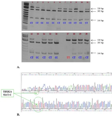
biolabs, Ipswich, MA, USA) restriction enzyme analysis on agarose gel (Fig. 1A) and further we
confirmed the results sequencing the TBXA2 gene (Fig. 1B).
Fig. 1. A. Restriction enzyme analysis on agarose gel. B. The human TBXA2R gene is located on
19p13.3, which spans 15 kbps containing three exons divided by two introns. The two variants of the
TBXA2R receptor have been described, based on the presence or absence of C924T polymorphism.
This polymorphism, defined by RsaI digestion on PCR product, has been confirmed by sequence
analysis.
For Western blot analysis, total proteins were extracted from lymphomonocyte isolated from whole
blood of selected patients using a Ficoll-Paque [17]. The cell pellets were lysed in a buffer containing
50 mM Tris.Cl pH 7.8, 1% Triton X100, 0.1% SDS, 250 mM NaCl, 5 mM EDTA, 100 mM NaF, 2
mM NaPPi, 2 mM Na3VO4, 1 mM PMSF. Cell lysates were loaded onto a 12% SDS-PAGE and the
separated proteins electrophoretically transferred to a polyvinylidene difluoride membrane (Bio-Rad,
Hercules, CA, USA). Membranes were incubated overnight at 4°C with a specific polyclonal
anti-TBXA2R Ab (Cayman Chemical, Ann Arbor, MI, USA). Membranes were then incubated with a
secondary anti-rabbit Ab (Cayman Chemical, Ann Arbor, MI, USA) for 1 h at room temperature. In
order to confirm that equal amounts of protein were loaded in each lane, the membranes were
incubated with an anti-actin antibody (Santa Cruz Biotechnology, Dallas, TX, USA) [18].
Immunocomplexes were visualized using the ECL detection system (GE Healthcare Life Sciences,
UK). Densitometric analysis was performed for the quantification of the immunoblots, using the
Molecular Analyst System (Bio Rad, Hercules, CA, USA).
Platelet aggregation
All patients were stratified on the basis of their genotype profile (CC, CT and TT) for the SNP C924T
and analyzed for platelet aggregation test. Blood samples were collected without stasis from the
antecubital vein with a 19 G butterfly needle, after 12–14 hrs of fasting. Each sample was
anti-coagulated with a 3.8% sodium citrate solution (1:10 v/v). The platelet-rich plasma (PRP) was
obtained by centrifugation at 160g for 10 min at room temperature. Platelet-poor plasma (PPP) was
prepared by further centrifugation of the remaining blood at 2,000g for 20 min. PRP was adjusted
with PPP to reach a platelet count of 250,000–300,000/ml. Platelet aggregation was induced by
challenging with Adenosine diphosphate (ADP) at 2 and 6 M, epinephrine at 0.6 and 10 g/ml and collagen at 1 and 9 mg/ml final concentrations. To evaluate platelet aggregation, according to the
Born method [19], a multichannel aggregation analyzer (ARKRAY) was used. Optical density for
PRP was set at 0% and for PPP at 100%. In addition, platelet aggregation induced by ADP
stimulation was also evaluated as closure time using a PFA-100 analyzer (Siemens Medical Solutions
Cell culture
The EBV-producing MEG-01 human cell line (ATCC) was grown in RPMI 1640 medium,
supplemented with 10% heat- inactivated fetal bovine serum (FBS), 2 mM L-glutamine and 1%
Pen-Strep at 37 °C in 5% CO2. The cells were seeded at 2.0 x 10 5
/ml and the experiment was performed in
triplicate. This cell clone was genotyped for heterozygosity at the T924C site.
Dose response with Actinomycin D on MEG-01 cell line
MEG-01 cells were seeded on six-well plates (2 x10⁵ cells/well) and treated with increasing doses (1
µM – 5 µM – 10 µM – 15 µM – 20 µM) of the transcription inhibitor Actinomycin D (Sigma Aldrich,
Saint Louis, MO, USA) at different exposure times (0, 1, 6, 12, 24 hours). After treatment, the cells
were further incubated at 37 °C in 5% CO2. Cell pellets were washed three times in PBS, centrifuged
(1,000 rpm for five minutes), the supernatant was removed and finally the cells were re-suspended
and counted using a Burker chamber and Trypan blue stain, for testing the viability.
Analysis of the steady state mRNA level of TBXA2R by qRT-PCR after Actinomycin D treatment on MEG-01 cell line
MEG-01 cells were seeded on Petri dishes (2x10⁵ cells/ml) and treated with 10 µM of Actinomycin D
at different exposure times (0, 1, 6, 12, 24 hours), as reported in several studies [20]. After treatment,
the cells were incubated at 37 °C in 5% CO2, washed two times with PBS and then lysed directly in
culture dishes using 1 mL of TRIZOL® Reagent. RNA isolation and extraction was performed using
the manufacturer’s protocol (Invitrogen, Carlsbad, CA, USA). The PCR primers for TBXA2R (+924C/T) allele-specific were respectively: forward for ―C‖ allele (F: 5’-CTGGACCCCTGGGTGTAT - 3’); forward for ―T‖ allele (F: - CTGGACCCCTGGGTGTAC);
reverse (R: 5’- GGAGAGTGCTTGGTAAAAGGATCA- 3’). The PCR primers for the TBXA2R preserved region outside of the C924T polymorphism, used as a reference gene, were: forward (F: 5’-
CCCAATCCAACCCGGG) and reverse (R: 5’- GGAGAGTGCTTGGTAAAAGGATCA) (Operon
molecules for life, Mountain View, CA, USA). Subsequently, we performed a qRT-PCR (Rotor-Gene
probes for the TBXA2R (+924C/T): for ―T‖ allele VIC-AGACACCCAGGGGTCCAG[MGB:minor
groove binding]; for ―C‖ allele FAM-GGACACCCAGGGGTCCAG[MGB]; for the TBXA2R
preserved region were: VIC-TCCAGGATCTGGTTCCA[MGB] and FAM-ACCCCCAACTCCTC
[MGB]). In addition, we used the GAPDH, as a qPCR reference gene, in order to evaluate the
efficiency of the assay (Supplementary Fig. S1).
Statistical analysis
Data are expressed as means ± SEM. Student t test or ANOVA followed by the appropriate post-hoc
test was used (Stat View 4.0 software, Abacus Concepts, Berkeley, CA, USA). P values < 0.05 were
considered statistically significant, with a confidence interval of 95%.
Results
Dose response with Actinomycin D on MEG-01 cell line
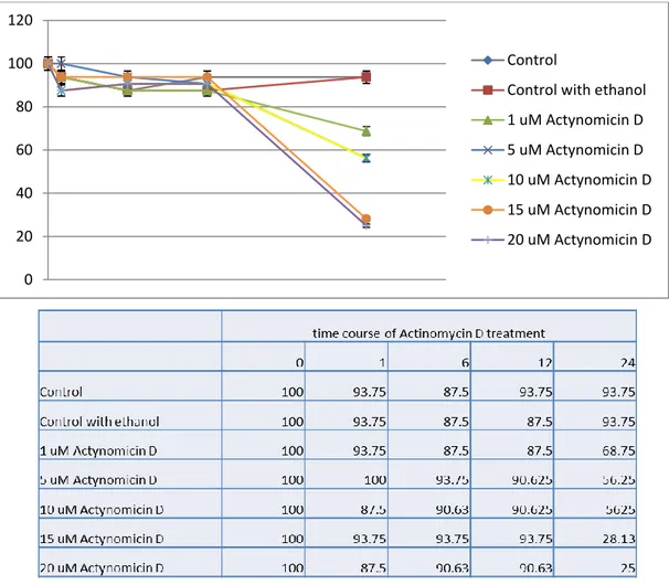
As shown in Fig. 2, a concentration of Actinomycin D of at least 5 M had a greater effect on cell viability. The decrease on cell viability was more evident on cells treated for 24 hr. The samples
treated with 5-10 M, 15 M and 20 M of Actinomycin D showed, a 44%, 72% and 75% in reduction of cell viability compared to a control group (p< 0.05), respectively. The samples treated
with 1 M of Actinomycin D didn’t show significant difference on cell viability as compared to the control group.
Fig. 2 Effect of Actinomycin D on cell viability. The decrease on cell viability is highly evident on
cells treated for 24 h, whereas samples treated with 5-10 M, 15 M and 20 M of Actinomycin D show a 44%, 72% and 75% in reduction of cell viability compared to the controls (p< 0.05),
respectively. Decrease in cell viability is more relevant for doses higher than 5 M and for exposure times longer than 24 h. The samples treated with 1 M of Actinomycin D didn’t show significant difference on cell viability as compared to the control group. The experiment was performed in
triplicate.
Analysis of the steady state mRNA level of TBXA2R by qRT-PCR after Actinomycin D treatment on MEG-01 cell line
0 20 40 60 80 100 120 Control
Control with ethanol 1 uM Actynomicin D 5 uM Actynomicin D 10 uM Actynomicin D 15 uM Actynomicin D 20 uM Actynomicin D
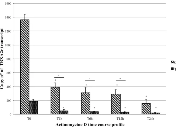
The influence of the C924T polymorphism on MEG-01 cells, after treatment with 10 M Actinomycin D, at different times, was analyzed. As shown in fig. 3 the TBXA2R polymorphism
expression was higher for the C allele compared to the T allele. We observed a significant decreased
expression of both alleles, when compared to the respective controls and lower levels of transcript
after 6 hours of treatment (p< 0.05). In addition the statistical analysis of the fold changes between the
C and T treated groups compared to the control ones showed that the mRNA stability was greater for
the C allele compared to the T allele, which instead evidenced a high degree of instability (Fig. 3).
Fig. 3. A significant decreased expression of C and T alleles was observed on MEG-01 cells treated
with 10 M of Actinomycin D at different exposure times when compared to the respective controls. *: p< 0.05 compared to the respective controls. The C and the T allele were down-regulated 3.27 and
3.87 fold (°: p= 0.013) in T1h groups, 4.62 and 5.33 fold (°: p= 0.023) in T6h, 4.89 and 6.86 fold (°:
p< 0.001) in T12h, 9.92 and 9.85 fold (p= 0.9) in T24h, when compared to the respective control
groups. This data suggests a higher instability of the mutant type transcript and an exponential
0 200 400 600 800 1000 1200 1400 1600 T0 T1h T6h T12h T24h C op y n ° of T B X A 2r t ra n sc ri p t
Actinomycine D time course profile
C T * * * * * * * * ° ° °
decrease of TBXA2R mRNA as early as 1 hour after treatment. Fold change = 2-∆∆Ct. The experiment
was performed in triplicate.
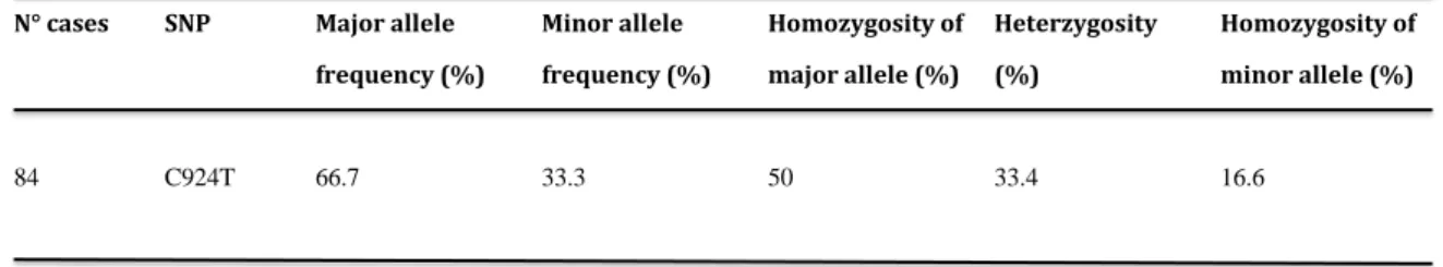
C924T allele frequency and patients’ genotype profile
The table 1 showed the allele frequency and the patients’ genotype profile of the C924T
polymorphism for all the patients recruited in this study. Specifically, we reported the presence of 42,
28 and 14 patients with a CC, CT and TT genotype, respectively.
Table 1. C924T polymorphism of the coding sequence of the TP receptor gene in an Italian
population study.
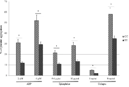
Platelet aggregation analysis
Platelet aggregation for the TBXA2R gene polymorphism C924T was analyzed in all patients,
stratified on the basis of their genotype profile (CC, CT and TT), using a PFA-100 analyzer. As
shown on table 2, the platelet aggregation induced by ADP was more effective on patients with at
least one C allele. Furthermore, these results point out that the platelet aggregation induced by ADP
was less efficient in the mutant genotype (TT), with respect to the wild type (n.r. 55-137 sec.). In
addition, platelet aggregation induced by challenging with Adenosine diphosphate (ADP) at 2 and 6
M, epinephrine at 0.6 and 10 g/ml and collagen at 1 and 9 mg/ml final concentrations was analyzed for the TBXA2R gene polymorphism in wild type patients (CC) and in mutated patients (TT) using a
multichannel aggregation analyzer (ARKRAY), according to the Born method (Fig. 4). These results
confirmed the data obtained by PFA-100 highlighting a less effective platelet aggregation on patients
with a TT genotype compared to those with a CC genotype.
N° cases SNP Major allele Minor allele Homozygosity of Heterzygosity Homozygosity of
frequency (%) frequency (%) major allele (%) (%) minor allele (%)
Table 2. Platelet aggregations analysis of CC, CT and TT patients for the polymorphism C924T of
the TBXA2R gene was evaluated as closure time using a PFA-100 analyzer. Patients with a TT
genotype showed a less efficient platelet aggregation compared to those with at least one C allele
(ANOVA p< 0.05; Bonferroni test between TT and CT or CC: p< 0.05)
Fig. 4. Platelet aggregation analysis of CC and TT patients for the polymorphism C924T of the
TBXA2R gene was evaluated using ADP at 2 and 6 M, epinephrine at 0.6 and 10 g/ml and collagen at 1 and 9 mg/ml final concentrations with using a multichannel aggregation analyzer
(ARKRAY), according to the Born method. * p< 0.05
TBXA2R protein expression
Genotype N° of cases Platelet aggregation (sec.) ANOVA (p)
CC 42 80.3 (± 2.1) < 0.05
CT 28 102 (± 8.4)
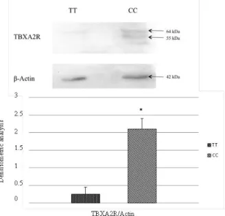
6 and 5 patients, respectively with a CC and a TT genotype based on the C924T polymorphism, were
randomly selected in order to evaluate the protein expression of the TP receptor (Fig. 5). The CC
patients showed a higher level of expression of TBXA2 receptor, with two different degrees of
glycosylation (55 and 64 kDa), when compared to patients with a TT genotype (p< 0.05). The data
showed a higher stability of the wild type transcript respect to the mutant type, confirming the results
obtained through the analysis of the steady state mRNA level of TBXA2R by qRT-PCR after
Actinomycin D treatment on MEG-01 cell line.
Fig. 5 The protein expression of TBXA2R on wild-type (CC) and mutant type (TT) patients was
analyzed. Wild type (CC) shows a higher expression level of TBXA2 receptor, recognizing two
different bands, with a different degree of glycosylation, (55 and 64 kDa), compared to the mutant
type (TT) (*: p< 0.05). This data strongly support the evidence of a higher stability of the wild type
transcript respect to the mutant type.
Previous studies have shown that leukocytes, platelets and endothelial cells (21, 22) are involved in
the biosynthesis of bioactive lipid mediators derived from arachidonic acid. For instance, stimulation
of neutrophils (23, 26) results in production of LTA4 [21, 22]. On the other hand, the reduction in
LTC4 biosynthesis observed in patients with unstable angina during 6-methylprednisolone (6-MP)
treatment could be explained by the glucocorticoid-dependent decrease of adhesion molecule
expression on blood and vascular cell surfaces [27].
Furthermore, a study by Vejar et al. examined the rate of thromboxane biosynthesis in the setting of
acute coronary syndromes through measurements of plasma levels of thromboxane B2, and of the
urinary excretion of major enzymatic metabolites such as 11-dehydro-thromboxane B2 and
2,3-dinor-thromboxane B2. This study demonstrated that the administration of low-doses of aspirin are
associated with statistically significant reduction in thromboxane biosynthesis by 70% [28].
Moreover, experimental and clinical studies have shown the characterization of variable patterns of
COX-2 expression as a key player in atherothrombosis and led to hypothesize a role for COX-2–
derived prostanoids in vascular disease progression and its thrombotic complications [29, 30]. The
results of morphological, pharmacological, and genetic studies of the human carotid plaque model are
consistent with the hypothesis that down-regulation of COX-2 expression in inflammatory cells may
protect against atherothrombosis in high-risk aspirin-treated patients [31, 32].
In light of the above findings, in the present study we focused our attention on the TBXA2 receptor as
an important contributor on atherosclerosis lesion formation and development. We investigated the
possibility that post-transcriptional mechanisms of regulation may be involved in the expression and
stability of TBXA2R, related to the C924T genotype. We used, as bio-molecular model, a T924C
heterozygosis cell clone derived from the MEG-01 cell line. The results evidenced that these cells
treated with increasing doses of actinomycin D had a reduction in viability. In particular, the decrease
in cell viability was more relevant for doses higher than 5 M and for exposure times longer than 24 h, which is consistent with a role for the TBXA2R in mediating cell survival. Furthermore, the
TBXA2R polymorphism expression was greater for the C allele compared to the T allele, regardless
of the actinomycin D exposure times. The analysis of the steady state mRNA level of TBXA2R by
the mRNA stability was greater for the wild type (CC) compared to the mutant type (TT), and the
latter showed a decrease of its transcript levels as early as 3 hours after treatment. This evidence
suggests a protective role for the TBXA2R TT genotype in the development and progression of
atherosclerosis lesions.
In order to better characterize the TBXA2R, its polymorphisms and the expression levels of the two
different alleles were studied using lymphomonocytes isolated from target patients. The Western blot
analysis showed that the wild type (CC) had a higher expression level of the TBXA2 receptor with
two different degrees of glycosylation (55 and 64 kDa), when compared to the mutant type (TT) on at
least twelve different genotyped patients. This data evidenced an increased instability of the mutant
(TT) type transcript in respect to the wild type, confirming the results obtained through the analysis of
the steady state mRNA level of TBXA2R by qRT-PCR after actinomycin D treatment of MEG-01
cell line. Moreover, a platelet aggregation analysis on selected patients, a wild (CC) and a mutant type
(TT) for the C924T polymorphism, was performed. The platelet aggregation induced by ADP,
epinephrine and collagen, at different concentrations, was less effective for the mutant type (TT). This
is consistent with a reduced thrombus formation and hemostasis. Thus, the instability of the TBXA2R
transcript and the associated reduction of platelet aggregation [33] might be associated with a
protective role for the TBXA2R TT genotype against atherothrombosis and its complications in
high-risk aspirin-treated patients. These findings could represent the basis for the prediction of plaque
formation and atherosclerosis by analyzing the patients’ genotype for the TBXA2R C924T
polymorphism. This approach could allow the identification of subjects more susceptible to
atherothrombosis, in particular among high-risk aspirin-treated patients.
Compliance with Ethical Standards
Ethical Approvals: Informed consent was given by patients whose retrospective data were used for
this study. In addition, according to the guidelines of the declaration of Helsinki for retrospective
studies, we notified such project to the University Department where experiments were conducted and
Conflicts of interest: none.
Funding: This project was partially funded by 60% Ateneo grants from Ministero dell’Università,
Italy to S.M. and E.T. We also received partial contribution to research expenses by the Department
References
1. Hansson GK. Inflammation, atherosclerosis, and coronary artery disease. N Engl J Med.
2005;352:1685–1695.
2. Zhang L, Tian CM, Liu QY, Wang SS, Wang X, Chen L, Feng Y, Guo LT. Clinical diagnosis
of carotid atherosclerostic plaque in hypertensive patients with high resolution magnetic
resonance angiography. J Biol Regul Homeost Agents. 2015 Apr-Jun;29(2):411-5.
3. Naghavi M, Libby P, Falk E, Casscells SW, Litovsky S, Rumberger J, et al. From vulnerable
plaque to vulnerable patient: a call for new definitions and risk assessment strategies: part I.
Circulation 2003;108:1664–1672.
4. Kobayashi T, Tahara Y, Matsumoto M, Iguchi M, Sano H, Murayama T et al. Roles of
thromboxane A (2) and prostacyclin in the development of atherosclerosis in apoE-deficient
mice. J Clin Invest 2004;114:784–794.
5. Cyrus T, Ding T, Praticò D. Expression of thromboxane synthase, prostacyclin synthase and
thromboxane receptor in atherosclerotic lesions: correlation with plaque composition.
Atherosclerosis 2010;208:376–381.
6. Cayatte AJ, Du Y, Oliver-Krasinski J, Lavielle G, Verbeuren TJ, Cohen RA. The
thromboxane receptor antagonist S18886 but not aspirin inhibits atherogenesis in apo
E-deficient mice: evidence that eicosanoids other than thromboxane contribute to
atherosclerosis. Arterioscler Thromb Vasc Biol. 2000;20:1724–1728.
7. Hock CE, Brezinski ME, Lefer AM. Anti-ischemic actions of a new thromboxane receptor
antagonist, SQ-29,548, in acute myocardial ischemia. Eur J Pharmacol. 1986;122:213–219.
8. Hamberg M, Svensson J, Samuelsson B. Thromboxanes: a new group of biologically active
compounds derived from prostaglandin endoperoxides. Proc Natl Acad Sci USA
1975;72:2994–2998.
9. Reale M, Barbacane RC, Frydas S, Anogianakis G, Trakatellis A, Dimitriadou D, Vacalis D,
beta induces thromboxane A2 release in polymorphonuclear leukocytes, macrophages and
platelets: effect of IL-1 receptor antagonist. Mol Cell Biochem. 1996 Jun 21;159(2):163-8.
10. Cipollone F, Toniato E, Martinotti S. A Polymorphism in the Cyclooxygenase 2 Gene as an
Inherited Protective Factor Against Myocardial Infarction and Stroke. JAMA 2004—Vol.
291, No. 18.
11. Needleman P, Moncada S, Bunting S, Vane JR, Hamberg M, Samuelsson B. Identification of
an enzyme in platelet microsomes which generates thromboxane A2 from prostaglandin
endoperoxides. Nature 1976;261:558–560.
12. Shen RF, Tai HH. Thromboxanes: synthase and receptors. J Biomed Sci. 1998;5:153–172.
13. Nusing RM, Hirata M, Kakizuka A, Eki T, Ozawa K, Narumiya S. Characterization and
chromosomal mapping of the human thromboxane A2 receptor gene. J Biol Chem.
1993;268:25253–25259.
14. Raychowdhury MK, Yukawa M, Collins LJ, McGrail SH, Kent KC, Ware JA. Alternative
splicing produces a divergent cytoplasmic tail in the human endothelial thromboxane A2
receptor. J Biol Chem. 1994;269:19256–19261.
15. Hirata T, Kakizuka A, Ushikubi F, Fuse I, Okuma M, Narumiya S. Arg60 to Leu mutation of
the human thromboxane A2 receptor in a dominantly inherited bleeding disorder. J Clin
Invest. 1994;94:1662–1667.
16. Unoki M, Furuta S, Onouchi Y, et al. Association studies of 33 single nucleotide
polymorphisms (SNPs) in 29 candidate genes for bronchial asthma: positive association a
T924C polymorphism in the thromboxane A2 receptor gene. Hum Genet. 2000;106:440–446.
17. Corkum CP et al. Immune cell subsets and their gene expression profiles from human PBMC
isolated by Vacutainer Cell Preparation Tube (CPT™) and standard density gradient. BMC Immunol. 2015;16(1):48.
18. Jakobsson J, Thorén S, Morgenstern R, et al. Identification of human prostaglandin E
synthase: a microsomal, glutathione-dependent, inducible enzyme, constituting a potential
19. Born GV. Aggregation of blood platelets by adenosine di-phosphate and its reversal. Nature.
1962;194:927-9.
20. Zajkowicz A et al. Actinomycin D and nutlin-3a synergistically promote phosphorylation of
p53 on serine 46 in cancer cell lines of different origin. Cell Signal. 2015;27(9):1677-87.
21. Fabre JE, Goulet JL, Riche E, et al. Transcellular biosynthesis contributes to the production of
leukotrienes during inflammatory responses in vivo. J Clin Invest. 2002;109:1373–1380.
22. Mastrangelo F, Sberna MT, Tettamanti L, Cantatore G, Tagliabue A, Gherlone E. Vascular
endothelial growth factor and nitric oxide synthase expression in human tooth germ
development. J Biol Regul Homeost Agents. 2016 Apr-Jun;30(2):421-32.
23. McGee JE, Fitzpatrick FA. Erythrocyte-neutrophil interactions: formation of leukotriene B4
by transcellular biosynthesis. Proc Natl Acad Sci USA. 1986;83:1349–1353.
24. Manukyan G, Petrek M, Navratilova Z, Margaryan S, Boyajyan A. Transcriptional activity of
neutrophils exposed to high doses of colchicine: short communication. J Biol Regul Homeost
Agents. 2015 Jan-Mar;29(1):125-30.
25. Żbikowska-Gotz M, Pałgan K, Gawrońska-Ukleja E, Kuźmiński A, Przybyszewski M, Socha
E, Bartuzi Z. Expression of IL-17A concentration and effector functions of peripheral blood
neutrophils in food allergy hypersensitivity patients. Int J Immunopathol Pharmacol. 2016
Mar;29(1):90-8. doi: 10.1177/0394632015617069.
26. Nacaroglu HT, İsgüder R, Bent S, et al. Can neutrophil/lymphocyte ratio be a novel
biomarker of inflammation in children with asthma? Eur J Inflamm. August 2016 14:
109-112, first published on July 21, 2016 doi:10.1177/1721727X16660558
27. Cronstein BN, Kimmel SC, Levin RI, et al. Mechanism for the anti-inflammatory effects of
corticosteroids: the glucocorticoid receptor regulates leukocyte adhesion to endothelial cells
and expression of endothelial-leukocyte adhesion molecule 1 and intercellular adhesion
28. Vejar M, Fragasso G, Hackett D, Lipkin PD, Maseri A, Born GVR, et al. Dissociation of
platelet activation and spontaneous myocardial ischemia in unstable angina. Thromb
Haemost. 1990;63:163-168.
29. McEver R. Adhesive interactions of leukocyte, platelets and vessel wall during hemostasis
and inflammation. Thromb Haemostas. 2001; 86:746–756.
30. Koki A, Khan NK, Woerner BM, Dannenberg AJ, Olson L, Seibert K, et al.
Cyclooxygenase-2 in human pathological disease. Adv Exp Med Biol. Cyclooxygenase-200Cyclooxygenase-2;66:13–18.
31. Burleigh ME, Babaev VR, Oates JA, Harris RC, Gautam S, Riendeau D, et al.
Cyclooxygenase-2 promotes early atherosclerotic lesion formation in LDL receptor–deficient
mice. Circulation 2002;105:1816–1823.
32. Eikelboom JW, Hirsh J, Weitz JI, Johnston M, Yi Q, Yusuf S. Aspirin-resistant thromboxane
biosynthesis and the risk of myocardial infarction, stroke, or cardiovascular death in patients
at high risk for cardiovascular events. Circulation 2002;105:1650–1655.
33. Duan HQ, Dong PS, Wang HL, Li ZJ, Du LJ, Zhao YW. Effect of prolonged dual
anti-platelet therapy on reducing myocardial infarction rate after percutaneous coronary