Components of a Neanderthal gut microbiome
recovered from fecal sediments from El Salt
Simone Rampelli
1,17
, Silvia Turroni
1,17
, Carolina Mallol
2,3,4
, Cristo Hernandez
2
, Bertila Galván
2
,
Ainara Sistiaga
5,6
, Elena Biagi
1
, Annalisa Astol
fi
7,16
, Patrizia Brigidi
8
, Stefano Benazzi
9,10
, Cecil M. Lewis Jr.
11,12
,
Christina Warinner
12,13
, Courtney A. Hofman
11,12
, Stephanie L. Schnorr
14,15,18
✉
& Marco Candela
1,18
✉
A comprehensive view of our evolutionary history cannot ignore the ancestral features of our
gut microbiota. To provide some glimpse into the past, we searched for human gut
micro-biome components in ancient DNA from 14 archeological sediments spanning four
strati-graphic units of El Salt Middle Paleolithic site (Spain), including layers of unit X, which has
yielded well-preserved Neanderthal occupation deposits dating around 50 kya. According to
our
findings, bacterial genera belonging to families known to be part of the modern human
gut microbiome are abundantly represented only across unit X samples, showing that
well-known bene
ficial gut commensals, such as Blautia, Dorea, Roseburia, Ruminococcus,
Faecali-bacterium and Bi
fidobacterium already populated the intestinal microbiome of Homo since as
far back as the last common ancestor between humans and Neanderthals.
https://doi.org/10.1038/s42003-021-01689-y
OPEN
1Unit of Microbiome Science and Biotechnology, Department of Pharmacy and Biotechnology, University of Bologna, Via Belmeloro 6, Bologna, Italy. 2Department of Geography and History, University of La Laguna, Campus de Guajara, La Laguna, Tenerife, Spain.3Archaeological Micromorphology and
Biomarker Research Lab, University of La Laguna, Avenida Astrofísico Francisco Sánchez 2, La Laguna, Tenerife, Spain.4ICArEHB - Interdisciplinary Center for Archaeology and the Evolution of Human Behaviour, Universidade do Algarve, Campus de Gambelas, Edificio 1, Faro, Portugal.5Earth, Atmospheric and Planetary Sciences Department, Massachusetts Institute of Technology, 77 Massachusetts Ave, Cambridge, MA, USA.6GLOBE Institute, Faculty of Health and Medical Sciences, University of Copenhagen, Oester Voldgade 5-7, Copenhagen, Denmark.7“Giorgio Prodi” Cancer Research Center, University of Bologna, Via Massarenti 11, Bologna, Italy.8Department of Medical and Surgical Sciences, University of Bologna, Via Massarenti 9, Bologna, Italy.
9Department of Cultural Heritage, University of Bologna, Via degli Ariani 1, Ravenna, Italy.10Department of Human Evolution, Max Planck Institute for
Evolutionary Anthropology, Deutscher Platz 6, Leipzig, Germany.11Laboratories of Molecular Anthropology and Microbiome Research, University of Oklahoma, 101 David L. Boren Blvd, Norman, OK, USA.12Department of Anthropology, University of Oklahoma, 455W Lindsey St, Norman, OK, USA.
13Department of Archaeogenetics, Max Planck Institute for the Science of Human History, Kahlaische Strasse 10, Jena, Germany.14Konrad Lorenz Institute
for Evolution and Cognition Research, Martinstraße 12, Klosterneuburg, Austria.15Department of Anthropology, University of Nevada, 4505S. Maryland
Pkwy, Las Vegas, NV, USA.16Present address: Department of Morphology, Surgery and Experimental Medicine, University of Ferrara, Via Fossato di Mortara 70, Ferrara, Italy.17These authors contributed equally: Simone Rampelli and Silvia Turroni.18These authors jointly supervised this work: Stephanie L. Schnorr and Marco Candela. ✉email:[email protected];[email protected]
123456789
O
ver the past decade, microbiome research has
high-lighted the crucial role that the gut microbiome plays in
human biology through its pleiotropic influence on
many physiological functions, such as human development,
immunity, metabolism and neurogenerative processes
1. This
body of knowledge has catalyzed interest in incorporating the
gut microbiome into our evolutionary history, as an adaptive
partner providing the necessary phenotypic plasticity to buffer
dietary and environmental changes. Studies aimed at exploring
the ancestral traits of the human gut microbiome are therefore
encouraged, as a unique evolutionary perspective to improve
our knowledge of gut microbiome assembly and interactions
with the human host
2.
The ancestral configuration of the human gut microbiome has
generally been inferred by microbiome data stemming from
contemporary populations found across all six human occupied
continents who adhere to traditional lifestyles, such as the Hadza
hunter-gatherers from Tanzania, the rural Bassa from Nigeria and
rural Papuans from Papua New Guinea, among others
3–11.
However, since this research involved modern populations, no
direct information on the ancient human gut microbiome
structure can actually be provided.
Alternatively, ancient DNA (aDNA) analysis based on
shot-gun metagenomic sequencing is emerging as an attractive and
reliable opportunity to directly investigate the microbial ecology
of our ancestors
12–15. Paleomicrobiological aDNA studies have
traditionally been conducted on dental calculus and bones
15–18,
providing landmark information on ancient pathogens and oral
microbial communities. However, given that stools are widely
acknowledged as a proxy of the gut microbiome structure
19, the
metagenomics of aDNA from paleofeces, also known as
coprolites, represents the only way to gain insight into the
ancient human gut microbiome
2. Pioneering studies in this
direction have been carried out, providing next-generation
sequencing data from modern human mummified intestinal
contents and paleofeces
20–24. Nevertheless, to the best of our
knowledge, paleofecal samples older than 8,000 years have never
being analyzed, leaving an important gap on the pre-historical
human gut microbiome configuration.
In this scenario, we attempted to identify ancient human gut
microbiome components by shotgun metagenomic analysis of
aDNA extracted from archeological sedimentary samples (ES1
to ES7) from the stratigraphic unit (SU) X (subunit Xb-H44) of
the Middle Paleolithic open-air site, El Salt (Alicante, Spain)
25(Fig.
1
). The archeological setting of El Salt yielded evidence of
recurrent occupation by Neanderthals, our closest evolutionary
relatives, dated between 60.7 ± 8.9 and 45.2 ± 3.4 kya
26,27. In
particular, the sedimentary samples ES1-7 have been previously
shown to include several millimetric phosphatic coprolites and
fecal lipid biomarkers, namely coprostanol and
5ß-stigmasta-nol, with proportions suggesting a human origin
25. These
samples therefore represent, to our knowledge, the oldest
known positive identification of human fecal matter. The
pre-sent work also includes an additional seven new archeological
sediments collected in 2018 as a control. Two were from SU X
(subunit Xa and Xb, respectively) and the others from
sur-rounding SUs, i.e., upper V (three samples), IX and XI (one
sample each) (Fig.
2
a). While SUs IX to XI are associated with
rich archaeological assemblages, upper SU V yielded very few
archaeological remains
26. We found that samples positive for
the presence of fecal biomarkers showed traces of both ancient
human mtDNA and ancient components of the modern human
gut microbiome. These components included so-called
“old
friends” and beneficial commensal inhabitants of modern
human guts, providing unique insights into their relevance to
the biology of the Homo lineage.
Results and discussion
Ancient DNA sequencing and damage assessment. DNA was
extracted from 14 archeological sedimentary samples and
pre-pared for shotgun metagenomics in a dedicated aDNA facility at
the Laboratories of Molecular Anthropology and Microbiome
Research in Norman (OK, USA) (see Methods). A total of
124,592,506 high-quality paired-end sequences were obtained by
Illumina NextSeq sequencing and analyzed for bacterial aDNA.
To remove contamination by modern DNA, which is one of the
major complications in studies of ancient samples
14,28,29, we
evaluated the DNA damage pattern as compared with
present-day DNA references. In particular, Skoglund and colleagues
12translated the pattern of cytosine deamination into a postmortem
degradation score (PMDS), which provides information on
whether a given sequence is likely to derive from a degraded
aDNA molecule. Reads were aligned against all bacterial genomes
of the NCBI database, and ancient bacterial reads were recovered
by setting PMDS > 5, to minimize the probability of a sequence
being from a present-day contaminating source
12. An average of
6,836 sequences per sample (range, 279–17,901) were retained,
corresponding to a small but consistent fraction of DNA being
ancient and derived from bacteria (mean ± SD, 0.069% ± 0.029%)
(Supplementary Table 1 and Supplementary Fig. 1). The same
procedure was applied to extraction, library and PCR blanks,
resulting in the retrieval of a minimal number of 144, 1, and 42
ancient bacterial sequences, respectively. Ancient reads from
blanks were assigned to 116 bacterial species that showed no
overlap with the sample dataset (Supplementary Data 1). When
comparing the fraction of reads with PMDS > 5 per million reads
between samples IX, Xa, ES1-7, Xb and XI (i.e., those positive for
the presence of fecal biomarkers and/or associated with rich
archaeological assemblages), and samples from SU V (i.e., with no
or very few archeological remains), the
first showed a greater
abundance of PMDS > 5 reads (p-value
= 0.01, Wilcoxon test)
(Supplementary Fig. 2), possibly as a result of the presence of
human fecal sediment.
Detection of ancient human mitochondrial DNA. In order to
detect human aDNA traces in our sample set, we searched for
human mitochondrial DNA (mtDNA) sequences in PMDS-filtered
metagenomes obtained from the 14 archeological sedimentary
samples. Ancient human mtDNA was detected in almost all ES1 to
ES7 samples from SU X (Fig.
2
b). No traces of mtDNA from other
animals were detected. To strengthen these
findings, all samples
were subjected to target capture of mtDNA with a Neanderthal bait
panel (Arbor Biosciences; see Methods), and subsequent sequencing
on Illumina NextSeq platform. Based on this analysis, ES1, ES2, ES5
and Xb samples tested positive for the presence of ancient human
mtDNA, showing >1000 human mtDNA reads with PMDS > 1,
breath of coverage >10%,
−Δ % ≥ 0.9 and modern contamination
less than 2% (Fig.
2
b and Supplementary Table 2)
30. Taken
toge-ther, this evidence strongly supports human origin for the El Salt
samples, particularly those from SU X
25.
Profiling of ancient prokaryotic DNA. As for prokaryotic
aDNA, seventeen bacterial and one archaeal phyla were identified
in the aDNA sedimentary record of El Salt, with different
representation across SUs (Fig.
3
). As expected for its wide
dis-tribution in nature
31,32, Actinobacteria is the most represented
phylum, with environmental species from Streptomycetaceae,
Pseudonocardiaceae, Micromonosporaceae, Nocardiaceae,
Myco-bacteriaceae, Microbacteriaceae and Nocardioidaceae families
being detected in almost all the SUs. Similarly, the vast majority
of sediment samples share a number of ancient sequences
assigned to Bacillaceae members, which are known to play
fundamental roles in soil ecology, where they can persist up to
thousands of years, if not longer, due to their ability to form
resistant endospores
33,34. Another large fraction of aDNA shared
by most SUs includes Proteobacteria constituents, especially from
Alphaproteobacteria (mainly Rhodobacteraceae, Rhodospirillaceae
and Sphingomonadaceae families), Betaproteobacteria (mainly
Comamonadaceae and Burkholderiaceae) and
Gammaproteo-bacteria (with Xanthomonadaceae) classes. Again, these are
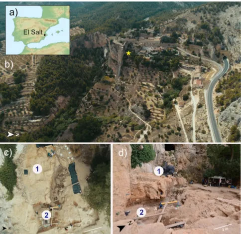
Fig. 1 The Middle Paleolithic site of El Salt (Spain). a, b General site setting. The yellow star marks the location of the excavated area. As can be seen in the photograph, the sedimentary deposit rests against a tall limestone wall.c, d Different views of the excavation area indicating the zones sampled for this study. Zone 1 includes samples V1-3 and Zone 2 all the rest.Fig. 2 Pleistocene stratigraphic sequence of El Salt (Units V-XI), chronometric dates of the sampled units, and evidence of traces of ancient human mitochondrial DNA. a The 14 sediment samples included in this study are shown in red. Samples ES1 to ES7 (subunit Xb-H44) are from Sistiaga et al.25.
b Red boxes, human mtDNA fragments as recovered from metagenomic sequencing data; red circles, confirmation of the presence of ancient human mtDNA through target capture and sequencing (please, see Methods for further details).
cosmopolitan bacteria commonly found in both terrestrial and
aquatic environments, as free-living organisms or symbionts in
different hosts
35,36. In light of their DNA damage pattern, it is
reasonable to assume that these are truly ancient environmental
bacteria that populated archeological sediments. The
con-tamination of archeological remains by environmental bacteria is
indeed well expected, as already documented in previous
paleo-microbiological aDNA studies
15,18,37. For the relative abundances
of bacterial families detected across the samples, please see
Sup-plementary Data 2.
Putative components of the Neanderthal gut microbiome.
Next, following an approach similar to Weyrich et al.
18, who
first characterized the oral microbiome from Neanderthal
dental calculus, we focused our analysis on intestinal
micro-organisms. Specifically, in order to identify potential ancient
human gut microbiome components, we searched for
bac-terial genera belonging to the 24 families that have recently
been indicated as being common to the gut microbiome of
hominids (i.e., Methanobacteriaceae, Bifidobacteriaceae,
Cor-iobacteriaceae, Bacteroidaceae, Porphyromonadaceae,
Pre-votellaceae, Rikenellaceae, Tannerellaceae, Enterococcaceae,
Lactobacillaceae, Streptococcaceae, Christensenellaceae,
Clos-tridiaceae, Eubacteriaceae, Lachnospiraceae, Oscillospiraceae,
Peptostreptococcaceae, Ruminococcaceae, Erysipelotrichaceae,
Veillonellaceae,
Desulfovibrionaceae,
Succinivibrionaceae,
Enterobacteriaceae and Spirochaetaceae)
38–45. Accordingly,
while harboring similar family-level gut microbiome profiles,
humans and non-human hominids, including our closest
living relatives—chimpanzees, can be differentiated on the
basis of the particular pattern of associated gut microbiome
genera (as well as species and strains) represented within
these families
45. This strong association between gut
micro-biome
composition
and
host
physiology—known as
phylosymbiosis—is believed to be universal in mammals,
essentially as a result of all the physical, chemical and
immunological factors that differentiate the intestine of the
host species (e.g., type of digestive organs, pH, oxygen level,
Fig. 3 Ancient bacteria in sediment samples from El Salt. The phylogenetic tree was built with representative sequences of bacterial genera for which at least one species was present with more than four hits in one sample. Different colors indicate different phyla (or classes for Proteobacteria) as follows: cyan, Actinobacteria; blue, Bacteroidetes; red, Alphaprotebacteria; brown, Gammaproteobacteria; pink, Betaproteobacteria; purple, Deltaproteobacteria; yellow, Acidobacteria; green, Firmicutes; light-yellow, Planctomycetes; orange, Euryarchaeota; grey, others (including Armatimonadetes, Chlorobi, Chloroflexi, Cyanobacteria, Deinococcus-Thermus, Fusobacteria, Gemmatimonadetes, Nitrospirae, Spirochaetes, Synergistetes and Verrucomicrobia). Bacterial taxa belonging to families common to the gut microbiome of hominids are highlighted at different taxonomic level. A, Alistipes; P, Prevotella; B, Bacteroides; PB, Parabacteroides.host-derived molecules and immune system)
46. According to
our
findings, 210 bacterial species belonging to
hominid-associated gut microbiome families, as listed above, are
represented in the aDNA from El Salt SU IX, X and XI, with
the highest detection rate in samples from SU X and,
parti-cularly, in ES1 to ES7 (Fig.
4
), for which a human-like host
origin had been previously suggested
25. In Supplementary
Fig. 3, we provide the overall compositional profile of the El
Salt samples from SU IX, X and XI restricted to the
hominid-associated gut microbiome families, while in Supplementary
Fig. 4, the proportions of these families are compared with
those of the samples with no or very few archeological
remains (i.e., from SU V). The compositional profile of
samples from SU IX, X and XI was next compared to publicly
available gut microbiomes from contemporary human
popu-lations as representative of different subsistence practices,
such as Hadza and Matses hunter-gatherers, Tunapuco rural
agriculturalists and western urbans from Italy and the US
7,47.
As shown by the Principal Coordinates Analysis of
Bray-Curtis distances between the family-level profiles
(Supple-mentary Fig. 5), the El Salt samples from SU IX, X and XI tend
to cluster closer to Tunapuco and Matses, resembling more
the
“ancestral” human gut microbiome of rural
agricultural-ists and hunter-gatherers than the urban western gut
micro-biome
7. However, as the degree of degradation of microbial
DNA in ancient samples might be different for various gut
microbiome components, any conclusions from these
com-positional data must be taken with due caution.
Further supporting a human-host origin of the bacterial
species belonging to the hominid-associated gut microbiome
families detected in the El Salt samples from SU IX, X and XI,
feces or gastrointestinal tract are the
first documented isolation
source for 91 species out of 210 (43.3%), with 60 of these being
classifiable as closely related to the human gut (Supplementary
Data 3). In the latter subgroup, we can count several species from
Lachnospiraceae (including well-known (beneficial) commensal
inhabitants of modern human guts, such as Blautia, Coprococcus,
Dorea, Fusicatenibacter and Roseburia spp.) and Ruminococcaeae
families. Particularly, within Ruminococcaceae, we detected
members of Anaerotruncus, Ruminococcus and Subdoligranulum
genera, and the butyrate producer Faecalibacterium, one of the
human commensal bacteria of greatest current interest, due to its
very promising potential as a biomarker of a healthy gut
microbiome
48. Most of the aforementioned bacterial genera have
been reported to account for the phylotypic diversity between
human and non-human hominids, showing strong bias towards
a human-host
45. It is also worth remembering that most of these
bacteria are able to produce short-chain fatty acids (mainly
acetate and butyrate) from the fermentation of indigestible
carbohydrates, through the establishment of complex syntrophic
networks. Short-chain fatty acids are today considered metabolic
and immunological gut microbiome players with a leading role
in human physiology
49. In addition, the Xb-H44 samples showed
a high number of hits for Bacteroides, Parabacteroides, Alistipes
and Bifidobacterium spp., other genera known to prevail in the
human gut microbiome
39,45. Interestingly, Bacteroides and
Bifidobacterium have been shown to exhibit patterns of
co-speciation with hominids
45. For Bifidobacterium, this is
particu-larly consistent with the propensity of this genus to be maternally
inherited across generations. Being capable of metabolizing milk
oligosaccharides and acting as a potent immunomodulator, the
presence of vertically transmitted Bifidobacterium spp. in the
infant gut could have provided important growth benefits to
infant Hominidae
50–52.
To further characterize the ancient microbial taxa detected in
the El Salt samples, we applied the HOPS
53-based approach
recently used by Jensen et al.
30. In short, all the reads were
first
annotated and, subsequently, the ancient origin of each taxon was
authenticated by computing three indicators: (i) the fraction of
reads with PMDS > 1, (ii) the negative difference proportion
(−Δ %) of PMDS > 1 reads, and (iii) their deamination rate at 5′.
Taxa showing more than 200 assigned reads, more than 50 reads
with PMDS > 1,
−Δ % = 1 and C-T transition at 5′ >10% were
Fig. 4 Ancient components of the human gut microbiome in sedimentsamples from El Salt. The heat map shows the hit number distribution across sediment samples from the different stratigraphic units. Only ancient bacterial species with≥ 2 hits in at least one sample were kept. See also Supplementary Data 3.
considered to be of ancient origin (see Table
1
and
Supplemen-tary Figs. 6–8 for MapDamage plots, coverage plots and edit
distance distribution)
30. This in-depth characterization of the
microbial metagenomic reads from the El Salt samples allowed us
to confirm the presence of several species belonging to the gut
microbiome families of hominids (including, among others,
Alistipes, Bifidobacterium, Desulfovibrio and Prevotella spp., and
Faecalibacterium prausnitzii), showing a read profile consistent
with their ancient origin.
As mentioned above, high amounts of coprostanol, a
metabolite formed through hydrogenation of cholesterol by
specific bacteria in the intestine of higher mammals, were found
in some of the El Salt sediments from SU X, with proportions
consistent with the presence of human fecal matter
25. We
therefore specifically looked for microorganisms capable of this
metabolism in the aDNA from El Salt samples. To date,
cholesterol-reducing capabilities associated with coprostanol
conversion in feces have been suggested for Bifidobacterium,
Collinsella, Bacteroides, Prevotella, Alistipes, Parabacteroides,
Enterococcus, Lactobacillus, Streptococcus, Eubacterium,
Lachnos-piraceae (e.g., Coprococcus and Roseburia) and Ruminococcaceae
(e.g., Anaerotruncus, Faecalibacterium, Ruminococcus and
Sub-doligranulum)
54–58, which were all detected, at variable but
substantial abundances, within the species belonging to the 24 gut
microbiome families (as defined above) in Xa and Xb-H44
subunits. While lending support to the presence of coprostanol in
the same layer as reported by Sistiaga et al.
25, our
findings on the
representation of potential cholesterol-reducing bacteria in
Neanderthal feces point to the microbial metabolism of
cholesterol as an important function of the human gut
microbiome for both modern and ancient humans, and suggest
that relatively higher cholesterol intake has been a feature of the
human diet at least since the Middle Pleistocene.
Finally, the remaining bacterial species belonging to the
hominid gut microbiome families identified in El Salt sediments
from SUs IX, X and XI could be sorted into two major source
categories: human (or animal) oral and/or pathobiont, and
environmental (see Supplementary Fig. 9). In particular,
possibly consistent with evidence of dental caries and
period-ontal disease in Neanderthals
18, we found traces of potential
opportunistic pathogens (e.g., Methanobrevibacter oralis,
Scar-dovia inopinata, Streptococcus parasanguinis, Streptococcus
sanguinis, Pseudoramibacter alactolyticus, Catonella morbi,
Johnsonella ignava, Lachnoanaerobaculum saburreum,
Shut-tleworthia satelles, Stomatobaculum longum, Treponema
mal-tophilum, Treponema medium, Treponema socranskii and
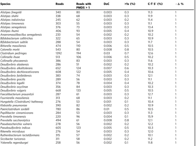
Table 1 List of the 36 most abundant microbial taxa identi
fied in the El Salt sediments, belonging to the hominid gut microbiome
families.
Species Reads Reads with
PMDS > 1 DoC >1x (%) C-T 5′ (%) −Δ % Alistipesfinegoldi 340 80 0.003 0.3 11.3 1 Alistipes shahii 338 68 0.003 0.3 11.9 1 Alistipes indistinctus 245 62 0.003 0.2 11.4 1 Alistipes timonensis 303 55 0.003 0.3 11.1 1 Alistipes senegalensis 376 73 0.003 0.3 13.4 1 Alistipes ihumii 406 93 0.005 0.4 10.9 1 Anaeromassilibacillus senegalensis 233 54 0.002 0.2 10.2 1 Bifidobacterium callitrichos 322 65 0.003 0.3 10.9 1 Bifidobacterium subtile 288 54 0.003 0.3 11.7 1 Bittarella massiliensis 474 110 0.006 0.5 10.5 1 Catonella morbi 543 80 0.008 0.8 10.5 1 Clostridium perfringes 1392 194 0.03 0.7 12.6 1 Collinsella ihuae 513 106 0.006 0.5 10.2 1 Collinsella phocaeensis 386 83 0.003 0.3 11.6 1 Desulfovibrio alaskensis 286 51 0.002 0.2 10.2 1 Desulfovibrio alkalitolerans 652 124 0.007 0.6 10.3 1 Desulfovibrio dechloracetivorans 608 122 0.005 0.4 10.6 1
Desulfovibrio fairfieldensis 383 74 0.003 0.3 12.1 1
Desulfovibrio gracilis 289 56 0.003 0.3 11.1 1 Desulfovibrio legallii 374 78 0.004 0.4 10.2 1 Desulfovibrio oxyclinae 356 84 0.003 0.3 10.3 1 Desulfovibrio vulgaris 668 133 0.005 0.5 10.5 1 Faecalibacterium prausnitzii 287 61 0.003 0.3 12.7 1 Fournierella massiliensis 313 68 0.003 0.2 11.1 1
Hungatella (Clostridium) hatheway 276 53 0.001 0.1 10.4 1
Klebsiella pneumoniae 390 82 0.002 0.2 10.9 1 Paeniclostridium sordelli 780 86 0.01 0.5 10.5 1 Papillibacter cinnamivorans 284 53 0.003 0.3 11.3 1 Prevotella timonensis 220 96 0.004 0.1 15.9 1 Prevotella saccharolytica 494 61 0.008 0.8 12.1 1 Pseudoescherichia vulneris 278 56 0.002 0.2 12.2 1 Pseudosulfovibrio indicus 605 123 0.005 0.5 10.3 1 Rikenella microfusus 276 54 0.003 0.3 12.0 1 Ruthenibacterium lactatiformans 315 57 0.002 0.2 10.1 1 Siccibacter turicensis 311 58 0.002 0.2 11.2 1 Yokenella regensburgei 258 56 0.002 0.1 11.8 1
Depth (DoC) and breadth of coverage (>1x) were calculated using BEDTools. Deamination rates at the 5’ ends of DNA fragments were calculated for the first 10 bases using mapDamage. −Δ % refers to the negative difference proportion introduced by Hübler et al.53. C-T (%) and−Δ % are computed on PMDS>1 reads.
Treponema vincentii), which have been associated with modern
oral and dental diseases in humans
59–68.
Expectedly, the samples from the upper part of SU V (which
are poor in archaeological remains) showed scarce and
incon-sistent presence of aDNA related to hominid-associated gut
microbiome bacterial families. The highest hit counts were found
for Clostridium perfringens, Paeniclostridium sordellii and
Tur-icibacter sanguinis, with the
first two being environmental
opportunistic microorganisms historically associated with human
gangrene and the last with acute appendicitis
69–71. These
findings
further support the presence of potential human-like gut
microbiome components as being unique to the samples from
Xa and Xb, the only sedimentary layers that to date have shown
traces of microscopic coprolites and fecal lipid biomarkers of
presumed archaic human origin.
In conclusion, by reconstructing ancient bacterial profiles from
El Salt Neanderthal feces-containing sediments, we propose the
existence of a core human gut microbiome with recognizable
coherence between Neanderthals and modern humans, whose
existence would pre-date the split between these two lineages, i.e.,
in the early Middle Pleistocene
72. Although the risk of fractional
contamination by modern DNA can never be ruled out and our
data must be taken with some caution, the identification of this
ancient human gut microbiome core supports the existence of
evolutionary symbioses with strong potential to have a major
impact on our health. In particular, the presence of known
short-chain fatty acid producers, such as Blautia, Dorea, Roseburia,
Rumunicoccus, Subdoligranulum, Faecalibacterium and
Bifidobac-terium, among the gut microbiome of Neanderthals, provides a
unique perspective on their relevance as keystone taxa to the
biology and health of the Homo lineage. While the former are
known to allow extra energy to be extracted from dietary
fiber
73,
strengthening the relevance of plant foods in human evolution,
Bifidobacterium could have provided benefits to archaic human
mothers and infants as a protective and immunomodulatory
microorganism. Furthermore, the detection of so-called
“old
friend” microorganisms
74as putative components of Neanderthal
gut microbiome (e.g., Spirochaetaceae, Prevotella and Desulfovibrio)
further supports the hypothesized ancestral nature of these human
gut microbiome members, which are now disappearing in
westernized populations
3–11. In the current scenario where we
are witnessing a wholescale loss of bacterial diversity in the gut
microbiome of the cultural
“west”, with the parallel rise in
dysbiosis-related autoimmune and inflammatory disorders
75, the
identification of evolutionarily integral taxa of the human
holobiont may benefit practical applications favoring their
retention among populations living in or transitioning to
increasingly microbially deplete contexts. Such therapeutic
applica-tions may in the near future include next-generation probiotics,
prebiotics or other gut microbiome-tailored dietary interventions.
Methods
Site and sampling. All samples used for this study were collected from the archaeological site of El Salt, Alicante, Spain. The archaeological team led by B. Galván conducted the excavations under a government permit and following the Spanish heritage law (No. 16/1985, 25 June). All excavated material including the sedimentary material is interpreted as archaeological material so no further permits are required for the presented study. Loose sediment samples (5–10 g) were col-lected in plastic vials using sterilized spoons (one per sample) after thoroughly cleaning the excavation surface with a vacuum cleaner in order to guarantee removal of any recent dust or sediment blown in from a different location. Lab safety masks and nitrile gloves were used at all times. The samples were collected from two different zones of the current El Salt excavation area (see Fig.1):
1. Zone 1. This is the upper excavation zone. Samples were collected from SU V, Facies 23 (one sample, V1) and Facies 24 (two samples, V2 and V3). This unit has been dated by OSL to 44.7 ± 3.2 ky BP76. Lithologically, it is
composed of massive, loose yellowish-brown calcareous silt with coarse sand and isolated larger limestone and travertine clasts. Facies 23 isfine-grained,
while Facies 24 (overlying Facies 23) also contains gravel. Unit V has yielded very few archaeological remains (bone fragments and technologically undiagnosticflint flakes).
2. Zone 2. This is the lower excavation zone. Samples were collected from SUs IX, Xa, Xb and XI, which are a stratified succession of sedimentary layers rich in Middle Paleolithic archaeological remains (charcoal, combustion features and burnt and unburnt bone andflint artifacts). From top to base: Unit IX (one sample): is the uppermost layer in this succession. It is discontinuous across the excavation area, comprising a series of dark brown-black sandy silt lenses.
Unit Xa (one sample): dated by TL to 52.3 ± 4.6 ky BP76, this is a
microstratified brownish-yellow deposit of loose calcareous silt sands with few larger clasts.
Unit Xb (eight samples): similar to Xa, also microstratified but darker (brown) andfiner-grained (sandy silts). Seven samples from this unit (ES1-7) were collected from a microstratified combustion structure (H44) at the top of this layer that yielded human fecal biomarkers25. The other sample was
collected from underlying sediment.
Unit XI (one sample): this is a layer of loose brown silty sand.
Ancient DNA extraction. All work was conducted at University of Oklahoma LMAMR ancient DNA laboratory according to the following protocols for coprolite-derived materials.
For DNA extraction, approximately 200 mg were subsampled from each sample material and incubated on a rotator with 400 µl of 0.5 M EDTA and 100 µl of proteinase K (QIAGEN) for 4 h. After that, the samples were subjected to bead-beating with 750 µl of PowerBead solution (QIAGEN) and then extracted using the MinElute PCR Purification kit (QIAGEN) with a modified protocol (method B) described in Hagan et al.77and based on Dabney et al.78, including two cleaning
steps beforefinal elution into two 30 μl of EB buffer (QIAGEN).
Library preparation and sequencing. Shotgun sequencing indexing libraries were constructed using the NEBNext DNA Library Prep Master Mix Set for 454 (New England Biolabs), following the“BEST” (Blunt-End-Single-Tube) method79, with
the hybridization of adapter oligos as per Meyer and Kircher80. Briefly, deaminated
(C to U) bases werefirst partially removed (UDG-half) by uracil DNA glycosylase treatment using USER enzyme81. End overhangs were repaired, creating blunt-end
phosphorylated regions for adapter ligation. Oligo adapters were ligated directly to blunt ends andfilled in to create priming sites for index primers. After purification with a MinElute column (QIAGEN), indexed libraries were generated in triplicate for each sample using unique forward and reverse barcoded primers. See Supple-mentary Table 3 for adapter and oligo sequences. The triplicates were pooled, cleaned using Agencourt AMPure XP magnetic beads (Beckman Coulter), and then run on a Fragment Analyzer (Advanced Analytical) using the high sensitivity NGS standard protocol. Samples containing adapter dimers below the main peak for putative authentic endogenous DNA (i.e., 200–250 bp)82, were further cleaned
using AMPure XP magnetic beads in a PEG/NaCl buffer83. Cleaned samples were
sequenced on Illumina NextSeq 500 platform (Illumina) at University of Bologna (Bologna, Italy), using paired-end 2 × 75 bp chemistry in order to obtain >1 Gbp of sequences per sample. Quality score exceeded Q30 for more than 95% of the sequenced bases. Sequencing data was pre-processed by retaining only merged reads matching the forward and reverse barcodes with no mismatches using AdapterRemoval84.
Bioinformatics analysis. Sequences were analyzed using Burrows-Wheeler Aligner (BWA) aln algorithm and the entire set of bacterial and archaeal genomes available through NCBI RefSeq (downloaded on November 15th, 2017). In particular, we
reduced the maximum accepted edit distance (i.e., the threshold of the maximum number of deletions, insertions, and substitutions needed to transform the reference sequence into the read sequence) to 1% (-n 0.01) and set the maximum number of gap opens (i.e., the threshold of the maximum number of gaps that can be initiated to match a given read to the reference) to 2, with long gap and seed length disabled (-e-1 -l 16500). These parameters are optimized for the specific types of errors gen-erated by postmortem DNA damage during the alignment of ancient DNA to modern references, as indicated by Schubert and colleagues85. The aligned reads were
furtherfiltered for mapping quality >20, and only the hits with the best unique match (X0= 1) were considered for analysis in order to minimize the number of false positives. In order to retrieve the entire phylogeny of the assignment, database sequences were previously annotated with the“Tax” tags of the NCBI database using the reference-annotator tool of the MEGAN utils package86. We then used the calmd
program of the samtools suite to recompute the MD tags (containing alignment information, such as mismatches) for all datasets.
To discriminate ancient DNA from modern-day contamination, we calculated the postmortem degradation score (PMDS) distributions12. Sequences with PMDS > 5
were considered ancient (over 5,000 years ago), as reported by Skoglund et al.12, and
used for further analysis. The outputs were transformed in sequence (.fasta) and annotation (.txt)files compatible with the QIIME command “make_otu_table.py”, in order to create a table that contained the phylogenetic classification and the
abundance as number of reads for each specific taxon. This table was then collapsed at family, genus and species level using the command“summarize_taxa.py”. The family-level relative abundance profiles of samples IX, Xa, ES1-7, Xb and XI were compared with publicly available data of the gut microbiota of human populations adhering to different subsistence strategies: urban Italians and Hadza hunter-gatherers from Tanzania (NCBI SRA, Bioproject ID PRJNA278393)47, urban US residents, Matses
hunter-gatherers and Tunapuco rural agriculturalists from Peru (NCBI SRA, BioProject ID PRJNA268964)7. Shotgun sequence datasets were downloaded and
processed as El Salt samples, without applying the PMDSfilter. 16S rRNA gene representative sequences of bacterial genera for which at least one species was present with more than 4 hits in one El Salt sample, were downloaded from the SILVA repository and used to build a phylogenetic tree by MUSCLE87and FastTree88. The
tree was visualized using the GraPhlAn software89. Finally, bacterial species belonging
to families that have recently been indicated as being common to the gut microbiome of hominids38–45, were specifically sought and visualized for their abundance across El
Salt samples by a heat map using the R software. Species membership in other source categories (i.e., human (or animal) oral and/or pathobiont, and environmental) was inferred by searching in PubMed the original article in which the taxonomy wasfirst assigned to that organism, as well as more recent articles reporting its habitat description.
Independent validation of taxonomic assignments. To validate the taxonomic assignments of the metagenomic reads recovered from the El Salt samples, we used the same procedure adopted by Jensen and colleagues30. Specifically, we combined
results from samples IX, Xa, ES1-7, Xb and XI (i.e., those positive for the presence of fecal biomarkers and/or associated with rich archaeological assemblages), then aligned the assigned reads to their respective reference genomes and examined edit distances, coverage distributions, and postmortem DNA damage patterns14,53. For
the 24 bacterial families identified as common to hominid gut microbiome, we chose to further investigate bacterial species with≥200 assigned reads (including strain-specific reads), for which at least 50 reads showed PMDS > 1 and at least one mismatch in thefirst 10 bases with respect to the reference genome. We then aligned the taxon-specific reads to the respective reference genome from the NCBI RefSeq database using bwa aln. MapDamage was used to estimate deamination rates (Supplementary Fig. 6)90. The breadth and depth of coverage were calculated
with bedtools91and visualized with Circos92(Supplementary Fig. 7). Edit distances
for all reads andfiltered for PMDS > 1 were extracted from the bam files with the samtools view93and plotted in R (Supplementary Fig. 8). The negative difference
proportion (−Δ %) was calculated considering the first 10 bases of reads with PMDS > 1. This metric was proposed by Hübler et al.53as a measure of decline in
the edit distance distribution, with a−Δ % value of 1 indicating a declining distribution associated with an ancient DNA profile. Indeed, correct taxonomic assignments generally show a continuously declining edit distance distribution with only a few mismatches, mostly resulting from aDNA damage or divergence of the ancient genome from the modern reference. On the other hand, the mapping to an incorrect reference is associated with an increased number of mismatches, high-lighted by the analysis of the edit distance distribution.
mtDNA analysis and contamination estimate. In order to detect human mtDNA, a similar procedure combining BWA (same parameters as above) and the reference-annotator tool of the MEGAN utils package, was applied to the entire set of mitochondrial sequences listed at the MitoSeqs website (https://www.mitomap. org/foswiki/bin/view/MITOMAP/MitoSeqs), including all the eukaryotic mito-chondrial sequences available at the NCBI database. Only taxa detected in ancient sequences (i.e., with PMDS > 5) with more than 2 hits and not present in the control samples were retained. This procedure allowed us to detect ancient human traces beyond any reasonable doubt, eventually discarding more sequences than necessary.
In parallel, capture-enrichment for mtDNA sequencing was performed on the indexed libraries with a Neanderthal bait panel, as per the manufacturer’s protocol (version 4.01, Arbor Biosciences). In short, libraries were denatured, blocked and incubated with baits for 48 h. After purification with streptavidin-coated magnetic beads, enriched libraries were amplified and concentrated, before being subjected to a second round of capture. Final libraries were sequenced on an Illumina NextSeq 500 platform (Illumina) at University of Bologna (Bologna, Italy), as described above. As for read processing, we used Schmutzi94to determine the endogenous consensus mtDNA
sequence and to estimate present-day human contamination. Reads were mapped to the mt-Neanderthal reference sequence (NC_011137) andfiltered for MAPQ ≥ 30. Haploid variants were called using the endoCaller program implemented in Schmutzi and only the variants with a posterior probability exceeding 50 on the PHRED scale (probability of error: 1/100,000) and breadth of coverage >10% of the total mitochondrial length were retained for further analysis. The PMDS profile of the reads was computed by PMDtools12. The negative difference proportion (−Δ %) was
calculated using only reads with PMDS > 153. Contamination estimates were obtained
using Schmutzi’s mtCont program and a database of putative modern contaminant mtDNA sequences. Samples with >1,000 PMDS > 1 reads, breadth of coverage >10%, −Δ % ≥ 0.9 and mtCont contamination less than 2% were considered to contain ancient human mtDNA.
Statistics and reproducibility. No replicates are included, all samples herein analyzed are unique.
Wilcoxon test was used to assess differences between samples IX, Xa, ES1-7, Xb and XI (i.e., those positive for the presence of fecal biomarkers and/or associated with rich archaeological assemblages), and samples from SU V (i.e., with no or very few archeological remains) in the number of PMDS > 5 reads, as well as in the relative abundances of the 24 families common to the gut microbiome of hominids38–45.
The significance of data separation in the Bray-Curtis-based Principal Coordinates Analysis between the family-level relative abundance profiles of samples IX, Xa, ES1-7, Xb and XI, and the gut microbiota of urban Italians and Hadza hunter-gatherers from Tanzania47, urban US residents, Matses
hunter-gatherers and Tunapuco rural agriculturalists from Peru7was tested using a
permutation test with pseudo-F ratio.
Reporting summary. Further information on research design is available in the Nature Research Reporting Summary linked to this article.
Data availability
Sequencing data are accessible at the European Nucleotide Archive (ENA; project ID PRJEB41665). Source data are available as Supplementary Data. All sediment samples are readily available from the authors, subject to exhaustion.
Received: 19 April 2020; Accepted: 5 January 2021;
References
1. Lynch, S. V. & Pedersen, O. The human intestinal microbiome in health and disease. N. Engl. J. Med. 375, 2369–2379 (2016).
2. Davenport, E. R. et al. The human microbiome in evolution. BMC Biol. 15, 127 (2017).
3. De Filippo, C. et al. Impact of diet in shaping gut microbiota revealed by a comparative study in children from Europe and rural Africa. Proc. Natl Acad. Sci. USA 107, 14691–14696 (2010).
4. Tyakht, A. V. et al. Human gut microbiota community structures in urban and rural populations in Russia. Nat. Commun. 4, 2469 (2013).
5. Schnorr, S. L. et al. Gut microbiome of the Hadza hunter-gatherers. Nat. Commun. 5, 3654 (2014).
6. Martínez, I. et al. The gut microbiota of rural Papua New Guineans: composition, diversity patterns, and ecological processes. Cell Rep. 11, 527–538 (2015).
7. Obregon-Tito, A. J. et al. Subsistence strategies in traditional societies distinguish gut microbiomes. Nat. Commun. 6, 6505 (2015).
8. Sankaranarayanan, K. et al. Gut microbiome diversity among Cheyenne and Arapaho individuals from Western Oklahoma. Curr. Biol. 25, 3161–3169 (2015).
9. Girard, C., Tromas, N., Amyot, M. & Shapiro, B. J. Gut microbiome of the Canadian Arctic Inuit. mSphere 2, e00297–e00316 (2017).
10. Ayeni, F. A. et al. Infant and adult gut microbiome and metabolome in rural Bassa and urban settlers from Nigeria. Cell Rep. 23, 3056–3067 (2018). 11. Jha, A. R. et al. Gut microbiome transition across a lifestyle gradient in
Himalaya. PLoS Biol. 16, e2005396 (2018).
12. Skoglund, P. et al. Separating endogenous ancient DNA from modern day contamination in a Siberian Neandertal. Proc. Natl Acad. Sci. USA 111, 2229–2234 (2014).
13. Moeller, A. H. et al. Cospeciation of gut microbiota with hominids. Science 353, 380–382 (2016).
14. Key, F. M., Posth, C., Krause, J., Herbig, A. & Bos, K. I. Mining metagenomic data sets for ancient DNA: recommended protocols for authentication. Trends Genet. 33, 508–520 (2017).
15. Philips, A. et al. Comprehensive analysis of microorganisms accompanying human archaeological remains. Gigascience 6, 1–13 (2017).
16. Warinner, C. et al. Pathogens and host immunity in the ancient human oral cavity. Nat. Genet. 46, 336–344 (2014).
17. Rasmussen, S. et al. Early divergent strains of Yersinia pestis in Eurasia 5,000 years ago. Cell 163, 571–582 (2015).
18. Weyrich, L. S. et al. Neanderthal behaviour, diet, and disease inferred from ancient DNA in dental calculus. Nature 544, 357–361 (2017).
19. Eckburg, P. B. et al. Diversity of the human intestinal microbialflora. Science 308, 1635–1638 (2005).
20. Tito, R. Y. et al. Phylotyping and functional analysis of two ancient human microbiomes. PLoS ONE 3, e3703 (2008).
21. Tito, R. Y. et al. Insights from characterizing extinct human gut microbiomes. PLoS ONE 7, e51146 (2012).
22. Lugli, G. A. et al. Ancient bacteria of the Ötzi’s microbiome: a genomic tale from the Copper Age. Microbiome 5, 5 (2017).
23. Santiago-Rodriguez, T. M. et al. Taxonomic and predicted metabolic profiles of the human gut microbiome in pre-Columbian mummies. FEMS Microbiol. Ecol. 92,fiw182 (2016).
24. Søe, M. J. et al. Ancient DNA from latrines in Northern Europe and the Middle East (500 BC-1700 AD) reveals past parasites and diet. PLoS ONE 13, e0195481 (2018).
25. Sistiaga, A., Mallol, C., Galván, B. & Summons, R. E. The Neanderthal meal: a new perspective using faecal biomarkers. PLoS ONE 9, e101045 (2014). 26. Galván, B. et al.“El Salt. The Last Neanderthals Of The Alicante Mountains
(Alcoy, Spain)” in Pleistocene and Holocene hunter-gatherers in Iberia and the Gibraltar Strait. The current archaeological record, 380–388 (R. Sala Ramos, Ed., Burgos, Univ. de Burgos y Fundación Atapuerca, 2014).
27. Garralda, M. D. et al. Neanderthals from El Salt (Alcoy, Spain) in the context of the latest Middle Palaeolithic populations from the southeast of the Iberian Peninsula. J. Hum. Evol. 75, 1–15 (2014).
28. Green, R. E. et al. Analysis of one million base pairs of Neanderthal DNA. Nature 444, 330–336 (2006).
29. Sampietro, M. L. et al. Tracking down human contamination in ancient human teeth. Mol. Biol. Evol. 23, 1801–1807 (2006).
30. Jensen, T. Z. T. et al. A 5700 year-old human genome and oral microbiome from chewed birch pitch. Nat. Commun. 10, 5520 (2019).
31. Barka, E. A. et al. Taxonomy, physiology, and natural products of Actinobacteria. Microbiol. Mol. Biol. Rev. 80, 1–43 (2015).
32. Lewin, G. R. et al. Evolution and ecology of Actinobacteria and their bioenergy applications. Annu. Rev. Microbiol. 70, 235–254 (2016).
33. Vreeland, R. H., Rosenzweig, W. D. & Powers, D. W. Isolation of a 250 million-year-old halotolerant bacterium from a primary salt crystal. Nature 407, 897–900 (2000).
34. Mandic-Mulec, I., Stefanic, P. & van Elsas, J. D. Ecology of Bacillaceae. Microbiol. Spectr. 3, TBS-0017-2013 (2015).
35. Aylward, F. O. et al. Comparison of 26 sphingomonad genomes reveals diverse environmental adaptations and biodegradative capabilities. Appl. Environ. Microbiol. 79, 3724–3733 (2013).
36. Simon, M. et al. Phylogenomics of Rhodobacteraceae reveals evolutionary adaptation to marine and non-marine habitats. ISME J. 11, 1483–1499 (2017). 37. Mann, A. E. et al. Differential preservation of endogenous human and
microbial DNA in dental calculus and dentin. Sci. Rep. 8, 9822 (2018). 38. Bittar, F. et al. Gorilla gorilla gorilla gut: a potential reservoir of pathogenic
bacteria as revealed using culturomics and molecular tools. Sci. Rep. 4, 7174 (2014).
39. Moeller, A. H. et al. Rapid changes in the gut microbiome during human evolution. Proc. Natl Acad. Sci. USA 111, 16431–16435 (2014).
40. Zhang, J. et al. A phylo-functional core of gut microbiota in healthy young Chinese cohorts across lifestyles, geography and ethnicities. ISME J. 9, 1979–1990 (2015).
41. Lloyd-Price, J., Abu-Ali, G. & Huttenhower, C. The healthy human microbiome. Genome Med. 8, 51 (2016).
42. Clayton, J. B. et al. The gut microbiome of nonhuman primates: Lessons in ecology and evolution. Am. J. Primatol. 80, e22867 (2018).
43. Hicks, A. L. et al. Gut microbiomes of wild great apesfluctuate seasonally in response to diet. Nat. Commun. 9, 1786 (2018).
44. Nishida, A. H. & Ochman, H. Rates of gut microbiome divergence in mammals. Mol. Ecol. 27, 1884–1897 (2018).
45. Nishida, A. H. & Ochman, H. A great-ape view of the gut microbiome. Nat. Rev. Genet. 20, 195–206 (2019).
46. Amato, K. R. et al. Evolutionary trends in host physiology outweigh dietary niche in structuring primate gut microbiomes. ISME J. 13, 576–587 (2019). 47. Rampelli, S. et al. Metagenome sequencing of the Hadza Hunter-Gatherer gut
microbiota. Curr. Biol. 25, 1682–1693 (2015).
48. Martín, R. et al. Functional characterization of novel Faecalibacterium prausnitzii strains isolated from healthy volunteers: a step forward in the use of F. prausnitzii as a next-generation probiotic. Front. Microbiol. 8, 1226 (2017).
49. Koh, A., De Vadder, F., Kovatcheva-Datchary, P. & Bäckhed, F. From dietary fiber to host physiology: short-chain fatty acids as key bacterial metabolites. Cell 165, 1332–1345 (2016).
50. O’Callaghan, A. & van Sinderen, D. Bifidobacteria and their role as members of the human gut microbiota. Front. Microbiol. 7, 925 (2016).
51. Milani, C. et al. Genomics of the genus Bifidobacterium reveals species-specific adaptation to the glycan-rich gut environment. Appl. Environ. Microbiol. 82, 980–991 (2015).
52. Lugli, G. A. et al. Reconstruction of the bifidobacterial pan-secretome reveals the network of extracellular interactions between bifidobacteria and the infant gut. Appl. Environ. Microbiol. 84, e00796–e00818 (2018).
53. Hübler, R. et al. HOPS: automated detection and authentication of pathogen DNA in archaeological remains. Genome Biol. 20, 280 (2019).
54. Lye, H. S., Rusul, G. & Liong, M. T. Removal of cholesterol by lactobacilli via incorporation and conversion to coprostanol. J. Dairy Sci. 93, 1383–1392 (2010).
55. Gérard, P. Metabolism of cholesterol and bile acids by the gut microbiota. Pathogens 3, 14–24 (2013).
56. Shimizu, M., Hashiguchi, M., Shiga, T., Tamura, H. O. & Mochizuki, M. Meta-analysis: effects of probiotic supplementation on lipid profiles in normal to mildly hypercholesterolemic individuals. PLoS ONE 10, e0139795 (2015). 57. Zanotti, I. et al. Evidence for cholesterol-lowering activity by Bifidobacterium
bifidum PRL2010 through gut microbiota modulation. Appl. Microbiol. Biotechnol. 99, 6813–6829 (2015).
58. Antharam, V. C. et al. An integrated metabolomic and microbiome analysis identified specific gut microbiota associated with fecal cholesterol and coprostanol in Clostridium difficile infection. PLoS ONE 11, e0148824 (2016). 59. Moore, L. V. & Moore, W. E. Oribaculum catoniae gen. nov., sp. nov.;
Catonella morbi gen. nov., sp. nov.; Hallella seregens gen. nov., sp. nov.; Johnsonella ignava gen. nov., sp. nov.; and Dialister pneumosintes gen. nov., comb. nov., nom. rev., anaerobic gram-negative bacilli from the human gingival crevice. Int. J. Syst. Bacteriol. 44, 187–192 (1994).
60. Crociani, F., Biavati, B., Alessandrini, A., Chiarini, C. & Scardovi, V. Bifidobacterium inopinatum sp. nov. and Bifidobacterium denticolens sp. nov., two new species isolated from human dental caries. Int. J. Syst. Bacteriol. 46, 564–571 (1996).
61. Willems, A. & Collins, M. D. Phylogenetic relationships of the genera Acetobacterium and Eubacterium sensu stricto and reclassification of Eubacterium alactolyticum as Pseudoramibacter alactolyticus gen. nov., comb. nov. Int. J. Syst. Bacteriol. 46, 1083–1087 (1996).
62. Willis, S. G. et al. Identification of seven Treponema species in health- and disease-associated dental plaque by nested PCR. J. Clin. Microbiol. 37, 867–869 (1999).
63. Becker, M. R. et al. Molecular analysis of bacterial species associated with childhood caries. J. Clin. Microbiol. 40, 1001–1009 (2002).
64. Downes, J., Munson, M. A., Radford, D. R., Spratt, D. A. & Wade, W. G. Shuttleworthia satelles gen. nov., sp. nov., isolated from the human oral cavity. Int. J. Syst. Evol. Microbiol. 52, 1469–1475 (2002).
65. Xu, P. et al. Genome of the opportunistic pathogen Streptococcus sanguinis. J. Bacteriol. 189, 3166–3175 (2007).
66. Horz, H. P. & Conrads, G. Methanogenic Archaea and oral infections - ways to unravel the black box. J. Oral Microbiol. 3;https://doi.org/10.3402/jom. v3i0.5940(2011).
67. Hedberg, M. E. et al. Lachnoanaerobaculum gen. nov., a new genus in the Lachnospiraceae: characterization of Lachnoanaerobaculum umeaense gen. nov., sp. nov., isolated from the human small intestine, and
Lachnoanaerobaculum orale sp. nov., isolated from saliva, and reclassification of Eubacterium saburreum (Prevot 1966) Holdeman and Moore 1970 as Lachnoanaerobaculum saburreum comb. nov. Int. J. Syst. Evol. Microbiol. 62, 2685–2690 (2012).
68. Sizova, M. V. et al. Stomatobaculum longum gen. nov., sp. nov., an obligately anaerobic bacterium from the human oral cavity. Int. J. Syst. Evol. Microbiol. 63, 1450–1456 (2013).
69. Welch, W. H. & Nuttall, G. H. F. A gas-producing bacillus (Bacillus aerogenes capsulatus, Nov, Spec.) capable of rapid development in the body after death. Bull. John Hopkins Hosp. Baltim. 3, 81–91 (1891).
70. Bosshard, P. P., Zbinden, R. & Altwegg, M. Turicibacter sanguinis gen. nov., sp. nov., a novel anaerobic, Gram-positive bacterium. Int. J. Syst. Evol. Microbiol. 52, 1263–1266 (2002).
71. Aldape, M. J., Bryant, A. E. & Stevens, D. L. Clostridium sordellii infection: epidemiology, clinicalfindings, and current perspectives on diagnosis and treatment. Clin. Infect. Dis. 43, 1436–1446 (2006).
72. Prüfer, K. et al. A high-coverage Neandertal genome from Vindija Cave in Croatia. Science 358, 655–658 (2017).
73. Kolodziejczyk, A. A., Zheng, D. & Elinav, E. Diet-microbiota interactions and personalized nutrition. Nat. Rev. Microbiol. 7, 742–753 (2019).
74. Rook, G. A. 99th Dahlem conference on infection, inflammation and chronic inflammatory disorders: darwinian medicine and the ‘hygiene’ or ‘old friends’ hypothesis. Clin. Exp. Immunol. 160, 70–79 (2010).
75. Duvallet, C., Gibbons, S. M., Gurry, T., Irizarry, R. A. & Alm, E. J. Meta-analysis of gut microbiome studies identifies disease-specific and shared responses. Nat. Commun. 8, 1784 (2017).
76. Galván, B. et al. New evidence of early Neanderthal disappearance in the Iberian Peninsula. J. Hum. Evol. 75, 16–27 (2014).
77. Hagan, R. W. et al. Comparison of extraction methods for recovering ancient microbial DNA from paleofeces. Am. J. Phys. Anthropol. 171, 275–284 (2019). 78. Dabney, J. et al. Complete mitochondrial genome sequence of a Middle
Pleistocene cave bear reconstructed from ultrashort DNA fragments. Proc. Natl Acad. Sci. USA 110, 15758–15763 (2013).
79. Carøe, C. et al. Single-tube library preparation for degraded DNA. Methods Ecol. Evol. 9, 410–419 (2018).
80. Meyer, M. & Kircher, M. Illumina sequencing library preparation for highly multiplexed target capture and sequencing. Cold Spring Harb. Protoc. 2010, pdb.prot5448 (2010).
81. Rohland, N., Harney, E., Mallick, S., Nordenfelt, S. & Reich, D. Partial uracil-DNA-glycosylase treatment for screening of ancient DNA. Philos. Trans. R. Soc. Lond. B Biol. Sci. 370, 20130624 (2015).
82. Ziesemer, K. A. et al. Intrinsic challenges in ancient microbiome
reconstruction using 16S rRNA gene amplification. Sci. Rep. 5, 16498 (2015). 83. Rohland, N. & Reich, D. Cost-effective, high-throughput DNA sequencing
libraries for multiplexed target capture. Genome Res. 22, 939–946 (2012). 84. Lindgreen, S. AdapterRemoval: easy cleaning of next-generation sequencing
reads. BMC Res. Notes 5, 337 (2012).
85. Schubert, M. et al. Improving ancient DNA read mapping against modern reference genomes. BMC Genomics 13, 178 (2012).
86. Huson, D. H. et al. MEGAN community edition - interactive exploration and analysis of large-scale microbiome sequencing data. PLoS Comput. Biol. 12, e1004957 (2016).
87. Edgar, R. C. MUSCLE: a multiple sequence alignment method with reduced time and space complexity. BMC Bioinformatics 5, 113 (2004).
88. Price, M. N., Dehal, P. S. & Arkin, A. P. FastTree: computing large minimum evolution trees with profiles instead of a distance matrix. Mol. Biol. Evol. 26, 1641–1650 (2009).
89. Asnicar, F., Weingart, G., Tickle, T. L., Huttenhower, C. & Segata, N. Compact graphical representation of phylogenetic data and metadata with GraPhlAn. PeerJ 3, e1029 (2015).
90. Jónsson, H., Ginolhac, A., Schubert, M., Johnson, P. L. & Orlando, L. mapDamage2.0: fast approximate Bayesian estimates of ancient DNA damage parameters. Bioinformatics 29, 1682–1684 (2013).
91. Quinlan, A. R. & Hall, I. M. BEDTools: aflexible suite of utilities for comparing genomic features. Bioinformatics 26, 841–842 (2010). 92. Krzywinski, M. et al. Circos: an information aesthetic for comparative
genomics. Genome Res. 19, 1639–1645 (2009).
93. Li, H. et al. The Sequence Alignment/Map format and SAMtools. Bioinformatics 25, 2078–2079 (2009).
94. Renaud, G., Slon, V., Duggan, A. T. & Kelso, J. Schmutzi: estimation of contamination and endogenous mitochondrial consensus calling for ancient DNA. Genome Biol. 16, 224 (2015).
Acknowledgements
We thank F. D’Amico (Department of Pharmacy and Biotechnology, University of Bologna, Bologna, Italy) and E. Cilli (Department of Cultural Heritage, University of Bologna, Ravenna, Italy) for their valuable help in library preparation. This research was supported by the US National Institutes of Health, grant number R01GM089886 (C.W.
and C.L.). S.B. was supported by the European Research Council (ERC) under the European Union’s Horizon 2020 research and innovation programme (grant agreement No 724046 - SUCCESS). Archaeological research at El Salt is funded by Spanish I+D Project HAR2008-06117/HIST (C.M., C.H. and B.G.).
Author contributions
M.C., S.T., S.R., S.L.S. and C.W.: conceptualization; C.M., C.H., B.G. and A.S.:field work, excavation, and sampling; S.L.S., C.A.H. and S.T.: DNA extraction and library pre-paration; A.A.: sequencing; S.R.: bioinformatics analysis; P.B., M.C., C.L., C.W. and S.B.: resources; M.C. and S.L.S.: supervision; M.C., S.T. and S.R.: writing—original draft; C.W., C.L., S.B., C.M., A.S., E.B., A.A., C.A.H. and S.L.S.: writing—review & editing. All authors gavefinal approval for publication.
Competing interests
The authors declare no competing interests.
Additional information
Supplementary informationThe online version contains supplementary material available athttps://doi.org/10.1038/s42003-021-01689-y.
Correspondenceand requests for materials should be addressed to S.L.S. or M.C.
Reprints and permission informationis available athttp://www.nature.com/reprints
Publisher’s note Springer Nature remains neutral with regard to jurisdictional claims in published maps and institutional affiliations.
Open Access This article is licensed under a Creative Commons Attribution 4.0 International License, which permits use, sharing, adaptation, distribution and reproduction in any medium or format, as long as you give appropriate credit to the original author(s) and the source, provide a link to the Creative Commons license, and indicate if changes were made. The images or other third party material in this article are included in the article’s Creative Commons license, unless indicated otherwise in a credit line to the material. If material is not included in the article’s Creative Commons license and your intended use is not permitted by statutory regulation or exceeds the permitted use, you will need to obtain permission directly from the copyright holder. To view a copy of this license, visithttp://creativecommons.org/ licenses/by/4.0/.