Research Article
Ticagrelor Conditioning Effects Are Not Additive to
Cardioprotection Induced by Direct NLRP3 Inflammasome
Inhibition: Role of RISK, NLRP3, and Redox Cascades
Claudia Penna
,
1Manuela Aragno
,
1Alessia Sofia Cento
,
1Saveria Femminò
,
1Isabella Russo
,
1Federica Dal Bello
,
2Fausto Chiazza
,
3Debora Collotta
,
3Gustavo Ferreira Alves
,
3Massimo Bertinaria
,
3Elisa Zicola
,
1Valentina Mercurio
,
4Claudio Medana
,
2Massimo Collino
,
3and Pasquale Pagliaro
11Department of Clinical and Biological Sciences, University of Turin, Turin, Italy
2Department of Molecular Biotechnology and Health Sciences, University of Turin, Turin, Italy 3Department of Drug Science and Technology, University of Turin, Turin, Italy
4Department of Translational Medical Sciences, Federico II University, Naples, Italy
Correspondence should be addressed to Massimo Collino; [email protected] and Pasquale Pagliaro; [email protected]
Received 6 April 2020; Revised 7 June 2020; Accepted 26 June 2020; Published 4 August 2020 Academic Editor: José P. Andrade
Copyright © 2020 Claudia Penna et al. This is an open access article distributed under the Creative Commons Attribution License, which permits unrestricted use, distribution, and reproduction in any medium, provided the original work is properly cited. Inhibition of either P2Y12 receptor or the nucleotide-binding oligomerization domain- (NOD-) like receptor pyrin domain containing 3 (NLRP3) inflammasome provides cardioprotective effects. Here, we investigate whether direct NLRP3 inflammasome inhibition exerts additive effects on myocardial protection induced by the P2Y12 receptor antagonist Ticagrelor. Ticagrelor (150 mg/kg) was orally administered to rats for three consecutive days. Then, isolated hearts underwent an ischemia/reperfusion (30 min ischemia/60 min reperfusion; IR) protocol. The selective NLRP3 inflammasome inhibitor INF (50μM) was infused before the IR protocol to the hearts from untreated animals or pretreated with Ticagrelor. In parallel experiments, the hearts isolated from untreated animals were perfused with Ticagrelor (3.70μM) before ischemia and subjected to IR. The hearts of animals pretreated with Ticagrelor showed a significantly reduced infarct size (IS, 49 ± 3% of area at risk, AAR) when compared to control IR group (69 ± 2% of AAR). Similarly, ex vivo administration of INF before the IR injury resulted in significant IS reduction (38 ± 3% of AAR). Myocardial IR induced the NLRP3 inflammasome complex formation, which was attenuated by either INF pretreatment ex vivo, or by repeated oral treatment with Ticagrelor. The beneficial effects induced by either treatment were associated with the protective Reperfusion Injury Salvage Kinase (RISK) pathway activation and redox defence upregulation. In contrast, no protective effects nor NLRP3/RISK modulation were recorded when Ticagrelor was administered before ischemia in isolated heart, indicating that Ticagrelor direct target is not in the myocardium. Our results confirm that Ticagrelor conditioning effects are likely mediated through platelets, but are not additives to the ones achieved by directly inhibiting NLRP3.
1. Introduction
Ischemic heart disease remains the leading cause of morbid-ity in the Western world, and the number of deaths from acute myocardial infarction (AMI) is also rapidly rising in the developing world. Although restoration of early blood
flow to the ischemic myocardium with thrombolysis is pres-ently the most effective therapy to limit infarct size, reperfu-sion alone is inadequate to salvage the damaged myocardium and may result in myocardial ischemia/reperfusion (IR) injury, which is characterized by excessive oxidative stress and inflammatory response [1–3]. In fact, as shown by both
Volume 2020, Article ID 9219825, 12 pages https://doi.org/10.1155/2020/9219825
preclinical and clinical studies, the excess myocardial cell death resulting from the restoration of blood and oxygen supply can contribute up to 50% of thefinal infarct size [1–3]. In clinical practice, P2Y12 adenosine disphosphate (ADP) receptor antagonists are standard of care in AMI patients undergoing primary percutaneous intervention. Sev-eral preclinical studies have convincingly shown that these drugs significantly protect against IR injury, suggesting that these pleiotropic effects could be even more important than their antiaggregant properties in this specific clinical setting [4–8]. Clinical trials have shown that the nonthienopyridine P2Y12 antagonists such as Ticagrelor and Cangrelor were associated to lower incidence of cardiovascular mortality, AMI, or stroke compared with the thienopyridine P2Y12 antagonists, Clopidogrel and Prasugrel [9]. These differences have been ascribed, at least in part, to better and more consis-tent pharmacokinetic profile of the nonthienopyridine P2Y12 antagonists (Ticagrelor and Cangrelor) that do not require hepatic P450-mediated metabolic conversion of the prodrug (e.g., Clopidogrel and Prasugrel) into active forms to ensure P2Y12 receptor inhibition. Moreover, Ticagrelor is the only P2Y12 antagonist that increases tissue adenosine levels via inhibition of the equilibrative nucleoside trans-porter 1 (ENT1) by protecting the extracellular adenosine from intracellular metabolism [10–12]. This effect has been suggested to further contribute to the drug-induced cardio-protection [13–15], despite a recently published paper cloud-ing this hypothesis [8].
Although different cell types (including endothelial cells [16]) express P2Y12 receptors, the conditioning effect of P2Y12 receptor-inhibitors has been attributed to the modu-lation of platelet sphingosine kinase activity and perhaps to sphingosine 1-phosphate (S1P) release [5, 17]. Since P2Y12 antagonists reduce infarct size but do not eliminate it, some other processes must be responsible of residual IR injury. Indeed, additive cardioprotective effects have been demon-strated by the combination of Ticagrelor and Rosuvastatin [13]. More recently, Audia et al. [4] demonstrated that a highly selective caspase-1 inhibitor provides additional and sustained infarct size reduction when added to Ticagrelor in preclinical models of IR injury. Caspase-1 activation is a crit-ical choke point for eliciting activation of the inflammatory cascade NLRP3 (NOD-like receptor family, pyrin domain-containing3) inflammasome. The NLRP3 inflammasome is a large multimeric protein complex which interacts with an apoptosis-associated speck-like protein including a caspase recruitment domain (ASC), thus recruiting and activating caspase-1, which in turn mediates the cleavage of inactive prointerleukin- (IL-) 1? and IL-18 into their active forms [18]. We and others have previously demonstrated the piv-otal role of the NLRP3 inflammasome in cardiometabolic disorders, including myocardial ischemia reperfusion injury, [19–23] and several NLRP3 inhibitors, including the small molecule INF we recently developed, have been tested in animal model of IR injury, showing salvage of part of the myocardium at risk [24, 25]. The cardioprotective role of NLRP3 inhibitors is attributable, at least in part, to their abil-ity to modify protective pathways and redox environment of cells [24, 26].
In the present study, we evaluate (1) the ability of Tica-grelor and INF, alone and in combination, to reduce infarct size following IR injury, (2) the potential mechanisms of cross-talk between the two drug treatments underlying their myocardial protection, and (3) the relevance of the presence of blood in mediating cardioprotective effects and the platelet mediators released after Ticagrelor exposure.
2. Materials and Methods
2.1. Ex Vivo Rat Model of Heart IR Injury. Male Wistar rats (Harlan Laboratories, Udine, Italy) 5–6 months old, reaching a body weight of 450–550 g, were anesthetized with sodium pentothal (50 mg/kg) by intraperitoneal injections and hepa-rinized (800 U/100 g b.w., i.m.) before being culled by cervical dislocation. The hearts were then rapidly excised, placed in an ice-cold buffer solution, and weighed. The excised hearts were rapidly perfused by the Langendorff technique with Krebs-Henseleit bicarbonate buffer containing (mM) NaCl 118, NaHCO325, KCl 4.7, KH2PO4 1.2, MgSO41.2, CaCl2 1.25, and Glucose 11. The buffer was gassed with 95% O2: 5% CO2. The hearts were perfused in constantflow mode to achieve a perfusion pressure of about 80 mmHg. To assess the conditions of experimental preparation, coronary perfu-sion pressure was monitored during all experiments [27], andflow rate was checked in a specific time period. The tem-perature of the perfusion system was maintained at 37°C. After a 30 min stabilization period, the hearts were subjected to a protocol of IR, which consisted in 30 min of global no-flow, normothermic ischemia followed by a period of 60 min of reperfusion. At the end of perfusion period, the hearts were rapidly removed from the perfusion apparatus and divided in two parts by a coronal section (perpendicular to the long axis). The apical part of the left ventricle (LV, less than 1/3 of ventricular mass) was frozen rapidly in liquid nitrogen and stored at -80°C and subsequently used for Western blot analysis; the basal part of the LV was used for infarct size assessment.
The protocol was approved by the Institutional Animal Care and Use Committee of the University of Turin and con-formed to the European Directive 2010/63/EU on the protec-tion of animals used for scientific purposes.
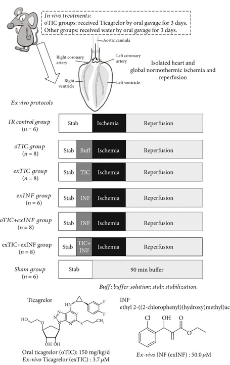
2.2. Drug Treatments. Rats (n = 6 − 8 per group) received water or Ticagrelor (TIC, 150 mg/kg/d) by oral gavage for 3 days (oTIC). Then, the isolated hearts were submitted to ischemia/reperfusion as described above (IR and oTIC groups). A subgroup of isolated hearts from oTIC rats were exposed to the selective NLRP3 inflammasome inhibitor INF (50μM) in the perfusate for 20 min before ischemia (oTIC+exINF). In a subsequent series of experiments, the isolated hearts from control rats were pretreated with 3.70μM Ticagrelor or 50 μM exINF or both in the perfusate for 20 min before ischemia (exTIC, exINF, and exTIC+exINF groups, respectively). After stabilization, sham hearts under-went 90 min perfusion only and served as control group (Figure 1).
A stock solution of 200 mM INF in DMSO was prepared and was then diluted at afinal concentration of 50 μM in the
perfusion buffer. The description of the synthesis of the inhibitor as well as the in vitro biological effects has been already published. INF is an acrylate derivative originally synthesized by Cocco et al. [28] and selected, among the tested compounds, as the most effective inhibitor of NLP3 activation (IC50 of 1:26 × 10−7M and 1:58 × 10−7M in LPS/ATP-triggered and LPS/nigericin-triggered pyropto-sis, respectively). As previously documented [29, 30], INF inhibits the NLRP3 ATPase activity of isolated human-recombinant NLRP3 protein as well as caspase-1 activation, and it acts as covalent NLRP3 inhibitor through irreversible binding to nucleophilic residues present in NLRP3, with a reactivity of 0:824 ± 0:017 M−1s−1, measured as
second-order rate constant (k2) for the reaction with cysteamine. Ticagrelor was dissolved at 3.70μM concentration in Krebs solution. The in vivo dose of Ticagrelor and the in vitro con-centrations of both Ticagrelor and INF were chosen accord-ing to previous studies demonstrataccord-ing their efficacy against myocardial IR injury [13, 24, 28, 31].
2.3. Infarct Size Assessment. Infarct areas were assessed at the end of the 60 min reperfusion with the nitro-blue-tetrazolium (NBT) technique. The basal part of the left ven-tricle was dissected by transverse sections into two/three slices. Following 20 min of incubation at 37°C in 0.1% solu-tion NBT (Sigma-Aldrich, St. Louis, MO, USA) in phosphate In vivo treatments:
oTIC groups: received Ticagrelor by oral gavage for 3 days. Other groups: received water by oral gavage for 3 days.
Ex vivo protocols
Buff: buffer solution; stab: stabilization. IR control group (n = 6) oTIC group (n = 8) exTIC group (n = 8) exINF group (n = 6) oTIC+exINF group (n = 8) exTIC+exINF group (n = 8) Sham group (n = 6)
Isolated heart and global normothermic ischemia and
reperfusion
Stab
Stab Buff
Reperfusion
Reperfusion
Stab TIC Reperfusion
Stab INF Reperfusion
Stab Stab INF Reperfusion Stab Ticagrelor Cl OH O N N CH3 F F HN N N N S HO OH OH O O INF ethyl 2-((2-chlorophenyl)(hydroxy)methyl)acrylate
Ex-vivo INF (exINF) : 50.0 𝜇M Oral ticagrelor (oTIC): 150 mg/kg/d
Ex-vivo Ticagrelor (exTIC) : 3.7 𝜇M TIC+ INF Reperfusion 90 min buffer Aortic cannula Left coronary artery Left ventricle Right ventricle Right coronary artery Ischemia Ischemia Ischemia Ischemia Ischemia Ischemia
Figure 1: Schematic representation of rat treatments in vivo and various protocols ex vivo. Rats received water or Ticagrelor (TIC) by oral gavage for 3 days; then, hearts were isolated and perfused. After stabilization, isolated hearts were submitted to specific treatment and then to global ischemia/reperfusion protocol.
buffer, unstained necrotic tissue was carefully separated from stained viable tissue by an independent observer, who was unaware of the protocols. Since the ischemia was global and we analyzed only the basal part of the ventricle, the necrotic mass was expressed as a percentage of the analyzed ischemic tissue [32].
2.4. Preparation of Tissue Extracts. As previously described [33], the heart apex was homogenized at 10% (w/v) in a Potter-Elvehjem homogenizer (Wheaton, NJ, USA) using a homogenization buffer (containing 20 mM HEPES, pH 7.9, 1 mM MgCl2, 0.5 mM EDTA, 1 mM EGTA, 1 mM dithio-threitol (DTT), 0.5 mM phenylmethyl sulphonyl fluoride (PMSF), 0.5% Nonidet P-40, phosphatase, and protease inhibitors) and centrifuged at 1300 × g for 5 min at 4°C. To obtain the cytosolic fraction, supernatants were removed and centrifuged at 16000 × g at 4°C for 40 minutes. The pel-leted nuclei were resuspended in extraction buffer containing 20 mM HEPES (pH 7.9), 1.5 mM MgCl2, 420 mM NaCl, 0.2 mM EDTA, 20% glycerol, 1 mM EGTA, phosphatase, and protease inhibitors and incubated in ice for 30 minutes followed by centrifugation at 16000 × g for 20 min at 4°C. The resulting supernatants containing nuclear proteins were carefully removed, and protein content was determined on both nuclear and cytosolic extracts using the bicinchoninic acid (BCA) protein assay following the manufacturer’s direc-tions (Therma Fisher Scientific, Rockford, IL). Protein extracts were stored at−80°C until use.
2.5. Determination of IL-1β in Heart Homogenates. Commer-cially available ELISA kit (R&D Systems, Abingdon, UK) was used to measure concentrations of IL-1β in tissue homoge-nates, according to the manufacturer’s instructions.
2.6. Western Blot Analysis. Equal amounts of total protein extracts were separated by SDS-PAGE and electrotransferred to nitrocellulose membrane (GE-Healthcare Europe, Milan, Italy). Membranes were probed with rabbit anti-NLRP3 (Abcam, Cambridge, UK), rabbit anti-caspase-1 (Santa Cruz Biotechnology, Dallas, TX, USA), mouse anti-Ser473Akt (Cell Signaling Technology, Danver, MA, USA), rabbit anti-total Akt (Cell Signaling Technology, Danver, MA, USA), rabbit anti-Ser9 GSK-3β (Abcam, Cambridge, UK), anti-total GSK-3β (Cell Signaling Technology, Danver, MA, USA), anti-Ser660PKC and total PKC (Santa Cruz Biotechnology, Dallas, TX, USA), SOD2 (Novus Biologicals, Centennial, CO, USA), and NRF2 (Thermo Fisher Scientific, Whaltam, MA, USA) followed by incubation with appropriate HRP-conjugated secondary antibodies (BioRad). Proteins were detected with Clarity Western ECL substrate (BioRad, Califor-nia, USA) and quantified by densitometry using analytic soft-ware (Quantity-One, BIO-RAD Image Lab Softsoft-ware.6.0.1.). Results were normalized with respect to densitometric value of mouse anti-tubulin (Abcam, Cambridge, UK), and autora-diograms showing statistically significant differences in terms of gel-loading homogeneity were excluded from the following biomarkers analyses.
2.7. Platelet Release of S1P and Adenosine. Fasting venous blood sample from four male healthy volunteers (mean age:
38 ± 2 years) was withdrawn without stasis and anticoagu-lated with citrate-dextrose solution (ACD, with the final ACD/blood ratio 1: 6 vol/vol). The human platelet study was authorized by“Comitato Etico Interaziendale San Luigi Gonzaga,” authorization n. 155/2017, and informed consent was obtained in accordance with the 1964 Declaration of Helsinki and its later amendments. The platelet-rich plasma, obtained by centrifugation at 100 × g for 20 min, underwent further centrifugation at 2000 × g for 10 min, and pellet was washed 2 times at 37°C in HEPES-Na buffer (mmol\L): 10 HEPES Na, 140 NaCl, 2.1 MgSO4, 10 D-glucose, and pH 7.4. Platelets were counted by automatic blood cells counter (Mythic 18, Orphèe, Switzerland) and resuspended to a final concentration of 2 × 10¹¹ cells/L in phosphate-buffered saline containing 1% BSA. The contamination of white blood cells was less than 1/104platelets. Platelet sam-ples were subjected to stirring (1200 rpm speed at 37°C) in both the absence and presence of Ticagrelor (5000 ng/mL, 30 min) or thrombin receptor-activating peptide (TRAP-6) (Mascia Brunelli, Monza, Milan, Italy) (10μmol/L, 8 min), then centrifuged at 4000 rpm for 10 min. Supernatants were stored at -20°C until sphingosine, S1P, and adenosine measurements.
2.8. Sample Preparation for UHPLC-Tandem Mass Analysis. 100μL of platelet samples and heart homogenates were added with 2 mL of 0.1% trifluoroacetic acid in chloroform/-methanol 1/1 and with internal standards (adenosine and S1P d7) at 300μg/L as final concentration. After vortex in g for 30 seconds, 0.5 mL of chloroform and 0.5 mL of water were added. After centrifugation, organic phase was recov-ered and extracted twice with 1 mL of chloroform. The solu-tion was dried overnight under vacuum (Centrivap, Labconco, Kansas City, MO, USA) and reconstituted with 100μL of eluents A/B 7/3.
2.9. UHPLC-Tandem Mass Analytical Method. The analyses of sphingolipids and adenosine were performed using a Nex-era (Shimadzu, Milan, Italy) UHPLC coupled through an ESI source to a Qtrap5500 triple quadrupole analyzer (Sciex, Milan, Italy).
The chromatographic separation was achieved with a Kinetex column (1.7μm, 100 × 2:1 mm, 100 Å, Phenomenex, Bologna, Italy) with 0.1% formic acid in water/acetonitrile 8/2 (eluent A) and 0.1% formic acid in isopropanol/acetoni-trile 8/2 (eluent B). The separation gradient was from 5 to 100% of B in 7 minutes, followed by column reconditioning. Flow rate was set at 400μL min-1, and injection volume was 3μL. The LC column effluent was delivered to the ESI ion source, using air as both 1 and 2 gasses (40 and 50 arbitrary units, respectively), and the ion voltage was 5.0 kV. Curtain gas (nitrogen) was 30 arbitrary units.
The multiple reaction monitoring (MRM) transitions and parameters were C18-Sph (m/z) 300@282 CE 13 V; C18-S1P (m/z) 380@264 CE 21 V; and adenosine (m/z) 268@136 CE 21 V. For the internal standard, the MRM tran-sition was C18-S1P d7 (m/z) 387@271 CE 19 V. The lower limit of detection (LLOQ) was 0.50μg/L for all analytes.
2.10. Materials. Unless otherwise stated, all compounds were purchased from the Sigma-Aldrich Company Ltd. (St. Louis, Missouri, USA).
2.11. Statistical Analysis. All values are expressed as means ± SEM and were analyzed by ANOVA test followed by Bon-ferroni’s posttest and Student’s t-test. A P value < 0.05 was considered statistically significant.
3. Results
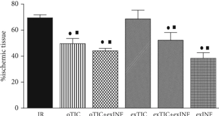
3.1. Infarct Size Was Reduced by Ticagrelor In Vivo but Not Ex Vivo. Rat hearts exposed to a 30 min global ischemia and 60 min reperfusion developed infarction which was 69:5 ± 2:3% of ischemic area at risk (AAR). Infarct size was significantly reduced by rat pretreatment for 3 days with Ticagrelor (oTIC, infarct size 49:6 ± 3:9% of AAR, P < 0:01 vs. IR group). No protective effects were recorded when Tica-grelor was added ex vivo to the perfusate of excised hearts from untreated animals (exTIC, infarct size 68:6 ± 3:0% of AAR), thus suggesting that Ticagrelor protection was only triggered in the intact organism (Figure 2). Coronary flow and perfusion pressure measured during stabilization in the IR group (10 ± 1 mL/min/g and 80 ± 2 mmHg, respectively) were not statistically different from those recorded in the treated groups, thus suggesting similar oxygen demands among groups.
3.2. Ticagrelor and INF Do Not Exert Additive Effects on Infarct Size Limitation. When INF was added ex vivo to the perfusate before ischemia (exINF), we observed a significant reduction in infarct size (exINF, infarct size 38:3 ± 3:0% of AAR,P < 0:05 vs. IR group), comparable to the one achieved by oTIC alone (Figure 2).
Interestingly, exINF did not exert additive effects on pro-tection against infarct size evoked by oral Ticagrelor pretreat-ment, as the reduction in infarct size evoked by the combination was almost identical to that obtained herein with either Ticagrelor or exINF alone (oTIC+exINF, infarct size 44:1 ± 1:9% of AAR, P < 0:05 vs. IR group). Similarly, no priming effects on exINF protection were recorded when Ticagrelor was coadministered ex vivo in the perfusate only (exTIC+exINF, infarct size 52:2 ± 2:7% of AAR).
3.3. Ticagrelor Pretreatment Prevented NLRP3 Inflammasome Activation and Downstream Signaling. Expression level and activation of the downstream signaling of NLRP3 in flamma-some were assessed by Western blotting analysis in protein extracts obtained from the apical portion of hearts pretreated or not with either Ticagrelor or INF and exposed to IR.
As expected, the INF pretreatment effectively reduced the IR-induced NLRP3 upregulation and activation, resulting in significant reduction of the cleaved active p10 subunit of caspase-1 (Figures 3(a) and 3(b)). As a consequence of reduced caspase-1 activation, the levels of IL-1β, that reached the highest concentrations after 60 min of reperfusion, showed a mild but still significant decrease in the exINF group (Figure 3(c)). Notably, both Western blotting analysis and ELISA assay demonstrated that similar inhibition of NLRP3 expression and activation could be reached when rats
were pretreated with Ticagrelor (oTIC), but not when Tica-grelor was added in the perfusate only (exTIC). Besides, no further NLRP3 inflammasome inhibition was recorded when INF was added in the perfusate of heart from rats previously exposed to Ticagrelor pretreatment (oTIC+exINF).
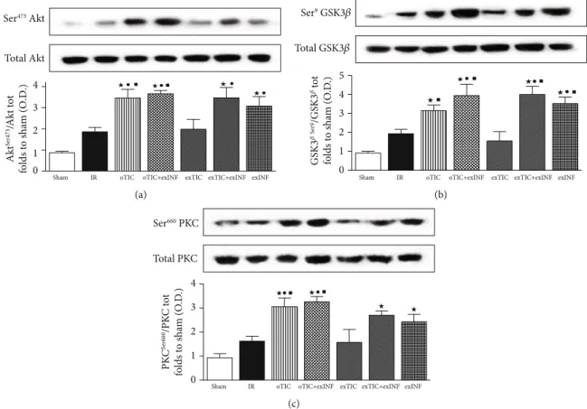
3.4. Risk Pathway Protective Activity Was Enhanced by Either Oral Ticagrelor Pretreatment Or INF Heart Exposure. Since RISK pathway is activated by both pre- and postconditioning treatments [34], we quantified expression and activity (in terms of phosphorylation) of its key members. After 60 min of reperfusion, slight but not significant increase in phos-phorylation rate of Akt, GSK-3β, and PKC (Figures 4(a)– 4(c), respectively) was recorded in untreated hearts exposed to IR protocol, when compared to the sham group. The phos-phorylation rates of Akt, GSK-3β, and PKC induced by the 60 min reperfusion were all increased massively in oTIC and exINF (bothP < 0:05 vs. sham; P = NS among groups). No additive effects were recorded when the two treatments were combined. Interestingly, ex vivo Ticagrelor exposure 20 min prior to ischemia only did not significantly modify the activation of the RISK pathway evoked by IR and/or INF. 3.5. Ticagrelor and INF Improved IR-Induced Antioxidant Response. SOD2 is an important endogenous antioxidant and provides protection against myocardial IR. Consistently with other studies [24], we found that IR led to increased expression of SOD2 (P < 0:05 vs. sham). All treatments blunted IR-induced SOD upregulation. However, in oTIC, oTIC+exINF, and exTIC+exINF groups, the levels of SOD2 were significantly lower than that of IR group (P < 0:05 vs. IR, Figure 5(a)). A reduction in SOD2 levels was also recorded in the heart of mice treated with INF only when compared to IR group, without reaching statistical signifi-cance (Figure 5(a)).
The Western blot analysis on the expression levels of the antioxidant transcription factor Nrf2 showed that its nuclear translocation was reduced by IR when compared to sham (Figure 5(b)). All treatments blunted IR-induced reduction of Nrf2 nuclear translocation. However, only in oTIC the
IR 0 20 40 60 80 %is chemic tissue
oTIC oTIC+exINF exTIC exTIC+exINF exINF
Figure 2: Infarct size. Data analyzed by one-way ANOVA followed by a Bonferroni post hoc test and expressed as mean ± SEM.n = 6 – 8 per group. Statistical significance: ●P < 0:05 vs. IR and ■P < 0:05vs. exTIC.
levels of Nrf2 nuclear translocation were significantly higher than in IR and similar to sham (Figure 5(b)).
3.6. Oral Ticagrelor Pretreatment Resulted in S1P and Adenosine Overaccumulation in the Heart. As shown in Figure 6, a marked increase in the myocardial concentration of either S1P or adenosine was recorded in the postischemic heart from rats exposed to the oral Ticagrelor pretreatment (oTIC) when compared to IR only, whereas ex vivo adminis-tration of Ticagrelor or exINF did not significantly affect their levels. Combining Ticagrelor and INF (oTIC+exINF) did not further increase S1P or adenosine accumulation in the heart in comparison to oTIC alone.
3.7. Ticagrelor Enhanced S1P and Adenosine Release from Platelets. Sphingosine, S1P, and adenosine were evaluated in the supernatant of human platelet samples subjected to stirring (1200 rpm speed at 37°C) with or without Ticagrelor (5000 ng/mL, 30 min) (Table 1). Ticagrelor-treated samples show high level of S1P in the supernatant and low level of sphingosine if compared to control (P < 0:05). Adenosine concentration increased significantly compared to control in the supernatant of Ticagrelor-treated platelets. The posi-tive control was obtained by incubating human platelet with TRAP-6 (10μmol/L, 8 min). The values of the release of S1P and adenosine in the supernatant in TRAP-6 group were sig-nificantly higher than control and Ticagrelor-treated plate-lets (data not shown).
4. Discussion
The present study further extends previousfindings on Tica-grelor cardioprotective effects, confirming that the protection was dependent upon its administration in vivo, as adding the P2Y12 antagonist ex vivo to the perfusate in excised hearts does not counteract the IR injury. Here, we confirm our pre-vious data [24] that the specific and direct inhibition of NLRP3 by exINF results in a significant reduction in infarct size. Most notably, adding INF just before ischemia does not further improve cardioprotection induced by Ticagrelor pre-treatment (3 days), with no significant effect of the combina-tion over each drug alone. We then aimed to assess whether Ticagrelor primes the isolated hearts exposed to the in flamma-some inhibitor. Administration of Ticagrelor to the perfusate ex vivo does not enhance the heart response to exINF, showing no interactions between the two treatments.
Indeed, Ticagrelor has been shown to modulate the expression on blood cells of toll-like receptors, key receptors involved in NLRP3 regulation [35]. Here, we show that the reduction in infarct size achieved by oral Ticagrelor is, at least in part, attributable to a cardioprotective effect mediated by the inhibition of the NLRP3 inflammasome pathway, as simi-lar inhibitory effects on the activation of pivotal markers of the inflammasome cascade were recorded when either pharmaco-logical tools (oral Ticagrelor or exINF) were used. With these two treatments, there is also an upregulation of the RISK path-way and a limitation of IR-induced oxidative stress.
NLRP3 Tubulin 1 0 2 3 4 NLRP3 fo lds t o sha m (O .D .)
Sham IR oTIC oTIC+exINF exTIC exTIC+exINF exINF
(a) Pro-caspase1 Tubulin Activated caspase-1 (p10) 0 Sham 2 4 6 8 A cti va te d/p ro caspas e-1 fo lds t o sha m (O .D .)
IR oTIC oTIC+exINF exTIC exTIC+exINF exINF
(b) 0 10 20 30 40 50 IL -1 𝛽 (pg/m g p ro tein)
Sham IR oTIC oTIC+exINF exTIC exTIC+exINF exINF
(c)
Figure 3: Western blotting analysis on (a) NLRP3, (b) pro- and activated caspase, and (c) quantification of IL1β by ELISA kit assay. Data analyzed by one-way ANOVA followed by a Bonferroni post hoc test and expressed as mean ± SEM. n = 6 – 8 per group. Statistical significance:●P < 0:05 vs. IR;★P < 0:05 vs. sham,■P < 0:05 vs. exTIC. Representative blots are shown of at least three different experiments.
To the best of our knowledge, so far, only another study has suggested that cardioprotection of Ticagrelor tested in models of acute myocardial injury can be partially
attribut-able to inhibition of mRNA levels of NLRP3 and IL-1beta in the heart of diabetic rats [13]. Here, we extended these observations to nondiabetic conditions and we documented Ser473 Akt Total Akt 0 1 2 3 4 Akt Ser473 /Akt tot
folds to sham (O.D.)
Sham IR oTIC oTIC+exINF exTIC exTIC+exINF exINF
(a) Ser9 GSK3𝛽 0 1 2 3 4 5 GS K3 𝛽 Se r9/GS K3 𝛽 tot fo lds t o sha m (O .D .) Total GSK3𝛽
Sham IR oTIC oTIC+exINF exTIC exTIC+exINF exINF
(b) Ser660 PKC Total PKC 0 1 2 3 4 PK C S er660 /PK C t o t fo lds t o sha m (O .D .)
Sham IR oTIC oTIC+exINF exTIC exTIC+exINF exINF
(c)
Figure 4: Western blotting analysis on (a) Akt, (b) GSK3β, and (c) PKC. Data analyzed by one-way ANOVA followed by a Bonferroni post hoc test and expressed as mean ± SEM.n = 6 – 8 per group. Statistical significance:●P < 0:05 vs. IR;★P < 0:05 vs. sham,■P < 0:05 vs. exTIC. Representative blots are shown of at least three different experiments.
SOD2 Tubulin 0 1 2 3 4 SO D2 fo lds t o sha m (O .D .)
Sham IR oTIC oTIC+exINF exTIC exTIC+exINF exINF
(a) PCNA Nuclear Nrf2 Tubulin Cytosolic Nrf2 0.0 0.5 1.0 1.5 NFR2 n uc leus/c yt os o l ra tio fo lds t o sha m (O .D .)
Sham IR oTIC oTIC+exINF exTIC exTIC+exINF exINF
(b)
Figure 5: Western blotting analysis on (a) SOD2 and (b) nuclear and cytosolic Nrf2. Data analyzed by one-way ANOVA followed by a Bonferroni post hoc test and expressed as mean ± SEM.n = 6 – 8 per group. Statistical significance:●P < 0:05 vs. IR;★P < 0:05 vs. sham, ■P < 0:05 vs. exTIC. Representative blots are shown of at least three different experiments.
that Ticagrelor, when administered to rats in vivo, evokes sig-nificant decrease of protein levels of NLRP3, resulting in lower activation of caspase-1, thus counteracting the IR-induced accumulation of active IL-1beta proteins in the heart.
A pharmacological approach with cardioprotective inhibitors has suggested that cardioprotection induced by P2Y12 antagonists is due to a conditioning phenomenon rather than to their antiplatelet effect [6]. In the presence of 0 50 100 150 Sp hin gosine (n g/mL)
Sham IR oTIC oTIC+exINF exTIC exTIC+exINF exINF
(a) 0 50 100 200 150 Sp hin gosine 1P (pg/mL)
Sham IR oTIC oTIC+exINF exTIC exTIC+exINF exINF
(b) 0 20 40 80 60 A denosine (n g/mL)
Sham IR oTIC oTIC+exINF exTIC exTIC+exINF exINF
(c)
Figure 6: Myocardial levels of sphingosine, sphingosine 1P (S-1P), and adenosine. Data analyzed by one-way ANOVA followed by a Bonferroni post hoc test and expressed as mean ± SEM.n = 6 – 8 per group. Statistical significance:●P < 0:05 vs. IR;★P < 0:05 vs. sham, ■P < 0:05 vs. exTIC.
Table 1: Level of sphingosine, sphingosine 1P (S-1P), and adenosine released by platelets exposed to Ticagrelor.
Sphingosine (ng/mL) S-1P (ng/mL) Adenosine (ng/mL)
Control 356:9 ± 44:3 94:3 ± 29:0 18:8 ± 5:8
Ticagrelor 68:4 ± 2:4∗ 300:7 ± 38:5∗ 48:8 ± 5:4∗
Level of sphingosine, S-1P, and adenosine, measured with UHPLC-tandem mass analysis, in the supernatant of human platelet subjected to stirring (1200 rpm
speed at 37°C) incubated in absence and presence of Ticagrelor (5000 ng/mL, 30 min). Data analyzed by Student’s t test and expressed as mean ± SEM; n = 6 − 8
sphingosine kinase inhibitor, Cangrelor’s antiplatelet effect seems intact; nevertheless, in vivo studies did not definitively rule out a contribution from the antiplatelet effect in limiting IR injury. Our study, in which the P2Y12 antagonist was administered in vivo and myocardial infarct subsequently induced ex vivo, in the absence of platelets, definitively con-firms that the protective effect is mainly due to a precondi-tioning effect in vivo that lasts throughout the IR procedure ex vivo. The lack of cardioprotection by Ticagrelor when administered in the ex vivo model further support the idea of a blood cell-mediated preconditioning effect [5, 8, 31]. Given the apparent dependence upon the presence of blood in mediating Ticagrelor effects [5, 17], it would seem logical to propose that the cardioprotective effect of Ticagrelor is dependent on platelets and likely the P2Y12 receptor. This is supported by previous observations showing that chemi-cally distinct P2Y12 antagonists have similar cardioprotec-tive properties [6, 16, 17]. We thus investigated the ability of Ticagrelor to affect platelet ability to release S1P, an essen-tial, bioactive lysophospholipid mediator that regulates vari-ous physiological functions such as lymphocyte trafficking, inflammation, and behavioural characteristics of the vascular system [36]. Platelets are among the major source of S1P in the circulation [17, 37], and platelet-derived S1P has been demonstrated to exert a critical role in the repair of pivotal microvascular structures during injury [38, 39]. Ticagrelor is unique in being an inhibitor of equilibrative nucleotide transporter 1 (ENT1) [6, 13, 14, 16]. Here, we confirm these effects in isolated human platelets, as revealed by an increase in S1P and adenosine release after exposure to Ticagrelor. Ticagrelor was previously demonstrated to raise tissue levels of adenosine, which is a known endogenous cardioprotective substance in pathophysiological conditions of the heart, including myocardial ischemia and heart failure [40, 41]. This effect has been suggested to involve inhibition of ENT1 in heart tissue [13–15]. However, so far, no experi-mental evidence of direct effects of Ticagrelor on ENT1 has been reported. Besides, ourfindings on the lack of cardiopro-tection by Ticagrelor on the isolated heart cloud this hypothe-sis of cardioprotection through interference with cardiac ENT 1 [8]. Thereby, its effects on increasing adenosine levels in heart tissue could derive from an effect of Ticagrelor on a sub-group of blood cells. Both erythrocytes and platelets are known to release different substances in blood stream, includ-ing adenosine and S1P [17, 42, 43]. Here, we demonstrated for thefirst time that Ticagrelor increased the levels of adenosine released from platelets, thus suggesting that this effect might contribute, together with S1P, to the cardioprotection recorded when Ticagrelor was administered in vivo only.
The contribution of blood-derived S1P and adenosine in mediating the myocardial protective effects of Ticagrelor was confirmed by showing that both S1P and adenosine reached the highest concentrations in the heart of rats orally exposed to Ticagrelor. While the increase in myocardial adenosine levels has been already documented in the IR heart of rats orally pretreated with Ticagrelor [13], so far, no direct myo-cardial detection of S1P or comparison of adenosine/S1P myocardial levels between in vivo vs. ex vivo treatments has been reported in the literature. Thus, our study adds a further
interesting piece of evidence on the ability of Ticagrelor to cause blood cells to release substances, which may contribute to cardioprotection. In fact, either S1P or adenosine has been already demonstrated to evoke protective effects throughout activation of the protective survival RISK pathway in the heart [44, 45]. As Ticagrelor protection seems to depend at least in part on the same signaling cascade modulate S1P and adenosine, it seemed likely that these endogenous components would be involved in Ticagrelor’s protective mechanism. Interestingly, both the treatment (Ticagrelor and exINF) in aerobic conditions do not affect myocardial perfusion, thus suggesting an unchanged oxygen demand in comparison to untreated hearts. Moreover, the levels of components of either NLRP3 or RISK pathways in sham ani-mals were lower than those detected in all IR groups (pro-tected and nonpro(pro-tected), thus suggesting that in aerobic conditions, the pharmacological treatments do not influence the myocardial metabolism, but may trigger mechanisms that will make the hearts more resistant to IR challenge boosting RISK activation in postischemic phase. Indeed, the RISK pathway is an intrinsic prosurvival signaling cascade evoked by IR itself which confers protection against the reperfusion insult by avoiding the opening of the mitochon-drial permeability transition pore at the onset of reperfusion [34, 46]. Potentiation of RISK activation in the early minutes of reperfusion contributes to cardioprotection induced by preconditioning protocols. Actually, in protected hearts, the phosphorylation of RISK enzymes peaks at 10-15 minutes of reperfusion and progressively wanes thereafter [34, 47– 49]. A two-threefold higher phosphorylation level after 60 min of reperfusion is a strong indication of kinase involve-ment in protection, especially if we consider that the reduc-tion in infarct size in protected hearts resulted in a ~20% increase in vital tissue when compared to control IR hearts. As previously documented [6], pharmacological inhibition of the RISK pathway blunted the protective effects of P2Y12 antagonists against IR, thus further supporting the hypothe-sis that their mechanisms of cardioprotection utilize specific signal transduction of myocardial protection rather than inhibition of intravascular coagulation. Our study extends these findings confirming cross-talk mechanisms linking NLRP3 inflammasome to RISK pathway in cardioprotection, which have been so far faintly suggested [50–52], but not convincingly demonstrated. Besides, it shows that Ticagrelor uses similar mechanisms of protection evoked by a NLRP3 inflammasome inhibitor, leading to activation of the RISK pathway. The lack of additive effects of the drug combination may be explained considering that additional protection can-not be induced by strategies that share common prosurvival signaling pathways such as the P2Y12 antagonist and the NLRP3 inflammasome inhibitor. However, previous studies demonstrated that treatment with a caspase-1 inhibitor prior to ischemia or reperfusion adds its protection to the one elic-ited by the P2Y12 antagonist, Cangrelor [4, 7]. Differently from INF that directly targets NLRP3 complex formation, direct caspase-1 inhibitors may influence not only inflamma-tory response but also glycolytic, mitochondrial, and pyrop-totic cell death [4]; thus, the additional beneficial effects recorded by these authors could be due to interference with
any of these pathways beyond the inhibition of the NLRP3 inflammasome-caspase axis. Another substantial difference between our study and that of Audia et al. [4] is that these authors had higher coronaryflow when the caspase-1 inhib-itor was used, while we perfused the hearts at constantflow, to avoidflow effects on IR injury.
The role of oxidative stress in contributing to IR injury is not clear [53, 54]. Overall, it seems that IR-dependent oxida-tive stress is reduced by all protecoxida-tive treatments. However, only Ticagrelor pretreatment displays a consistent effect in limiting the oxidative component as suggested by the signif-icant increase in nuclear levels of Nrf2 and by the downregu-lation of SOD2. Nevertheless, also for this mechanism, there are not apparent differences between Ticagrelor and INF. Moreover, the antioxidant effect of Ticagrelor given orally seems stronger than that observed when it is given ex vivo.
5. Limitation of the Study
Our results confirm Ticagrelor conditioning effects, which are not additives to the cardioprotection achieved by directly inhibiting NLRP3. It is likely that this lack of additive effect is due to the activation of RISK pathway by both treatments. Other studies demonstrated additive effect when P2Y12 and a downstream NLRP3 factor, namely, caspase-1, were inhib-ited [4, 7], thus confirming that the cross-talk between NLRP3 and RISK cardioprotective pathways is quite complex [36]. Besides, here, we did not test the impact of the proposed pharmacological treatments on the tested signaling cascades at basal condition. Therefore, further studies are needed to fully elucidate the cross-talk among mechanisms linking NLRP3 complex, redox state, and RISK pathway. Finally, we must consider that all experimental paradigms have dis-advantages and dis-advantages. For instance, we used gavage to administer Ticagrelor instead of a spontaneous intake which is more physiological, as gavage guarantees a more constant dosage, which is recommendable in cardioprotection studies [55]. Determining which blood-derived factors mediate Ticagrelor-induced cardioprotection was beyond the scope of this study. Nevertheless, the fact that Ticagrelor increases platelet release of both adenosine and S1P suggests these fac-tors as important players that deserve further investigations.
6. Conclusions
In conclusion, we confirm that Ticagrelor requires the pres-ence of blood to act as conditioning agent. Importantly, we demonstrate that the cardioprotective effects of Ticagrelor are not due to a direct action on the myocardial tissue nor to its antiaggregating effect, whereas the NLRP3 inhibitor, INF, is able to act directly on the heart. Nevertheless, these two drugs given before ischemia activate a similar protective pathway, involving RISK pathway and redox modulation, without additive cardioprotective effects.
Abbreviations
RISK: Reperfusion Injury Salvage Kinase
IR: Ischemia/reperfusion
AMI: Acute myocardial infarction
NLRP3: Nucleotide-binding oligomerization domain-(NOD-) like receptor pyrin domain containing 3
IS: Infarct size
AAR: Area at risk
INF: NLRP3 inflammasome inhibitor.
Data Availability
The data used to support thefindings of this study are avail-able from the corresponding author (MC and PP) upon request.
Conflicts of Interest
The authors declare that they have no conflicts of interest.
Authors’ Contributions
Claudia Penna and Manuela Aragno are sharedfirst authors of this study. PP and MC drafted thefirst version of the man-uscript and supervised the writing. CP and MA made the revision and statistical management of paper and figures. EZ and SF made the isolated hearts. ASC, MA, MC, and FC made the WB analysis. IR isolated human platelets. CM and FDB made the HPLC analysis. MB and GFA made INF. All authors evaluated retrieved papers and their refer-ence lists to identify additional relevant articles. All authors actively contributed to the study, revised the manuscript, and approved the final version of the manuscript. Claudia Penna, Manuela Aragno, Massimo Collino, and Pasquale Pagliaro contributed equally to this works. Pasquale PAGLIARO and Massimo COLLINO are shared senior authors of this study.
Acknowledgments
This study was funded by the University of Turin, Ricerca Locale Ex-60% (Grants: PAGP_RILO_16_01; PENC_RILO, ARAM_RILO, COLM_RILO) and by MIUR (PAGP_ FFABR_17_01 and PENC_FFABR_17_01, COLM_FFABR_ 17_01).
References
[1] D. J. Hausenloy, S. B. Ong, and D. M. Yellon,“The mitochon-drial permeability transition pore as a target for precondition-ing and postconditionprecondition-ing,” Basic Research in Cardiology, vol. 104, no. 2, pp. 189–202, 2009.
[2] P. Pagliaro, F. Moro, F. Tullio, M. G. Perrelli, and C. Penna, “Cardioprotective pathways during reperfusion: focus on redox signaling and other modalities of cell signaling,” Antiox-idants & Redox Signaling, vol. 14, no. 5, pp. 833–850, 2011. [3] C. Penna, M. G. Perrelli, and P. Pagliaro,“Mitochondrial
path-ways, permeability transition pore, and redox signaling in car-dioprotection: therapeutic implications,” Antioxidants & Redox Signaling, vol. 18, no. 5, pp. 556–599, 2013.
[4] J. P. Audia, X. M. Yang, E. S. Crockett et al.,“Caspase-1 inhi-bition by VX-765 administered at reperfusion in P2Y12 recep-tor antagonist-treated rats provides long-term reduction in
myocardial infarct size and preservation of ventricular func-tion,” Basic Research in Cardiology, vol. 113, no. 5, 2018. [5] M. V. Cohen, X. M. Yang, J. White, D. M. Yellon, R. M. Bell,
and J. M. Downey, “Cangrelor-mediated cardioprotection requires platelets and sphingosine phosphorylation,” Cardio-vascular Drugs and Therapy, vol. 30, no. 2, pp. 229–232, 2016. [6] X. M. Yang, Y. Liu, L. Cui et al.,“Platelet P2Y12blockers confer direct postconditioning-like protection in reperfused rabbit hearts,” Journal of Cardiovascular Pharmacology and Thera-peutics, vol. 18, no. 3, pp. 251–262, 2013.
[7] X. M. Yang, J. M. Downey, M. V. Cohen, N. A. Housley, D. F. Alvarez, and J. P. Audia,“The highly selective caspase-1 inhib-itor VX-765 provides additive protection against myocardial infarction in rat hearts when combined with a platelet inhibi-tor,” Journal of Cardiovascular Pharmacology and Therapeu-tics, vol. 22, no. 6, pp. 574–578, 2017.
[8] X. M. Yang, S. Gadde, J. P. Audia, D. F. Alvarez, J. M. Downey, and M. V. Cohen, “Ticagrelor does not protect isolated rat hearts, thus clouding its proposed cardioprotective role through ENT 1 in heart tissue,” Journal of Cardiovascular Pharmacology and Therapeutics, vol. 24, no. 4, pp. 371–376, 2019.
[9] R. Koski and B. Kennedy,“Comparative review of oral P2Y12 inhibitors,” P & T : a peer-reviewed journal for formulary man-agement, vol. 43, no. 6, pp. 352–357, 2018.
[10] D. Armstrong, C. Summers, L. Ewart, S. Nylander, J. E. Sid-away, and J. J. J. van Giezen,“Characterization of the adeno-sine pharmacology of ticagrelor reveals therapeutically relevant inhibition of equilibrative nucleoside transporter 1,” Journal of Cardiovascular Pharmacology and Therapeutics, vol. 19, no. 2, pp. 209–219, 2014.
[11] L. Bonello, M. Laine, N. Kipson et al., “Ticagrelor increases adenosine plasma concentration in patients with an acute cor-onary syndrome,” Journal of the American College of Cardiol-ogy, vol. 63, no. 9, pp. 872–877, 2014.
[12] M. Cattaneo, R. Schulz, and S. Nylander, “Adenosine-medi-ated effects of ticagrelor: evidence and potential clinical rele-vance,” Journal of the American College of Cardiology, vol. 63, no. 23, pp. 2503–2509, 2014.
[13] Y. Birnbaum, G. D. Birnbaum, I. Birnbaum, S. Nylander, and Y. Ye,“Ticagrelor and rosuvastatin have additive cardiopro-tective effects via adenosine,” Cardiovascular Drugs and Ther-apy, vol. 30, no. 6, pp. 539–550, 2016.
[14] G. Vilahur, M. Gutiérrez, L. Casani et al.,“Protective effects of ticagrelor on myocardial injury after infarction,” Circulation, vol. 134, no. 22, pp. 1708–1719, 2016.
[15] Y. Ye, G. D. Birnbaum, J. R. Perez-Polo, M. K. Nanhwan, S. Nylander, and Y. Birnbaum,“Ticagrelor protects the heart against reperfusion injury and improves remodeling after myocardial infarction,” Arteriosclerosis, Thrombosis, and Vas-cular Biology, vol. 35, no. 8, pp. 1805–1814, 2015.
[16] S. Nylander and R. Schulz,“Effects of P2Y12receptor antago-nists beyond platelet inhibition-comparison of ticagrelor with thienopyridines,” British Journal of Pharmacology, vol. 173, no. 7, pp. 1163–1178, 2016.
[17] S. M. Davidson, I. Andreadou, L. Barile et al., “Circulating blood cells and extracellular vesicles in acute cardioprotec-tion,” Cardiovascular Research, vol. 115, no. 7, pp. 1156– 1166, 2019.
[18] R. Mastrocola, M. Aragno, G. Alloatti, M. Collino, C. Penna, and P. Pagliaro,“Metaflammation: tissue-specific alterations of the NLRP3 inflammasome platform in metabolic
syn-drome,” Current Medicinal Chemistry, vol. 25, no. 11, pp. 1294–1310, 2018.
[19] F. Chiazza, A. Couturier-Maillard, E. Benetti et al.,“Targeting the NLRP3 inflammasome to reduce diet-induced metabolic abnormalities in mice,” Molecular Medicine, vol. 21, no. 1, pp. 1025–1037, 2015.
[20] M. Collino, E. Benetti, M. Rogazzo et al.,“Reversal of the del-eterious effects of chronic dietary HFCS-55 intake by PPAR-δ agonism correlates with impaired NLRP3 inflammasome acti-vation,” Biochemical Pharmacology, vol. 85, no. 2, pp. 257–264, 2013.
[21] S. Cannito, E. Morello, C. Bocca et al.,“Microvesicles released from fat-laden cells promote activation of hepatocellular NLRP3 inflammasome: a pro-inflammatory link between lipo-toxicity and non-alcoholic steatohepatitis,” PLoS One, P. Strnad, Ed., vol. 12, no. 3, article e0172575, 2017.
[22] E. Benetti, R. Mastrocola, G. Vitarelli et al.,“Empagliflozin pro-tects against diet-induced NLRP-3 inflammasome activation and lipid accumulation,” The Journal of Pharmacology and Experimental Therapeutics, vol. 359, no. 1, pp. 45–53, 2016. [23] D. Nigro, F. Menotti, A. S. Cento et al.,“Chronic
administra-tion of saturated fats and fructose differently affect SREBP activity resulting in different modulation of Nrf2 and Nlrp3 inflammasome pathways in mice liver,” The Journal of Nutri-tional Biochemistry, vol. 42, pp. 160–171, 2017.
[24] R. Mastrocola, C. Penna, F. Tullio et al., “Pharmacological inhibition of NLRP3 inflammasome attenuates myocardial ischemia/reperfusion injury by activation of RISK and mito-chondrial pathways,” Oxidative Medicine and Cellular Longev-ity, vol. 2016, Article ID 5271251, 11 pages, 2016.
[25] S. Toldo, A. G. Mauro, Z. Cutter et al.,“The NLRP3 inflamma-some inhibitor, OLT1177 (dapansutrile), reduces infarct size and preserves contractile function after ischemia reperfusion injury in the mouse,” Journal of Cardiovascular Pharmacology, vol. 73, no. 4, pp. 215–222, 2019.
[26] R. Mastrocola, M. Collino, C. Penna et al.,“Maladaptive mod-ulations of NLRP3 inflammasome and cardioprotective path-ways are involved in diet-induced exacerbation of myocardial ischemia/reperfusion injury in mice,” Oxidative Medicine and Cellular Longevity, vol. 2016, Article ID 3480637, 12 pages, 2016.
[27] H. E. Bøtker, D. Hausenloy, I. Andreadou et al., “Practical guidelines for rigor and reproducibility in preclinical and clin-ical studies on cardioprotection,” Basic Research in Cardiology, vol. 113, no. 5, pp. 39–39, 2018.
[28] M. Cocco, D. Garella, A. StiloDi et al.,“Electrophilic warhead-based design of compounds preventing NLRP3 in flammasome-dependent pyroptosis,” Journal of Medicinal Chemistry, vol. 57, no. 24, pp. 10366–10382, 2014.
[29] M. Cocco, G. Miglio, M. Giorgis et al.,“Design, synthesis, and evaluation of acrylamide derivatives as direct NLRP3 inflam-masome inhibitors,” ChemMedChem, vol. 11, no. 16, pp. 1790–1803, 2016.
[30] M. Cocco, C. Pellegrini, H. Martínez-Banaclocha et al., “Devel-opment of an acrylate derivative targeting the NLRP3 in flam-masome for the treatment of inflammatory bowel disease,” Journal of Medicinal Chemistry, vol. 60, no. 9, pp. 3656– 3671, 2017.
[31] A. Moulias, I. Xanthopoulou, and D. Alexopoulos,“Does tica-grelor improve endothelial function?,” Journal of Cardiovascu-lar Pharmacology and Therapeutics, vol. 24, no. 1, pp. 11–17, 2019.
[32] C. Penna, F. Tullio, A. Merlino et al.,“Postconditioning car-dioprotection against infarct size and post-ischemic systolic dysfunction is influenced by gender,” Basic Research in Car-diology, vol. 104, no. 4, pp. 390–402, 2009.
[33] S. M. Coldewey, E. Benetti, M. Collino et al., “Elevation of serum sphingosine-1-phosphate attenuates impaired cardiac function in experimental sepsis,” Scientific Reports, vol. 6, no. 1, 2016.
[34] X. Rossello and D. M. Yellon, “The RISK pathway and beyond,” Basic Research in Cardiology, vol. 113, no. 1, 2018. [35] A. J. van der Ven, N. Riksen, G. Rongen et al.,“Differential
effects of platelets and platelet inhibition by ticagrelor on TLR2- and TLR4-mediated inflammatory responses,” Throm-bosis and Haemostasis, vol. 113, no. 5, pp. 1035–1045, 2015. [36] F. Tukijan, M. Chandrakanthan, and L. N. Nguyen,“The
sig-nalling roles of sphingosine-1-phosphate derived from red blood cells and platelets,” British Journal of Pharmacology, vol. 175, no. 19, pp. 3741–3746, 2018.
[37] N. Urtz, F. Gaertner, M.-L. von Bruehl et al.,“Sphingosine 1-phosphate produced by sphingosine kinase 2 intrinsically con-trols platelet aggregation in vitro and in vivo,” Circulation Research, vol. 117, no. 4, pp. 376–387, 2015.
[38] R. L. Proia and T. Hla,“Emerging biology of sphingosine-1-phosphate: its role in pathogenesis and therapy,” The Journal of Clinical Investigation, vol. 125, no. 4, pp. 1379–1387, 2015. [39] Z. Wang, K. Chen, Y. Han et al.,“Irisin protects heart against ischemia-reperfusion injury through a SOD2-dependent mito-chondria mechanism,” Journal of Cardiovascular Pharmacol-ogy, vol. 72, no. 6, pp. 259–269, 2018.
[40] M. V. Cohen, C. P. Baines, and J. M. Downey,“Ischemic pre-conditioning: from adenosine receptor to KATP channel,” Annual Review of Physiology, vol. 62, pp. 79–109, 2000. [41] M. Kitakaze and M. Hori,“Adenosine therapy: a new approach
to chronic heart failure,” Expert Opinion on Investigational Drugs, vol. 9, no. 11, pp. 2519–2535, 2000.
[42] E. M. Golebiewska and A. W. Poole,“Platelet secretion: from haemostasis to wound healing and beyond,” Blood Reviews, vol. 29, no. 3, pp. 153–162, 2015.
[43] J. Öhman, R. Kudira, S. Albinsson, B. Olde, and D. Erlinge, “Ticagrelor induces adenosine triphosphate release from human red blood cells,” Biochemical and Biophysical Research Communications, vol. 418, no. 4, pp. 754–758, 2012. [44] S. J. Somers, M. Frias, L. Lacerda, L. H. Opie, and S. Lecour,
“Interplay between SAFE and RISK pathways in sphingosine-1-phosphate-induced cardioprotection,” Cardiovascular Drugs and Therapy, vol. 26, no. 3, pp. 227–237, 2012.
[45] W. J. Zang, L. Sun, and X. J. Yu,“Cardioprotection of ischemic postconditioning and pharmacological post-treatment with adenosine or acetylcholine,” Acta physiologica Sinica, vol. 59, no. 5, pp. 593–600, 2007.
[46] D. J. Hausenloy and D. M. Yellon,“New directions for protect-ing the heart against ischaemia-reperfusion injury: targetprotect-ing the Reperfusion Injury Salvage Kinase (RISK)-pathway,” Car-diovascular Research, vol. 61, no. 3, pp. 448–460, 2004. [47] P. Ferdinandy, D. J. Hausenloy, G. Heusch, G. F. Baxter, and
R. Schulz, “Interaction of risk factors, comorbidities, and comedications with ischemia/reperfusion injury and cardio-protection by preconditioning, postconditioning, and remote conditioning,” Pharmacological Reviews, vol. 66, no. 4, pp. 1142–1174, 2014.
[48] C. Penna, C. Angotti, and P. Pagliaro,“Protein S-nitrosylation in preconditioning and postconditioning,” Experimental Biol-ogy and Medicine, vol. 239, no. 6, pp. 647–662, 2014. [49] M. V. Cohen and J. M. Downey, “Signalling pathways and
mechanisms of protection in pre- and postconditioning: his-torical perspective and lessons for the future,” British Journal of Pharmacology, vol. 172, no. 8, pp. 1913–1932, 2015. [50] S. Femminò, P. Pagliaro, and C. Penna,“Obesity and
cardio-protection,” Current Medicinal Chemistry, vol. 27, no. 2, pp. 230–239, 2020.
[51] Ø. Sandanger, E. Gao, T. Ranheim et al.,“NLRP3 inflamma-some activation during myocardial ischemia reperfusion is cardioprotective,” Biochemical and Biophysical Research Com-munications, vol. 469, no. 4, pp. 1012–1020, 2016.
[52] C. J. Zuurbier,“NLRP3 inflammasome in cardioprotective sig-naling,” Journal of Cardiovascular Pharmacology, vol. 74, no. 4, pp. 271–275, 2019.
[53] H. Y. Sun, N. P. Wang, F. Kerendi et al.,“Hypoxic postcondi-tioning reduces cardiomyocyte loss by inhibiting ROS genera-tion and intracellular Ca2+ overload,” American Journal of Physiology. Heart and Circulatory Physiology, vol. 288, no. 4, pp. H1900–H1908, 2005.
[54] Z. Xu, M. V. Cohen, J. M. Downey, T. L. vanden Hoek, and Z. Yao,“Attenuation of oxidant stress during reoxygenation by AMP 579 in cardiomyocytes,” American Journal of Physiol-ogy. Heart and Circulatory Physiology, vol. 281, no. 6, pp. H2585–H2589, 2001.
[55] X. M. Yang, L. Cui, A. Alhammouri, J. M. Downey, and M. V. Cohen,“Triple therapy greatly increases myocardial salvage during ischemia/reperfusion in the in situ rat heart,” Cardio-vascular Drugs and Therapy, vol. 27, no. 5, pp. 403–412, 2013.