Acute heart failure in patients with acute
aortic syndrome: pathophysiology and
clinical–prognostic implications
Fabio Vagnarelli
1, Anna Corsini
1, Massimiliano Lorenzini
1, Davide Pacini
2,
Marinella Ferlito
1, Letizia Bacchi Reggiani
1, Simone Longhi
1, Samuele Nanni
1,
Giulia Norscini
1, Laura Cinti
1, Giulia Bugani
1, Ferdinando Pasquale
1, Elena Biagini
1,
Francesco Grigioni
1, Roberto Di Bartolomeo
2, Marco Marini
3, Gian Piero Perna
3,
Giovanni Melandri
1, and Claudio Rapezzi
1*
1Cardiology, Department of Experimental Diagnostic and Specialty Medicine, Alma Mater Studiorum-University of Bologna, Bologna, Italy;2Cardiac Surgery, Department of Experimental Diagnostic and Specialty Medicine, Alma Mater Studiorum-University of Bologna, Bologna, Italy; and3Cardiology, Cardiovascular Department, ‘Ospedali Riuniti di Ancona’, Ancona, Italy
Received 13 May 2015; revised 14 June 2015; accepted 24 June 2015 ; online publish-ahead-of-print 27 July 2015
Aims Although acute heart failure (AHF) is a potential complication of acute aortic syndromes (AAS), its clinical details and management implications have been scarcely evaluated. This study aimed to assess prevalence, pathophysiological mechanisms, impact on treatment, and in-hospital mortality of AHF in AAS.
... Methods
and results
Data were collected from a prospective AAS registry (398 patients diagnosed between 2000 and 2013). Patients with AHF were identified by the presence of dyspnoea as the presentation symptom or radiological signs of pulmonary congestion or cardiogenic shock, including patients with cardiac tamponade (CT). AHF frequency was 28% (Stanford type A 32% vs. type B 20%, P = 0.01). Four mechanisms leading to AHF were identified, alone or in combination: CT (26%), aortic regurgitation (25%), myocardial ischaemia (17%), and hypertensive crisis (10%). In type A patients, aortic regurgitation and CT were the most frequent mechanisms, whereas myocardial ischaemia and hypertensive crisis were the most frequent in type B patients. Although no difference was noted for diagnostic times, AHF at presentation led to a longer surgical delay in type A AAS. In-hospital mortality was higher in patients with AHF compared with those without (34% vs. 17%, P< 0.001). After multivariable analysis, AHF was associated with increased risk of in-hospital death (adjusted odds ratio 1.97, 95% confidence interval 1.14–3.36, P = 0.014).
...
Conclusion AHF occurs in more than a quarter of patients with AAS of both type A and type B, is due to a variety of
pathophysiological mechanisms, and is associated with increased surgical delay and in-hospital mortality.
...
Keywords Acute heart failure • Acute aortic syndromes • Cardiogenic shock • Cardiac tamponade
Introduction
Acute heart failure (AHF) is rightly regarded not as a single disease but as a syndrome that can be caused by different mechanisms and different diseases. Although it is known that aortic dissection is one of the possible causes of AHF,1,2the literature on the subject
*Corresponding author: Cardiology, Department of Experimental Diagnostic and Specialty Medicine, Alma Mater Studiorum-University of Bologna, Via G. Massarenti 9, 40138 Bologna, Italy. Tel: +39 051 349858, Fax: +39 051 344859, Email: [email protected]
...
consists mainly of case reports.3–8 The only systematic approach
to this issue dates back to 10 years ago.2 A research letter
published in 2005 summarizes findings from the IRAD registry, but only partially specifies the mechanisms leading to AHF. Since then, diagnostic tools and surgical techniques have evolved enor-mously so that an in-depth analysis of this serious complication
of acute aortic syndromes (AAS) in the current ‘era’ could be useful.
Using the data from a prospective metropolitan AAS registry, we aimed to assess the frequency of AHF in AAS, characterize the patients’ clinical and instrumental profile, explore the patho-physiological mechanisms underlying the condition, and evaluate the impact on treatment and in-hospital mortality.
Methods
Setting, patients, and data collection
Our registry (AESA, Archivio Elettronico Sindromi Aortiche acute) includes data from all consecutive patients referred to our Institution between 2000 and 2013 who received a final diagnosis of spontaneous AAS. The S. Orsola-Malpighi University Hospital is the referral centre for AAS treatment in a metropolitan hospital network that covers Bologna and its surrounding areas (catchment area ∼1 000 000 inhabitants).The database contains information on patient demographics, history, clinical presentation, physical findings, laboratory findings, imaging study results, details of medical and surgical treatment, and patient outcome, including mortality. In accordance with inter-national guidelines and the internal protocol at our institution, the diagnosis of AAS was confirmed by computed tomography (CT) scan in the vast majority of cases (93.5%). Baseline charac-teristics included ‘classic’ risk factors for AAS and cardiovascular/ non-cardiovascular co-morbidities. Pain features and presentation symptoms were reported in detail. Two experienced cardiologists blindly reviewed all the ECGs. Laboratory findings included data on cardiac troponin assay, when performed according to the standard protocol used in the chest pain unit (until 2010 the standard test was used, and was then replaced by a high sensitivity assay). Imaging was interpreted by specialized radiologists and echocardiographers, and entered on the data form. Helical CT, transoesophageal/transthoracic echocardiography (TEE/TTE), magnetic resonance imaging, and/or angiography were obtained and reviewed.
The following relevant diagnostic time intervals were also recorded: (i) symptom onset to presentation at any hospital; (ii) hospital presen-tation to final AAS diagnosis; and (iii) global diagnostic delay (symptom onset to final AAS diagnosis at any hospital). Surgical delay (for Stan-ford type A) was defined as the time between symptom onset and the operating room.
With a method comparable with that of the IRAD registry, diagnos-tic time intervals were recorded prospectively during the initial phases of hospitalization.9The ‘time of final diagnosis’ was defined as the time
when the first demonstration of the aortic lesion was documented on an imaging examination and recorded.
Patients with symptom onset>14 days at hospital presentation were not included in the registry. AAS (aortic dissection, penetrating ulcer, and intramural haematoma) was defined according to the Stanford classification.
In all cases (presenting at either a hub or a spoke centre), the diagnosis was confirmed by a multidisciplinary team that included a cardiologist, cardio-thoracic surgeon, and cardiovascular radiologist.
The investigation conforms with the principles outlined in the Declaration of Helsinki. The study was approved by the local ethics committee, and all patients provided written informed consent. ...
...
Definitions and mechanisms
Patients with AHF were identified by the presence of dyspnoea as the presentation symptom or radiological signs of pulmonary congestion or cardiogenic shock, including patients with cardiac tamponade. Shock was defined as sustained hypotension (systolic blood pressure
<90 mmHg for >30 min) accompanied by clinical signs of
periph-eral/cerebral hypoperfusion,10 despite adequate LV filling pressure.
The standard definition for cardiac tamponade was used.11
Clinical and instrumental data of patients with AHF were systemat-ically reviewed in order to identify the mechanisms leading to AHF. A distinction between ‘main’ and ‘contributing’ mechanisms was made by two cardiologists on a case-by-case basis using the following hierarchy: cardiac tamponade, severe aortic regurgitation, myocardial ischaemia, and hypertensive crisis.
An ECG was considered to be acute coronary syndrome (ACS)-like in the presence of≥1 of the following characteristics: (i) ST-segment elevation in two contiguous leads with the cut-off point≥0.1 mV in all leads other than V2–V3, where the cut-off point was≥0.2 mV; (ii) horizontal or down-sloping ST-segment depression≥0.05 mV in two contiguous leads; and (iii) T-wave inversion≥0.1 mV in two contiguous leads.
The diagnosis of troponin positivity using standard cardiac troponin T (cTnT) testing was made in the presence of at least one value of cTnT
>30 ng/L (10% coefficient of variation cut-off). When high sensitivity
cTnT (HS-cTnT) was used, the diagnosis of troponin positivity was made in the presence of at least one value of HS-cTnT>14 ng/L (99th percentile, upper reference limit).
Myocardial ischaemia was defined by the presence of ACS-like ECG findings and/or troponin positivity.
Aortic regurgitation was evaluated semi-quantitatively with TTE/TEE using the proximal regurgitant jet height/LV outflow tract diameter ratio12 and the vena contracta method in selected cases,13 and was
considered a possible mechanism of AHF only when graded severe or moderate to severe. Mechanisms leading to aortic regurgitation in type A AAS were classified according to Movsowitz et al.14
A hypertensive crisis was defined according to current Euro-pean Society of Cardiology (ESC) guidelines on arterial hyperten-sion (systolic blood pressure>180 mmHg or diastolic blood pressure
>110 mmHg).15
Pleural effusion was diagnosed by chest X-ray or CT scan. Pericardial effusion was diagnosed by TTE/TEE cardiac CT, or magnetic resonance imaging. Periaortic haematoma was diagnosed by TTE/TEE, CT, or magnetic resonance imaging.16
Statistical analysis
Categorical data were expressed as proportions, and continuous vari-ables reported as mean ± SD or median [interquartile range (IQR)], as appropriate. The𝜒2 test for categorical variables was used to
com-pare groups. The two-tailed Student t-test was used to comcom-pare nor-mally distributed continuous variables. Comparison of non-nornor-mally distributed variables was conducted using the Mann–Whitney U-test. We explored the association between diagnostic delay and patient clinical–instrumental profile. In order to identify unusually long diag-nostic times, we used the 75th percentile of in-hospital delay as the cut-off, in keeping with previous analyses.17
Logistic regression analysis was performed to identify predictors of in-hospital delay and in-hospital mortality. Non-correlated variables with P< 0.2 at the univariate analyses were included in the multivariate
analysis. Model discrimination was assessed with the c-statistic, and model calibration was assessed with the Hosmer–Lemeshow statistic. A P-value<0.05 in the two-tailed tests was considered significant. All analyses were performed with the STATA/SE 12.1 software for Windows (StataCorp LP, College Station, TX, USA).
Results
Frequency and profile of patients
presenting with acute heart failure
During the study period, a total of 398 patients received a final diagnosis of spontaneous AAS and were entered into the AESA Registry.
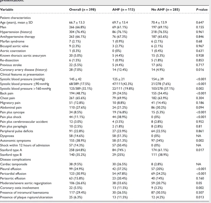
Epidemiological, clinical, instrumental, and outcome findings of patients presenting with/without AHF are shown in Tables 1 and 2. The overall frequency of AHF among patients with AAS was 28% (113/398); presentation with AHF was more common in patients with Stanford type A AAS (84/258, 32%) vs. Stanford type B (29/140, 20%) (P = 0.01). Regarding clinical history, prior CAD was the only feature observed more often among patients presenting with AHF. These patients were more likely to present significant aortic regurgitation, pleural effusion, and ACS-like ECG findings. On the other hand, patients without AHF had a higher systolic blood pressure and reported back or abdominal pain more frequently.
Pathophysiological mechanism
A characterization of the possible mechanism(s) underlying AHF was obtained in 89/113 patients presenting with AHF. In type A patients, aortic insufficiency was the single most frequent mecha-nism (alone or in combination), followed by cardiac tamponade, whereas myocardial ischaemia and hypertensive crisis were the leading causes of AHF in type B patients (Table 3).
Among the 38 patients with aortic regurgitation, a spectrum of causes was identified, as a single mechanism or in combination: pre-existing aortic valve disease (bicuspid aortic valve or degen-erative leaflet thickening) in 5 cases, incomplete leaflet closure due to dilatation of the sino-tubular junction in 23 patients, aor-tic leaflet prolapse/disruption in 20, and prolapse of the dissection flap through aortic valve orifice producing a ‘funnel effect’ in 6.
Diagnostic delay
Median global diagnostic delay (time to diagnosis) was 307 (IQR 180–900) min. Median pre-hospital (time to presentation) and in-hospital delays were 90 (IQR 50–210) min and 166 (IQR 90–353) min, respectively (Table 2). The median time from symp-tom onset to presentation was shorter among patients with AHF, while no difference was noted for both in-hospital and global diag-nostic times (Table 2; Figure 1A and B). Presentation with AHF was associated with increased surgical delay in type A AAS patients (Figure 1B).
Table 4 shows the results of univariate/multivariable analysis of
predictors of in-hospital diagnostic delay. Excess risk was related to ...
...
...
pleural effusion, whereas back pain and pulse deficit were protec-tive from late in-hospital diagnosis. AHF as a clinical presentation of AAS did not influence in-hospital diagnostic time [odds ratio (OR) 1.43, 95% confidence interval (CI) 0.88–2.32, P = 0.152].
In-hospital outcome
Among patients with type A, 219 underwent surgical treatment. Forty-five patients (21%) underwent ascending aorta replacement, 121 (55%) ascending aorta and hemiarch, 2 (1%) ascending aorta and partial arch, and 51 (23%) ascending aorta and total arch replacement. Four patients (2%) underwent classic elephant trunk procedure, while the frozen elephant trunk procedure was used in 4 cases (2%). Associated procedures included: Bentall procedure in 73 patients (33%), aortic valve replacement in 15 (7%), David procedure in 7 (3%), and coronary artery bypass graft in 9 (4%) patients. Intra-operative mortality was 0.9%.
A total of 66 (47.1%) type B patients underwent endovascu-lar/surgical treatment: 53 (80.3%) patients underwent placement of endoprosthesis of the descending aorta, while 13 (19.7%) were treated with aortic graft.
Overall in-hospital mortality was 21.8% (26.3% for type A, 13.6% for type B). Among patients presenting with AHF, overall mortality was twice that of patients without AHF (Table 2), mainly due to an excess risk in type A patients. Independent predictors of in-hospital mortality in the overall population are reported in Table 5 (results of multivariable analysis performed in the type A subgroup are reported in the Supplementary material online, Tables S1 and S2). AHF was an independent risk factor in conjunction with age, Stanford type A, pleural effusion, ACS-like ECG findings, and pulse deficit. Surgery or endovascular treatment were protective.
Discussion
The main findings of our analysis are that AHF occurs in more than a quarter of patients with AAS of both type A and type B, is due to a variety of pathophysiological mechanisms, and is associated with increased surgical delay and in-hospital mortality.
The study population of our single-centre series is comparable with that of the largest available AAS registry, the IRAD registry, specifically with regard to age (mean 66.7 years), male prevalence (67%), relative frequency of Stanford type A, as well as frequency and distribution of signs and symptoms at presentation.18A history
of hypertension was the most frequent risk factor (76%), while Marfan syndrome and bicuspid aortic valve were found only in 2.1% and 2.3% of patients, respectively.
Presentation with AHF occurred in 28% of our population, rang-ing from 20% among type B patients to 32% among those with type A AAS. The prevalence reported in IRAD is decidedly lower (6%), but the discrepancy can be explained by differences in the definition of AHF. In the study by Januzzi et al.,2 the diagnosis of
AHF was based on the impression of the managing physicians as noted in the IRAD case report form. In this series, all patients with dyspnoea as presentation or radiological signs of pulmonary con-gestion or cardiogenic shock were considered to have AHF, in an
Table 1 Baseline clinical characteristics in the overall study population according to acute heart failure at presentation.
Variable Overall (n= 398) AHF (n= 113) No AHF (n= 285) P-value . . . .
Patient characteristics
Age (years), mean ± SD 66.7 ± 13.3 69.7 ± 13.4 70.4 ± 13.9 0.647
Men 266 (66.8%) 69 (61.1%) 197 (69.1%) 0.155
Hypertension (history) 304 (76.4%) 86 (76.1%) 218 (76.5%) 0.961
Antihypertensive therapy 263 (66.1%) 76 (67.3%) 187 (65.6%) 0.846
Marfan syndrome 7 (2.1%) 1 (0.9%) 6 (2.1%) 0.68
Bicuspid aortic valve 9 (2.3%) 3 (2.7%) 6 (2.1%) 0.967
Aortic coarctation 1 (0.3%) 0 (0%) 1 (0.4%) 0.631
Known thoracic aortic aneurysm 20 (5.0%) 5 (4.4%) 15 (5.3%) 0.928
Re-dissection 6 (1.5%) 1 (0.9%) 5 (1.8%) 0.853
Previous stroke 22 (5.5%) 5 (4.4%) 17 (6%) 0.717
Coronary artery disease (history) 28 (7.0%) 14 (12.4%) 14 (4.9%) 0.016
Clinical features at presentation
Systolic blood pressure (mmHg) 145 ± 42 125 ± 21 154 ± 39 <0.001
Systolic blood pressure≤90 mmHg 68/389 (17.5%) 47/111(42.3%) 21/278 (7.6%) <0.001
Systolic blood pressure>160 mmHg 125/389 (32.1%) 22/111 (19.8%) 103/278 (37.1%) 0.002
Back pain 194 (48.7%) 39 (34.5%) 155 (54.4%) <0.001
Chest pain 261 (65.6%) 79 (69.9%) 182 (63.9%) 0.304
Migratory pain 51 (12.8%) 10 (8.8%) 41 (14.4%) 0.186
Abdominal pain 110 (27.6%) 24 (21.2%) 86 (30.2%) 0.094
Pain plus syncope 34 (8.5%) 19 (16.8%) 15 (5.3%) <0.001
Pain plus shock 44 (11.1%) 44 (38.9%) 0 (0%) <0.001
Pain plus cerebrovascular accident 12 (3.0%) 4 (3.5%) 8 (2.8%) 0.952
Pain plus paraplegia 10 (2.5%) 2 (1.8%) 8 (2.8%) 0.81
Peripheral pulse deficits 91 (22.8%) 27 (23.9%) 64 (22.5%) 0.861
Dyspnoea 58 (14.6%) 58 (51.3%) 0 (0%) NA
Autonomic symptoms 155 (38.9%) 58 (51.3%) 97 (34%) 0.002
Shock within 12 hours of admission 57 (14.3%) 57 (50.4%) 0 (0%) NA
Stanford type A 258 (64.8%) 84 (74% ) 174 (61.1%) 0.017 Stanford type B 140 (35.2%) 29 (25%) 111 (38.9%) Disease complications Cardiac tamponade 38 (9.5%) 30 (26.5%) 8 (2.8%) NA Pleural effusion 99 (24.9%) 42 (37.2%) 57 (20%) <0.001 Pericardial effusion 123 (30.9%) 54 (47.8%) 69 (24.2%) <0.001 Periaortic effusion 63 (15.8%) 23 (20.4%) 40 (14%) 0.160
Moderate/severe aortic regurgitation 106 (26.6%) 38 (33.6%) 59 (20.7%) 0.05
Coronary ostia involvement 22 (5.5%) 13 (11.5%) 9 (3.2%) 0.002
Presence of intramural haematoma 117 (29.4%) 30 (26.5%) 87 (30.5%) 0.507
Presence of plaque rupture/ulceration 25 (6.3%) 13 (11.5%) 12 (4.2%) 0.013
AHF, acute heart failure; NA, not applicable.
attempt to assume as much as possible the unbiased perspective of a physician evaluating an acutely ill patient with AHF, before he reaches a diagnosis. Interestingly, both categories of AHF proposed by ESC guidelines1are represented in our study: 56/113 patients
with AHF (49%) presented with pulmonary congestion/oedema without shock; 57/113 patients (51%) presented with hypotension, hypoperfusion, or shock. When faced with either AHF presen-tation (and an appropriate clinical context), the physician should therefore consider the possibility of AAS.
Although not specifically aimed at investigating the mechanistic aspects, our registry offers several insights into the possible ...
mechanisms underlying AHF during AAS due to the prospective collection of many clinical and instrumental variables from all patients, including standard ECG, troponin values, TTE, and TEE.
First it should be noted that the frequency of the possible mech-anisms differs between type A and type B (Table 3). In type A AAS, aortic regurgitation and cardiac tamponade are the main causes of AHF. In the context of AAS, cardiac tamponade may rapidly lead to death, but it can also occur over a relatively long period of time, leading to progressive heart failure and shock at presentation.
Aortic regurgitation may be due to a variety of mechanisms14
Table 2 Instrumental examinations, treatment, and outcome in the overall study population according to the presence of acute heart failure at presentation
Variable Overall (n= 398) AHF (n= 113) No AHF (n= 285) P-value . . . . Instrumental examinations Computed tomography 372 (93.5%) 99 (87.6%) 273 (95.8%) 0.006 Transoesophageal echocardiography 87 (21.8%) 29 (25.7%) 58 (20.4%) 0.307 Transthoracic echocardiography 222 (55.8%) 63 (55.8%) 159 (55.8%) 0.916 Chest X-Ray 237 (59.5%) 78 (69%) 159 (55.8%) 0.021 Abdominal ultrasound 78 (19.6%) 21 (18.6%) 57 (20%) 0.856
Magnetic resonance imaging 20 (5.0%) 7 (6.2%) 13 (4.6%) 0.676
Angiography 42 (10.6%) 11 (9.7%) 31 (10.9%) 0.878
ACS-like ECG 102 (25.6%) 38 (33.6%) 64 (22.5%) 0.03
Troponin positivity 70/248 (28.2%) 25/69 (36.2%) 45/179 (25.1%) 0.114
Treatment
Surgery/endovascular 287 (72.1%) 85 (75.2%) 202 (70.9%) 0.455
Only medical treatment 111 (27.9%) 28 (24.8%) 83 (29.1%)
Outcome
In-hospital death (overall) 87 (21.8%) 39 (34.5%) 48 (16.8%) <0.001
Type A 34/84 (40.1%) 34/174 (19.5%) <0.001
Type B 5/29 (17%) 14/111 (12%) 0.731
In-hospital death of patients surgically treated patients 55 (13.8%) 27 (23.9%) 28 (9.8%) <0.001
In-hospital death of patients treated with medical therapy 32 (8.0%) 12 (10.6%) 20 (7%) 0.324 Delays (median, Q1–Q3)
Pre-hospital delay*, min 90 (50–210) 73 (41–180) 90 (60–210) 0.05
In-hospital delay, min 166 (90–353) 209 (92–510) 160 (86–322) NS
Global delay**, min 307 (180–900) 333 (180–1112) 300 (193–840) 0.86
ACS, acute coronary syndrome; AHF, acute heart failure. *Time from symptom onset to presentation.
**Time from symptom onset to diagnosis.
Table 3 Mechanism of acute heart failure in acute aortic syndrome
Main mechanism Contributing mechanism
. . . . . . . .
Overall (n= 113) Type A (n= 84) Type B (n= 29) Overall (n= 113) Type A (n= 84) Type B (n= 29) . . . . Cardiac tamponade 30/113 (26%) 30/84 (36%) 0/29 (0%) 0/113 (0%) 0/84 (0%) 0/29 (0%) Aortic regurgitation 29/113 (25%) 29/84 (35%) 0/29 (0%) 9/113 (8%) 9/84 (11%) 0/29 (0%) Myocardial ischemia 19/113 (17%) 12/84 (14%) 7/29 (24%) 29/113(26%) 29/84 (35%) 0/29 (0%) Hypertensive crisis 11/113 (10%) 1/84 (1%) 10/29 (34%) 10/113 (9%) 4/84 (5%) 6/29 (20%) Unknown 24/113 (21%) 12/84 (14%) 12/29 (41%)
dilation, relative to the aortic annulus, causing leaflet tethering and a persistent diastolic orifice; (ii) extension of the dissection into the aortic root and disruption of normal leaflet attachment to the aortic wall, thereby resulting in leaflet prolapse and eccentric regurgitation; (iii) prolapse of the dissection flap through the aortic valve orifice; and (iv) pre-existing aortic valve disease (bicuspid aortic valve or degenerative leaflet thickening).
Myocardial ischaemia, which leads to LV systolic or diastolic dysfunction, may be related to a clear anatomical obstruction of at least one coronary artery due to the dissection of a coronary artery or to the diastolic apposition of the flap to the ostium. In the remaining cases, the mechanism, albeit undefined, is probably multifactorial and includes acute pressure overload in patients with or without pre-existing coronary disease. ...
Although most patients with AHF in our series had type A AAS, this study shows that as many as 25% of patients with AHF had a distal dissection; when AHF is a presenting symptom of type B AAS, this may be due to myocardial ischaemia or hypertensive crisis.
Indeed, one-third patients with AHF showed ACS-like ECG abnormalities and/or troponin T positivity, irrespective of Stanford subtype.
The clinical profile of patients with AHF is similar to that of patients without AHF with regard to age and risk factors (Table 1). On the other hand, AHF patients are more likely to have type A AAS and to have lower blood pressure, and are less likely to present with back pain; the pain, however, is more frequently associated with syncope, and a pleural effusion is more common.
Figure 1 Time to presentation (median value, hours), time to diagnosis (median value, hours), and time to surgery (median value, hours) in the overall study population (A) and in Stanford type A acute aortic syndrome (B) according to the presence of acute heart failure (AHF).
Table 4 Univariate and multivariate analysis for late in-hospital diagnosis (cut-off>75th percentile, 406 min)
Univariate analysis Multivariate analysis
. . . .
Variable OR (95% CI) P-value OR (95% CI) P-value
. . . . Pleural effusion 2.1 (1.28–3.44) 0.003 2.17 (1.31–3.6) 0.003 Pericardial effusion 1.67 (1.04–2.68) 0.033 Acute heart failure 1.43 (0.88–2.32) 0.152 Male gender 0.75 (0.47–1.21) 0.236 Pulse deficit 0.50 (0.27–0.92) 0.027 0.56 (0.30–1.05) 0.003 Back pain 0.48 (0.31–0.77) 0.002 0.51 (0.32–0.81) 0.005
CI, confidence interval; OR, odds ratio.
Although some of these findings (such as dyspnoea and pleu-ral effusion) could theoretically lead to a longer in-hospital delay,17
median time to diagnosis was not significantly different between patients presenting with/without AHF, and AHF was not identi-fied as an independent predictor of late in-hospital diagnosis at multivariable analysis (Table 4). These results are consistent with previous findings from the IRAD registry.2 ...
It is possible that the overall perception of a more critical con-dition of AHF patients by the physician led to a faster diagnostic work-up, and that this compensated an initial delay in hypothesiz-ing AAS.
Patients with AHF tended to have a shorter median time from symptom onset to presentation; this was probably due to a perception of greater severity of the condition that led the patients to seek medical attention sooner. Conversely, in our study, as in the IRAD registry,2 median time to surgical treatment
(when performed) was longer among patients presenting with AHF (Figure 1B). Although the exact explanation of this finding is not clear, it could be argued that this delay was due to the increased complexity of management of these patients and the attempt to stabilize them before surgical treatment. Presentation with AHF is an incremental risk factor for in-hospital mortality of type A AAS patients (both operated and not operated) probably due to a greater degree of pre-operative multiorgan damage.
Limitations
Our prospective registry includes data from a single hub centre operating in a rather densely populated urban area with a hub and spoke organization of long duration. The findings regarding hospital
Table 5 Univariate and multivariate analysis for in-hospital mortality of acute aortic syndrome patients Univariate analysis Multivariate analysis
. . . . . . . .
Variable OR (95% CI) P-value OR (95% CI) P-value
. . . .
Stanford type A 2.28 (1.30–3.98) 0.004 3.22 (1.65–6.22) 0.001
Acute heart failure 2.60 (1.58–4.27) <0.001 1.97 (1.14–3.36) 0.014
Pleural effusion 2.27 (1.36–3.78) 0.002 1.80 (1.03–3.20) 0.043
ACS-like ECG 2.14 (1.29–3.56) 0.003 1.81 (1.03–3.11) 0.037
Pericardial effusion 1.82 (1.11–2.98) 0.018
Troponin positivity 1.63 (0.86–3.09) 0.131
Pulse deficit 1.5 (0.87–2.56) 0.142 1.70 (0.91–3.01) 0.08
Age (for each 1 year increase) 1.04 (1.02–1.06) <0.001 1.03 (1.02–1.05) 0.007
Surgery/EVAR 0.44 (0.22–0.68) 0.001 0.41 (0.21–0.77) 0.006
Male gender 0.63 (0.39–1.03) 0.067
ACS, acute coronary syndrome; CI, confidence interval; EVAR, endovascular aneurysm repair; OR, odds ratio.
Figure 2 Case 1 (A/B): transoesophageal echocardiogram (longitudinal section for the LV outflow). (A) Intimal flap (arrow) prolapsing into the aortic valve during diastole (‘funnel effect’) and causing severe aortic regurgitation (B). Case 2 (C/D): transoesophageal echocardiogram (cross-section of the aortic root at the level of the aortic cusps) shows the flap (arrow) involving the non-coronary cusp and partially the right coronary cusp (C). Longitudinal section for the LV outflow showing the prolapse of the right coronary cusp (D); the flap (arrow) is visible within the ascending aorta.
arrival times therefore cannot be generalized to more challenging geographic settings. Inevitably, this registry included only patients who reached a final diagnosis of AAS and could not include patients that never received a diagnosis of AAS, or had a post-mortem diagnosis. Data on LV systolic function were not available in the majority of cases and were therefore not analysed. The registry covers a relatively long period of time (13 years) during which ...
some (minor) technical and organizational variations occurred that do not emerge from our averaged data. Finally, dyspnoea is not necessarily a sign of AHF but its interpretation has an intrinsic and unresolvable margin of uncertainty. We considered it a sign of AHF even in the absence of radiological pulmonary congestion since the presence of dyspnoea per se can influence the physician’s diagnostic suspicion.
Conclusions
Acute heart failure occurs in more than a quarter of patients with AAS of both type A and type B, and is associated with increased surgical delay and in-hospital mortality. AHF is due to a variety of pathophysiological mechanisms including cardiac tamponade, aortic regurgitation, myocardial ischaemia, and hypertensive crisis. Awareness of the frequency and potential mechanisms of AHF in AAS is essential to guide physicians in this complex and challenging disease.
Supplementary Information
Additional Supporting Information may be found in the online version of this article:
Table S1. Univariate and multivariate analysis for in-hospital
mortality of type A AAS patients.
Table S2. Univariate and multivariate analysis for in-hospital
mortality in type A surgically treated patients.
References
1. McMurray JJ, Adamopoulos S, Anker SD, Auricchio A, Bohm M, Dickstein K, Falk V, Filippatos G, Fonseca C, Gomez-Sanchez MA, Jaarsma T, Kober L, Lip GY, Maggioni AP, Parkhomenko A, Pieske BM, Popescu BA, Ronnevik PK, Rutten FH, Schwitter J, Seferovic P, Stepinska J, Trindade PT, Voors AA, Zannad F, Zeiher A, Bax JJ, Baumgartner H, Ceconi C, Dean V, Deaton C, Fagard R, Funck-Brentano C, Hasdai D, Hoes A, Kirchhof P, Knuuti J, Kolh P, McDonagh T, Moulin C, Reiner Z, Sechtem U, Sirnes PA, Tendera M, Torbicki A, Vahanian A, Windecker S, Bonet LA, Avraamides P, Ben Lamin HA, Brignole M, Coca A, Cowburn P, Dargie H, Elliott P, Flachskampf FA, Guida GF, Hardman S, Iung B, Merkely B, Mueller C, Nanas JN, Nielsen OW, Orn S, Parissis JT, Ponikowski P. ESC guidelines for the diagnosis and treatment of acute and chronic heart failure 2012: the Task Force for the Diagnosis and Treatment of Acute and Chronic Heart Failure 2012 of the European Society of Cardiology. Developed in collaboration with the Heart Failure Association (HFA) of the ESC. Eur J Heart Fail 2012;14:803–869. 2. Jannuzzi JL, Eagle KA, Cooper JV, Fang J, Sechtem U, Myrmel T, Evangelista A, Oh
JK, Llovet A, O’Gara PT, Nienaber CA, Isselbacher EM. Acute aortic dissection presenting with congestive heart failure: results from the International registry of acute aortic dissection. J Am Coll Cardiol 2005;46:733–735.
3. Agrawal S, Longfield S, George G. Congestive cardiac failure and aortic dissection in a young man with Marfan’s syndrome. J Accid Emerg Med 1994;11:259–260. ...
...
4. Niitsuma Y, Takahara Y, Sudo Y, Nakano H. Acute type A aortic dissection associated with an aortic annular abscess. Ann Thorac Surg 2001;72:2136–2137. 5. Oliveira JS, Bestetti RB, Marin-Neto JA, Costa RS, Carneiro JJ. Ruptured aortic dissection into the left atrium: a rare cause of congestive heart failure. Am Heart J 1991;121:936–938.
6. Patsouras D, Argyri O, Siminilakis S, Michalis L, Sideris D. Aortic dissection with aorto-left atrial fistula formation soon after aortic valve replacement: a lethal complication diagnosed by transthoracic and transesophageal echocardiography. J Am Soc Echocardiogr 2002;15:1409–1411.
7. Shimazaki Y, Uesho K, Takeda F, Nakashima K, Inui K. Concomitant David’s operation and total arch replacement for acute type A aortic dissection. Jpn J Thorac Cardiovasc Surg 2003;51:609–611.
8. Van Camp G, Liebens I, Silance PG, Cham B, Vandenbossche JL. Ruptured aortic dissection into the left atrium which presented as congestive heart failure and was diagnosed by transoesophageal echocardiography. Br Heart J 1994;72:400–402. 9. Harris KM, Strauss CE, Eagle KA, Hirsch AT, Isselbacher EM, Tsai TT, Shiran
H, Fattori R, Evangelista A, Cooper JV, Montgomery DG, Froehlich JB, Nien-aber CA. Correlates of delayed recognition and treatment of acute type A aortic dissection: the international registry of acute aortic dissection. Circulation 2011;124:1911–1918.
10. Hollenberg SM, Kavinski CJ, Parillo JE. Cardiogenic shock. Ann Intern Med 1999;131:47–59.
11. Spodick DH. Acute cardiac tamponade. N Engl J Med 2003;349:684–690. 12. Perry GJ, Helmcke F, Nanda NC, Byard C, Soto B. Evaluation of aortic
insuffi-ciency by Doppler color flow mapping. J Am Coll Cardiol 1987;9:952–959. 13. Tribouilloy CM, Enriquez-Sarano M, Bailey KR, Seward JB, Tajik AJ. Assessment
of severity of aortic regurgitation using the width of the vena contracta: a clinical color Doppler imaging study. Circulation 2000;102:558–564.
14. Movsowitz HD, Levine RA, Hilgenberg AD, Isselbacher EM. Transesophageal echocardiographic description of the mechanisms of aortic regurgitation in acute type A aortic dissection: implications for aortic valve repair. J Am Coll Cardiol 2000;36:884–890.
15. Mancia G, Fagard R, Narkiewicz K, Redon J, Zanchetti A, Böhm M, Christiaens T, Cifkova R, De Backer G, Dominiczak A, Galderisi M, Grobbee DE, Jaarsma T,Kirchhof P, Kjeldsen SE, Laurent S, Manolis AJ, Nilsson PM, Ruilope LM, Schmieder RE, Sirnes PA, Sleight P, Viigimaa M, Waeber B, Zannad F, Redon J, Dominiczak A, Narkiewicz K, Nilsson PM, Burnier M, Viigimaa M, Ambrosioni E, Caufield M, Coca A, Olsen MH, Schmieder RE, Tsioufis C, van de Borne P,Zamorano JL, Achenbach S, Baumgartner H. 2013 ESH/ESC Guidelines for the management of arterial hypertension. Eur Heart J 2013;34:2159–2219. 16. Evangelista A, Mukherjee D, Mehta RH, O’Gara PT, Fattori R, Cooper JV, Smith
DE, Oh JK, Hutchinson S, Sechtem U. Internation Registry of Aortic dissection Investigators. Acute intramural hematoma of the aorta: a mystery in evolution. Circulation 2005;111:1063–1070.
17. Rapezzi C, Longhi S, Graziosi M, Biagini E, Terzi F, Cooke RT, Quarta C, Sangiorgi D, Ciliberti P, Di Pasquale G, Branzi A. Risk factors for diagnostic delay in acute aortic dissection. Am J Cardiol 2008;102:1399–1406.
18. Tsai TT, Trimarchi S, Nienaber CA. Acute aortic dissection: perspectives from the international registry of Acute Aortic Dissection (IRAD). Eur J Vasc Endovasc Surg 2009;37:149–159.