UNIVERSITÀ DEGLI STUDI DELLA TUSCIA,VITERBO
DIPARTIMENTO DI ECOLOGIA E BIOLOGIA
Corso di Dottorato di Ricerca in
GENETICA E BIOLOGIA CELLULARE – XXVIII Ciclo.
MEAT AGING: THE ‘OMICS’ INTEGRATION TO THE ANALYSIS OF THE BIOCHEMICAL EVOLUTION DRIVING THE MUSCLE-TO-MEAT CONVERSION
PROCESS.
BIO/11
Tesi di dottorato di:
Dott. Alessandro Lana
Coordinatore del corso
Tutore
ABSTRACT………....page 1 PART 1 – A GENERAL INTRODUCTION………..page 5
1.1 AN INTRODUCTION TO MEAT STRUCTURE………....page 6
-1.1.1 Composition of mammalian muscle………page 7 -1.1.2 Basic muscle structure: from whole muscle to individual fibers……..page 8 -1.1.3 Muscle proteins………...page 9 -1.1.4 Muscle fiber organelles………page 10
1.2 UNDERSTANDING THE MUSCLE PHYSIOLOGY BEHIND THE
CONVER CONVERSION INTO MEAT……..………....page 11
-1.2.1 Physiology of muscle activation………..page 11
-1.2.2 Muscle metabolism……….page 13
-1.2.2a Glycogen and glucose: glycogenolysis and glycolysis……...page 14
-1.2.2b The phosphagen system………....page 17
1.3 MEAT SCIENCE AND THE OMICS TECHNOLOGIES………...page 20
-1.3.1 Proteomics………...page 21
-1.3.1a 2D Gel electrophoresis………....page 21
-1.3.1b Two-dimensional HPLC………..page 22
-1.3.1c Quantitative approaches with mass spectrometry…………....page 23
-1.3.3 Metabolomics………..page 25
-1.3.4 Omics strategies: their integration in systems biology………page 26
REFERENCES………...page 28
PART 2 – PROLONGED MEAT AGING: THE METABOLOMIC INVESTIGATION
COMPLETECOMPLETES THE PHYSICAL CHARACTERIZATION…...….…….page 34
2.1 INTRODUCTION………...page 35
2.2 MATERIALS AND METHODS………..page 40
- 2.2.1 Classical standard analyses………...page 40
- 2.2.1a Microbiological analysis………...page 40
- 2.2.1b Chemical and physical analysis………page 40
- 2.2.2 Omics analysis………..page 42
- 2.2.2a 1D SDS–PAGE LC–MS/MS………...page 42
- 2.2.2b Metabolomics………...page 44
- 2.2.2c Statistics………...page 45
2.3 RESULTS AND DISCUSSION………...page 46
- 2.3.1 Microbiological results………page 46
- 2.3.4 Metabolomics results……….………..page 54
- 2.3.5 Statistical analysis results………...page 66
2.4 CONCLUSIONS………...page 71
2.5 CONCLUSIONS AS A STARTING POINT………....page 72
- 2.5.1 Sources of ROS in skeletal muscle……….page 72
- 2.5.2 Oxidative stress effects……….page 74
- 2.5.3 ROS can trigger both autophagy and apoptosis……….page 75
REFERENCES………page 79
PART 3 – MEAT AGING: THE OMIC INVESTIGATION SUGGESTS THE
APOPTOTIC BAPOPTOTIC BEHAVIOR OF MEAT CELLS………page 88
3.1 INTRODUCTION………....page 89
3.2 MATERIALS AND METHODS………...page 89
- 3.2.1 Proteomics……….page 90
- 3.2.1a Sample preparation………...page 90
- 3.2.1b 2D-PAGE………..page 90
- 3.2.1c Image analysis………page 91
- 3.2.2a TiO2 enrichment and ETD / neutral loss analysis………….page 93
- 3.2.3 Metabolomics………...page 93
3.3 RESULTS AND DISCUSSION………page 94
- 3.3.1 Proteolysis………..………page 95
- 3.3.2 Energetic evolution……….page 97
- 3.3.3 Omic clues towards apoptosis………page 106
3.4 CONCLUSIONS………..page 114
1 ABSTRACT
My PhD thesis is centered on the analysis of the ‘muscle-to-meat’ conversion process in the Piedmontese beef breed by an ‘omic’ approach. This process takes place when a farm animal is slaughtered and its muscles are left to a resting period, during which they develop the qualities of the final product, as to say, the eatable meat.
In my PhD thesis I have described the muscle conversion to meat throughout a 44 days – time course analysis, primarily relying on the variations of some key metabolites, and of the proteome by means of differential 1D and 2D gel electrophoresis coupled with MS protein identification. Moreover, I intertwined the classical, physical measurements for meat quality evaluation with the omic data.
After the rigor mortis (when muscle becomes meat and reaches its highest toughness), a proteolytic process takes place that promotes meat tenderization; the proteomic overview documents changes in sarcoplasmic and myofibrillar proteins. My results show the rapid impairing of creatine/phosphocreatine system, as well as of the aerobic production of ATP. Glycolysis becomes the only source of ATP for the meat cells; the increasing oxidative stress is the cause of the metabolic changes (meat cells try to counteract it), such as the increase of pentose phosphate pathway intermediates as to gain reducing power in the form of NADPH (necessary for the recovery of reduced glutathione). The statistical processing of physical and metabolomic measurements along the time points indicated the possibility to use some metabolites as markers for meat tenderness (in particular, serine, arginine and glutamate).
2
In the second part of my thesis, a more in-depth omic analysis showed that meat cells likely execute the apoptotic mechanisms when the oxidative stress inevitably becomes overwhelming. The proteomic results gave clues of apoptosis in the time course trend of annexin A2, Phosphatidylethanolamine Binding Protein, DJ1 protein, Heat Shock Protein B6, Adenylate Kinase, crystalline αB, and 31 kDa actin fragment; the metabolomic results gave clues of apoptosis looking at the general metabolic behavior and at particular key metabolites (such as nitrotyrosine and taurine); the phosphoproteomic results gave clues of apoptosis in cryαB, HSPB6, myosin 2, synaptopodin 2 and in the phosphorylation of some metabolic enzymes.
3
ABSTRACT (IN ITALIANO)
La mia tesi di dottorato è focalizzata sull’analisi, tramite approccio omico, del processo di conversione del muscolo in carne nella razza bovina Piemontese. Questo processo ha inizio nel momento della macellazione dell’animale da allevamento, quando i suoi muscoli vengono lasciati ad un periodo di riposo durante il quale essi sviluppano le qualità del prodotto finale, cioè la carne commestibile.
Nella mia tesi ho descritto la conversione del muscolo a carne attraverso un’analisi temporale del periodo di frollatura (44 giorni), principalmente considerando la variazione del proteoma, attraverso gel 1D e 2D con identificazione in MS, e di alcuni metaboliti chiave. Inoltre, ho integrato i dati omici con le classiche misure fisiche adottate nella valutazione della qualità della carne.
Dopo il rigor mortis (momento in cui il muscolo diviene carne e raggiunge la sua massima durezza), ha inizio un processo proteolico che promuove l’intenerimento della carne; la panoramica proteomica testimonia cambiamenti nelle proteine miofibrillari e sarcoplasmatiche. I miei dati mostrano il rapido degrado del sistema creatina/fosfocreatina, così come della produzione aerobica di ATP. La glicolisi diviene l’unica fonte di ATP per le cellule della carne; lo stress ossidativo in costante aumento, che le cellule della carne tentano di contrastare, è la causa di stravolgimenti metabolici quali l’aumento degli intermedi della via dei pentosofosfati allo scopo di ottenere potere riducente sotto forma di NADPH (necessario al recupero di glutatione ridotto). L’elaborazione statistica dei dati fisici e metabolomici raccolti lungo alcuni punti temporali suggeriscono la possibilità di utilizzare alcuni metaboliti come marker per la tenerezza della carne (serina, arginina e glutammato in particolare).
4
Nella seconda parte della tesi, un’analisi omica maggiormente approfondita mostra il probabile comportamento apoptotico delle cellule della carne nel momento in cui lo stress ossidativo diviene insostenibile. I risultati proteomici hanno fornito indizi al riguardo nel trend temporale di alcune proteine differenziali quali annessina A2, PEBP, DJ1, HSPB6, adenilato chinasi, crystalline αB, e frammento di 31 kDa dell’actina; la stessa metabolomica ha fornito indizi in questa direzione, sia nell’interpretazione del comportamento metabolico generale che di particolari metaboliti chiave (quali nitrotirosina e taurina); gli indizi forniti dalla fosfoproteomica si reggono sull’analisi temporale di cry αB, HSPB6, miosina 2, sinaptopodina 2 e di alcuni enzimi metabolici chiave.
5
A GENERAL INTRODUCTION
The omnivorous diet is at the base of the human nutrition. Meat likely began quite early to play an important part during the human evolution, as it has been suggested by Leonard et al. (2002), because the development of our sophisticated brains that need a large proportion of the whole body’s energy intake could be permitted only by the exploitation of energy-rich foods like meat. After the period of the progresses in hunting techniques, the domestication of animals and the animal husbandry guarantee a more reliable source of meat, at the same time reducing the number of animal species from which it was obtained; nowadays, the utilized sources of meat are turkeys, geese, ducks, ostrich, farmed fish, goats, sheep, pigs, horses, rabbits, bovines, with various new species taken into account.
However, the most important meat-producing species are poultry, sheep, pigs and cattles, the first one giving the ‘white meat’ while the remaining giving the ‘red meat’. Sheep are the most important in the Near East, pigs in Far East, and beef plays the role of the main character in North and South America, Africa and Europe (Warriss, 2010). Beside the classical division into red and white meat, also ‘processed’ meats have to be grouped in a specific class, including cured and smoked meats, ham, bacon, sausages, hamburgers, salami and tinned meat. For the purpose of my thesis, the mention of red meat from here on will refer only to red meat which is unprocessed. As evidenced by Linseisen et al. (2002), the EPIC (European Prospective Investigation into Cancer and Nutrition) investigation reports the daily red, white and processed meat consumption data for the principal European countries (Table 1). It is
6
evident that red meat is the most important in terms of consumption in all the considered nations (except for Germany), so the economic impact is strong, and the growing interest is so explained.
1.1 AN INTRODUCTION TO MEAT STRUCTURE
Meat is defined by the Codex Alimentarius (2005) as “All parts of an animal that are intended for, or have been judged as safe and suitable for, human consumption”; a more intuitive definition for meat is ‘the flesh of an animal used as food’. When speaking of meat, everyone is surely leaded to associate it to ‘muscle’; it is a simple, but also simplistic, association, because there exist a number of striking differences between muscle and meat. However, muscle is the starting point of meat production. The muscular tissue is one of the highest examples of structural organization found in the animal body which can perform a wide range of mechanical functions. Muscle cells are necessary for locomotion, for the movement of limbs and other gross movements, but also coordination and balance maintenance are among their tasks; the association of muscle movement and metabolism permits other different functions like the support to the blood circulation and the body temperature maintenance. Living muscle cells are able to undergo striking intracellular changes, a characteristic that heavily influences their response after the death of the organism. Their organization, structure
7
and metabolism are intertwined aspects that ensure the maintenance of muscle integrity both during contraction and during the moments following death (the early postmortem period), the latter being pivotal in the frame of meat transformation.
-1.1.1 Composition of mammalian muscle. (Table 2). Water is the major constituent of muscle (US Department of Agriculture, 2008), about 75%, but in postmortem muscle this percentage is variable into the range of 65-80%; it is the principal component of the extracellular fluid into the muscle and of the sarcoplasmic fluid, functioning as thermoregulator, as medium for muscular metabolic processes and as intramuscular/extramuscular transporter. The protein component is the second biggest one (16-22%), whose principal task is the contractile process; the myofibrillar proteins, insoluble at low ionic strength, are divided into contractile proteins (directly involved in movement) and regulatory proteins (involved in the regulation of the interactions between the contractile proteins). Another group is constituted by the sarcoplasmic proteins (soluble at low ionic strength), which are signaling proteins and important enzymes intervening into the cellular metabolism/remodeling. The muscular lipid content is variable (1-13%), depending on age, nutritional level, muscle type; however, it shows an inverse proportion with water (Callow, 1948). Some lipid is contained into the muscle cells, but the greatest percentage is located between the muscle bundles. Carbohydrates make up a relatively small percentage of muscular tissue (0.5-1.5%), but they are extremely important in the context of the normal and postmortem metabolism; the quantitatively and qualitatively most important muscular carbohydrate is glycogen. The picture is completed by the presence of non-protein nitrogenous compounds, such as nucleotides, free amino acids, and creatine metabolites.
8
-1.1.2 Basic muscle structure: from whole muscle to individual fibers. (Figure 1). The arrangement of muscle tissue, where the myofibers and the connective tissue are intertwined, is one of the most organized. Muscle fibers are multinucleated and postmitotic, and each nucleus is the responsible for the control of the protein synthesis necessary for that specific region (nuclear domain; Hikida, 2011) of the myofibril. A whole muscle is surrounded by the epimysium, a layer of connective tissue; the perimysium surrounds the bundles of fibers within the muscle, while the sarcolemma (cell membrane) surrounds any single muscle fiber.
Many proteins physically associate the sarcolemma with the internal myofilament structure, in particular with actin and myosin.
Skeletal muscles are highly heterogeneous, because of the functional relationship with their specific task. The diversity is not simply at the gross level, but even in the size and the
FIGURE 1
From muscle to its functional units.
9
number of the cells; for example, we have hundreds of muscle cells forming the tensor tympani, whereas over a million forming the gastrocnemius (Feinstein et al., 1955). The number of cells has a strong influence on the functionality of the living muscle, but it is also of extreme importance for meat quality.
-1.1.3 Muscle proteins. (Figure 2). Each muscle fiber is made of thousands of myofibrils and contains billions of myofilaments, whose highly ordered pattern of assembly forms sarcomeres (the basic contractile units), containing more than 65 proteins (Fraterman et al., 2007). The striated appearance of the muscular cells is the result of the particular sarcomeric structure: protein dense A-bands and less dense I-bands alternate within the myofibril. In the middle of the I-bands, there are dark lines called Z-bands; the sarcomere is the structure put between two consecutive Z-lines, so it is constituted by Z-line/half I-band/A-band/half I-band/Z-line. In a relaxed muscle cell, the length of the sarcomere is approximately 2.2 µm, and the number of these repeated units determines the length of the myofibril (Wickiewicz et al., 1983). Proteinacious filaments, known as intermediate filaments, connect the myofibril with the
Z-FIGURE 2
. A schematic view of the sarcomeric unit. MyoHC = myosin heavy chain; MyoLC = myosin light chain; TNN = troponin.10
line, while other protein structures, costameres, attach the sarcolemma to the outermost myofibrils (Robson et al., 2004). Myofibrils are formed by many myofilaments, divided into two principal types classified as thick and thin; a third one is primarily composed by the titin protein (Ma et al., 2006). The I-bands are made up by thin filaments, while the A-bands are characterized by the overlap of thin and thick filaments, principally constituted by myosin and actin, respectively, and together they form approximately the 70-80% of the total protein content of a single fiber. Myosin is the main molecular motor and up to eleven sarcomeric myosin genes have been described in mammals. Other fundamental proteins, involved into the activation process of the myofilament sliding and force generation, are troponin T, I, C and tropomyosin. Titin is a large elastic protein that attaches to the Z-disk and to myosin to give stability and the correct alignment to the thick filament. Nebulin, on the other hand, is integrated with other proteins in the thin filaments (Ottenheim & Granzier, 2010). Titin and nebulin are principally involved into the maintenance of the integrity of the sarcomere, influencing passive tension and stiffness of any single cell. The principal proteins that form the Z-disk are α-actinin, filamin, cypher protein 3 and desmin.
-1.1.4 Muscle fiber organelles. The muscular functions are strictly dependent on the organelles network, where we can find the transverse tubular system (T tubule), the sarcoplasmic reticulum and the mitochondria; the exact amount of these elements varies with the fiber type. The T tubule system is of fundamental importance for the conduction of the action potential from the exterior to the interior of the muscle cell, ensuring that the excitation spread uniformly throughout the fiber (Jayashinghe & Launikonis, 2013).
The sarcoplasmic reticulum is responsible for the management of the intracellular concentration of calcium (storage, release and reuptake after activation). The terminal cisternae of the sarcoplasmic reticulum are in close contact with the T tubule system and contain calcium ions; each T tubule is flanked on both sides by two cisternae, forming a
11
structure called triad. Two proteins are of particular importance in maintaining calcium homeostasis: Ca2+-ATPase (SERCA), responsible for the calcium reuptake into the cistern after muscle activation, and calsequestrin, which binds calcium loosely within the sarcoplasmic reticulum.
The mitochondria network is highly spatially organized in all the three dimensions throughout the cell; it supplies the cell with the energy necessary for muscle actions when oxygen is made available to the fiber (Dahl et al., 2014); the spatial disposition of the mitochondria, located closer to the sarcolemma, is organized in such a way that the diffusion distance for oxygen transported by capillaries is reduced; it is particularly useful during aerobic exercise, which increases the demand for oxygen. Another population of mitochondria is located in the intermyofibrillar space. A muscle used to heavy exertions has a greatest number of mitochondria with respect to a sedentary one, because of the mitochondrial biogenesis process, regulated by the peroxisome proliferator-activated receptor γ coactivator 1α (Yan et al., 2012).
1.2 UNDERSTANDING THE MUSCLE PHYSIOLOGY BEHIND THE
CONVERSION INTO MEAT.
-1.2.1 Physiology of muscle activation. The generation of muscular force is the result of the so-called excitation-contraction coupling, which relies in the coordination of two processes: the transmission of the nerve impulse to the triad, with the consequent release of calcium from the cisternae of the sarcolasmic reticulum, and the formation of the actin/myosin cross-bridges. When the action potential arrives to the muscle fiber membrane, the T tubule system conduces it to the interior of the muscle cell; in the triad, the close proximity of the T tubule and the terminal cisternae permits to a voltage sensor subunit of the dihydropiridine receptors
12
on the T tubule to open and to allow an inward current of calcium (Rebbeck et al., 2014) that in turn triggers the opening of the ryanodine receptors in the terminal cisternae, leading to a massive release of calcium into the sarcoplasm. At this point, Ca2+ is picked up by troponin C, able to induce a conformational change in troponin I that leads to its dislocation from the thin
filaments (I as ‘inhibitory’, definition due to its inhibitory action against the formation of the actomyosin complex without nervous stimulus); if no action potential arrives, the tropomyosin complex sterically obstructs the myosin binding sites on actin, but after Ca2+ reception by troponin C, troponin T (the Tropomyosin-binding element of the heterotrimeric troponin complex) transmits the conformational change to the tropomyosin complex, making actin able to bind myosin. The actomyosin complex is able to dissociate only in the presence of ATP, because of the conformational change induced by ATP binding on a specific site of the myosin globular head; this domain has an ATPase activity, able to hydrolyze ATP into
13
ADP and inorganic phosphate. The released energy is conserved into the myosin structure, where ADP and Pi are initially maintained. The so-called ‘power stroke’ (figure 3) begins at the release of Pi from myosin crossbridge, event that allows the tension unloading by means of the mechanical movement of myosin, which pushes back the linked actin filament: the result is the sliding of thin and thick filament, the shortening of sarcomeres and contraction. At this step ADP is bound to the myosin head, and this conformational status allows only a weak binding to the actin filament; when ADP is released, the binding becomes really strong and essentially irreversible without another incoming ATP molecule. This is the explanation for the onset of rigor mortis, which is the starting point of the muscle-to-meat conversion.
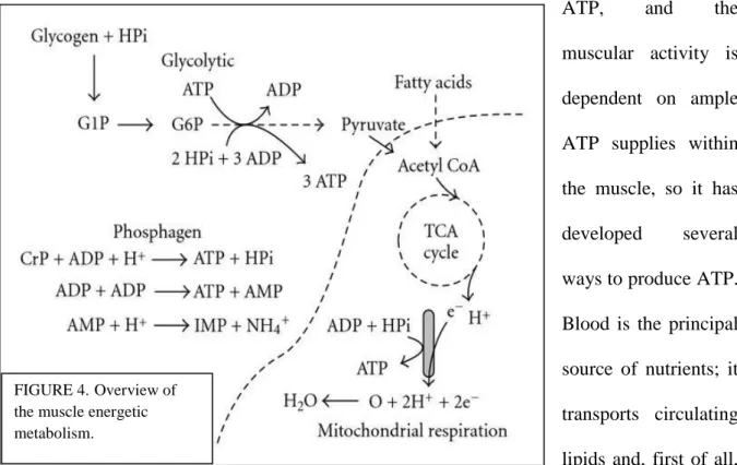
-1.2.2 Muscle metabolism. (Figure 4). In an actively exercising animal, up to the 90% of the oxygen consumption of the whole body is accounted by the muscular tissue; the muscle’s metabolic rate can increase of as much as 200% from the resting state (Hargreaves & Thompson, 1999). Obviously, central to the existence of the muscle cell is the production of ATP, and the muscular activity is dependent on ample ATP supplies within the muscle, so it has developed several ways to produce ATP. Blood is the principal source of nutrients; it transports circulating lipids and, first of all, glucose, but muscle cells can employ energy precursors reserves such as glycogen, lipids and
FIGURE 4.Overview of
the muscle energetic metabolism.
14
phosphagens (ATP, phosphocreatine); the choice between the exploitation of the internal or circulating resources is dependent on the activity the muscle is carrying on: when the muscular task is not intense, it preferentially consumes the energy sources picked up from the blood stream, or the lipids contained in its cells (by means of a completely aerobic metabolism). When the muscular activity is of higher intensities, the ATP is very rapidly consumed, and the cells make use of the intracellular reserves of phosphagens and glycogen, which are burnt very rapidly, leading to fatigue. The ATP concentration within the muscle is critical; the relaxation of the fibers can occur when this value is approximately above the 30% of the resting stores, also because the removal of calcium from the sarcoplasm is an ATP-dependent mechanism (Hargreaves & Thompson, 1999).
-1.2.2a Glycogen and glucose: glycogenolysis and glycolysis. Glucose and glycogen are the preferred substrates for muscular energy production, and they can be utilized in an aerobic (oxidative phosphorylation) or anaerobic way (anaerobic glycolysis); lipids are molecules with a very high level of energetic density, but it is curious to underline that despite they are very efficient with respect to the amount of ATP that can be produced per unit of substrate, their rate of ATP synthesis is much slower compared to the ATP production rate of glycogen (about the half, 1.5mmol/kg/sec for free fatty acids versus 3mmol/kg/sec for the aerobic use of glycogen versus 5mmol/kg/sec for the anaerobic use of glycogen) (Joanisse, 2004). Over time, during muscular activity, the muscle glycogen stores diminishes and, as a consequence, the reliance on it diminishes, while the glucose import into myofibers increases; however, if the activity is heavy, little or no exogenous glucose is used for ATP production, and the reliance on glycogen proportionally increases with the increase of the effort (due to the shift toward the anaerobic catabolism). Glycogen is synthesized in the muscle at rest; diet has a heavy influence on the content of whole-body glycogen storage. Glycogen breakdown (figure 4) is under hormonal control, with the cAMP-dependent activation of phosphorylase kinase
15
by epinephrine, and it is further stimulated by the release of Ca2+ from the sarcoplasmic reticulum during contraction (Allen & Westerblad, 2001). The breakdown of glycogen directly involves two enzymes: glycogen phosphorylase (GP) and glycogen debranching enzyme; the rate of this process is limited by the availability of HPO42-, that is not present at pH 5, due to
the H2PO4 pKa (7.2). The glycogen
phosphorylase is in an inactive form during the muscular resting period, while it is activated by epinephrine, a stimulus that wakes up a signaling cascade involving calcium, cAMP, cAMP-activated protein kinase and phosphorylase kinase; glucose 6 phosphate and ATP exert an inhibitory effect on GP. HPO42- or AMP bind to an effector site of GP,
increasing the acceptance of glycogen at the active site. It is worth noting the amplification of the signal in this pathway: the relative concentrations of the three successive enzymes of the cascade (cAMP-dependent protein kinase, phosphorylase kinase and GP) occur as molar ratios of 1:10:240, including adrenalin at a ratio of 1x10⁻⁴, and a single molecule of epinephrine bound to a specific receptor on a muscle fiber results in 400’000 glucose-1P units cleaved by the GP per second (Lodish, 2000).
The generation of ATP in skeletal muscle with an adequate oxygen supply is dependent on fatty acid β-oxidation, on the pyruvate dehydrogenase complex reactions, on the Krebs cycle, on the electron transport chain and on the oxidative phosphorylation. Acetyl-CoA is the central molecule of the aerobic behavior, able to undergo the Krebs cycle reactions and to give reducing equivalents of NADH and FADH2; these molecules are then used by the
electron transport chain to drive the proton gradient across the inner mitochondrial membrane,
FIGURE 4. Hormonal stimulation of glycogen breakdown
16
leading to the recharge of ATP from ADP; oxygen is the final electron acceptor. The volume density of the mitochondria within the cell, the capacity for oxygen transport and storage within the myofiber (dependent on the intracellular levels of myoglobin) determine the capacity for oxidative phosphorylation; the controlling elements of the mitochondrial respiration vary with the state of the myofiber, as well as with its composition. However, major contributors are the concentrations of free ADP, Pi, the NADH/NAD+ ratio, calcium
levels and oxygen availability; the relative contribution of all these factors show a different importance under different conditions.
The carbon units of glucose encounter a key step at the level of pyruvate: it can be completely broken down in an oxidative way entering the mitochondria by means of the pyruvate dehydrogenase complex, giving CO2 and H2O as endproducts and yielding 36 mol of ATP per
mole of glucose. By contrast, if the catabolic choice of the muscle cell is anaerobic (like it is in postmortem muscle), pyruvate does not enter the mitochondria, and it is converted into lactate, yielding only 2 mol of ATP per mol of glucose. So, there is an evident gap between the ratio of ATP moles aerobically produced per mole of substrate and the ratio of ATP moles anaerobically produced, but, reminding the higher speed of ATP production in a time interval, it is not correct to judge the anaerobic metabolism as less convenient: the particular activity, the availability of nutrients and oxygen and so on (in other words, the context), are the deciding factors for the aerobic or the anaerobic behavior in the living muscle, while the only choice for the postmortem muscle is to enter the anaerobic metabolism when oxygen reservoirs are exhausted.
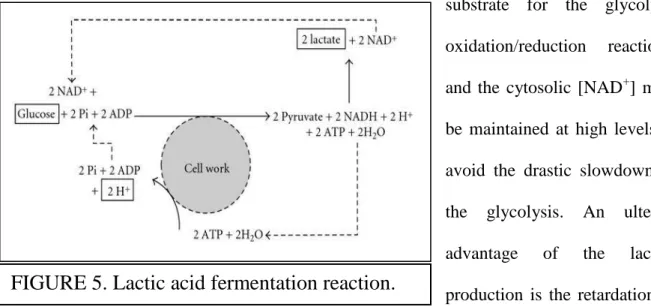
The role of the anaerobic production of lactate is important to understand in the contexts of both living and dead muscles, because many reactions occurring in the postmortem muscles resemble the physiologic reactions of a living muscle subjected to a strong anaerobiosis. The
17
complete oxidative consumption of glucose is impaired, because the pyruvate production occurs at rates that exceed the mitochondrial take-up of pyruvate. The reaction
° pyruvate + NADH + H+ → lactate + NAD+ ,
catalyzed by the lactate dehydrogenase (LDH), is pivotal for the prevention of the reduction in the rate of glycolytic ATP regeneration: it permits to remove pyruvate (figure 5), to sustain a high-rate glycolysis, and to regenerate cytosolic NAD+ (Robergs et al., 2004). The latter is the substrate for the glycolytic oxidation/reduction reactions, and the cytosolic [NAD+] must be maintained at high levels to avoid the drastic slowdown of the glycolysis. An ulterior advantage of the lactate production is the retardation of muscular acidosis: the LDH employs two electrons and one proton from NADH, and a second H+ from the environment, so it has a metabolic proton buffering function.
-1.2.2b The phosphagen system. Muscle contraction depends on the free energy released from the breakdown of ATP. Despite the strong need for ATP, the ATP store within the muscle cells are not so large (approximately 8 mmol/kg wet weight of muscle), and, as I have briefly explained, they have to produce it by means of the catabolism of energetic molecules (fatty acids, glycogen); here it is important to stress that muscle cells rely on a sensitive control system to rapidly increase ATP production when it is needed, and that they are unique in this frame as they can vary their metabolic rate to a greater extent than the cells of other tissues
18
(Glaister, 2005). Indeed, muscular tissue is able to buffer even a 1000-fold increase in ATP demand, maintaining the stability of the ATP concentration (the decrease does not exceed 2 mmol/kg wet weight of muscle during these conditions) (Spriet, 1992). The levels of ATP and associated molecules, such as ADP, AMP and Pi, are an essential requirement for muscle cell
function. The fluctuations of their concentration are the sensors for muscular energetic situation; an ATP reduction leads to the rapid development of fatigue in vital muscles, and to rigor mortis in dead muscles (Vollestad & Sejersted, 1988). Muscular cells are able to respond to a sudden increase in ATP demand, and beside the two major systems for ATP resynthesis (glycolysis and mitochondrial respiration), there is a third system in muscle cells, called ‘Phosphagen system’.
Three principal enzymes are involved into this system, participating to three different but intertwined reactions:
°Creatine kinase: Creatine phosphate + ADP + H+ → ATP + creatine
°Adenylate kinase: ADP + ADP → ATP + AMP
°AMP deaminase: AMP + H+ → IMP + NH4+
The first and the second reaction both produce ATP, but the creatine kinase reaction has by far the greatest capacity for ATP regeneration. In the living muscle, creatine phosphate shows a relatively high concentration with respect to ATP (up to 100 mmol/kg dry muscle weight, depending on the fiber type, against the 25 mmol/kg for ATP; Joanisse, 2004), but a relatively low concentration with respect to glycogen (500 mmol/kg). The creatine kinase reaction is reversible, so it can be adapted to the ATP request: when it is strong, the phosphate group is transferred from pCr to ADP, and when it is weak, pCr supplies can be readily restored. In a living muscle, the creatine kinase reaction shows also the advantage of consuming a proton, thus reducing the muscle cell acidosis associated with anaerobic glycolysis. The principal role
19
of pCr in skeletal muscle is that of an energy buffer operating when the ATP demand overcomes the ATP production by the mitochondrial respiration. The so-called ‘creatine/pCr shuttle’ acts as a cellular energy transport system (based on the existence of different cytosolic and mitochondrial creatine kinase isoforms [figure 6]) that gives to Cr and pCr the roles of energy transporters: Cr becomes pCr in the mitochondria, and after its diffusion in the cytosol, it releases the phosphate group for the recharge of ADP into ATP, employed in the contraction process (Wallimann et al, 1992). So the Cr/pCr system is a low threshold ADP sensor that functions to maintain [ATP]/[ADP] ratios in subcellular locations where creatine kinase is functionally coupled to ATP-consuming and ATP-producing pathways; when Cr reacts with ATP in the mitochondria, the increase in local [ADP] stimulates the mitochondrial respiration (Greenhaff, 2001). In the physiological postmortem conditions, however, the pCr system is not a major contributor to metabolism, because of the rapid depletion of pCr supplies.
An important feature of the adenylate kinase reaction is the production of AMP. It is a strong allosteric activator for the glycogen phosphorylase, starting point of the glycogenolysis, as well as a strong activator for the phosphofructokinase, a key enzyme for the glycolytic destiny of the glucose 6P molecules.
The AMP deaminase reaction does not regenerate ATP. However, it is correct to insert it into the phosphagen system, because the AMP conversion into IMP is necessary for the maintenance of the phosphate transfer potential within the muscle (Atkinson, 1977). If the [ADP] and [AMP] are kept at a low level, even a small reduction in [ATP] is sufficient to release the quantity of free energy necessary to fuel the muscular contraction.
20
This brief excursus about the muscular metabolism has to be kept in mind to understand the striking changes that characterize the physiology of the muscle during its conversion into meat.
In the following section, I will introduce the reader to the importance of the ‘omics’ technologies in the field of meat science.
1.3 MEAT SCIENCE AND THE OMICS TECHNOLOGIES.
Meat science, a branch of farm animal science, is a leading field of research with a strong economic importance; the target is the production of a high-quality meat to attract the largest number of consumers. Meat quality is an ensemble of many features; the end-consumers judge the different meat products in particular on the basis of flavor, juiciness and tenderness (Bendixen et al., 2011). The ‘classical’, standard methods for the characterization of meat products mainly consisted in physical measurements such as Warner-Bratzler shear force (an assay for meat resistance to cut, as to ‘measure’ tenderness), Minolta values (for meat color) and pH. In order to understand the molecular basis of meat development, many biochemical researches have been conducted over the last decades (Paredi et al., 2012; Huff-Lonergan et al., 2010). Gradually, the biochemical investigations integrated the physical measurements, beginning with the spectrophotometric determination of myofibrillary fragmentation indexes that measured the increasing concentrations of peptides and amino acids released by the proteolytic processes in the postmortem muscle (Olson et al., 1976). The last two decades have seen the strong technological growth of the field of ‘omics’ sciences; genomics,
21
transcriptomics, proteomics and metabolomics have been engaged in meat science, and the scientific community is directing many efforts in an attempt to join the different techniques in an organic fashion.
-1.3.1 Proteomics. The first example of omics application to meat science was the genomic investigation of genome associations with meat-quality parameters; gradually, the interest shifted towards the use of mRNA quantitative assays like PCR or microarrays as to appreciate the changes of the genomic expression (Xu et al., 2013). The natural integration with the proteomic technology has been rapidly reached, and big strides have been achieved towards the completion of the biological understanding underlying muscle-to-meat conversion (D’Alessandro & Zolla, 2013; Hollung et al., 2007). Beside the improvements of the proteomic methodologies like protein isolation and fractionation, bioinformatic tools have begun to give a strong contribution, relying on the consolidation and expansion of databases covering animal-genome sequences.
-1.3.1a 2D Gel electrophoresis. The separation of the proteome components of a biological sample by means of gel-based techniques have been firmly established over the last thirty years. Muscles are made of proteins, so the association between meat science and gel electrophoresis has been easily reached. 2D gels are probably the most employed in meat characterization. Depending on the project and hypothesis of a particular meat experiment, different strategies for extraction and pre-fractionation of muscle proteins before 2D-separation could be considered. Some muscle proteins are part of large protein aggregates, like the myofibrillar proteins, some are localized in membranes and others are cytosolic enzymes. With the help of pre-fractionation of the proteins based on solubilization and extraction procedures, the enrichment of soluble sarcoplasmic or myofibrillar proteins can be achieved. These proteins will have very different chemical properties which can be used to isolate the proteins in different fractions (Righetti et al., 2005). Several issues should be
22
considered before developing a sample preparation strategy. It is advisable to keep sample preparation as simple as possible to avoid protein losses. However, if only a subset of the proteins in the tissue or cells is of interest, pre-fractionation can be employed during sample preparation.
The approach of the proteomic experiments is the opposite with respect to the approach of the traditional, physical experiments: the latter are based on few measurements and variables over many animals, while the former are based on many measurements and variables over a limited number of animals, due to the heavy work-load. In this context, the statistical methodologies play a pivotal role in the validation of the results, and multivariate analysis such as principal component (PCA) are becoming an integral part of proteomic (and in general, omic) experiment (Jacobsen et al., 2007).
Beside the classical 2D-gels, recent technical improvements like the adoption of fluorophores for sample labeling, have permitted the simultaneous analysis of multiple samples in the same gel, a compromise that can abate technical variability issues and increase the sensitivity of the assay (0.25 ng for DIGE) (Jia et al., 2009)
-1.3.1b Two-dimensional HPLC. This technique employs an online combination of ion-exchange and reversed-phase strategies to separate peptide samples; the online elution into a mass spectrometer allows the direct peptide-based identification and their absolute or relative quantification. 2D-HPLC has the potential for a successful application in the field of meat qualities investigation, but it has not hitherto found a systematic use for these purposes; it could be complementary to electrophoresis analysis of more hydrophobic or very high molecular weight proteins, given the technical difficulties of electrophoresis with these species.
23
-1.3.1c Quantitative approaches with mass spectrometry. The MS-based techniques for the quantification of targeted proteins may be inserted into meat science as validation strategies for potential protein biomarkers of a particular meat quality. The AQUA (Absolute QUAntification of proteins) strategy allows the determination of absolute proteins against synthetic standards. The emPAI (Exponentially Modified Protein Abundance Index) is the logarithmic correlation between protein abundance and the relative intensity of the arbitrary ion count given by the MS analyzer (Cifuentes, 2010). Alternative approaches are ICAT (Isotope Coded Affinity Tag), iTRAQ (Isobaric Tag for Relative and Absolute Quantitation) and SILAC (Stable-Isotope Labeling by Amino acids in cell Culture), which employ isotopic labeling; they have been built as to avoid some inherent quantitative limits of the label-free strategies, coming from the wide range of physicochemical properties that can influence the MS signal for a specific peptide. The stable-isotope-labeled peptide used during the isotopic labeling experiments shows the same chemistry of the non-labeled counterpart, so they have the same reactions to the experiment steps, but their ratio (that provides the quantitative information about the relative abundance of a specific protein) can be calculated since they remain distinguishable in terms of molecular weight (Cifuentes, 2010).
The advantages of these gel-free based approaches are the higher reproducibility, accuracy and swiftness, with the drawbacks of the costs of MS equipments and the need for strong expertise and training either for data acquisition and processing.
-1.3.1d Post-translational modifications. Meat researchers have begun to demonstrate a clear interest for PTM in the field of meat quality investigations, due to the strong influence that a PTM can exert on the final role of a protein. Sumoylation, ubiquitination, glycosylation and phosphorylation showed the greatest importance in meat science (D’Alessandro & Zolla, 2013). Their detection requires preparative techniques such as the 2D-HPLC, with the serial combination of strong cation-exchange or reversed phase columns (Edelmann, 2011), or the
24
selective enrichment of specific peptides characterized by a particular PTM, for example the glycopeptides captured by lectin columns or the phosphopeptides enriched with Immobilized Metal Affinity Chromatography (IMAC) or TiO2 microcolumns. The Electron Transfer
Dissociation mass spectrometry (ETD) is adequate for PTM detection, overcoming the limits of the CID MS analysis that causes the loss, and so the undetectability, of the side chains of the modified peptides (Mikesh et al., 2006).
In the field of meat science, phosphorylation surely deserves a particular mention; during the muscle-to-meat conversion process, ATP reservoirs are completely exhausted, and given that ATP is the token used for the phosphorylation of proteins, it is easy to hypothesize that the normal phosphorylation pattern of the muscle fibers is impaired, reflecting the changes in tissue structure and in its physiologic metabolism; many glycolytic enzymes are subjected to the modulation by means of phosphorylation, and it has been shown for many metabolic key enzymes involved in the anaerobic metabolism of aging meat (D’Alessandro et al., 2012). The recent MS advances in the identification and quantification of phosphopeptides may be a useful tool for the assessment of methodologies applied to meat production chain for the improvements of meat tenderness, as it has been shown a probable connection between low-voltage electrical stimulation of carcasses, postmortem glycolytic rates and phosphorylation levels in pig meat (Huang et al, 2011).
-1.3.2 Redox proteomics and oxidative modifications. The metabolic impairment suffered by the slaughtered muscle cells leads to an extended redox modification of muscle proteins which heavily influences the organoleptic properties of the meat (D’Alessandro & Zolla, 2013). Once again, ETD mass spectrometry can perform the investigation on protein sulfonation, oxidation or nytrosylation (Mikesh et al., 2006), as well as the analysis of ROS-induced protein fragmentation; however, these interesting topics have been hitherto underinvestigated.
25
-1.3.3 Metabolomics. Mass spectrometry has unparalleled specificity and sensitivity, high resolution and wide dynamic range, and the potential to enable comprehensive qualitative and quantitative measurements of large-scale small-molecular metabolites in complex biological samples; for this reason, MS has emerged as the foremost technology in metabolomics studies, and as a relatively new mean to investigate food at its metabolome level; food analysis regarding the quality and the safety of the food has become a topic of enormous interest to consumers. Thanks to the potential of MS-based metabolomics, meat industry has paid increasing attention to comprehensive determination of the complex meat composition in raw materials, semi-manufactured and final products involved in authenticity, functionality, quality and safety issues (Herrero et al., 2012). Meat science is always in search for new means and technologies to employ for the improvement of final meat products qualities; given the complexity of the biological mechanisms underlying the develop of meat characteristics, MS-based metabolomics can represent a new, leading approach to the analysis of the muscle-to-meat conversion process. Beside the more commonly used omic techniques, metabolomics could furnish another point of view; the integrated pictures of proteome, genome, transcriptome, phosphoproteome and metabolome could help to delve into the physiology of the postmortem muscle. Muscle-to-meat conversion starts soon after animal slaughter; the bleeding promotes deoxygenation, anoxia and the loss of nutrients supply. The metabolism suffers a sort of revolution, because of the switch to the anaerobiosis. It is clear that in this context, the analysis of the metabolome could be extraordinarily informative, especially when performed by means of MS; until a few years ago, pH monitoring and spectrophotometric approaches constituted the strategy of choice, limited by the small number of metabolites roughly detected at the quantitative level. The technological improvements of MS, and the bioinformatic advancements (with the increasing completeness and accuracy of metabolite-spectra databases), allow the elaboration of raw mass metabolite-spectra for feature detection, peak
26
alignment, tentative identification, isotopic distribution and relative quantification, so exponentially enlarging the range of the detected metabolites as to follow a bigger part of the entire metabolome. MS-based metabolomic have been successfully applied to investigate several biological issues in meat science (D’Alessandro & Zolla, 2013; Bowker et al., 2000).
-1.3.4 Omics strategies: their integration in systems biology. The comprehension of the biochemistry and molecular mechanisms leading to the develop of meat qualities cannot be complete if we simply collect the bits of information from separate omics disciplines. Meat scientists are beginning to integrate all the collected pieces of the puzzle, in order to propose a complete, unified overview of the molecular phenomena underpinning meat production events. Muscle-to-meat conversion process involves pH, metabolism, glycolytic enzymes, PTMs, proteases, fragmentation of the muscular structure, ROS; these aspects are all integrated in a single concept where each piece interacts to some extent with at least some of the others. A systems-biology approach is opposed to a reductionist approach; it permits to find out specific characteristics appreciable only as emerging properties of the general system and not of the distinct parts. Applied to meat science, it could represent a step toward the building of a definitive model for meat qualities development, which is a multifactorial process, affected by the characteristics of the living animal (species, breed, gender, age [D’Alessandro et al., 2011]), rearing environment (Kwasiborski et al., 2008), feeding regimen, modalities of transportation to the abattoir and slaughter (Yamane et al., 2006), postmortem treatments (Li et al., 2012), bleeding, hanging, deboning procedures and cooling rate (Bertram et al., 2001). Clearly, a unique model for meat formation is utopian. However, models that could be valid for specific species (more often, specific breeds) appear to be more reliable: centuries of selection of livestock for breeding have narrowed the genetic diversity of the animals, while other variables could be constantly checked during laboratory experiments.
27
The conversion of muscle into meat as a whole and the postmortem development of the eating quality are far from being understood. Improvement of our knowledge about the underlying mechanisms is particularly faced to the large biological variability of meat qualities and to the non-identification of their major determinants. Research tools to investigate the in toto biochemical mechanism, such as the integrated omics device, can play a major role in farm animal and meat science. Given the intricate scenario of this process, my PhD thesis wanted to be an attempt to establish a few milestones as to delve into the events leading from muscle to meat, exploiting some of the discussed potentialities of omics technologies.
28 REFERENCES
Allen DG, Westerblad H (2001). Role of phosphate and calcium stores in muscle fatigue. Journal of Physiology 536:657-665.
Atkinson DE (1977). Cellular Energy Metabolism and Its Regulation, Academic Press, New York, NY, USA, 1st edition.
Bendixen E, Danielsen M, Hollung K, Gianazza E, Miller I (2011). Farm animal proteomics – a review. J. Proteomics 74(3):282–293.
Bertram HC, Donstrup S, Karlsson AH, Andersen HJ, Stodkilde-Jorgensen H (2001). Postmortem energy metabolism and pH development in porcine M. longissimus dorsi as affected by by two different cooling regimes. A (31)P-NMR spectroscopic study. Magnetic Resonance Imaging 19(7):993-1000.
Bowker BC, Grant AL, Forrest JC, Gerrard DE. Muscle metabolism and PSE pork (2000). Journal of Animal Science 79:1-8.
Callow, EH (1948). Comparative studies of meat. II. Changes in the carcass during growth and fattening and their relation to the chemical composition of the fatty and muscular tissues. Journal of Agricultural Science 38:174.
Cifuentes A (2010). Food science, foodomics and capillary electromigration methods, Electrophoresis 31(13):2091.
29
D’Alessandro A, Marrocco C, Zolla V, D’Andrea M, Zolla L (2011). Meat quality of the longissimus lumborum muscle of Casertana and Large White pigs: metabolomics and proteomics intertwined. Journal of Proteomics 75(2):610-627.
D’Alessandro A, Rinalducci S, Marrocco C, Zolla V, Napolitano F, Zolla L, Love me tender: an Omics window on the bovine meat tenderness network. Journal of Proteomics 75(14):4360–4380.
D’Alessandro A, Zolla L (2013). Meat science: From proteomics to integrated omics towards system biology. Journal of Proteomics 78:558-577.
Dahl R, Larsen S, Dohlmann TL, Qvortrup K, Helge JW, Dela F, Prats C (2015). Three-dimensional reconstruction of the human skeletal muscle mitochondrial network as a tool to assess mitochondrial content and structural organization. Acta Physiol (Oxf) 213(1):145-155.
Edelmann MJ (2011). Strong cation-exchange chromatography in analysis of posttranslational modifications: innovations and perspectives. Journal of Biomedicine and Biotechnology 2011:936508.
Feinstein B, Lindegard B, Nyman E, Wohlfart G (1955). Morphologic studies of motor units in normal human muscles. Acta Anatomica 23:127-142.
Fraterman S, Zeiger U, Khurana TS, Wilm M, Rubinstein NA (2007). Quantitative proteomic profiling of sarcomere associated proteins in limb and extraocular muscle allotypes. Molecular Cell Proteomics, 6:728-737.
Glaister M (2005). Multiple sprint work: physiological responses, mechanisms of fatigue and the influence of aerobic fitness. Sports Medicine 35(9):757–777.
30
Greenhaff PL (2001). The creatine-phosphocreatine system: there's more than one song in its repertoire. Journal of Physiology 537(3):657.
Hargreaves M, Thompson M (eds) (1999). Biochemistry of Exercise, 10. Champaign, III: Human kinetics.
Herrero M, Simo V, Garcia-Canas V, Ibanez E, Cifuentes A (2012). Foodomics: MS-based strategies in modern food science and nutrition. Mass Spectrometry Reviews 31:49-69.
Hikida RS (2011). Aging changes in satellite cells and their functions. Current Aging Science 4:279-297.
Hollung K, Veiseth E, Jia X, Faegerstad EM, Hildrum KI (2007). Application of proteomics to understand the molecular mechanisms behind meat quality. Meat Science 77(1):97-104.
Huang H, Larsen MR, Karlsson AH, Pomponio L, Costa LN, Lametsch R (2011). Gel-based phosphoproteomics analysis of sarcoplasmic proteins in postmortem porcine muscle with pH decline rate and time differences. Proteomics 11(20):4063–4076.
Huff Lonergan E, Zhang W, Lonergan SM (2010). Biochemistry of postmortem muscle – lessons on mechanisms of meat tenderization. Meat Science 86(1):184-195.
Jacobsen S, Grove H, Jensen KN, Sørensen HA, Jessen F, Hollung K et al (2007). Multivariate analysis of two-dimensional gel electrophoresis protein patterns - Practical approaches. Electrophoresis 28:1289–1299.
Jayashinhe ID, Launikonis BS (2013). Three-dimensional reconstruction and analysis of the tubular system if vertebrate skeletal muscle. Journal of Cell Science 126:4048-4058.
31
Jia X, Veiseth-Kent E, Grove H, Kuziora P, Aass L, Hildrum KI, Hollung K (2009). Peroxiredoxin-6 – a potential protein marker for meat tenderness in bovine longissimus thoracis muscle. Journal of Animal Science 87(7):2391– 2399.
Joanisse DR (2004). Skeletal muscle metabolism and plasticity. In Functional Metabolism, Regulation and Adaptation, Storey KB (ed), 295-318. Hoboken NJ: Wiley-Liss.
Kwasiborski A, Sayd T, Chambon C, Santè-Lhoutellier V, Rocha D, Terlouw C (2008). Pig longissimus lumborum proteome: Part I. Effects of genetic background, rearing environment and gender. Meat Science 80(4):968-981.
Leonard WR, Robertson ML, Snodgrass JJ, Kuzawa CW (2002). Metabolic correlates of hominid brain evolution. Comparative Biochemistry and Physiology Part A 136, 5-15.
Li CB, Li J, Zhou GH, Lametsch R, Ertbjerg P, Bruggeman DA, Huang HG, Karlsson AH, Hviid M, Lundstrom K (2012). Electrical stimulation affects metabolic enzyme phosphorylation, protease activation, and meat tenderization in beef. Journal of Animal Science 90(5):1638-1649.
Linseisen J, Kesse E, Slimani N, Bueno-de Mesquito HB, Ocke MC, Skeie G, et al (2002). Meat consumption in the European Prospective Investigation into Cancer and Nutrition (EPIC) cohorts: Results from 24-hour dietary recalls Public Health Nutrition, 5(6B)1243– 1258.
Lodish H (2000). Molecular Cell Biology, 4th Edition; Section 20.6, Second Messengers.
Ma K, Forbes JG, Gutierrez-Cruz G, Wang K (2006). Titin as a glant scaffold for integrating stress and src homology domain 3-mediated signaling pathways – the clustering of novel overlap ligand motifs in the elastic pevk segment. Journal of Biological Chemistry 281:27539-27556.
32
Mikesh LM; Ueberheid B, Chi A, Coon JJ, Syka JEP, Shabanowitz J, Hunt DF (2006). The utility of ETD mass spectrometry in proteomic analysis. Biochimica et Biophysica Acta 1764(12):1811-1822.
Olson DG, Parrish Jr FC, Stromer MH (1976). Myofibril fragmentation and shear force resistance of three bovine muscles during postmortem storage. Journal of Food Science 41:1036-1041.
Ottenheim CAC, Granzier H (2010). Lifiting the nebula: novel insights into skeletal muscle contractility. Physiology 25:304-310.
Paredi G, Raboni S, Bendixen E, de Almeida AM, Mozzarelli A (2012). ‘’Muscle to meat’’ molecular events and technological transformations: the proteomics insight, Journal of Proteomics 75(14):4275–4289.
Rebbeck RT, Karunasekara Y, Board PG, Beard NA, Casarotto MG, Dulhunty AF (2014). Skeletal muscle excitation–contraction coupling: who are the dancing partners? Int J Biochem Cell Biol 48:28–38.
Righetti PG, Castagna A, Antonioli P, Boschetti E (2005). Prefractionation techniques in proteome analysis: the mining tools of the third millennium. Electrophoresis 26:297–319.
Robergs RA, Ghiasvand F, Parker D (2004). Biochemistry of exercise-induced metabolic acidosis. American Journal of Physiology 287(3):502–516.
Robson RM, Huiatt TW, Bellin RM (2004). Muscle intermediate filament proteins. Methods of Cell Biology 78:519-553.
Spriet LL (1992). Anaerobic metabolism in human skeletal muscle during short-term, intense activity. Canadian Journal of Physiology and Pharmacology 70(1):157–165.
33
US Department of Agriculture, ARS (2008). USDA national nutrient database for standard reference, release 21, Nurient Data Laboratory Home Page. http://www.ars.usda.gov/Services/docs.htm?docid=8964.
Vollestad NK, Sejersted OM (1988). Biochemical correlates of fatigue. A brief review. European Journal of Applied Physiology and Occupational Physiology,57(3):336– 347.
Walliman T, Wyss M, Brdiczka D, Nicolay K, Eppenberger HM (1992). Intracellular compartmentation, structure and function of creatine kinase isoenzymes in tissues with high and fluctuating energy demands: the 'phosphocreatine circuit' for cellular energy homeostasis. Biochem J 281(1):21–40.
Warriss PD. Meat science: an introductory text (2010). Cabi Series, Modular Texts. Cabi Ed., 234 pages.
Wickiewicz TL, Roy RR, Powell PL, Edgerton V (1983). Muscle architecture of the lower limb. Clinical Orthopedics and Related Research 179:277.
Xu H, Xu Y, Liang X, Wang Y, Jin F, Liu D, Ma Y, Yuan H, Song X, Zeng W (2013). Porcine skeletal muscle differentially expressed gene ATP5B: molecular characterization, expression patterns, and association analysis with meat quality traits, Mammalian Genome 24(3-4):142-150.
Yamane H, Kanouchi H, Arimizu G, Obi T, Oka T (2006). Increases in pig major acute-phase protein in wasting pigs brought to the abattoir. Journal of Veterinary Medical Science 68(5):511-513.
Yan Z, Lira VA, Greene NP (2012). Exercise training-induced regulation of mitochondrial quality. Exercise and Sport Science Reviews 40:159-164.
34
PART 2
PROLONGED MEAT AGING: THE METABOLOMIC
INVESTIGATION COMPLETES THE PHYSICAL
35 2.1 INTRODUCTION
My research on muscle-to-meat conversion process has been conducted on a particular bovine meat, the Piedmontese. The Piedmontese is a typical breed of cattle from the region of Piedmont, in north-west Italy. The breed originated when migrating Zebu (Bos indicus) cattle crossed with the autochthonous Aurochs, approximately 25 thousand years ago (Piedmontese – Origins of Breed. Breeds of Livestock, 2014). However, processes of natural selection and domestication, especially from the late nineteenth century, resulted in the selection of characteristic postpartum hypertrophic muscle growth (‘‘double muscling’’), a peculiarity that stems from the inherited inactivation of the myostatin gene and favors muscle growth in this breed (Wheeler et al., 2001). Piedmontese has been historically considered a triple feature/ attribute/characteristic (meat, milk and work). In 1976 the Piedmontese breed became a specialized variety for meat. The individuals reach the average slaughter weight (males 550– 650 kg, females 400–450 kg) in about 14–18 months. The Piedmontese breed is mainly known for its superior yields of lean and tender meat. In comparison to other breeds, the double-muscled Piedmontese meat is characterized by higher water and protein contents. Normally the intramuscular fat content is about 1% or lower. Consequently, the triacylglycerol content is greatly reduced, resulting in lower fat deposition, with a positive
36
increase of the polyunsaturated/saturated fatty acid ratio (Brugiapaglia et al., 2014). The meat of the hypertrophied Piedmontese animals is also very tender, because of a large reduction in muscle collagen and a lower proportion of stable non-reducible cross-links (Destefanis & Barge, 1993; Destefanis & Brugiapaglia, 1993). As a result, the Piedmontese is amongst the most important Italian autochthonous beef breed, and it contributes for 37% to the beef production and for about 50% to the gross sealable product in Piedmont, with approximately 300,000 heads of cattle (Destefanis, Barge et al., 1993; Destefanis, Brugiapaglia et al., 1993). Tenderness is a key factor influencing consumers’ repurchase intention, and biochemical models have been proposed over the years to describe the main events driving muscle to meat conversion and meat tenderization processes (Ouali et al., 2006; D’Alessandro & Zolla, 2013a). Overall, the complexity of the process of muscle to meat conversion process can be summarized in three main steps, with (i) a short pre-rigor phase during which muscle still remains excitable; (ii) the rigor phase, during which high energy phosphate compounds (ATP, phosphocreatine) and glycogen are exhausted, while tissue reaches its maximum toughness; and (iii) the post-rigor tenderizing phase, largely depending on ageing duration and temperature, muscle types, individual animals and animal species (Becila et al., 2010). During phase three, tenderization is driven by the activity of proteases (calpains, cathepsins, proteasomes, caspases, serinpeptidases and metalloproteases) on skeletal muscle (Ouali et al., 2006; D’Alessandro & Zolla, 2013a). Other than proteolysis, non-enzymatic aspects such as temperature, pH, calcium concentration, sarcomere length, and connective tissue/collagen content of the muscles can all affect meat quality, as these variables have an impact on proteolytic activity in the muscle (Ouali et al., 2006; D’Alessandro & Zolla, 2013a). Lepetit & Culioli (1992) defined tenderness of meat as ‘the ease, perceived by the consumer, with which meat structure is disorganized during mastication’; there is not a strict definition in physical terms, but it depends directly on the mechanical properties of the meat. The most used among
37
all the standard parameters is the Warner-Bratzler shear force (Lepetit & Culioli, 1992), measured in the homonymous test; until the last decades, it was empirically employed to evaluate the effect of different treatments on tenderness values (i.e. feeding, breed, etc.). Despite their practical usefulness, Warner-Bratzler shear force test and other similar techniques were not able to give an accurate description of the biological mechanisms, and the first part of my PhD research has exploited the synergy between the classical meat techniques and the proteomics and metabolomics potentialities.
All the biochemical factors that influence tenderness can be summarized in three groups: background toughness, postmortem toughness and postmortem (and post-rigor) tenderization. The first one is defined as ‘the resistance to shearing of the unshortened muscle’ (Marsh & Leet, 1966), and this is principally due to the muscular connective tissue component, in particular to the organization of the perimysium (that shows a general correlation with the tenderness of muscles in chicken and beef [Strandine et al., 1949]). This typology of toughness is not dependent on postmortem events, while the toughening and the tenderization phases are related to the actual muscle-to-meat conversion process; the postmortem toughening, as previously explained, depends on the sarcomere shortening of the rigor mortis onset: from this moment on, muscle can be defined as meat (despite a ‘young’ meat). Here I want to underline that the toughening mechanism shows a lower degree of variability (in carcasses under similar processing conditions) with respect to the tenderization phase, which on the contrary is characterized by a large variation in both rate and extent, very often resulting in the inconsistency of meat tenderness found at the consumer level (Koohmaraie & Geesink, 2006).
Juiciness is strictly correlated to water-holding capacity of meat. WHC is defined as the ability to retain water even though external pressures (for example, heating or gravity) are
38
applied to it. The mechanism by which water is lost from meat is influenced by both the pH of the tissue and by the amount of space in the muscle cell; for example, an acidic pH causes the denaturation of some muscle proteins leading to reduction in their water holding capacity. This happens because the myofibrillar components expel the fluid into the extracellular space which increases in volume (Warriss, 2000). When such meat is cut the fluid is released resulting in the exudates. A large amount of exudates reflects poor water holding capacity (as found in the so-called Pale Soft Exudative meats).
Meat color mostly depends on myoglobin in the sarcoplasm of muscular fibers. This protein is an unstable chemical compound and when the oxygen availability is high, it changes to oxymyoglobin giving meat with a bright red color. On the contrary, if the oxygen tension is low, an oxidation reaction happens and metmyoglobin of brown color is formed. Currently, food color is measured in terms of CIE L*, a*, b* values, hue angle and chroma. The L* a* b*, or CIELab, color space is an international standard for color measurement: L* is the lightness component, which ranges from 0 to 100 (from black to white) and the parameters a* (redness) (from green if negative to red if positive) and b* (yellowness) (from blue if negative to yellow if positive) are two chromatic components which range from –120 to + 120 (Yam & Papadakis, 2004).
While the molecular mechanisms underlying muscle to meat conversion and meat tenderization have yet to be fully disclosed, precious insights have been gained during the last few years upon the introduction of omics technologies in the field of farm animal proteomics (D’Alessandro & Zolla, 2012; Ibáñez et al., 2013; for a more detailed discussion, refer to the first part of the thesis) and, in particular, in meat science (D’Alessandro & Zolla, 2013b), and their integration with standard physical assays to investigate meat quality parameters (for example, on pig and bovine meat quality [D’Alessandro, Gevi & Zolla, 2011; D’Alessandro,