EDITORIALS
Functional Role of In
flammasome Activation in a Subset of Obese
Nonsmoking Patients with Severe Asthma
In a study presented in this issue of theJournal,Lachowicz-Scroggins and coworkers (pp. 1076–1085) elegantly investigated the role of extracellular DNA (eDNA), neutrophil extracellular traps (NETs), and inflammasome activation in nonsmoking patients with severe asthma (1). The authors examined induced sputum from 399 obese patients with asthma (66% females, mean [6SD] age, 48 6 1 yr; body mass index [BMI], 33 6 9; 89% under regular treatment with inhaled corticosteroids, 62% under regular treatment with a high dose [not otherwise specified] of inhaled corticosteroids, and 16% under regular treatment with systemic glucocorticoids; comorbidities [including bronchiectasis] and their treatment not reported) from the Severe Asthma Research Program-3 cohort and two control groups of 59 (61% females; mean age, 386 1 yr) and 35 (51% females; mean age, 40 6 1 yr) healthy overweight subjects with a mean (6SD) BMI of 25 6 6 and 276 5, respectively. None of the subjects were current smokers or had a significant history of smoking. The main results of this study were that 13% of the patients with severe asthma had a high eDNA sputum level (.3.8 mg/ml), and this subset of patients had the worst asthma control (and also a frequent history of mucus hypersecretion and frequent use of oral glucocorticoids for maintenance of asthma control). The presence of increased sputum eDNA was also associated with sputum neutrophilic inflammation, increased NET formation, and inflammasome activation.In vitro NETs cause human bronchial epithelial cell cytotoxicity that is preventable by DNase treatment.
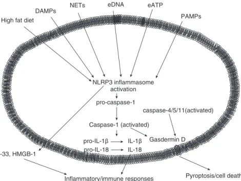
NLRs (nucleotide oligomerization domain–like receptors) are a particular type of intracellular pattern recognition receptor that recognize pathogen-associated molecular patterns and the host-derived signals called danger-associated molecular patterns (2) (Figure 1). A typical inflammasome is composed of an NLR, an adaptor protein such as ASC, and an effector caspase that activates proinflammatory cytokines. Within the NLRP3 complex, autocatalytic cleavage of procaspase 1 to active caspase-1 enables the removal of IL-1b and IL-18 prosequences, resulting in biologically active forms and thereby initiating T-helper cell type 1 (Th1) and Th17 adaptive immune responses (2, 3) (Figure 1). Gasdermin D (membrane pore-forming) activation by cleavage from both caspase-1 and caspase-4/5/11 causes a form of programmed cell death termed pyroptosis (4). NRLP3 inflammasome activation also causes the release of alarmins (molecules released from a damaged or diseased cell that can stimulate a sterile immune or inflammatory response)
such as IL-33 (another member of the IL-1 family, which acts through the receptor ST2) and atypical chemokines such as HMGB-1 (5). The NLRP3 inflammasome is activated by many stimuli, including NETs and double-stranded DNA (2) (Figure 1).
NETs are composed of decondensed chromatinfibers (containing double-stranded DNA) coated with antimicrobial granular and cytoplasmic proteins such as myeloperoxidase, neutrophil elastase, anda-defensins (6). Neutrophil extrusion of NETs may induce tissue damage and inflammasome activation, and neutrophils use an inflammasome- and gasdermin D– dependent mechanism to activate NETosis as a defense response against cytosolic bacteria (7).
This topic has been partially addressed in previous studies. In a female mouse model, NLRP3 inflammasome responses were found to drive experimental severe, steroid-resistant asthma (8). The U-BIOPRED (Unbiased Biomarkers in Prediction of Respiratory Disease Outcomes) Consortia Project Team demonstrated inflammasome activation in induced sputum from patients (smoking and nonsmoking) with severe asthma compared with nonsmoking patients with mild/moderate asthma and healthy control subjects that was associated with sputum neutrophilia and increased IL-1b protein levels (9). In that study, most (64%) of the nonsmoking patients with severe asthma were overweight females (mean BMI 29) (9), and neutrophilic severe asthma was defined using a cutoff of induced sputum neutrophils of mean > 74% (9), compared with a median of 51% (interquartile range 34–74) induced sputum neutrophils in the study by Lachowicz-Scroggins and coworkers (1).
There are some potential biases in the study by Lachowicz-Scroggins and coworkers. First, all of the patients with severe asthma included in their study were obese and compared only with healthy overweight control subjects, and not also with cohorts of nonobese subjects with asthma of different severities and healthy obese control subjects. In fact, obesity alone may influence NET formation (10) and NLRP3 inflammasome activation (11), and weight loss decreases peripheral blood neutrophil NET formation and inflammasome activation (12, 13). In one recent study, sputum NLRP3 mRNA and IL-1b protein levels were found to be significantly increased in obese versus nonobese patients with stable asthma (13). Furthermore, the authors provided no information about the diet of their subjects, and it has been demonstrated that in obese patients with stable asthma an acute challenge with a meal high in saturated fatty acids increases their sputum neutrophilia (13).
Another potential bias may arise from the glucocorticoid (inhaled and systemic) treatment used in the study. NET formation was shown to be downregulated by glucocorticoids in an animal model of asthma (14), whereas intriguing data from other animal
This article is open access and distributed under the terms of the Creative Commons Attribution Non-Commercial No Derivatives License 4.0 (http://creativecommons.org/licenses/by-nc-nd/4.0/). For commercial usage and reprints, please contact Diane Gern ([email protected]).
Originally Published in Press as DOI: 10.1164/rccm.201903-0667ED on March 25, 2019
Am J Respir Crit Care Med Vol 199, Iss 9, pp 1045–1060, May 1, 2019 Internet address: www.atsjournals.org
models suggest that glucocorticoids may be mandatory for inflammasome activation (15). It is worth noting that only 13% of the patients with severe asthma had a high eDNA sputum level, and this percentage is somewhat similar to that observed in the same patients under regular treatment with systemic glucocorticoids.
Future studies should also use better techniques, such as sputosorption (16), and measure the whole spectrum of
inflammasome activation, including IL-1Ra (17), IL-33, gasdermin D, and HMGB-1. These new and exciting data have potential therapeutic implications for this subset of obese nonsmoking patients with severe asthma. For example, the effect of fasting on asthma inflammasome activation was investigated in a recently completed pilot observational clinical study (https://clinicaltrials. gov/NCT02471300). Currently approved inhibitors of IL‐1b that may be tested in controlled clinical trials include anakinra (recombinant human IL‐1R antagonist [18]), rilonacept (recombinant soluble IL‐1R), and canakinumab (humanized anti–IL‐1b monoclonal antibody [19]). Two selective NLRP3 inflammasome inhibitors are b‐hydroxybutyrate (a ketone body produced during periods of starvation [20]) and the oral small molecule MCC950 (8). A potential advantage of these selective NLRP3 inflammasome inhibitors over IL‐1b blockers is that NLRP3‐specific agents may also target IL‐18 but permit IL‐1b activation by other inflammasomes. This may be important for the response to infection and other cellular processes, and also target the noncanonical effects of the inflammasome, which may be advantageous for controlling inflammasome‐mediated diseases (21).n
Author disclosures are available with the text of this article at www.atsjournals.org.
Acknowledgment: The authors thank Mr. Angelo Zodda and Ms. Franca Mollica for their technical assistance in the preparation of this editorial. Paolo Ruggeri, M.D.
Gaetano Caramori, M.D., Ph.D. Pneumologia, Dipartimento BIOMORF Universit `a degli Studi di Messina Messina, Italy
ORCID IDs: 0000-0003-0379-9869 (P.R.); 0000-0002-9807-327X (G.C.).
References
1. Lachowicz-Scroggins ME, Dunican EM, Charbit AR, Raymond W, Looney MR, Peters MC, et al.; National Heart, Lung, and Blood Institute Severe Asthma Research Program-3 Investigators. Extracellular DNA, neutrophil extracellular traps, and inflammasome activation in severe asthma. Am J Respir Crit Care Med 2019; 199:1076–1085.
2. Strowig T, Henao-Mejia J, Elinav E, Flavell R. Inflammasomes in health and disease. Nature 2012;481:278–286.
3. Di Stefano A, Caramori G, Barczyk A, Vicari C, Brun P, Zanini A, et al. Innate immunity but not NLRP3 inflammasome activation correlates with severity of stable COPD. Thorax 2014;69:516–524.
4. Shi J, Gao W, Shao F. Pyroptosis: gasdermin-mediated programmed necrotic cell death. Trends Biochem Sci 2017;42:245–254. 5. Kapurniotu A, Gokce O, Bernhagen J. The multitasking potential of
alarmins and atypical chemokines. Front Med (Lausanne) 2019;6:3. 6. Porto BN, Stein RT. Neutrophil extracellular traps in pulmonary diseases:
too much of a good thing? Front Immunol 2016;7:311–322. 7. Chen KW, Monteleone M, Boucher D, Sollberger G, Ramnath D, Condon
ND, et al. Noncanonical inflammasome signaling elicits gasdermin D-dependent neutrophil extracellular traps. Sci Immunol 2018;3: eaar6676.
8. Kim RY, Pinkerton JW, Essilfie AT, Robertson AAB, Baines KJ, Brown AC, et al. Role for NLRP3 inflammasome-mediated, IL-1b-dependent
DAMPs
PAMPs High fat diet
NLRP3 inflammasome activation pro-caspase-1 Caspase-1 (activated) Pyroptosis/cell death Inflammatory/immune responses pro-IL-1β pro-IL-18 IL-1β IL-18 IL-33, HMGB-1 eDNA eATP NETs caspase-4/5/11(activated) Gasdermin D
Figure 1. The main stimuli involved in inflammasome activation and related intra- and extracellular functional responses. DAMPs = damage-associated molecular patterns; eATP = extracellular ATP; eDNA = extracellular DNA; HMGB-1 = high-mobility group box protein-1; NETs = neutrophil extracellular traps; NLRP3 = nucleotide-binding oligomerization domain–like receptor family, pyrin domain–containing 3; PAMPs = pathogen-associated molecular patterns.
1046 American Journal of Respiratory and Critical Care Medicine Volume 199 Number 9
|
May 1 2019responses in severe, steroid-resistant asthma. Am J Respir Crit Care Med 2017;196:283–297.
9. Rossios C, Pavlidis S, Hoda U, Kuo CH, Wiegman C, Russell K, et al.; Unbiased Biomarkers for the Prediction of Respiratory Diseases Outcomes (U-BIOPRED) Consortia Project Team. Sputum transcriptomics reveal upregulation of IL-1 receptor family members in patients with severe asthma. J Allergy Clin Immunol 2018;141: 560–570.
10. Wang H, Wang Q, Venugopal J, Wang J, Kleiman K, Guo C, et al. Obesity-induced endothelial dysfunction is prevented by neutrophil extracellular trap inhibition. Sci Rep 2018;8:4881–4887.
11. Vandanmagsar B, Youm YH, Ravussin A, Galgani JE, Stadler K, Mynatt RL, et al. The NLRP3 inflammasome instigates obesity-induced inflammation and insulin resistance. Nat Med 2011;17: 179–188.
12. Roberts HM, Grant MM, Hubber N, Super P, Singhal R, Chapple ILC. Impact of bariatric surgical intervention on peripheral blood neutrophil (PBN) function in obesity. Obes Surg 2018;28:1611–1621. 13. Wood LG, Li Q, Scott HA, Rutting S, Berthon BS, Gibson PG, et al.
Saturated fatty acids, obesity, and the nucleotide oligomerization domain-like receptor protein 3 (NLRP3) inflammasome in asthmatic patients. J Allergy Clin Immunol 2019;143:305–315.
14. Vargas A, Boivin R, Cano P, Murcia Y, Bazin I, Lavoie JP. Neutrophil extracellular traps are downregulated by glucocorticosteroids in lungs in an equine model of asthma. Respir Res 2017;18:207–217. 15. Sobesky JL, D’Angelo HM, Weber MD, Anderson ND, Frank MG,
Watkins LR, et al. Glucocorticoids mediate short-term high-fat diet
induction of neuroinflammatory priming, the NLRP3 inflammasome, and the danger signal HMGB1. eNeuro 2016;3:ENEURO.0113-16.2016
16. Melo JT Jr, Tunstall T, Pizzichini MMM, Maurici R, Rocha CC, Dal-Pizzol F, et al. IL-5 levels in nasosorption and sputosorption correlate with sputum eosinophilia in allergic asthma. Am J Respir Crit Care Med 2019;199:240–243.
17. Gao P, Gibson PG, Baines KJ, Yang IA, Upham JW, Reynolds PN, et al. Anti-inflammatory deficiencies in neutrophilic asthma: reduced galectin-3 and IL-1RA/IL-1b. Respir Res 2015;16:5–14. 18. Hernandez ML, Mills K, Almond M, Todoric K, Aleman MM, Zhang H,
et al. IL-1 receptor antagonist reduces endotoxin-induced airway inflammation in healthy volunteers. J Allergy Clin Immunol 2015;135: 379–385.
19. Baldwin AG, Brough D, Freeman S. Inhibiting the inflammasome: a chemical perspective. J Med Chem 2016;59:1691–1710. 20. Johnson JB, Summer W, Cutler RG, Martin B, Hyun DH, Dixit VD,
et al. Alternate day calorie restriction improves clinicalfindings and reduces markers of oxidative stress and inflammation in overweight adults with moderate asthma. Free Radic Biol Med 2007; 42:665–674. [Published erratum appears in Free Radic Biol Med 43: 1348.]
21. Levy M, Thaiss CA, Elinav E. Taming the inflammasome. Nat Med 2015; 21:213–215.
Copyright© 2019 by the American Thoracic Society
On Trapped Air and Trapped Blood in Chronic Obstructive
Pulmonary Disease
Cardiopulmonary interactions in chronic obstructive pulmonary disease (COPD) involve many pathophysiologic mechanisms and are thus a complexfield of research (1, 2). An increasingly studied mechanism over the last decade is the effect of lung hyperinflation on cardiac filling and the associated consequences for exercise tolerance and physical activity (3–6).
Because all available long-acting bronchodilators used for maintenance therapy in chronic obstructive pulmonary disease (COPD) are effective lung deflators, it would be logical to also look for the positive effects of lung deflation on cardiac filling. However, hurdles related to technology and uncertainties about the appropriate endpoints have prevented a timely development of protocols. Such protocols would be much more complex than simply measuring lung volumes by body plethysmography. It was only 3 years ago that Stone and colleagues, in a landmark study, showed that lung deflation by monobronchodilator therapy plus an inhaled corticosteroid improves biventricularfilling and cardiac output in patients with COPD and hyperinflation (7). Last year, Hohlfeld and colleagues confirmed these findings by using dual bronchodilation in patients with COPD whose lungs were slightly more hyperinflated than those examined in the study by Stone
and colleagues (8). Through careful patient selection based on hyperinflation, and a more effective lung deflation therapy by dual bronchodilation, Hohlfeld and colleagues showed even more pronounced effects on cardiacfilling and cardiac output (8). These impressive improvements in the study of cardiacfilling, which might indirectly imply an improved pulmonary blood volume circulation, allowed the authors to also study the effects of lung deflation on pulmonary perfusion. In a follow-up study presented in this issue of theJournal, Vogel-Claussen and colleagues (pp. 1086–1096) used functional lung magnetic resonance imaging to measure regional pulmonary ventilation (9).
For me, the current (“secondary”) analyses are even more important than the previous primary analyses, which confirmed what had been shown before. So why are the current analyses important and clearly not secondary, as formally stated in the statistical analysis plan in the protocol? It is because they help us understand the mechanisms by which bronchodilators improve cardiacfilling. Based on data derived from chest X-ray imaging and right heart catheterization with wedge pressure measurements, it was previously speculated that lung hyperinflation prevents the extension of the heart within the cardiac fossa during diastolicfilling (10). It now seems that this is an effect of lung hyperinflation on pulmonary perfusion rather than an improvement in the anatomical position of the heart. Furthermore, the improvements of pulmonary perfusion seen with lung deflation in the current analyses by Vogel-Claussen and colleagues seem to occur predominantly at the microvasculature site rather than at the macrovasculature site (9). This implies that reduced air trapping
This article is open access and distributed under the terms of the Creative Commons Attribution Non-Commercial No Derivatives License 4.0 (http://creativecommons.org/licenses/by-nc-nd/4.0/). For commercial usage and reprints, please contact Diane Gern ([email protected]).
Originally Published in Press as DOI: 10.1164/rccm.201901-0061ED on January 29, 2019