University of Catania
International PhD in Translational Biomedicine XXVIII cycle
PhD COORDINATOR PROF. DANIELE FILIPPO CONDORELLI
ALESSANDRO SINOPOLI
_______
Molecular mechanisms of A
recognition and neuroprotection by
the glycoconjugate β-sheet-breaker peptide Ac-LPFFD-Th.
PhD
T
H E S I S
PhD Supervisor :
Prof. Daniele F. Condorelli
PhD Co-Supervisor:
Dott. Giuseppe Pappalardo
TABLE OF CONTENTS
Abstract ... 4
CHAPTER 1: Introduction ... 5
1.1 Protein folding, misfolding and amyloidoses. ... 5
1.2 Alzheimer’s disease and Amyloid-β ... 9
1.3 Therapeutic strategies to inhibit amyloid aggregation process ... 19
CHAPTER 2: Material and methods ... 26
CHAPTER 3: Results ... 34
3.1 ThT assay ... 34
3.2 Dynamic light scattering ... 40
3.3 Recognition of the full-length Aβ monomeric form (ESI and limited proteolysis) ... 43
3.4 NMR Interaction studies between Ac-LPFFD-Th with Aβ-16-28 and Aβ1-40 ... 49
3.5 Aß/AßOs WESTERN BLOT analysis ... 59
3.6 Neuroprotective activity of the beta sheet breaker-peptides ... 60
3.7 In Vivo study, Y-maze assay ... 64
CHAPTER 4: Discussion ... 66 Conclusions ... 70 References ... 71
Abstract
Inhibition of amyloid formation may represent a promising therapeutic approach for the treatment of neurodegenerative diseases. To this regard, peptide-based inhibitors of Aβ aggregation have been widely investigated with a particular emphasis to those derived from original amyloid sequences.
The experimental work described in the present PhD Thesis aims at outlining the molecular mechanisms underlying the antifibrillogenic and neuroprotective action exhibited by a new class of trehalose conjugated pentapeptides. Trehalose (Th), a non-reducing disaccharide of α-(D)glucose, has been demonstrated to be effective in preventing the aggregation of several proteins. We figured out that the development of hybrid compounds may provide new molecules with improved properties that might sinergically increase the potency of their single moieties.
Since it has been demonstrated that Aβoligomers are the toxic species, it becomes a priority to counteract the Aβ self-assembly because of its doubly dangerous effect associated with oligomers generation and removal of the neurotrophic monomeric form of Aβ1-42 peptide.
Starting from the well-known neuroprotective action of the LPFFD peptide, (C. Soto, Nat Med. 1998) we investigated whether the Ac-LPFFD-Th peptide would act as an “A monomer-stabilizer” thus exerting a neuroprotective activity. In support of this hypothesis, time-resolved proteolysis and ESI-MS experiments, performed during my PhD experimental work, showed a direct interaction between these -breaker peptides and Aβmonomers.
In this work, the C-terminal trehalose conjugated Ac-LPFFD-Th derivative ability to recognize and bind low molecular weight aggregated forms of Aβ has been investigated by means of different biophysical techniques, including Th-T fluorescence, DLS, ESI-MS and NMR. Furthermore, biological assays on murine cortical primary neuronal cultures were performed in order to clarify and further characterize the mechanism of cytoprotection exhibited by the Ac-LPFFD-Th.
In this PhD thesis, we demonstrated that Ac-LPFFD-Th modifies the aggregation features of Aβ and protects neurons from Aβ oligomers’ toxic insult.
Introduction
1.1 PROTEIN FOLDING, MISFOLDING and AMYLOIDOSIS
.Proteins after their synthesis in the cell, must be converted into tightly folded compact structures in order to function. Many of these structures are astonishingly intricate, the fact that folding is usually exceptionally efficient is an outstanding evidence of the power of evolutionary biology. Given that there are 20 different naturally occurring amino acids, the total possible number of different proteins with the average size of those in our bodies is enormous. The properties of the natural proteins are not typical of random sequences, but have been selected through evolutionary pressure in order to have specific characteristics of which the capacity to fold to unique structures and therefore to generate enormous selectivity, in addition to the diversity in their functions. (1)
How proteins find their distinctive native folded states from the information enclosed in their sequences is a question at the heart of molecular biology.(1)
The mechanism by which also the smaller and easier proteins fold to unique structures has been defined in detail. There are strong evidences that the native state of a protein corresponds, except in very exceptional circumstances, to the most stable structure under physiological conditions.
However, the total number of possible conformations of a protein is so great that it would take an enormous amount of time to find this particular structure throughout a systematic search of all the conformational space.(2,3)
Now it is clear that the folding of a protein does not involve a succession of compulsory steps between well-defined partially folded states, but rather a stochastic search of the many conformations accessible to a polypeptide chain. In principle, the inherent fluctuations in the conformation of an unfolded polypeptide chain allow even residues at very different positions in the amino acid sequence to come in contact with the others. Since the correct (native-like) interactions between the different residues are generally more stable than the incorrect (non-native) ones, such a search mechanism is, in theory, able to find the lowest energy structure possible between all the different conformations. (1-4)
It is clear that this process is particularly efficient for those sequences that have been selected during evolution to fold into globular structures, and in fact, only a very little number of all possible conformations needs be sampled during the search process. This stochastic description of protein folding introduces the concept of an “energy landscape”. (1) This describes the free energy of the polypeptide chain as a function of its conformational properties. In order to enable a protein to fold correctly and efficiently, the landscape required has been associated to a funnel because the conformational space easily accessible to the polypeptide chain becomes more and more restricted as the native state is closer (figure 1). (1-4)
The elevated degree of disorder of the polypeptide chain is reduced as the folding progresses because there is a gain in enthalpy related with all the stable native-like interactions that are created. This gain in enthalpy can counterbalance the decreasing in entropy of the polypeptide chain as the structure becomes more ordered. (5a) Even though the details of the way in which the different proteins can fold may appear to differ considerably, in terms of the rates of folding and the type of species populated during the folding process, the fundamental features of this general mechanism can be considered to be universal. (1,5)
Hence globular proteins mainly fold by minimizing the non-polar surface that is exposed to water, while at the same time forming hydrogen interactions for buried backbone groups, typically in the form of secondary structures such as α-helices, β-sheets, and tight turns. (5b)
Figure 1. Protein folding and proteostasis. (Nature 2011, 475, 324–332).
The folding of some proteins in vivo appears to be co-translational, it means that the folding begins when thegrowing chain is still being synthesized on the ribosome (6). Because of the complexity of the folding process, misfolding is an inherent feature of it for all proteins, particularly under unfavorable environment conditions.
Misfolding can roughly be defined as reaching a state that has a significant proportion of nonnative interactions between residues and whose properties differ considerably from those of a similar state having native-like interactions. The cellular levels of many chaperones are, as a matter of fact, significantly increased during cellular stress, from that their common designation as heat-shock proteins (Hsps). Some of these molecular chaperones act to capture misfolded proteins, or even some types of aggregates, and so giving them another opportunity to fold correctly. (7)
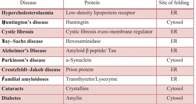
One of the better known characteristic features of many of the misfolding diseases is that they often result in to the deposition of proteins in form of amyloid fibrils, stable, ordered, filamentous protein aggregates. A significant number of human diseases, including neurodegenerative diseases such as Alzheimer’s disease, Parkinson’s diseases, prion diseases, Amyotrophic lateral sclerosis (ALS), Type 2 diabetes mellitus and several other pathological conditions share a common feature. In each of these pathologies, protein or peptide changes from their natural soluble state into insoluble fibrils, which aggregate and accumulate in a variety of tissues and organs. Although approximately 20 different proteins are known to be involved in the amyloidoses, they are generally unrelated in terms of sequence or structure. Despite these differences in sequence and structure, the fibrils formed from these different peptides or proteins in diverse pathologies share many common properties. Those are a core cross-β-sheet structure in which continuous β-sheets are formed with β-strands running perpendicular to the long axis of the fibrils. (8) All fibrils have similar morphologies, being twisted, rope-like structures, reflecting a filamentous substructure. (9) T a b l e 1 . M
Table 1. Protein misfolding in conformational diseases
Disease Protein Site of folding
Hypercholesterolaemia Low-density lipoprotein receptor ER
Huntington’s disease Huntingtin Cytosol
Cystic fibrosis Cystic fibrosis trans-membrane regulator ER
Tay–Sachs disease Hexosaminidase ER
Alzheimer’s Disease Amyloid β peptide/ Tau ER
Parkinson’s disease α-Synuclein Cytosol
Creutzfeldt–Jakob disease Prion protein ER
Familial amyloidoses Transthyretin/Lysozyme ER
Cataracts Crystallins Cytosol
1.2 ALZHEIMER’S DISEASE and AMYLOID-β.
Alzheimer disease (AD) is the most prevalent neurodegenerative disease affecting more than 35 million people worldwide. (10)
Despite considerable research, there is no cure for this pathology yet and available treatments are only symptomatic.
AD is characterized by progressive loss of memory, declining cognitive function, decreased physical function and ultimately death. It represents one of the main public health challenge of growing significance among the aged population. (11)
The hallmarks of AD are the amyloid plaques, protein aggregates deposited in the brain, and neurofibrillary tangles, intraneuronal bundles of paired abnormally phosphorylated tau proteins.
The generation and aggregation of Aβ activate a complex pathologic cascade leading to neuronal dysfunction, inflammation and neuronal loss. This cascade plays a central role in pathogenesis and is commonly referred to as the amyloid cascade hypothesis. (12) The amyloid hypothesis states that the 39–43 amino acid amyloid-β-peptides (Aβ), the main constituent of amyloid plaques, are responsible for the initiation of a neurotoxic cascade that ultimately leads to neuronal death and dementia. (12)
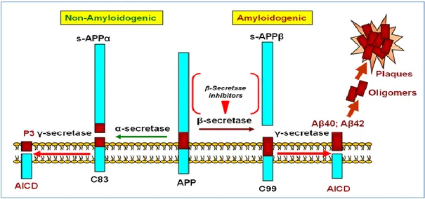
The Aβ peptides are generated by the cleavage of amyloid precursor protein (APP), a type I integral transmembrane protein. The processing of APP takes place via amyloidogenic and non-amyloidogenic pathway (figure 2).
The normal non-amyloidogenic pathway cleaves APP at amino acid positions 16 and 17, generating a secreted APP derivative sAPPa and a shorter C-terminal fragment (CTF) of 83 amino acids (C83). C83 is subsequently cleaved by γ-secretase to form a non-toxic 3 kDa peptide (p3). Although Aβ is produced normally in the CNS, these α- and β-pathways may compete for APP substrate. APP is believed to be cleaved preferentially through this pathway in normal brain, with the balance shifting to the amyloidogenic pathway in AD. In the amyloidogenic pathway, cleavage occurs within the ecto-domain of APP mediated by β-site APP cleaving enzyme β-secretase (Bace-1, memapsin-2, ASP-2). Cleavage occurs at methionine (position 671) and aspartic acid (position 672) to generate a secreted amino terminal APP derivative (sAPPβ) and a membrane-inserted CTF (β-CTF) of 99 amino acids in length (C99).
C99 is further cleaved by the intramembranous γ-secretase cleavage at 711–713 to the catalytic subunit presenilin, to facilitate cleavage within the hydrophobic lipid bilayer. This cleavage generates the Aβ at the C-terminus and liberates the Aβ peptide into the extracellular space. (13)
Figure 2. Amyloid cascade hypothesis (C. Zhang. Discovery Medicine, 2012, 14, 189-197)
Due to its biophysical properties, under certain condition Aβ 1-40/42 may self-aggregate into different forms, from 4 kDa monomers to dimers, trimers, tetramers, dodecamers, higher-order oligomers, protofibrils, and finally mature fibrils. Fibrils are responsible for the formation of plaques in AD brains. Several environmental factors, such us pH, metal ions and oxidative stress may contribute to the destabilization of the native random coil Aß conformation leading it to aggregation.
Although there are accumulating evidence that soluble oligomers are the most cytotoxic form of Aβ, it is still unclear which size and morphology of the aggregates exert neurotoxicity; it has been observed that there is a decrease in toxicity when Aβ1-42 oligomers grow in size. (14)
A new pathological role for Aβ in AD has been observed due to its essential function in the synapse. It is supposed that Aβ may play a variety of physiological roles. Proposed functions of Aβ include neural metabolism control of synaptic activity, memory consolidation, trophic and neuronal survival, cholesterol transport.
As a matter of fact it seems that the protective or negative effects of Aβ depends by its relative concentration as well as the age-related changes in the cellular environment. (15)
In the human brain, Aβ is generally found in the interstitial fluid; considering its significant affinity for metal ions, due to the presence of three His in the N-terminal part, Aβ could have a function in removing unbound iron, copper and zinc from the extracellular space, stimulating an inflammatory response in microglia which would in turn promote rapid phagocytosis of the peptide.
Therefore, the binding Aβ to metal ions could be a mechanism to remove potentially hazardous metal ions from the extracellular space and to facilitate their clearance from the brain. (16) Moreover it was demonstrated that monomeric Aβ1–40 interacting with metal ions quenches the transition metal-mediated oxygen radical generation, e exerting a neuroprotective effect on neurons. Noteworthy the oligomeric forms of Aβ loose this protective property. These results point out new ideas about the dualistic biological function of Aβ. As a monomer it behaves as an antioxidant molecule, preventing the generation of oxygen radicals, while oligomeric or aggregated forms not only lose antioxidant activity but also contribute to the generation of ROS. (17)
Recent work has shown that monomeric Aβ1–42 in the 30-100 nM concentration range is able to activate the survival pathway in cultures of rat cortical neurons in condition of trophic deprivation, it also protects neurons against NMDA-mediated excitotoxicity cell-death. This effect was mediated by the activation of IGF1/Insulin receptor which trigger the PI-3-K (phosphatidylinositol-3-kinase) pathway. Furthermore, it was shown an increased expression of the antiapoptotic protein Bcl-2.
On the other hand, the Aβ peptide carrying the mutation (E22G) the so-called “Arctic mutation” which is characterized by a rapid aggregation kinetic, it shows a complete loss of neuroprotective activity.(18)
Inflammation is a very important marker in AD as it has been shown in recent works which suggest that, once inflammation is initiated in response to neurodegeneration this may actively contribute to disease progression (figure 3).
Numerous neuroinflammatory mediators are involved such as: cytokines, chemokines, complement activators and inhibitors, radical oxygen species (ROS) and inflammatory enzymes, including the cyclooxygenase-2 (COX-2) as well as the inducible nitric oxide synthase (iNOS). (19,20)
Figure 3. Inflammatory process in Alzheimer's Disease (M.A. Meraz-Ríos et all. Front. Integr.
Neurosci., 2013)
It has been shown that Aβ can specifically bind to phospholipid bilayers with relatively high affinity suggesting that the cell membrane of neurons may be a primary target of Aβ. Numerous membrane models including lipid monolayers, bilayers and liposomes, have been used to examine the interactions between Ab and membrane surfaces. The interactions between lipid bilayers and Aβ have been studied using a variety of biophysical techniques, including fluorescence spectroscopy, DSC, CD and NMR. (21a)
It was suggested that the head-group charge of the phospholipids contributes to the association between Aβ and the membrane via electrostatic interactions.
Aβ has been reported to form ionic pores, directly leading to cell death or starting the apoptotic signaling interfering with the regulation of calcium homeostasis. (21b,22)
Aβ is also able to alter the physicochemical properties of neuronal membranes (e.g. membrane fluidity), and so inducing membrane destabilization and permeabilization. Both membrane-Aβ interactions and toxicity seem to be modulated by the lipid composition, particularly cholesterol and ganglioside, which are speculated to be the major components in the lipid-raft domains of the plasma membrane. It has been reported that cholesterol enhances the binding of Aβ to a lipid bilayer and facilitates Aβ aggregation. (23a) However, the role of cholesterol in AD remains controversial. Inconsistent data and conclusions reported in the literature make it difficult to determine the exact mechanism underlying the membrane-associated toxicity of Aβ. The lack of a clear and detailed description of Aβ-membrane interactions hinders a complete understanding of Aβ toxicity in AD pathogenesis. (23b)
Several works indicate that the Aβ forms which interact with the membranes, leading to the formation of pores, are associated to microscopic structures similar to very small fibrils, but not to unstructured forms of Aβ. These results strongly suggest that Aβ oligomers are directly involved in destabilization and/or perforations of the synaptic membranes. (24)
It has been demonstrated that amyloid fibrils, usually considered as highly stable and biologically inert structures, can be destabilized and easily reverted to soluble and highly toxic Aβ smaller aggregates by the biological lipids present in the brain. (25) This suggests that part of the critical balance between toxic and inert Aβ pools could be determined by the relative amounts of lipids in the direct environment of the plaques. Surprisingly, the identified toxic species share many properties with the oligomeric aggregates in terms of biophysical and cell biological behavior. Different biophysical assays show how these structures are quite heterogeneous in nature, and the size distribution of these oligomeric aggregates ranges from 80 to 500 kDa. Lipids are apparently promoting the equilibrium toward the protofibrillar pools, inducing toxicity of the amyloid mixture.
Furthermore there is an evidence that membrane composition and properties play a critical role in Aβ cytotoxicity associated with its conformational changes and aggregation into oligomers and fibrils. (25)
A wide range of AD synapto-toxic Aβ oligomeric sizes have been identified. How oligomer size precisely is related to the disease process has not been clarified and recent work shows that the wide range of Aβ oligomers may have a specific conformation in common.
These findings suggest that Aβ oligomer size may not be the only AD-inducing factor but both oligomer size and structural conformation act as toxic parameters in AD progress. Previous studies targeting the Aβ toxic species in AD have usually highlighted only one of these aspects. Instead it is necessary a multi disciplinary approach where oligomer size and structural characteristics should be taken into consideration together. (26) In the typical cross-β-structure, hydrogen bonds are oriented parallel to the fibril axis, with the β -strands running perpendicular to the fibril’s axis, usually from 2 to 9 protofilaments form fibrils. However, the amyloid formation is a very complex process, biophysical studies performed in presence of component of the extracellular matrix such as glycosaminoglycans and well-defined model membranes have shown that a variety of amyloidogenic peptides and protein readily adopt helical structures when interacting with surfaces. These generated helical intermediates generated appear to play an important role in amyloid growth in which helix-mediated association will lead to a high local concentration of an aggregation prone sequence, and so favoring intermolecular β-sheet formation. (27) It has been reported that monomeric Aβ1–40 shows a β-hairpin comprising residues 17–36, in which the 17-23 and the 30-36 fragments make intramolecular backbone hydrogen bonds to form the two central strands of the sheet. Both faces of the β-hairpin are predominantly apolar, for this reason they are buried inside a large hydrophobic tunnel-like cavity with the ‘‘interior’’ face of the hairpin containing Leu-17, Phe-19, Ile-32, Leu-34, and Val-36 side chains docked into the cleft to form a large intermolecular hydrophobic core. It can be speculated that the β-hairpin constitutes an intermediate conformation on the formation of amyloid fibrils. Therefore oligomers might form by hydrophobic interactions of β-hairpins and remain soluble as a consequence of the hydrophilic surface due to the hydrogen bond capacity of the exposed peptide backbones.
Fibril seeds could subsequently be generated by a concerted conformational transition toward intermolecular β-sheets. (28)
Amyloid aggregation is described by a sigmoidal curve and is considered to be a nucleation–polymerization reaction where the monomer addition steps are assumed to be thermodynamically unfavorable until a critical nucleus is formed.
However, aggregation is a thermodynamically favorable process during the polymerization stage. The critical nucleus is defined as the least thermodynamically stable species in solution, which is the oligomer of minimal size capable of initiating further growth. The nucleus can also be defined as the aggregate size after which the association rate exceeds the dissociation rate for the first time. In addition to the homogeneous nucleation, heterogeneous nucleation (or seeding) can also take place on the surface of existing polymers. Furthermore, aggregation can be further accelerated by the fragmentation of existing aggregates. For some proteins with specific distributions of polar and hydrophobic residues (e.g. for Aβ1–40 peptide), fibrillation can start only above a certain critical micelle concentration at which the peptide micelles are formed.
The formation of these micelles represents a crucial step, as fibrils nucleate inside them and then grow by irreversible binding of monomers to fibril ends.
In addition to the amyloid fibrils discussed above, proteins can self-assemble to form several other types of aggregate such as amorphous aggregates. Amorphous aggregates are typically formed faster than fibrils. There is no special conformational prerequisite for amorphous aggregation to occur, and many destabilized and partially unfolded proteins precipitate out of solution in a form of amorphous aggregate. On the other hand, fibrillation requires special conditions that promote the formation of the specific amyloidogenic conformations. Therefore the conformations of monomers at different aggregation stages are not identical, and a given protein can self-assemble into various aggregated forms, depending on the peculiarities of its environment. Aβ oligomers have shown a wide range molecular mass distribution (from <10 to >100 kDa) in the AD brain (figure 4). In vitro studies have revealed that Aβ dimers were three times more toxic than monomers, in addition to Aβ tetramers that were 13 times more toxic, as well as the SDS-stable Aβ nonamers and dodecamers which were shown to induce a significant fall-off in the spatial memory performance and can be associated with deleterious effects on cognition. (29)
Figure 4. Different morphologies of Aβ aggregates (FEBS J. 2010, 277, 2940–2953)
The structure of Aβ fibrils is determined by constraints due to the monomer’s conformation, interestingly, fibril structure formation is determinate also by the kinetic of the process rather than the thermodynamically lowest energy state.
It was hypothesized that the predominant fibrillar structure would be the one with the fastest kinetics of formation even though more thermodynamically stable states might exist. (30)
It’s important to emphasize that amyloid aggregation properties are significantly different between the two principal Aβ forms, although both peptides aggregate into fibrils in vitro, Aβ1-42 does so more rapidly than Aβ1-40. The hydrophobicity of the residues at positions 41 and 42 is the major contributor to the enhanced amyloidogenicity of Aβ1-42 relative to Aβ1-40 (figure 5). That would be consistent with residues Ile41 and Ala42 taking place in the buried core of fibrils. Additionally these aminoacids have a good β-sheet propensity.
In Aβ1-42 these residues, together with Val39 and Val40 (which are also hydrophobic β-sheet formers), may form a β-strand at early stages of aggregation, hence lower the kinetic barrier for the aggregation of Aβ1-42 relative to Aβ1-40. (31) In addition the C-terminus of Aβ1-42 is more structured than that of Aβ1-40.
Figure 5. Aβ’s aminoacid sequence
An interesting work asserts that Aβ1-40 inhibits Aβ1-42 oligomerization through the formation of a hetero-oligomer, typically a stable mixed tetramer via dimer condensation, composed of equal parts of Aβ1-40 and Aβ1-42. This suggests that Aβ1-40, in a healthy human brain, actually sequesters Aβ1-42 thus preventing its further oligomerization and formation of the putative dodecamer toxic form, and consequently potentially deterring the development of AD. (32)
Furthermore, increasing evidences suggest that the ratio of Aβ1-40 to Aβ1-42, rather than the total amount of Aβ, is an important determinant of aggregation, fibrillogenesis, and toxicity. (33)
Elucidation of the different mechanisms involved in aggregation, conformational transitions as well as in deep knowledge of fibrils and oligomers structure; will help us in the rational design of aggregation inhibitors.
For many years, the fibrillar Aβ assemblies, in analogy to what seen in amyloid plaques, have been considered the main responsible for the neurodegenation associated with AD. However, the quantity and temporal progression of amyloid plaques do not correlate well with the clinical evolution of the disease. (34)
An invariant and early feature of AD is the synapse loss and there is a strong correlation between the extent of synapse loss and the severity of dementia. Alterations in synaptic connectivity and/or strength have been observed in transgenic mouse models of AD before the appearance of senile plaques. (35,36)
In the past decade of research emerged a wide consensus pointing out the soluble non-fibrillar Aβ assemblies as the major cause of synapto toxicity in the early phases of the pathology. (8)
The pathogenic relevance of natural Aβ oligomers is supported by the observation that their formation is increased by expressing AD-causing mutations within APP or presenilin genes in recombinant cells.
Indeed, oligomers have been detected in CSF and in brain tissues of AD patients, where the level of soluble Aβ species appear to correlate with disease progression. (37-43) When micro-injected in living rats or added in vitro to hippocampal slices, natural oligomers of human Aβ are acutely toxic on synaptic functions. Soluble forms of Aβ have been shown to bind with high specificity to excitatory synapses and recently Steinerman et al. correlated AD symptoms with membrane-bound Aβ1-42 pools. (44)
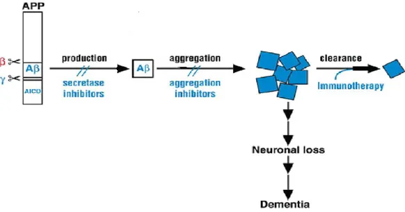
Therefore to prevent the side effects of this amyloid aggregation pathway, research efforts should aimed at intervening at three different “steps” of the process, as shown in Figure 6:
1) Inhibiting Aβ production using small molecules or peptide/peptidomimetic which block the two secretases who produce Aβ from APP
2) Inhibiting the aggregation process itself
3) Increasing the clearance of the different aggregated forms using specific antibody
1.3
Therapeutic strategies to inhibit amyloid aggregation process.
At present there is no cure for amyloidoses. Available therapeutic interferences are just symptomatic. (45)Immune approaches have been tried for the treatment of AD, but the accompanying inflammatory process in the brain was severe enough for this approach to be abandoned. (46)
Also efforts have been made to inhibit the secretases responsible for the production of Aβ peptides but anyway these approaches have not been successful so far. Interference with the Notch signaling pathway is the greatest concern when developing gamma-secretase inhibitors; this make this protein not a good target to treat AD. (47)
Meanwhile Bace-1 has become an approved target in the discovery and development of medicines against AD because it is rate limiting for this disease process and a partial inhibition of this protein would result in a benign phenotype. (48)
The problem is that Bace1 seems to be a very challenging target for medicinal chemistry. First, Bace-1 has a large hydrophobic substrate-binding site designed to fit polypeptides, thus making it difficult to inhibit the enzyme with small non-peptidic compounds that have desirable drug-like characteristics. Ideally, Bace-1 inhibitor drugs should be molecules with a molecular weight <500, orally bioavailable, metabolically stable, intrinsically potent and highly selective for Bace-1. Compounds must also be hydrophobic enough to penetrate both plasma and intracellular membranes to gain access to the lumen of the compartment where the Bace-1 active site is localized. Finally, efficacious Bace-1 drugs would need to efficiently cross the blood-brain barrier and achieve a high concentration in the cerebral parenchyma. (49)
In such a scenario molecules able to prevent the basic molecular recognition process underlying the formation of Aβ early intermediates, would be the most valuable candidates for the treatment and the prevention of AD. A number of small molecules, carbohydrate and peptide-based inhibitors, have been shown to hinder A aggregation and toxicity.
However to inhibit the formation of those toxic Aβ forms, understanding the details of the aggregation/fibrillation mechanism at the molecular level is crucial to develop an approach in which this process is inhibited.
Since numerous studies suggest that small oligomers, intermediate aggregate species formed during the first steps of the amyloid aggregation process, are the neurotoxic species in amyloid disease, agents that prevent or reverse their formation may in principle attenuate cytotoxicity. (50)
Many strategies to inhibit the amyloid aggregation process have been attempted. Some of them involve small molecules, carbohydrate (51) peptides and peptidomimetics. (52, 53)
1.3.1 Small molecules inhibitor
Various small molecules with specific primary targets have been shown also to
interact with amyloidogenic protein and influence their aggregation. Tetracycline have been shown to prevent the formation of Alzheimer’s amyloid
aggregates beside the capacity to de-polymerize the Aβ1-42 fibril. (54) In addition having a well-defined antibiotic activity, tetracyclines are potent antiamyloidogenic agents. In the C. elegans model of AD, tetracyclines reduced the Aβ deposition, reduced the oxidative stress and delayed the disease onset. (55) The antifibrillogenic activity of tetracyclines appears to be different with respect to the protein involved. Galantamine exhibited a dose dependent inhibition of aggregation of Aβ1-40 and Aβ 42. The cytotoxicity and apoptosis were significantly reduced. (56)
The chemical structure is important in the antiamyloidogenic property of these drug molecules in fact small variations can make the difference. Resveratrol has been observed to convert fibrillar intermediates into high molecular weight and unstructured aggregates. It did not accelerate the aggregation of monomers or soluble oligomers. Molecular dynamics simulations show that resveratrol interferes with the aggregation process by inhibiting the lateral growth and not the elongation. Inter sheet side chain interactions are interfered with resveratrol. (57)
Numerous small molecules are known to be efficient in preventing amyloid aggregation. Most of them have a central fused aromatic ring structure. More often they have many hydroxyl substitutions. The are many sources of these compounds.
A general remark describing their essential features will be made here. These compounds have the structural and chemical complementarity to interact with amyloid structures.
They are also good antioxidants since amyloid aggregation and oxidative damage are always involved in amyloid diseases. For those reasons compounds with antiaggregation and antioxidant activities are most probably better drug candidates. In vitro and in vivo experiments establish that these compounds can indeed reduce the aggregation of amyloids, sometimes reverse the aggregation and attenuate the oxidative damage. A various number of natural products such as curcumin, epigallocatechin gallate, extract from green and black tea, polyphenolic preparation from grape seeds and baicalein, a flavonoid extracted from the Chinese herb Oroxylum indicum, have been tested in vitro and in vivo. They have shown antifibrillation and antioxidant effects. (58)
1.3.2 Carbohydrate Inhibitors
Carbohydrate have been used as additives in protein folding experiments due to their well known effect in stabilize protein structure. The use of these sugars and their property has been extended to inhibit protein aggregation. This approach has been tried in many amyloid diseases such as Alzheimer, Parkinson and Huntington’s diseases. The use of sugar in inhibiting protein aggregation consists in combining β-strand breaking property and enhancement of aqueous solubility. (58)
Trehalose has been established as protein’s antistress molecule. It is used in protein folding experiments. It has a stabilizing role during heat denaturation or freeze-drying of proteins. The water substitution hypothesis is invoked in explaining the protective role of trehalose. The higher solubility and lower price make trehalose a tempting proposition as a therapeutic agent. Trehalose interacts through direct and solvent mediated interactions to weaken the inter-peptide interactions between Aβ chains. The surrounding layer of hydrophilic trehalose reduces the peptide-peptide hydrophobic interactions thereby reducing the aggregation. (59,60)
Glycodendrimers with sulfated glucosamine mimicking glycosaminoglycan suppress the fibril formation by Aβ peptides (Aβ1-42, Aβ1-40 and Aβ25-35). The effect depends on the nature of the carbohydrate moiety.
The acidic oligosaccharide sugar chain (AOSC), extracted from the brown algae Echlonia kurome, reduced the Aβ induced toxicity in the SH-SY5Y cell line and primarily cortical neurons. It interacts with monomeric Aβ and oligomeric Aβ. (58,61)
A stereoisomer of the sugar inositol, scyllo-inositol, has been shown to inhibit Aβ1-42 amyloid aggregation by directly interacting with the Aβ peptide. Many scyllo-inositol derivatives containing deoxy, fluoro, chloro and methoxy substitutions have been tested. The 1-deoxy-1-fluoro- and 1,4- dimethyl-scyllo-inositols were effective in inhibiting the Aβ1-42 fibrillation. This compound is going under clinical evaluations. It has been shown that scyllo-inositol, in CSF, decrease oligomeric Aβ1-42 concentration. Another sugar found in thermophilic microorganisms, alpha-d-mannosylglycerate (MG), has been observed to protect proteins against various structure-destabilizing conditions such as freezing, heat and drying. The efficacy of MG in preventing Aβ fibrillation and neurotoxicity in neuroblastoma cells has been shown as well. (58,61)
1.3.3 Peptide inhibitor
Peptides are molecules endowed with different characteristic that makes them interesting in drug discovery. Peptides are usually less toxic, more soluble and more specific than small organic molecules. Small peptide sequences encompassing the hydrophobic regions of the sequence of the aggregation-prone protein may interfere with the fibril formation process. (62)
This is like a competition between two molecules, one is the native protein/peptide and the other one is a smaller part of it. They compete to the same peptide segment, which forms the amyloid aggregate.
Since it is known that the major force driving amyloid aggregation is hydrophobicity, adding some charged residues to the ends of the recognition motif could be a disrupting element. (63)
Different works have show that at least three lysines are required as an appropriate disrupting element the compound (KLVFFKKKK) showed activity in altering fibril morphology and reducing cellular toxicity in vitro.
The compound with an anionic disrupting element KLVFFEEEE, has been observed having similar effects, while the neutral compound KLVFFSSSS was inefficient, suggesting that the charged nature of the disrupting element is critical. (63)
Different teams studied the incorporation of N methyl-amino acids into peptides as β-sheet disrupting elements. (64) The idea behind is that one side presents a hydrogen-bonding 'complementary' face to the protein, with the other side having methyl groups in place of backbone NH groups, thus presenting a 'blocking' face. N-methylated peptides corresponding to 16-22 and subsequently 16-20 sequence of Aβ have been studied. These peptides can prevent Aβ fibrils formation. N-methylation contributes in addition to make the peptides more resistant to proteolysis than normal peptides. Kapurniotu and colleagues synthesized an Api-28 analog constrained by an internal cycle between residues Lysl7 and Ala21. This modified peptide inhibited Aβ aggregation and cytotoxicity. (65)
β-sheet breakers peptides (BSB) have appeared years ago as the prototype class of compounds inhibiting Aβ aggregation. The β-sheet breaker approach uses the Aβ self-recognition motif (17-20) to achieve binding and specificity but, at sometime, replacing a residue important for forming β-sheets with an amino acid thermodynamically unable to fit inside β structure makes the aggregation process unfavorable. (66)
Valine at position 18 of Aβ plays an important role on stabilizing β -sheet folding in Aβ but it seems not to be necessary for self-recognition. Consequently, this amino acid has been replaced by proline, an aminoacid residue that due to of its particular chemical structure is an efficient β-sheet breaker. The β-sheet breaker peptide LPFFD has been developed by Soto and tested in vitro, in cellular model as well as in vivo. The in vitro activity was verified using both the Th-T binding assay, a commonly used fluorometric method to quantify amyloid, and qualitatively using electron microscopy to study the fibril morphology. Two animal models have been employed to monitor the activity of β-sheet breaker peptides in vivo. In both of them there were prevention of the formation of fibrillar lesion. (67)
The main problem of small peptides like the β-sheet breakers, it is that they are very easily degraded by peptidase and exhibit very short half-lives in vivo. (68)
For these reasons the synthesis of modified BSB peptides is necessary in medicinal chemistry.
Studies on the aggregation process of Aβ, identified the critical region involved in amyloid fibril formation; this has been mapped within the hydrophobic core at the residues 16-20 (KLVFF) of Aβ. (69) Designed peptides based on this recognition motif bind to the homologous sequence of Aβ thereby preventing its self-association.
Despite the good in vitro activity found for some of these compounds, their therapeutic usefulness is questioned due to their tendency to self-aggregate and to the risk of being incorporated into amyloid fibril.
Therefore, modified synthetic peptides based on the sequence of this central region of Aβ conjugated with the sugar trehalose, have been previously considered in our lab. (70) We used trehalose because it has been demonstrated to be effective in preventing the aggregation of numerous proteins including Aβ (71,72).
The antifibrillogenic ability of trehalose might stem from its preferential exclusion effect from the peptide surface. Consequently, threalose could stabilize the secondary structures of monomers, making the inter-peptide interactions/aggregation unfavorable. Yet, the thin hydration layer between Aβ and trehalose clusters around the peptide, thereby weakening the hydrophobic interactions between peptide chains.
More recently a novel function of trehalose as autophagy activator has been reported. (73) This feature makes trehalose particularly interesting for application in protein-misfolding disease such as AD, in which the consequent enhancement of the clearance of damaged organelles and aggregates could be beneficial. It is expected that, the development of hybrid compounds may provide new molecules with improved properties that might sinergically increase the potency of their single moieties. As said before, earlier work was focused on the synthesis and neuroprotective activity of a novel class of trehalose conjugated LPFFD derivatives.
The disaccharide moiety was introduced in different regions of the aminoacid sequence (i.e. N- or C-Terminus or at the aspartic acid side chain) to endow these systems with optimal bioavailability in terms of higher stability toward proteolytic degradation within biological fluids and hence better opportunities for potential clinical trials. (70)
The C-terminus trehalose-conjugated peptide showed the highest stability toward proteolytic degradation in rat brain homogenate. (70)
Based on previous studies, in this PhD work we report further insight into the Aβ1-42 recognition process and neuroprotective action of the C-terminus trehalose-conjugated Ac-LPFFD-Th, a member of the above mentioned novel class of compounds.
We found that Ac-LPFFD-Th preferentially interacts with a specific region of the Aaminoacid sequence and interferes with the early events of A’s self-aggregation.
On the whole, our results suggest that hampering A seeding capacity may constitute a viable and effective way to keep benign monomeric Aout from toxic oligomer formation. This would eventually lead to a protection against neurodegeneration and disease progression in AD. (70,74)
2. Materials and Methods
2.1 Peptide Synthesis
Peptides were assembled using the microwave-assisted solid phase peptide synthesis strategy on a Liberty Peptide Synthesiser. All Fmoc-amino acids were introduced according to the TBTU/HOBT/DIEA activation method. All syntheses were carried out under a 4-fold excess of amino acid. Removal of Fmoc protection during synthesis was achieved by means of 20% piperidine solution in DMF. The following instrumental conditions were used for each coupling cycle: microwave power 25Watts, reaction temperature 75◦C, coupling time 300 s.
The instrumental conditions used for the deprotection cycles were: microwave power 25 Watts, reaction temperature 75 ◦C, deprotection time 180 s. The acetylation was carried using a 20% acetic anhydride in DMF containing 5% DIEA.
Ac-LPFFD-NH2 and Ac-LFPDF-NH2 (Scrambled peptide)
These peptides were assembled using a Novabiochem TGR resin (substitution0.22 mmol/g). After the completion of the coupling cycles, the peptidyl resin peptide-TGR was treated with a mixture of Acetic Anhydride 20% in DMF for 1 h at room temperature to give the acetylated peptides. Then the peptides were cleaved from the resin using a mixture of Trifluoracetic acid (TFA) Triisopropyl-silane, water (95/2.5/2.5 v/v, 1h room temperature). The solution containing the free peptide was filtered, to remove the solid resin and concentrated in vacuo. The peptides were precipitated with cold diethyl ether, then filtered and dried under vacuum.
The resulting crude peptides were purified by RP-HPLC and characterized by ESI-MS. HPLC for Ac-LPFFD-NH2 [from 0 to 5 min isocratic elution with 100% A, then linear gradient from 0% to 30% B in 15 min, finally isocratic elution with 30% B; Rt = 28.37 min]. ESI-MS [obsd: m/z (M+ H)+ 679.3; (M + Na)+ 701.5; calcd for C35H46N6O8: 678.33]. NH2 H N CH C H2C O CH CH3 CH3 N C O N H CH C H2C O H N CH C H2C O H N CH C H2C O C OH O C O H3C C O H3C CH C CH2 O CH CH3 CH3 H N CH C CH2 O N C O NH CH C H2C O C HO O N H CH C H2C NH2 O H N
Ac-LPFFD-Th (Th-CT)
For the synthesis of the C-term derivate it has been used the Fmoc-Asp(Wang LL)-OAll resin (0.36 mmol/g, 0.1 mmol scale). After the completion of the synthesis of Ac-LPFFD(Wang LL)-OAll precursor, the dried peptidyl resin was treated with 10 ml of a CHCl3/AcOH/N-methyl-morpholine (37:2:1) mixture containing 0.4 mmol of Pd(PPh3)4, to selectively remove the allyl protecting group according to the resin provider (novabiochem). The resulting peptidyl resin with the free α-carboxy group of Asp was allowed to react with Th-NH2 (0.4 mmol) in the presence of HOBt (0.4 mmol), TBTU (0.4 mmol) and DIPEA (0.4 mmol) in DMF (5 ml) solution. The obtained trehalose-conjugated peptidyl resin was then treated with the TFA/TIS/H2O (95/2.5/2.5 v/v) mixture, the solution was filtered, concentrated under vaccum and the product precipitated with cold diethyl ether.
The obtained crude trehalose-peptide was purified by RP-HPLC and characterized by ESI-MS. HPLC [from 0 to 5 min isocratic elution with 100% A, then linear gradient from 0% to 35% B in 20 min, finally isocratic elution with 35% B; Rt = 28.0 min]. ESI-MS [obsd: m/z (M+Na)+ 1025.6; (M+K)+ 1041.1; calcd for C47H66N6O18)
O H HO H HO H OH OH H NH O H OH H OH H HOHO H OH H N CH C H2C O CH CH3 CH3 N C O N H CH C H2C O H N CH C H2C O H N CH C H2C O C OH O C O H3C Ac-LPFFD-Th 2.2 Sample preparation
The Aβ1-42 lyophilized peptide was dissolved in TFA (1 mg/ml) and sonicated in a water bath sonicator for 10 min. Then the TFA was evaporated under a gentle stream of argon and 1 ml HEXA FLUORO ISOPROPANOL (HFIP) was added to the peptide. After 1 h incubation at 37°C, the peptide solution was dried under a stream of argon, the peptide film was dissolved in 2 ml HFIP, dried under argon stream to remove ANY remaining trace of TFA, again dissolved in 1 ml HFIP and frozen at -30 °C for 4 o 5 hours, then lyophilized overnight.
The lyophilized sample was dissolved in 10 mM phosphate buffer pH 7.4 to a concentration of 15 M, ready for the DLS or Th-T measurements. In the case of biological experiments the lyophilized sample was dissolved in Dimethylsulfoxide (DMSO) at a 5mM concentration stock solution.
2.3 Thioflavin-T (Th-T) fluorescence assay for fibril formation
Amyloid growth kinetics were monitored by Th-T binding. A VarioSkan Flash from Thermo Scientific fluorescence 96-wells plate reader was used for the Th-T measurements. To minimize evaporation effects the multiwells plate was sealed by a transparent heat-resistant plastic film. Readings were taken every 10 min, after weak shaking for 10 s, over a window of time of 1400 min. Fluorescence excitation was at 440 nm and emission detected at 480 nm.
To minimize errors during sample preparation we freeze-dried the aliquots of monomerized Aβ1-42 and directly into each well of the plate. All Th-T experiments were carried out at pH 7.4, at 37 °C in 10mM phosphate buffer. The Th-T and Aβ1-42 concentration were 45 µM and 15µM respectively. Measuraments were carried out at 5 or 20 fold molar excess of the BSB peptides respect at to the Aβ1-42. All experiments were performed in quadruplicate.
2.4 Dynamic Light Scattering (DLS) experiments
DLS measurements were carried out on a Zetasizer Nano ZS (Malvern Instruments, UK) equipped for backscattering at 1738 with a 633 nm He–Ne laser. Aβ1-42 solutions (15 µm) were incubated at pH 7.4 in 10mM phosphate buffer at 37°C for 24 h either in the absence or in the presence of the peptide at the same molar ratio as for ThT assays. All DLS measurements were carried out at t=0h and t=24h and run using automated, optimal measurement times and laser attenuation settings.
The recorded correlation functions were converted into size distributions by using Dispersion Technology Software (DTS).
The software gives interpretations of the data collected for the sample such as intensity, volume and number distribution graphs, as well as statistical analysis for each. By using Mie theory the intensity distribution is converted into volume and number distributions.
2.5 Limited proteolysis and ESI experiment
Limited proteolysis experiments were performed to investigate the site of interaction between Aβ monomers and the beta sheet-breaker peptides. The experiments were carried out by co-incubating monomerized β-amyloid with the Ac-LPFFD-Th peptide at 37 °C in 10 mM ammonium bicarbonate buffer (pH 7.4) in the presence of trypsin (5 min) or insulin degrading enzyme (IDE, 60 min). ESI measurements were performed in presence of both the LPFFD and the Scrambled peptide in the same condition as well. The Aβ1-42 concentration was established at 50 M in presence of equimolar concentration of the Ac-LPFFD-Th peptide. An enzyme to substrate ratio of 1:10 (w/w) with respect to Aβ was established. After digestion the samples were removed from the incubator and directly injected in the ESI source to immediately observe the proteolysis-derived fragments. The ESI-MS experiments were performed by using a Finnigan LCQ DECA XP PLUS ion trap spectrometer operating in the positive ion mode and equipped with an orthogonal ESI source (Thermo Electron Corporation, USA).
Sample solutions were injected into the ion source at a flow-rate of 5 μl/min, using nitrogen as a drying gas. The mass spectrometer operated with a capillary voltage of 46 V and capillary temperature of 250 °C, while the spray voltage was 4.3 kV.
2.6 NMR Interaction studies
NMR spectra were recorded at 298 K on a VarianUNITY INOVA 600 spectrometer, equipped with a cold-probe.
The following samples were analyzed: 1) Ac-LPFFD-Th peptide (600 µM concentration), 2) Ac-LPFFD-Th peptide (600 µM concentration) plus A1-40 (250 µM concentration); 3) Ac-LPFFD-Th peptide (600 µM concentration) plus A1-40 (30 µM concentration). Lyophilized samples of Ac-LPFFD-Th peptide alone and in complex, were dissolved into the NMR buffer consisting of 50 mM sodium phosphate, 150 mM NaCl at pH=7.0 and 10% of D2O (99.8% d, Armar Scientific,
Switzerland); samples total volumes equal to 600 µL were implemented.
Excitation sculpting was used for water suppression (1); 1D [1H] proton experiments were recorded with a relaxation delay d1=1.5 s and 64-128 scans; 2D [1H, 1H] TOCSY (Total Correlation Spectroscopy)
(2) (70 ms mixing time) and NOESY (Nuclear Overhauser Enhancement Spectroscopy) (3) (350 ms mixing times) experiments were typically recorded with 16-64 scans, 128-256 FIDs in the 1 dimension, 1024-2048 data points in the 2
dimension.
1D STD (Saturation Transfer Difference) experiments (4) were performed with samples 1, 2 and 3 as well; STD experiments were recorded with 4096 scans with an ensemble of Gaussian shaped pulses of 50 ms each with a 1 ms inter-pulse delay, for a total saturation time of 2 s and by alternating on-resonance irradiation at -1.0 ppm and off-resonance irradiation at 30 ppm. NMR data were processed with the software VNMRJ (Varian by Agilent Technologies, Italy). 2D NMR spectra were analyzed with NEASY, as implemented in Cara, (http://www.nmr.ch/).
2.7 Western Blot
10-12 mg of each unheated peptide sample were diluted in 4X Bolt LDS Sample Buffer without reducing agents or in Tricine Sample buffer, and size fractionated by SDS PAGE using pre-cast 4–12% Bolt Bis-tris gels (Life technologies) running in MES buffer or by native PAGE using 10-20% SDS polyacrylamide Tris-Tricine gels (Life technologies).
Peptides were transferred onto a 0.2 mm nitrocellulose membrane (Hybond ECL, Amersham Italia) using the semi-wet transfer unit Mini Blot Module (Life technologies). Membranes were blocked in Odissey blocking buffer (Li-COR Biosciences) and were incubated at 4°C o.n. with either 1:1,000 MAb 6E10 (against the amino-terminus of amyloid-ß Covance) or 1:500 mAb 4G8 (against the mid-region of amyloid-ß Covance) in 1:1 odissey bocking buffer/PBS-Tween. Secondary goat rabbit labeled with IR dye 680 (1:20000 Li-COR Biosciences) or goat anti-mouse labeled with IRdye 800 (1:20.000 Li-COR Biosciences) were used at RT for 45 min. Hybridization signals were detected with the Odyssey Infrared Imaging System (LI-COR Biosciences). As discussed in the text, bands at 4 kDa correspond to Aß monomers whereas those at 8 kDa and at 12 kDa correspond to Aß dimers and Aß trimers, collectively named LDS/SDS-stable low MW Aß Os (due to the analysis method involving LDS in the sample buffer and SDS in the polyacrylamide gel), largely characterized in 7PA2 cells.
When required, membranes were stripped using New blot nitro stripping buffer (Licor) for 30–180 min at room temperature, depending on antibody affinity.
2.8 Pure neuronal cultures preparation
Cultures of pure cortical neurons were obtained from E15 rat embryos according to a well-established method that allows the growth of more than 99% pure neuronal population. (75)
Cortical cells were dissected, mechanically dissociated and seeded on medium consisting of DMEM F12 (1:1) supplemented with the following components: 10mg/ml bovine serum albumin, 10µg/ml Insulin, 100µg/ml trasferrin, 100µM putresceine, 20nM progesterone, 30nM selenium, 2mM glutamine, 6mg/ml glucose, 50U/ml penicillin and 50µg/ml streptomycin. Cortical cells were plated on 24-well plates pre-coated with 0.1 mg/ml poly-D-Lysine and incubated at 37°C with 5% CO2
in a humidified atmosphere. Cytosine-β-D-arabinofuranoside (10µM) was added to the cultures 18 h after plating to avoid the proliferation of non-neuronal elements and was kept for 3 days before medium replacement.
Aβ1-42 monomers, Ac-LPFFD-NH2 and Ac-LPFFD-Th peptides were added to
mature neuronal cultures between 6 and 8 days in vitro.
2.9 Mixed Neuronal Cultures preparation
Cultures of mixed cortical cells, containing both neurons and glia, were obtained from rats at embryonic day of 17 and grown as described previously. (76)
Briefly, after dissection, cortical cells were plated onto Poly-D-Lysine coated 24-well plate in Eagle’s minimal Essential medium (MEM) supplemented with 10% heat inactivated horse serum, 10% heat inactivated fetal bovine serum , 2mM glutamine and 15mM Glucose. After 5-7 days in vitro, glial cells division was inhibited by exposure to 10µM Cytosine-β-D-arabinofuranoside and the cells were shifted into a maintenance serum free medium. Medium was subsequently replaced twice per week. Mature cultures (14-16 days in vitro) were used for the study.
2.10 Assessment of NMDA toxicity in culture
Mixed cortical cultures at maturation were exposed to 300 µM NMDA for 10 min at room temperature in a HEPES-buffered salt solution (120mm NaCl, 5.4mM KCl , 1.8mM CaCl2, 15mM Glucose, 20mM Hepes). Aβ monomers and the tested
peptides were added in combination with NMDA . After extensive washing, cultures were incubated for 24hr into a maintenance medium. Neuronal toxicity was examined 24 h later by light microscopy and quantified after staining with trypan blue (0.4% for 5 min). Stained neurons were counted from three random fields/well.
2.11 MTT assay
Ac-LPFFD-NH2 and Ac-LPFFD-Th peptides were applied to mature neuronal
cultures between 6 and 8 days in vitro at final concentration of 100nM. During the experiment, cells were grown in absence of Insulin support and maintained for 48h at 37°C in 5% CO2. To assess cell viability cells were incubated with
[3-(4,5-Dimethylthiazol-2-yl)-2,5-Diphenyltetrazolium Bromide] (MTT 0,9 mg/ml final concentration) for 2h at 37°C. A solubilization solution containing 20% sodium dodecyl sulfate (SDS) was added and formazan production was evaluated in a plate reader at 560nm wavelength. To test the ability of trehalose-conjugated peptides to stabilize Aβ monomers, we incubated 100µM of fresh prepared monomeric Aβ at 4°C for 24h in the presence or in absence of Ac-LPFFD-NH2 and its
trehalose-conjugated derivative.
After incubation, a pure neuronal culture switched to an insulin deprived medium, was treated with peptide solutions at final concentration of 100nM. Cells were maintained for 48h and viability was assessed by MTT assay.
2.12 In Vivo assay Animals and surgery
After habituation to the vivarium conditions, male C57BL/6J mice (3-4 months) underwent stereotaxic surgery under deep isoflurane anesthesia. One injection cannula connected via a catheter to a 5 ml Hamilton syringe was first aimed at the right lateral ventricle using the following coordinates: anteroposterior (AP) relative
to bregma, –1.0 mm; lateral (L) to midline, 1.3 mm; ventral (V) from the skull surface, -2.0 mm. A 4 µl solution of oligomeric forms of Aβos or Aβos +LPFFD-th or phosphate buffered saline (PBS) used as vehicle was infused at a rate of 0.5 μl/min with an injection pump controlling the syringe (Harvard Apparatus, Holliston, MA, USA). For mice infused with lidocaine (4% in artificial cerebrospinal fluid (aCSF), Sigma-Aldrich), bilateral guide cannulas were implanted into the dorsal hippocampus as previously described. (77) Lidocaine (0.5 ml per side) was delivered by means of cannulas connected to a 5 ml syringe mounted on a perfusion pump. Experimental procedures complied with official European Guidelines for the care and use of laboratory animals (directive 2010/63/UE) and were approved by the ethical committee of the University of Bordeaux (protocol A50120159).
Spatial recognition memory
The Y-maze two-trial procedure is routinely used to examine spatial recognition memory and takes advantage of the innate tendency of rodents to explore novel environments. (78) To increase its spatial cognitive demand, we adapted it to the 8-arm radial maze in which only three 8-arms were used to form a Y-shape (90°-135°-135° between the arms). Each arm was 62 cm long and 12 cm wide and radiated from a central platform (32 cm in diameter). Training was composed of the exploration (encoding) phase and the recognition phase, which were separated by various inter-trial intervals (ITIs). During the encoding trial, one of the arms was blocked.
The mouse was positioned on the central platform of the maze and allowed to explore the two available arms during 10 min. During the recognition trial, the exploration time of each of the three arms was automatically recorded during a 5 min period with the entrance into an arm scored when the first half of the body was inside the arm.
Mice normally tend to explore the previously blocked arm (novel arm) of the maze more often than the previously accessible (familiar) ones. Discriminating the novel arm from the two familiar arms is thus considered as an index of spatial recognition memory. Memory performance was expressed as the percentage of time spent in novel arm ((seconds in novel arm)/(seconds in previously visited arms + seconds in novel arm) x 100).
3. Results
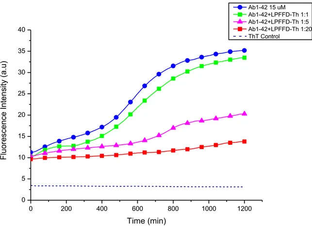
3.1 ThT assay
To compare the antiaggregating ability of the studied pentapepeptides, we carried out the rapid and specific assay based on the Th-T is fluorescence emission upon its binding to amyloid aggregates.
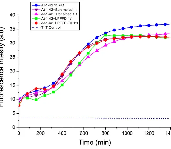
The results obtained by using the tested peptides in the presence of Aβ1-42 were compared to those of Aβ1-42 alone. Samples containing the peptides and Aβ at different molar ratios were co-incubated and co-lyophilized before dissolution in 200 µL of a 45 µM Th-T solution in 10 mM phosphate buffer at pH 7.4.
Interpretation of fluorescence data relies on the ability of Th-T to selectively detect amyloid structures, exhibiting a dramatic increase in fluorescent brightness. The more extended the aggregation is, the more intense is the emission of Th-T (λ= 480 nm); Th-T fluorescence originates only from the dye bound to amyloid substances because dye binding is linked to the presence of the cross-β structure. Th-T did not binds uniformly to the β-sheet surface, but preferentially interacts with channels formed by aromatic residues. (25)
We performed a Th-T assay of Aβ1-42 alone or in presence of the studied BSB peptides using different Aβ/BSB ratios. The Aβ1-42 alone as shown in figures 7, 8, 9 (blue curve) exhibits the classical sigmoid curve where after a lag-phase of approximately 300 minutes it starts to aggregate in fibrillar forms. It is clear from Figures 7, 8 and 9 that the Ac-LPFFD-Th shows the highest antifibrillogenic activity. This is clear already from figure 8, where at the Aβ/peptides ratio was 1:5, the LPFFD-Th is the most active. Looking at Figure 9, it can be also seen, apart a strong decrement in the whole fluorescence intensity with respect to Aβ, that the presence of this glycopeptide slows down the aggregation process through a lengthening of the lag phase.
In other words, the interaction between Aβ and the Ac-LPFFD-Th peptide not only greatly decreases the amyloidogenic process, but it seems that this peptide inhibits the early events of the aggregation process. In Figure 10 is shown the effect of the Ac-LPFFD-Th peptide at the three concentrations tested.
It is apparent a concentration dependent effect; in fact an almost flat curve is observed at the 1:20 molar ratio (300 µM), with no appearance of the exponential growth phase during the 21 hours of the kinetic assay.
Figure 7. Fibril formation kinetics of Aβ1-42 monitored by Th-T fluorescence. The concentration of Aβ1-42 was 15 μM in 10mM phosphate buffer solution pH 7.4 at 37 °C. The measurements in presence of all of the peptides/Trehalose were carried out at equimolar concentration of the peptides/Trehalose with respect toAβ1-42.
0 200 400 600 800 1000 1200 1400 0 5 10 15 20 25 30 35 40
Fluo
re
sc
en
ce In
te
sity (a.
u)
Time (min)
Ab1-42 15 uM Ab1-42+Scrambled 1:1 Ab1-42+Trehalose 1:1 Ab1-42+LPFFD 1:1 Ab1-42+LPFFD-Th 1:1 ThT ControlFigure 8. Fibril formation kinetics of Aβ1-42 monitored by Th-T fluorescence. The concentration of Aβ1-42 was 15 μM in 10mM phosphate buffer solution pH 7.4 at 37 °C. The measurements in presence of all of the peptides/Trehalose were carried out at 5 fold molar excess of the peptides/Trehalose with respect toAβ1-42.
0 200 400 600 800 1000 1200 1400 0 5 10 15 20 25 30 35 40 F luo re sce nce In te nsity ( a.u ) Time (min) Ab1-42 15uM Ab1-42+Trehalose 1:5 Ab1-42+LPFFD 1:5 Ab1-42+LPFFD-Th 1:5 Ab1-42+Scrambled 1:5 ThT Control
Figure 9. Fibril formation kinetics of Aβ1-42 monitored by Th-T fluorescence. The concentration of Aβ1-42 was 15 μM in 10mM phosphate buffer solution pH 7.4 at 37 °C. The measurements in presence of all of the peptides/Trehalose were carried out at 20 fold molar excess of the peptides/Trehalose with respect toAβ1-42.
0 200 400 600 800 1000 1200 0 5 10 15 20 25 30 35 40 Flu or esce nce In ten sity (a .u) Time (min) A 1-42 15 uM A 1-42+ Scrambled 1:20 A 1-42+ Trehalose 1:20 A 1-42+ Ac-LPFFD 1:20 A 1-42+ Ac-LPFFD-Th 1:20 ThT Control
Figure 10. Fibril formation kinetics of Aβ1-42 monitored by Th-T fluorescence. The concentration of Aβ1-42 was 15 μM in 10mM phosphate buffer solution pH 7.4 at 37 °C. The measurements in presence of the peptide Ac-LPFFD-Th were carried out at 1,5,20 fold molar excess of the peptide with respect toAβ1-42.
As expected the scrambled peptide has little effect on the Aβ aggregation process, in fact the Th-T curve is similar to that one observed for Aβ alone (Figure 7,8,9).
Notably, trehalose alone shows a slight effect in slowering the lag phase of Aβ aggregation resulting in a decreasing on the aggregates formation (Figure 7,8,9). We wanted also to investigate if the fibrillation inhibition effect observed on Aβ1-42 from Ac-LPFFD-Th was specific. For this reason we used another amyloidogenic peptide, IAPP (Islet Amyloid Polypeptide). This 37 aminoacids peptide is involved in type II diabetes mellitus where it is found as the mayor component in amyloid deposit in pancreatic β-cell of patients affected with T2DM.
0 200 400 600 800 1000 1200 0 5 10 15 20 25 30 35 40 F lu ore scen ce In te nsi ty (a .u ) Time (min) Ab1-42 15 uM Ab1-42+LPFFD-Th 1:1 Ab1-42+LPFFD-Th 1:5 Ab1-42+LPFFD-Th 1:20 ThT Control
hIAPP is an amyloidogenic peptide which shows about 25% sequence identity and 50% sequence similarity with Aβ peptide and morphology of amyloid fibrils for these reason we used hIAPP to perform a ThT assay in presence of our beta-sheet breaker peptide. (79,80)
From the ThT fluorescence data we can observe as how hIAPP 20 M shows a fast fibrillation process with a lag phase of a bit less than 4h in agreement with our previous work. (80)
hIAPP, in the presence of Ac-LPFFD-Th 20 molar fold in excess, does not inhibit the hIAPP fibril formation, but rather promotes hIAPP aggregations. (Figure 11)
Figure 11 Fibril formation kinetics of hIAPP monitored by Th-T fluorescence. The concentration of hIAPP was 20 μM in 10mM phosphate buffer solution pH 7.4 at 37 °C. The measurements in the presence of the peptide Ac-LPFFD-Th were carried out at 5,20 fold molar excess of the peptide with respect to hIAPP.