CASE REPORT
Endoscopic ultrasound-guided ethanol ablation of
pancreatic neuroendocrine tumours: A case study and
literature review
Elia Armellini, Stefano F Crinò, Marco Ballarè, Socrate Pallio, Pietro Occhipinti
Elia Armellini, Stefano F Crinò, Marco Ballarè, SocratePallio, Pietro Occhipinti, Gastroenterology Division, Azienda Ospedaliero Universitaria “Maggiore della Carità”, 28100 Novara, Italy
Author contributions: Armellini E performed the endoscopic procedures; Ballarè M, Pallio S and Occhipinti P collected and analyzed the data; Armellini E and Crinò SF wrote the paper and contributed equally to this manuscript.
Institutional review board statement: This case report was exempt from the Institutional Review Board standards at our Institution.
Informed consent statement: The patient involved in this study gave his written informed consent authorizing use and disclosure of his protected health information.
Conflict-of-interest statement: All the authors have no conflicts of interests to declare.
Open-Access: This article is an open-access article which was selected by an in-house editor and fully peer-reviewed by external reviewers. It is distributed in accordance with the Creative Commons Attribution Non Commercial (CC BY-NC 4.0) license, which permits others to distribute, remix, adapt, build upon this work non-commercially, and license their derivative works on different terms, provided the original work is properly cited and the use is non-commercial. See: http://creativecommons.org/ licenses/by-nc/4.0/
Correspondence to: Elia Armellini, MD, Gastroenterology Division, Azienda Ospedaliero Universitaria “Maggiore della Carità”, Corso Mazzini 18, 28100 Novara,
Italy. [email protected] Telephone: +39-32-13733206 Fax: +39-32-13733345 Received:May 8, 2015
Peer-review started: May 11, 2015 First decision: July 25, 2015 Revised: August 20, 2015 Accepted: October 12, 2015
Article in press: October 13, 2015 Published online: February 10, 2016
Abstract
Here we offer a review of the literature regarding endoscopic ultrasound-guided ethanol ablation for pancreatic neuroendocrine tumours and describe the case of a cystic tumour completely ablated after a multisession procedure. A total of 35 PubMed indexed cases of treated functioning and non-functioning pancreatic neuroendocrine tumours resulted from our search, 29 of which are well-documented and summarised. Endoscopic ultrasound-guided ethanol ablation appears as a local, minimally invasive treatment of pancreatic neuroendocrine tumours, suitable for selected patients. This technique appears feasible, relatively safe and efficient, especially when applied to symptom relief in functioning tumours, aiming at loss of endocrine secretion. For non-functioning tumours, where the goal is complete tissue ablation, eus guided ethanol ablation can provide good results for patients who are unfit for surgery or for those who refuse surgical resection. Its role in “fit for surgery” patients requires assessment through further studies.
Key words: Endoscopic ultrasound; Pancreatic neuro-endocrine tumour; Endoscopic ultrasound-guided injection; Ethanol; Tumour ablation
© The Author(s) 2016. Published by Baishideng Publishing
Group Inc. All rights reserved.
Core tip: We report a complete review of the literature about endoscopic ultrasound-guided ethanol ablation for pancreatic neuroendocrine tumours. The case of a cystic tumour completely ablated after a multisession procedure is described. On long term follow-up a durable DOI: 10.4253/wjge.v8.i3.192 © 2016 Baishideng Publishing Group Inc. All rights reserved.
remission of the tumour was obtained; a complete image gallery showing the pre and post-treatment appearance is available. The technical aspects, clinical success and complication rates related to this kind of procedures are described.
Armellini E, Crinò SF, Ballarè M, Pallio S, Occhipinti P. Endoscopic ultrasound-guided ethanol ablation of pancreatic neuroendocrine tumours: A case study and literature review.
World J Gastrointest Endosc2016; 8(3): 192-197 Available from: URL: http://www.wjgnet.com/1948-5190/full/v8/i3/192. htm DOI: http://dx.doi.org/10.4253/wjge.v8.i3.192
INTRODUCTION
In recent years the improvement of diagnostic and therapeutic technologies has led to less invasive treatments in any field of medicine with a shift from surgery to imaging guided treatments.
Endoscopic ultrasonography (EUS) has demonstrated excellent diagnostic accuracy for bilio-pancreatic district diseases and high safety and precision when applied for operative purposes. Along the years this peculiarity has made of EUS an optimal technique for imaging and cytological diagnosis, as well as for execution of more advanced procedures (i.e., drainages and local treatments).
The current management of T1 and T2 pancreatic neuroendocrine tumours (pNETs ) is somewhat similar to that of most pancreatic tumours (surgical resection), with a considerable economic burden and post-operative complications. However we are dealing with a pathology that offers a better prognosis and that is potentially responsive to local treatments[1,2].
Neuroendocrine tumours arise from cells present in the diffuse endocrine system and can be found throughout the body. They are most commonly located in the gastrointestinal tract and lung but are also found in the pancreas[3]. The 2010 World Health Organization
(WHO) classification divides the pNETs in three grades (G1, G2 and G3) on the basis of Ki-67 nuclear antigen expression (< 2%; 2%-20% and > 20%) and mitotic rate (< 2; 2-20 and > 20). Biopsy is most commonly used to assess the grade of the tumour. According to the TNM, the tumour is classified as T1a (< 1 cm), T1b (1-2 cm) and T2 (larger than 2 cm); T3 and T4 are locally advanced tumours (Table 1).
Tumour grading and tumour stage are the main prognostic factors of pNETs. Well and moderately differentiated have a significantly better survival com-pared to poorly differentiated neuroendocrine car-cinomas.
pNETs are also classified as functioning and non-functioning depending on the secretion of specific hor mones. Functioning tumours are commonly associated with a specific hormonal syndrome directly related to a hormone secreted by the neoplasm such as insulinomas
with ipoglicemia, gastrinomas with Zollinger–Ellison or carcinoid syndrome. Most non-functioning tumours occur in the head of the pancreas and produce mass effect symptoms. When small, they are usually incidentally discovered due to the incremental use of high-level diagnostic imaging.
EUS is the optimal diagnostic modality and can provide a biopsy specimen for histological confirmation and differentiation grade. The EUS image is usually of a solid, ipoechoic, round and smooth nodule, sometimes with a cystic central component (bull’s eye appearance). To date, the management of pancreatic sporadic, small (< 2 cm), asymptomatic, low-grade (G1) NETs suggests a “wait and see” strategy. Surgical resection of non-functioning pNETs is actually recommended for large (> 2 cm) or G2-G3 lesions[4]. For patients unfit for
surgery due to high-risk comorbidity or for those who refuse resection, the EUS-guided ethanol ablation has been reported in a few cases[5] as a local and minimally
invasive therapy.
CASE REPORT
A 58-year-old man with essential hypertension and recent onset of glucose intolerance was referred for a
Grade Ki-67 index (%) Mitotic count/10 HPF
G1 ≤ 2 < 2
G2 3-20 2-20
G3 > 20 > 20
TNM Size (cm) Muscularis propria
invasion
T1a < 1 _
T1b 1-2 _
T2 > 2 +
Table 1 World Health Organization classification of pan-creatic neuroendocrine tumors
Accordingly to the WHO classification 2010, the higher grade is assumed if the Ki-67 index and mitotic count differ; in the WHO 2010 TNM, the tumor is classified as T2 if it is larger than 2 cm in diameter or if it invades the muscularis propria. T3 and T4 tumors are locally aggressive tumors. WHO: World Health Organization; HPF: High-power field.
Figure 1 Abdominal magnetic resonance imaging demonstrating a round, well-demarcated nodule of the pancreatic tail. The 22 mm lesion (calipers)
transabdominal ultrasonography (US). Other laboratory test results including levels of carcinoembryonic anti-gen and carbohydrate antianti-gen were all within normal ranges. The US session diagnosed a focal lesion on the pancreatic tail. An abdominal magnetic resonance image showed a 22 mm nodule with peripheral hypervascularization (Figure 1), and EUS confirmed a “bull’s-eye” appearance nodule with peripheral hypervascular pattern via power Doppler and a central cystic component. The EUS-guided FNA of the lesion confirmed the diagnosis of pNET. The Ki67 proliferative index was > 5% to yield a G2 grade. However, because the patient adamantly refused surgical resection, we decided to ablate the lesion via EUS-guided ethanol injection.
After aspiration of the cystic component, a mean volume of 1.7 mL of 95% ethanol per session was injected into the tumour and re-aspirated using a 25-gauge needle (Echo-tip ultra, Cook, Limerick, Ireland) through a linear array echoendoscope (Figure 2). Three treatment sessions over six months were performed to ablate the nodule (Figure 3).
The hospitalization time was 2 d for each session. The patient experienced mild pancreatitis in 2 out of 3 sessions - that resolved with standard-of-care. No major or late complications were observed. After 24 mo, we achieved a durable and complete remission of
the tumour as shown by CT and EUS morphological imaging (Figure 4).
DISCUSSION
Most diagnosed pNETs are non-functioning tumours (90.8%); the remaining 9% are malignant functioning tumours such as gastrinomas (4.2%), insulinomas (2.5%), glucagonomas (1.6%), and VIPomas (0.9%). Although commonly perceived to be indolent tumours, they exhibit a broad range of growth rates, malignant potential, and overall prognosis. Most patients with pNETs (60%-70%) present with metastatic disease at diagnosis. Following surgical resection, the 5-year cumulative survival for pNETs other than insulinomas is roughly 65% with a 10-year survival of 45%[6]
.
Patients with incidental diagnosis of pNETs with a tumour size < 2 cm and low-grade (G1) dysplasia have a 5-year overall survival of 100% with a minimal risk of recurrence[6]. In this setting, a “wait and see” policy is
recommended.
On the contrary, surgical resection is the standard treatment for functioning and non-functioning G2-G3 pNETs. However, this is associated with a high risk of complications. Even when performed in high-volume centres, typical pancreatic resections (pancreati-coduodenectomy or distal pancreatectomy) have a mortality rate of about 5% with complications ranging from 40% to 50%[7]. This is particularly common in the
elderly or patients with comorbidities. Typical pancreatic resections are also associated with a high incidence of exocrine and endocrine insufficiency.
In an attempt to reduce complications and pan-creatic impairment, new parenchyma-sparing resection techniques such as enucleation and middle pancreatec-tomy (resection of the central part of the gland) have been applied to small tumours[8]. Although pancreatic
head tumour enucleation resulted in decreased operative time and length of hospitalization, the 5-year survival and overall morbidity and mortality were comparable to standard surgical resection even for small pNETs[9]. To
date, no alternative treatment has been standardized for patients unfit for surgery or for those who refuse
A
B
Figure 2 Endoscopic ultrasound appearances before (A) and after (B) treatment (white arrow).
Figure 3 Computed tomography scan showing thin residual hypervas-cular tissue (white arrow) two months after the first treatment.
function of tumour size. For small (≤ 20 mm) tumours, Qin et al[19] suggested that the volume be calculated
as follows. For round tumours, the volume of ethanol corresponds to half the tumour size; for oval or irregular tumours, the volume of ethanol is (major axis + minor axis of the tumour)/2. A 1.0 mL syringe should be used for precise injection.
In terms of therapeutic outcomes, differentiation of functioning and non-functioning tumours seems to be very important. For small functioning symptomatic G1 tumours, the aim of the ablation is the symptom relief. For non-functioning tumours, the treatment goal is complete ablation of the lesion as confirmed by imaging.
Including the case here described, this technique achieved clinical success (complete symptom resolution) in 100% of 19 functioning tumours with a mean follow-up of 13.6 mo (range 2-38). Ethanol ablation is less effective for non-functioning tumours with a reported success (complete radiological ablation) of 70% (7/10 tumours were ablated, one lost to follow-up) with a mean follow-up of 13.4 mo (range 3-24) (Table 2). The reason is unclear but it might be due to a “debulking” effect in functioning ones, resulting in loss of endocrine resection.
In the recent decades, EUS has evolved into a useful therapeutic tool for treating a broad range of tumours. EUS-guided injection has been applied both as a pancreatic cancer treatment aimed at controlling pain through nerve blockade as well as a solid tumour therapy for the introduction of brachytherapy seeds and viral vectors or as a tool for ablation therapy[10,11].
The pNET EUS-guided ethanol ablation is a new, less invasive therapeutic option although it remains rare.
A PubMed literature review showed 26 patients affected by small pNETs (maximum diameter of 21 mm) who underwent EUS-guided ethanol ablation[12-21]
including 19 functioning and 10 non-functioning tumours (Table 2). The number of patients treated by this tecnique progressively increased from 2006 to 2015 (Figure 5).
Conscious sedation is generally reported during the procedure. A mean hospitalization time of 2 d/session is usually necessary even in the absence of complications.
Technical success is reported in 100% of cases; a 22 or 25 gauge needle was generally used to inject a small volume of ethanol with a range between 0.2 and 8 mL per session. The choice of ethanol volume is a
A
B
Figure 4 Twenty-four months follow-up. A: Computed tomography scan showing absence of hypervascular tissue around a small hypodense area (white arrow); B:
Endoscopic ultrasound scan of the pancreatic tail demonstrating poorly defined hyperechoic tissue (fibrosis) with posterior shadow (caliper).
2006 Jürgensen[12] 2008 Muscatiello - Deprez[13,14] 2011 Vleggaar[15] Levy -2012 Schnack[16,17] 2014
Bor - Qin-Paik[18-20]Park[21-described case]2015
14 12 10 8 6 4 2 0
Figure 5 Reported endoscopic ultrasound-guided ethanol ablation procedures over time. Literature review showed a progressive increase of performed
ablate based both on the grading and the age of the patient. Moreover it is worth noting that FNA cytology may underestimate the staging based on surgical specimens. Physicians should be very cautious in using FNA specimens to classify a tumour as low-grade[22].
Consequently our treatment aimed at the complete ablation of the lesion while sparing the pancreatic parenchyma. The nodule we treated had a cystic central component, which has not yet been described in the literature for pNET EUS-guidance ablation. A tecnique similar to that described for cystic neoplasm ablation (ethanol injection and reaspiration) was used.
In conclusion, based on our case study and literature review, we find that this technique is feasible, relatively safe and efficient when applied to symptom relief in functioning tumours. However, the long-term outcomes remain unknown. For non-functioning tumours, it can provide good results for patients unfit for surgery or for those who refuse surgical resection. Its role in “fit for surgery” patients is still undefined and larger comparative studies with long-term follow-up are needed to assess its role.
COMMENTS
Case characteristics
The authors describe a procedure of eus guided ethanol ablation along three sessions for a cystic pancreatic neuroendocrine tumours (pNET).
Clinical diagnosis
Incidental focal lesion of the pancreatic tail with endoscopic ultrasound (EUS) “bull’s eye appearance” and peripheral hypervascularization, suspicious for neuroendocrine tumour.
Differential diagnosis
Other focal lesions of the pancreas.Laboratory diagnosis
No lab abnormality including levels of carcinoembryonic antigen and carbohy drate antigen, but recent onset of glucose intolerance.
Imaging diagnosis
Abdominal ultrasound, endoscopic ultrasound, magnetic resonance, EUS guided FNA.
Pathological diagnosis
Neuroendocrine tumor, G2, Ki67 proliferative index > 5%.
Treatment
The authors treated the patient by EUSguided ethanol injection along three sessions.
Related reports
For patients unfit for surgery due to high-risk comorbidity or for those who refuse resection EUSguided ethanol ablation has been reported in a few cases.
Term explanation
pNETS: Pancreatic neuroendocrine tumours; EUS: Endoscopic ultrasound.
Experiences and lessons
The authors find that EUS guided ethanol ablation is relatively safe and efficient secretion, although with persistent viable tissue, or to a
more aggressive histological grading of non-functioning tumours. Unfortunately, lesion grading was not available in most of the reviewed cases.
Few early complications (within one week) are reported: 7 mild pancreatitis cases were observed (16.2%) out of 43 procedures. One (2.3%) major early complication was described[13]: A pancreatic necrotic
lesion that was likely caused by ethanol effusion. It was managed by laparoscopic necrosectomy.
Two (4.6%) late complications occurred: One hematoma and ulceration of the duodenal wall[14] and
main pancreatic duct stricture[21]. These were managed
by endoscopic retrograde cholangiopancreatography and stent placement (Table 3).
In our case, we achieved a diagnosis of a non-functioning pNET with moderate dysplasia, grade (G2), established on the basis of biopsy (Ki67 > 5%) in a 58-year-old male who refused surgery. We decided to
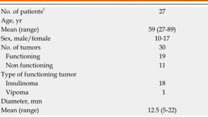
No. of patients1 27 Age, yr Mean (range) 59 (27-89) Sex, male/female 10-17 No. of tumors 30 Functioning 19 Non functioning 11
Type of functioning tumor
Insulinoma 18
Vipoma 1
Diameter, mm
Mean (range) 12.5 (5-22)
Table 2 Patient demographic information and baseline characteristics of the tumours
No. of treatment session per tumor
Mean (range) 1.43 (1-3) Alcohol volume, mL Mean (range) 1.83 (0.18-8) Technical success, n (%) 30/30 (100) Clinical success1, n (%) Functioning 19/19 (100) Non functioning2 7/10 (70) Adverse events3, n (%) 11 (25.5)
Early (within one week), n (%) 9 (21)
Pancreatic necrotic lesion 1 (2.3)
Mild pancreatitis 7 (16.2)
Abdominal pain 1 (2.3)
Late, n (%) 2 (4.6)
Hematoma and ulceration of the duodenal wall
1 (2.3)
Main pancreatic duct stricture 1 (2.3)
Follow-up, mo
Mean (range) 13.4 (2-38)
Table 3 Procedural outcomes
1Clinical success: Symptom resolution for functioning tumours and
radiological ablation for non-functioning tumour; 2One non functioning
tumor was lost to follow-up; 3Adverse events percentage is intended in
relation to procedure number.
COMMENTS
22474439 DOI: 10.1155/2012/503098]
11 Jin Z, Du Y, Li Z, Jiang Y, Chen J, Liu Y. Endoscopic ultrasono-graphy-guided interstitial implantation of iodine 125-seeds combined with chemotherapy in the treatment of unresectable pancreatic carcinoma: a prospective pilot study. Endoscopy 2008; 40: 314-320 [PMID: 18283622 DOI: 10.1055/s-2007-995476]
12 Jürgensen C, Schuppan D, Neser F, Ernstberger J, Junghans U, Stölzel U. EUS-guided alcohol ablation of an insulinoma. Gastrointest Endosc 2006; 63: 1059-1062 [PMID: 16733126] 13 Muscatiello N, Salcuni A, Macarini L, Cignarelli M, Prencipe S,
di Maso M, Castriota M, D’Agnessa V, Ierardi E. Treatment of a pancreatic endocrine tumor by ethanol injection guided by endo-scopic ultrasound. Endoscopy 2008; 40 Suppl 2: E258-E259 [PMID: 19090457 DOI: 10.1055/s-2007-966962]
14 Deprez PH, Claessens A, Borbath I, Gigot JF, Maiter D. Successful endoscopic ultrasound-guided ethanol ablation of a sporadic insulinoma. Acta Gastroenterol Belg 2008; 71: 333-337 [PMID: 19198582]
15 Vleggaar FP, Bij de Vaate EA, Valk GD, Leguit RJ, Siersema PD. Endoscopic ultrasound-guided ethanol ablation of a symptomatic sporadic insulinoma. Endoscopy 2011; 43 Suppl 2 UCTN: E328-E329 [PMID: 22020710 DOI: 10.1055/s-0030-1256775] 16 Levy MJ, Thompson GB, Topazian MD, Callstrom MR, Grant
CS, Vella A. US-guided ethanol ablation of insulinomas: a new treatment option. Gastrointest Endosc 2012; 75: 200-206 [PMID: 22078104 DOI: 10.1016/j.gie.2011.09.019]
17 Schnack C, Hansen CØ, Beck-Nielsen H, Mortensen PM. Treatment of insulinomas with alcoholic ablation. Ugeskr Laeger 2012; 174: 501-502 [PMID: 22348674]
18 Bor R, Farkas K, Bálint A, Molnár T, Nagy F, Valkusz Z, Sepp K, Tiszlavicz L, Hamar S, Szepes Z. [Endoscopic ultrasound-guided ethanol ablation: an alternative option for the treatment of pancreatic insulinoma]. Orv Hetil 2014; 155: 1647-1651 [PMID: 25282110 DOI: 10.1556/OH.2014.30012]
19 Qin SY, Lu XP, Jiang HX. EUS-guided ethanol ablation of insuli-nomas: case series and literature review. Medicine (Baltimore) 2014;
93: e85 [PMID: 25255024 DOI: 10.1097/MD.0000000000000085]
20 Paik WH, Seo DW, Dhir VK, Wang HPO. Mo1373 EUS-Guided Ethanol Ablation of Small Solid Pancreatic Neoplasm. Gastroin test Endosc 2014; 79 Suppl 5: Page AB413 [DOI: 10.1016/ j.gie.2014.02.550]
21 Park do H, Choi JH, Oh D, Lee SS, Seo DW, Lee SK, Kim MH. Endoscopic ultrasonography-guided ethanol ablation for small pancreatic neuroendocrine tumors: results of a pilot study. Clin Endosc 2015; 48: 158-164 [PMID: 25844345 DOI: 10.5946/ce.2015.48.2.158] 22 Vinayek R, Capurso G, Larghi A. Grading of EUS-FNA cytologic
specimens from patients with pancreatic neuroendocrine neoplasms: it is time move to tissue core biopsy? Gland Surg 2014; 3: 222-225 [PMID: 25493252 DOI: 10.3978/j.issn.2227-684X.2014.07.03]
P- Reviewer: Ghosn M, Sadik R S- Editor: Ji FF L- Editor: A E- Editor: Wu HL
for the treatment of pNETs in patients unfit for surgery or for those who refuse surgical resection. Its role in “fit for surgery” patients is still undefined.
Peer-review
A well written paper having a clear endpoint and objectives. The review of the literature is complete and presented in an attractive way.
REFERENCES
1 Clift AK, Frilling A. Management of patients with hepatic
metastases from neuroendocrine tumors. Ann Saudi Med 2014; 34: 279-290 [PMID: 25811199 DOI: 10.5144/0256-4947.2014.279] 2 de Baere T, Deschamps F, Tselikas L, Ducreux M, Planchard D,
Pearson E, Berdelou A, Leboulleux S, Elias D, Baudin E. GEP-NETS update: Interventional radiology: role in the treatment of liver metastases from GEP-NETs. Eur J Endocrinol 2015; 172: R151-R166 [PMID: 25385817 DOI: 10.1530/EJE-14-0630] 3 Metz DC, Jensen RT. Gastrointestinal neuroendocrine tumors:
pancreatic endocrine tumors. Gastroenterology 2008; 135: 1469-1492 [PMID: 18703061 DOI: 10.1053/j.gastro.2008.05.047] 4 Partelli S, Maurizi A, Tamburrino D, Baldoni A, Polenta V, Crippa
S, Falconi M. GEP-NETS update: a review on surgery of gastro-entero-pancreatic neuroendocrine tumors. Eur J Endocrinol 2014;
171: R153-R162 [PMID: 24920289 DOI: 10.1530/EJE-14-0173]
5 Zhang WY, Li ZS, Jin ZD. Endoscopic ultrasound-guided ethanol
ablation therapy for tumors. World J Gastroenterol 2013; 19: 3397-3403 [PMID: 23801831 DOI: 10.3748/wjg.v19.i22.3397] 6 de Wilde RF, Edil BH, Hruban RH, Maitra A. Well-differentiated
pancreatic neuroendocrine tumors: from genetics to therapy. Nat Rev Gastroenterol Hepatol 2012; 9: 199-208 [PMID: 22310917 DOI: 10.1038/nrgastro.2012.9]
7 Bettini R, Partelli S, Boninsegna L, Capelli P, Crippa S, Pederzoli
P, Scarpa A, Falconi M. Tumor size correlates with malignancy in nonfunctioning pancreatic endocrine tumor. Surgery 2011; 150: 75-82 [PMID: 21683859 DOI: 10.1016/j.surg.2011.02.022] 8 Falconi M, Zerbi A, Crippa S, Balzano G, Boninsegna L, Capitanio
V, Bassi C, Di Carlo V, Pederzoli P. Parenchyma-preserving resections for small nonfunctioning pancreatic endocrine tumors. Ann Surg Oncol 2010; 17: 1621-1627 [PMID: 20162460 DOI: 10.1245/ s10434-010-0949-8]
9 Pitt SC, Pitt HA, Baker MS, Christians K, Touzios JG, Kiely JM,
Weber SM, Wilson SD, Howard TJ, Talamonti MS, Rikkers LF. Small pancreatic and periampullary neuroendocrine tumors: resect or enucleate? J Gastrointest Surg 2009; 13: 1692-1698 [PMID: 19548038 DOI: 10.1007/s11605-009-0946-z]
10 Wiechowska-Kozłowska A, Boer K, Wójcicki M, Milkiewicz P.
The efficacy and safety of endoscopic ultrasound-guided celiac plexus neurolysis for treatment of pain in patients with pancreatic cancer. Gastroenterol Res Pract 2012; 2012: 503098 [PMID: