Original Paper
This is an Open Access article licensed under the terms of the Creative Commons Attribution-NonCommercial 3.0 Unported license (CC BY-NC) (www.karger.com/OA-license), applicable to the online version of the article only. Distribution permitted for non-commercial purposes only.
Copyright © 2015 S. Karger AG, Basel
Department of Biological and Environmental Sciences, University of Messina, (Italy) Tel. +39 090 6765214, Fax +39 090 394030, E-Mail [email protected]
Angela Marino,
Curcumin Protects –SH Groups and
Sulphate Transport after Oxidative Damage
in Human Erythrocytes
Rossana Morabitoa Giuseppe Fallitib Antonella Geracic Giuseppa La Spadad
Angela Marinod
aDepartment of Human and Social Sciences, University of Messina, Italy; bClinical Pathology, A.O.O.R.
“Papardo-Piemonte”, Messina, Italy; cBromatech, Giarre (CT), Italy; dDepartment of Biological and
Environmental Sciences, University of Messina, Messina, Italy
Key Words
Erythrocytes • Band 3 protein • Oxidative stress • pH • NEM • Curcumin Abstract
Background/Aims: Erythrocytes, continuously exposed to oxygen pressure and toxic
compounds, are sensitive to oxidative stress, namely acting on integral Band 3 protein, with consequences on cell membranes deformability and anion transport efficiency. The aim of the present investigation, conducted on human erythrocytes, is to verify whether curcumin (1 or 10µM), a natural compound with proved antioxidant properties, may counteract Band 3-mediated anion transport alterations due to oxidative stress. Methods: Oxidative conditions were induced by exposure to, alternatively, either 2 mM N-ethylmaleimide (NEM) or pH-modified solutions (6.5 and 8.5). Rate constant for SO4= uptake and -SH groups estimation were
measured to verify the effect of oxidative stress on anion transport efficiency and erythrocyte membranes. Results: After the exposure of erythrocytes to, alternatively, NEM or pH-modified solutions, a significant decrease in both rate constant for SO4= uptake and -SH groups was
observed, which was prevented by curcumin, with a dose–dependent effect. Conclusions: Our results show that: i) the decreased efficiency of anion transport may be due to changes in Band 3 protein structure caused by cysteine -SH groups oxidation, especially after exposure to NEM and pH 6.5; ii) 10 µM Curcumin is effective in protecting erythrocytes from oxidative stress events at level of cell membrane transport.
Introduction
Oxidative stress is an imbalance between free radicals production and efficiency of cell antioxidant defense system [1], provoking damages at level of DNA and cell membrane lipids
[2, 3]. Reactive Oxygen Species (ROS) are produced as a consequence of electron transfer in oxidative phosphorylation during aerobic metabolism [4] and their activity is balanced by enzymatic and non-enzymatic antioxidants, acting through different mechanisms, such as free radicals scavenging or chelating ion metals [5].
Since erythrocytes are susceptible to free radicals-induced oxidative damages, they may be considered as a good model to verify the effects of oxidative conditions on living cells, also providing a useful basis for in vitro investigations. One of the most studied physiological features of erythrocytes is Band 3 protein-mediated anion transport [6], including the rapid Cl-/HCO
3- exchange, essential to blood for eliminating CO2 across erythrocyte membranes [7].
Band 3 protein consists of a membrane domain, specifically mediating anion exchange, and a cytoplasmic domain, which mainly contributes to the protein–protein interactions, by coupling the lipid bilayer to the underlying cytoskeleton, through cysteine –SH groups [8]. Since the cytoplasmic domain has been shown to reversibly interact with haemoglobin [9, 10], revealing a link between Band 3 protein and intracellular components, it is reasonable to suggest that alterations at level of Band 3 protein may affect, not only anion exchange and ion balance across cell membrane, but also efficiency of O2 transport and cell membrane deformability [11]. In this latter regard, the intracellular magnesium has been proved to contribute to the visco-elastic properties of Band 3 protein [12], and, in turn, to erythrocytes membrane deformability.
On this basis and taking into account that Band 3 protein has been considered as a tool to monitor the effect of metabolites, drugs, toxins or pathological states like hypertension and diabetes on erythrocytes [13-15], the aim of the present investigation is to verify the effect of different pH values of the external medium or, alternatively, the effect of N-ethylmaleimide (NEM), known as an oxidant compound [16], on both anion transport and –SH groups of Band 3 protein in human erythrocytes. For this purpose, the rate constant for SO4= has been
used as an index of anion transport efficiency through Band 3 protein, since its measurement is a more controllable method to investigate anion transport [17]. Quantification of Band 3 protein –SH groups has been used as an index of oxidative damage [15]. Moreover, the effect elicited by both experimental media at different pH values and NEM has been verified in presence of curcumin (1,7-bis(4-hydroxy-3-methoxyphenyl)-1E,6E-heptadiene-3,5-dione or diferuloyl methane)), whose biological activity, including antioxidant properties, has been already demonstrated [18, 19].
Curcumin, also known as Indian saffron, turmeric yellow or curry powder, a yellow hydrophobic pigment deriving from the rhizome (turmeric) of Curcuma longa herb, has been shown to provide several beneficial effects [20] and has been long used in Indian Ayurvedic medicine and as food-coloring agent [21].
Materials and Methods
Erythrocytes preparation
Human blood was obtained by venipuncture from healthy volunteers after informed consent, collected in EDTA tubes, washed in a medium containing 10 mM HEPES, 140 mM NaCl, 5 mM KCl, pH 7.4, 300±5mOsm (henceforth referred to as pH 7.4 isotonic buffer) and centrifuged three times for 5 min at 2000 g (ALC centrifuge). The buffy coat was carefully removed at each step and erythrocytes suspended at different hematocrit (0.05%, 3%, 10%), according to the following experimental protocols: hemolytic assay, SO4=
transport measurement and determination of -SHgroups.
Hemolytic assay
To exclude that experimental solutions, i.e. solutions at different pH values or, alternatively, 2 mM NEM or curcumin, may provoke lysis of human erythrocytes, hemolytic assay was performed [22] before SO4= rate constant measurement and –SH groups determination. Briefly, human erythrocytes, after washing,
were suspended to 0.05% hematocrit in, alternatively, pH – modified isotonic buffer (10 mM HEPES, 140 mM NaCl, 5 mM KCl, pH 6.5 or 8.5), or pH 7.4 isotonic buffer plus 2mM NEM or isotonic buffer (10 mM HEPES, 140 mM NaCl, 5 mM KCl, pH 7.4) plus curcumin at different concentrations (1 or 10 µM). In any case
erythrocytes were incubated for 1 h at 25°C and then centrifuged (5 min, 2000 g). The supernatant deriving from each experimental condition (A) was spectrophotometrically read at 414 nm (Beckman DU 640). The optical absorbance of (A) was then compared to the optical absorbance observed after maximal lysis (B) induced by addition of distilled water. The hemolysis percentage was calculated using the formula: A/B x 100.
SO4= transport measurement
According to the turbidimetric method [17], human erythrocytes were re-suspended to 3% hematocrit in 50 ml isotonic medium (henceforth referred to as SO4= medium), having the following composition in mM:
118 Na2SO4, 10 HEPES, 15 glucose (pH 7.4; 300±5mOsm).
Control conditions
To measure the rate constant for SO4= transport in control conditions, at specified time intervals
(respectively 5-15-30-45-60-90 and 120 min), 5 ml erythrocytes suspension were removed, added to a test tube containing 5µM 4,4’-diisothiocyanato-stilbene-2,2’-disulfonate (DIDS), used as stopping medium, and kept under ice. After the last sample withdrawal, erythrocytes were washed three times in a pH 7.4 isotonic buffer at 0 °C to remove the outside SO4= and then hemolysed by distilled water and perchloric
acid (4% v/v final concentration in 1 ml/sample). The membranes were discarded by centrifugation (4000 g, 10 min, 4 °C) and SO4= ions contained in the supernatant were precipitated as follows: 1 ml glycerol plus
distilled water solution (1:1), 0.5 ml 4 M NaCl in HCl (hydrochloric acid 37%) solution (12:1) and 1 ml 1.24 M BaCl2.2H
2O solution were sequentially added to 1 ml supernatant containing SO4=. Within a few minutes,
SO4= levels were spectrophotometrically read at 425 nm wavelength. Using a calibrated standard curve
obtained by precipitating known SO4= concentrations, the absorption was converted to mM of intracellular
SO4=, necessary to calculate the rate constant in min-1, derived from the following equation: C
t = C∞ (1-e -rt) + C
0, where Ct, C∞ and C0 represent the intracellular SO4= concentrations measured at time t, 0 and ∞
respectively; e indicates Nepero number (2.7182818) and r is a constant.
Experimental protocols
After the assessment of control conditions, the rate constant for SO4= transport was measured in
different experimental conditions, as described in the following protocols:
Curcumin treatment
To test the possible effect of curcumin on SO4= transport, erythrocytes at 3 % hematocrit were
centrifuged and re-suspended in 50 ml pH 7.4 isotonic buffer plus curcumin at different concentrations (1 or 10 µM, alternatively). Samples were incubated at 25 °C for 1 h, centrifuged (4000g, 5 min) and finally re-suspended in 50 ml SO4= medium plus curcumin (1 or 10µM, alternatively) at 25 °C. At fixed time intervals
(respectively 5-15-30-45-60-90 and 120 min), 5 ml samples were withdrawn and handled as reported in
SO4= transport measurement, to finally measure SO
4= uptake kinetics. Exposure to medium at different pH values or to NEM
For this protocol, the effect of solutions at different pH values or, alternatively, the effect of 2 mM NEM was tested.
pH: erythrocytes at 3% hematocrit were centrifuged, re-suspended in pH-modified isotonic buffer
incubated at 25 °C for 30 min , centrifuged (4000g, 5 min) and then re-suspended in 50 ml SO4= medium at
different pH values (6.5 or 8.5, alternatively). At fixed time intervals (respectively 5-15-30-45-60-90 and 120 min), samples (5 ml each) were withdrawn and handled as reported in SO4= transport measurement, to finally measure SO4= uptake kinetics.
NEM: erythrocytes at 3 % hematocrit were centrifuged, re-suspended in 50 ml pH 7.4 isotonic buffer
plus 2 mM NEM, then incubated at 25°C for 30 min, centrifuged (4000g, 5 min) and re-suspended in 50 ml SO4= medium plus 2 mM NEM. At fixed time intervals (respectively 5-15-30-45-60-90 and 120 min), samples
(5 ml each) were withdrawn and handled as reported in SO4= transport measurement, to finally measure SO4=
uptake kinetics.
Curcumin treatment of erythrocytes exposed to different pH values or to NEM
pH and curcumin: erythrocytes at 3 % hematocrit were centrifuged and re-suspended in 50 ml pH
°C for 1h, centrifuged (4000g, 5 min) and then re-suspended in 50 ml SO4 medium plus curcumin (1 or alternatively 10 µM) at different pH values (6.5 or 8.5 alternatively). Samples (5 ml each) were withdrawn at fixed time intervals (respectively 5-15-30-45-60-90 and 120 min) and handled as reported in SO4= transport
measurement, to finally measure SO4= transport kinetics.
NEM and curcumin: erythrocytes at 3 % hematocrit were centrifuged, re-suspended in 50 ml pH 7.4
isotonic buffer plus curcumin (1 or alternatively 10 µM) and incubated at 25 °C for 30 min. NEM (2 mM) was then added and samples were further incubated at 25 °C for 30 min. Erythrocytes were then centrifuged (4000g, 5 min), re-suspended in 50 ml SO4= medium plus curcumin (1 or, alternatively, 10 µM) and 2 mM
NEM, withdrawn at fixed time intervals (respectively 5-15-30-45-60-90 and 120 min) and handled as reported in SO4= transport measurement, to finally measure SO4= transport kinetics.
-SH groups determination
Erythrocytes, after exposure to external medium at different pH values (6.5, 7.4, 8.5) or to 2 mM NEM, with or without curcumin (1 or 10 µM), were addressed to –SH groups determination technique [15, 23], after Band 3 protein isolation. For this purpose, erythrocytes were re-suspended to 10% hematocrit in 20 ml pH 7.4 isotonic buffer and then hemolyzed by re-suspension in 20 ml of cold hypotonic buffer (5mM sodium phosphate, pH 7.4). Membranes were obtained by centrifugations at 20,000 g for 30 min at 0°C (Beckman J2-21 refrigerated centrifuge). The process was repeated with the same hypotonic buffer to discard haemoglobin. The obtained pellet (erythrocyte membranes) was then incubated with 0.1 M NaOH (1:9) for 30 min at 0°C, in presence of 0.2 mM dithiothreitol (DTT) and 20 µg/ml Phenylmethylsulfonil fluoride (PMSF). After incubation, samples were centrifuged at 56,000 g for 30 min at 0°C. The pellet (0.2 ml) containing Band 3 protein was washed thrice with 5 mM sodium phosphate, pH 8.0 and solubilized by incubation in 0.3 ml of 20% sodium dodecyl sulphate (SDS) reagent and 3 ml of 100 mM sodium phosphate (pH 8.0) for 30 min at 37 °C. The solubilized pellets were further incubated with 0.1 ml of 10 mM 5,5'-dithiobis-(2-nitrobenzoic acid) (DTNB) in 100 mM sodium phosphate (pH 8.0) for 20 min at 37 °C. DTNB reacts specifically with thiol groups, producing a highly coloured yellow anion. Levels of -SH groups in the suspension were spectrophotometrically read at 412 nm, using the molar extinction coefficient at 13,600 [23], and % decrease of –SH groups respect to untreated erythrocytes was considered.
Reagents
All chemicals were purchased from Sigma (Milan, Italy). Curcumin (Curcumin (1,7-bis(4-hydroxy-3-methoxyphenyl)-1E,6E-heptadiene-3,5-dione, MW=368.4, “pro analysis” purity grade, Sigma, code C7727 (Milan, Italy) was kindly provided by Prof. S. Cuzzocrea (University of Messina). For stock solutions: curcumin was dissolved in DMSO 0.5%; NEM and DIDS were dissolved in distilled water.
Experimental data and statistics
Data are shown as mean values ± standard error of the means (S.E.M.). Each data set is derived from at least five individual experiments. Significance of the differences was tested using a Student's
t test. For multiple comparisons, one-way analysis of variance (ANOVA), followed by Dunnett's post hoc
test or Bonferroni’s post hoc test were used to determine significant differences. p<0.001 was considered statistically significant.
Results
Hemolytic assay
Erythrocytes treated with different concentrations of curcumin (1 or 10 µM) did exhibit neither morphological alterations (checked under light microscope, 400x magnification), nor hemolytic response after 1 h incubation, as assessed by spectrophotometrical measure (data not shown). The same result was observed when erythrocytes were exposed to alternatively, isotonic buffer at different pH values (6.5 and 8.5) or to 2 mM NEM. Since the majority of in vitro studies have been performed with curcumin dissolved in DMSO [24], due to the solubility properties of this powder, our protocols required to treat erythrocytes, in a separate set of experiments, with DMSO (up to 0.005% v/v), which did not elicit any detrimental effect (data not show).
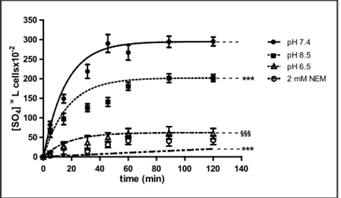
The kinetics of SO4= uptake in
erythrocytes exposed to medium at different pH values (pH 7.4, 6.5, 8.5) or, alternatively to 2 mM NEM, are shown in Fig. 1, while the rate constant for SO4= uptake are reported in Table 1.
SO4= transport in control erythrocytes
(pH 7.4) increased steeply at the initial stage and reached equilibrium in 30 min (Fig. 1), exhibiting a rate constant of 0.063±0.001. Erythrocytes incubated in SO4= medium did not exhibit any
morphological change, as shown in Fig. 2.
SO4= transport in, alternatively, pH
6.5- or 2 mM NEM-treated erythrocytes was significantly inhibited with respect to the control conditions (pH 7.4) (Fig. 1, p<0.001), as well as the rate constant for SO4= uptake (0.035±0.001 in pH 6.5
medium and 0.001±0.003 in presence of NEM) (Tab. 1, p<0.001).
Erythrocytes exposed to pH 8.5 medium exhibited SO4= transport
kinetics (Fig 1, p<0.001) as well as a rate constant for SO4= uptake (0.058±0.005,
Table 1, p<0.001) significantly reduced with respect to untreated cells, albeit significantly higher than what observed in pH 6.5- and NEM-treated erythrocytes (Fig. 1, p<0.001; Table 1
0 20 40 60 80 100 120 140 0 50 100 150 200 250 300 350 pH 7.4 pH 8.5 -pH 6.5 -2 mM NEM - - - -*** §§§ *** time (min) [SO 4 ] =L ce lls x10 -2
Fig. 1. Time course of SO4= uptake in human
erythrocytes measured in a medium at
dif-ferent pH values (pH 7.4, 6.5, 8.5) or treated with 2 mM NEM. Bars represent the mean
± SEM from at least 8 experiments, where
***p<0.001 significantly different versus con-trol (pH 7.4 medium), §§§ p<0.001 significantly
different versus control (pH 7.4 medium) and versus pH 8.5, as determined by one way ANOVA followed by Dunnett's post hoc test, by comparing all values, at all time points.
Fig. 2. Light microscope picture of human erythrocytes
in-cubated in SO4= medium (400x magnification).
Table 1. Rate constant (min-1) of SO
4= uptake in human
erythrocytes measured in a medium at different pH values (7.4; 6.5; 8.5) or treated with 2 mM NEM. Data are pre-sented as means ± SEM of at least 8 experiments, where ***p<0.001 significantly different versus control (pH 7.4 medium), ), §§§ p<0.001 significantly different versus con-trol (pH 7.4 medium) and versus pH 8.5, as determined by one way ANOVA followed by Dunnett's post hoc test
p<0.001).
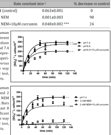
In order to verify the effect of curcumin on SO4= uptake, erythrocytes were exposed to
pH 6.5 medium plus 1 µM curcumin. At this concentration, kinetics of SO4= uptake and the
corresponding rate constant were unchanged with respect to pH 6.5 treatment (data not shown). Curcumin (1 µM) was also ineffective in restoring rate constant for SO4= uptake in
both pH 8.5- and NEM-treated cells. So, higher curcumin concentrations were used.
Fig. 3 shows that 10 µM curcumin dissolved in pH 6.5 medium significantly increased SO4= uptake, with respect to pH 6.5 conditions (p<0.001) and the corresponding rate constant
for SO4= uptake was significantly higher than that observed in pH 6.5 conditions (Table 2,
Upon light microscope observations, notable morphological alterations were observed after incubation of erythrocytes in pH 6.5 medium, for 5 and 90 min (Fig. 4A-4B, see arrows). These changes to the cell shape were abolished by 10 µM curcumin, at both 5 and 90 min of treatment (Fig. 4a, 4b).
In a separate protocol, erythrocytes exposed to pH 8.5 medium plus 10 µM curcumin exhibited a SO4= transport efficiency significantly higher with respect to pH 8.5-treated
erythrocytes (Fig. 5, p<0.001) and a rate constant for SO4= uptake comparable to that one
observed in control conditions (pH 7.4) (Table 3, p<0.001).
0 20 40 60 80 100 120 140 0 50 100 150 200 250 300 350 pH 7.4 pH 6.5 -pH 6.5+10 µM curcumin -*** time (min) [SO 4 ] =L ce lls x10 -2
Fig. 3. Time course of SO4 uptake in
hu-man erythrocytes exposed to medium
at different pH values (7.4 and 6.5) and
to pH 6.5 medium plus 10µM curcumin.
Data in pH 6.5 and 7.4 as reported in Fig. 1 and Table 1. Bars represent the mean ±
SEM from at least 8 experiments, where
***p<0.001 significantly different versus pH 6.5 medium, as determined by one way ANOVA followed by Dunnett's post
hoc test, by comparing all values, at all
time points.
Table 2. Rate constant (min-1) of SO
4= uptake in human erythrocytes in a medium
at different pH values (7.4, 6.5) and pH 6.5 plus 10µM curcumin. Data are pre-sented as means ± SEM of at least 8 experiments where ***p<0.001 significant
versus pH 6.5 medium, as determined by one way ANOVA followed by Dunnett's post hoc test
A
B
a
b
Fig. 4. Light microscope
observations of human erythrocytes exposed to pH 6.5 medium for 5 min (A) and 90 min (B), and exposed to pH 6.5 medium plus 10 µM curcumin for 5 min (a) and 90 min (b). 400x magnification. Ar-rows indicate erythrocytes morphological alterations. Treatment with curcumin impairs cell shape changes due to pH 6.5 exposure.
Light microscope observations of pH 8.5-treated erythrocytes showed slight modifications in cell shape, with respect to untreated erythrocytes (data not shown).
In a further protocol, to verify whether what observed after exposure to pH 6.5 was due to an oxidative effect, erythrocytes were exposed to 2 mM NEM, as an oxidant agent. This treatment significantly decreased both SO4= transport efficiency (Fig. 6, p<0.001) and rate
constant for SO4= uptake (Table 4, p<0.001), with respect to untreated erythrocytes.
Treatment of erythrocytes with 10 µM curcumin, before adding 2 mM NEM, increased SO4= transport efficiency, reaching values comparable to those observed in control medium
(pH 7.4) (Fig. 6, p<0.001). The rate constant for SO4= uptake was also brought towards
control values (Table 4, p<0.001).
Table 3. Rate constant (min-1) of SO
4= uptake in human erythrocytes in
medium at different pH values (7.4, 8.5) and pH 8.5 plus 10µM curcu-min. Data are presented as means ± SEM of at least 8 experiments where, ***p<0.001 significant versus pH 8.5 medium, as determined by one way ANOVA followed by Dunnett's post hoc test
0 20 40 60 80 100 120 140 0 50 100 150 200 250 300 350 pH 7.4 pH 8.5 pH 8.5+10 µM curcumin -- -- -- *** time (min) [SO 4 ] =L ce lls x10 -2
Fig. 5. Time course of SO4=uptake in human
erythrocytes exposed to medium at different
pH values (7.4, 8.5) and to pH 8.5 medium plus 10µM curcumin. Data in pH 8.5 and 7.4 as reported in Fig. 1 and Table 1. Bars repre-sent the mean ± SEM from at least 8
experi-ments, where ***p<0.001 significant versus
pH 8.5 medium, as determined by one way ANOVA followed by Dunnett's post hoc test, by comparing all values, at all time points.
0 20 40 60 80 100 120 140 0 50 100 150 200 250 300 350 pH 7.4 2 mM NEM 2 mM NEM+10 µM curcumin -- -- -- *** time (min) [SO 4 ] =L ce lls x10 -2
Fig. 6. Time course of SO4= uptake in human
erythrocytes exposed to 2 mM NEM and 2
mM NEM plus 10µM curcumin. Data in NEM and 7.4 as reported in Fig. 1 and Table 1. Bars represent the mean ± SEM from at least 8
experiments, where ***p<0.001 significant
versus 2 mM NEM, as determined by one way
ANOVA followed by Dunnett's post hoc test, by comparing all values, at all time points.
Table 4. Rate constant (min-1) of SO
4= uptake in human erythrocytes
treated with 2 mM NEM or 2 mM NEM plus 10µM curcumin. Data are presented as means ± SEM of at least 8 experiments, where ***p<0.001 significant versus 2 mM NEM, as determined by one way ANOVA followed by Dunnett's post hoc test
Upon light microscope observations in SO4= medium plus 2 mM NEM, for 5 and 90
min without curcumin, erythrocytes exhibited notable morphological alterations (Fig. 7A, 7B, see arrows) which were totally prevented by 10 µM curcumin treatment at any time of observation (Fig. 7a, 7b).
–SH groups determination
–SH groups estimation, as an indicator of possible alterations in the tertiary structure of integral membrane proteins, has been considered. Fig. 8 shows a significant reduction in–SH groups after exposure of erythrocytes to pH 6.5 medium (p<0.001), with respect to control condition (pH 7.4). Treatment with 10 µM curcumin, dissolved in a pH 6.5 medium, brought –SH groups levels back to control values (Fig. 8, p<0.001). With regard to pH 8.5, with or without 10 µM curcumin, –SH groups levels were unchanged, if compared with control (Fig. 8).
A a
B b
Fig. 7. Light microscope
observations of human erythrocytes incubated in SO4= medium containing 2
mM NEM for 5 min (A) and 90 min (B), or incubated in SO4=medium
contain-ing 2 mM NEM plus 10 µM curcumin for 5 min (a) and 90 min (b). 400x magni-fication. Arrows indicate erythrocytes morphological alterations which were to-tally prevented by curcumin treatment. 0 20 40 60 80 100 10 µM curcumin pH 7.4 pH 6.5 10 µM curcumin pH 6.5 pH 8.5 10 µM curcumin pH 8.5 pH 7.4 §§§ *** % -S H gr ou ps
Fig. 8. -SH groups estimation in human
erythrocytes exposed to medium at
differ-ent pH values (7.4, 6.5, 8.5) without or with
10 µM curcumin. Bars represent the mean ±
SEM of 8 experiments where §§§p<0.001
sig-nificant versus pH 7.4 medium, ***p<0.001 significant versus pH 6.5 and pH 7.4 medi-um, as determined by one way ANOVA fol-lowed by Bonferroni’s post hoc test.
0 20 40 60 80 100 10 µM curcumin pH 7.4 2 mM NEM 10 µM curcumin+2 mM NEM pH 7.4 pH 7.4 *** §§§ # # # % -S H gr ou ps
Fig. 9. -SH groups estimation in human
erythrocytes exposed to 2 mM NEM in pH
7.4 medium without or with 10 µM cur-cumin. Bars represent the mean ± SEM of 8
experiments, where §§§ p<0.001 significant
versus pH 7.4 medium, *** p<0.001
signifi-cant versus 2 mM NEM, # # # p<0.001
sig-nificant versus pH 7.4 medium plus 10 µM
curcumin, determined by one way ANOVA followed by Bonferroni's post hoc test.
To verify whether pH 6.5 effect on erythrocytes membrane was due to oxidative events, -SH groups were estimated after 2 mM NEM treatment in pH 7.4 medium, with or without 10 µM curcumin. As depicted in Fig. 9, –SH groups levels were significantly reduced by NEM treatment with respect to control erythrocytes (p<0.001), whereas 10 µM curcumin, before adding 2 mM NEM, significantly increased –SH groups levels, with respect to erythrocytes exposed to 2 mM NEM alone, although significantly lower than control values (Fig. 9, p<0.001).
Discussion
Anion influx through Band 3 protein in human erythrocytes has been here studied under different experimental conditions and measured by SO4= uptake through this protein,
a faster method than Cl- uptake measurement to monitor anion influx [17]. Each human
erythrocyte contains ~1 million copies of Band 3 protein [25], possesses high membrane concentration of polyunsaturated fatty acids and play a role in carrying oxygen, being, thus, a major target for free radical attack [26]. The hypothesis is that the exposure of erythrocytes to different pH values in the external medium or to oxidant agents, such as NEM, may affect both cytoskeletal and integral membrane proteins, including Band 3 protein, leading to membrane destabilization and ionic imbalance, possibly via oxidative damage. The cross link between Band 3 protein and other cytoplasmic proteins has been previously reported [8].
It has been already described [27] that the acidification or alkalinisation of the external medium may interfere with -SH groups of cell membrane proteins, provoking cell membrane deformability, with activation of K-Cl co-transport and inhibition of anion transport. Moreover, Ivanov and co-workers [28] argued that, in an acidic medium, hemoglobin is oxidized, provoking oxyradical formation which, albeit buffered by the intrinsic antioxidant system of the erythrocyte, may induce cell membrane damage. These latter authors observed oxidative damage of erythrocytes membrane after cytosol acidification, finally resulting in a hemolytic event, which was impaired by applying antioxidants (catalase and superoxide dismutase) in the external medium.
The present investigation moves a step forward, by using an isotonic buffer at pH 6.5, higher than that one considered by Ivanov and collaborators [28], not hemolytic for human erythrocytes, and, what is more, monitoring membrane proteins, specifically Band 3 protein, whose function is assessed by rate constant of SO4= uptake measurement [17].
The present study, taking inspiration from this background, also aims to verify whether natural antioxidant compounds can counteract the reduction in anion exchange efficiency, thus protecting -SH groups, namely at level of Band 3 protein so abundant in erythrocytes membrane, and cell membrane visco-elastic properties from oxidative damage.
Here we show that SO4= influx is significantly reduced in erythrocytes exposed to pH
6.5, thus impairing the capability of this protein to regulate anion transport. This issue is supported by light microscope observations and estimation of both rate constant for SO4=
uptake and Band 3 protein -SH groups. In detail, cell shape of treated erythrocytes during exposure to pH 6.5 was irregular, suggesting a notable re-arrangement of the lipid bilayer, albeit not leading to hemolysis. Furthermore, after incubation in pH 6.5 medium, the rate constant for anion transport was significantly reduced as well as -SH groups, with respect to untreated erythrocytes.
The effect of low pH (pH<6) on erythrocytes has been already reported by Ivanov and co-workers [28], correlating the hemolysis detected in low pH-treated erythrocytes to anion transport modifications. These authors observed a pre-lytic cell swelling in pH<6-treated erythrocytes, probably due to hemoglobin, buffering the entry of protons from the external medium, then followed by the hemolytic phase.
Our observations, performed on erythrocytes exposed to pH 6.5, tell about cell shape alteration with no hemolytic event. This different response, with respect to what reported by Ivanov and collaborators [28], can be attributed to the different pH value used for the present experiments. At any rate, after incubation of cells at pH 6.5, alterations in terms of
Band 3 protein activity were seen, since the rate constant for SO4 uptake was significantly reduced, if compared with that one of untreated cells. This finding could be explained in terms of oxidative events associated to the acidification of the external medium, in line with what already argued by Ivanov and collaborators. These authors described that an acidic medium may interfere with hemoglobin (Hb), an oxidative-sensitive protein abundant in erythrocytes, thus generating oxidative products, chemically reactive and capable to diffusing from Cytosol to extracellular medium, thus inducing oxidative damage on membranes. The reduction in SO4= uptake correlated to oxidative events and elicited by pH 6.5 exposure,
can be corroborated by the significant reduction in –SH groups observed in pH 6.5-treated erythrocytes, with respect to control conditions.
With regard to the exposure of erythrocytes to pH 8.5, changes in cells shape were seen, comparable to those detected in pH 6.5-treated erythrocytes (data not shown). Moreover, the rate constant for SO4= uptake in erythrocytes exposed to pH 8.5 was also significantly
reduced, if compared with control conditions. So, apparently, the effect of pH acidification on erythrocytes was comparable to that one elicited by alkalinisation. Nevertheless, the rate constant in pH 8.5-treated erythrocytes was significantly higher than that one observed in pH 6.5-treated erythrocytes and, accordingly, –SH groups estimation in pH 8.5- was higher than that one observed in pH 6.5-treated erythrocytes, as well as comparable to control conditions, hence suggesting a reduction in Band 3 protein activity in both experimental conditions (pH 6.5 and 8.5) and a significant oxidative damage only in pH 6.5 condition.
Cell membrane damages by low pH values of the external medium, as already said, have been previously proved by Ivanov and co-workers [28], pointing out that the injury is associated to oxidative stress. These authors in particular showed that hemolysis under pH 4.5 exposure was mediated by anion transport, as totally prevented by treatment of erythrocytes with DIDS, a known inhibitor of Cl-/HCO
3- exchanger [29]. Our results add
more information to Ivanov findings, proving that acid pH >4.5 (pH 6.5) did not produce any hemolytic event, but damaged erythrocyte membrane in terms of cell shape alterations and anion transport decreased efficiency, as attested by, respectively, light microscope observations and reduction in rate constant for SO4= uptake.
At this point, a further step taken by the present study was to correlate Band 3 protein activity to oxidative damage of its –SH groups after exposure of erythrocytes to pH 6.5. To confirm this issue, in a separate experimental protocol, erythrocytes were treated with NEM, an alkylating compound with proved oxidant properties [16]. Observations conducted on 2 mM NEM-treated erythrocytes excluded any hemolytic effect, while light microscope checking revealed an abnormally deformed cell membrane. Moreover, similarly to what observed in pH 6.5-treated erythrocytes, both rate constant for SO4= uptake and levels of –SH
groups of Band 3 protein were dramatically reduced, if compared with control erythrocytes. Our observations agree with what already reported by Salhany and collaborators [30], demonstrating that treatment with thiol-oxidizing and alkylating agents, such as diamide, NEM or orthovanadate alters erythrocyte redox state and affects membrane transport systems. In this regard, that –SH groups-oxidizing agents can cause cross-linking and reduction in –SH groups of integral proteins in human erythrocytes Band 3 has been already proved [31].
On this basis, we may conclude that pH modifications in the external medium (namely pH 6.5) and oxidative agents, such as NEM, reduce Band 3 protein function via oxidation of –SH groups, confirming that the external medium critically interferes with erythrocytes membrane and transport systems.
To further support the hypothesis that oxidative stress in both pH 6.5- and NEM-treated erythrocytes plays a major role in reducing Band 3 protein efficiency, curcumin, a natural antioxidant compound [32], has been used. The role of natural products in human healthcare has a great impact on population and is an interesting issue still debated by many authors. In this regard, 80% of individuals declare to choose natural products for their healthcare needs [32].
Curcumin is the principal curcuminoid component of turmeric. The other major curcuminoids are demethoxycurcumin and bis-demethoxycurcumin. Traditionally, turmeric has been used as a spice to provide curry, with its distinctive yellow color and flavor, and, in folk medicine, as therapeutic preparations in different parts of the world [18, 33]. Both in
vitro and in vivo investigations suggest a wide range of potential therapeutic or preventive
effects associated with curcumin [20] and defining the possible target of curcumin is a complex still unsolved question. In this regard, Goel and co-authors [34 and ref therein] report about curcumin binding to different molecules, including growth factor receptors, glutathione, protein kinase C, lipoxygenase, transcription factors like nuclear factor-kB (NF-kB), peroxisome proliferator-activated receptor-g (PPAR-γ) or multiple genes/ pathways involved in apoptosis and cell adhesion. An interaction of curcumin with membrane transport system has been also considered, though not completely clarified [35].
Treatment with 1µM curcumin of human erythrocytes in a pH modified medium (pH 6.5 or 8.5), or, alternatively, in a NEM-containing medium, neither prevented the reduction of rate constant for SO4= uptake nor protected -SH groups from oxidation. Conversely, when 10µM
curcumin was used, erythrocytes exhibited a rate constant for SO4= transport significantly
higher than that one measured in erythrocytes exposed to a pH 6.5 medium or to NEM. Moreover, 10µM curcumin protected -SH groups from oxidation provoked by both pH 6.5 and NEM treatment. On this basis, we could conclude that curcumin elicits an antioxidant effect, protecting Band 3 protein function from the detrimental action of oxidative stress.
With regard to pH 8.5, treatment of erythrocytes with 10 µM curcumin, from one hand restored SO4= transport capacity, while , from the other hand, did not modify the oxidation
state of –SH groups, which remained comparable to –SH groups estimated both in control and in pH 8.5-treated erythrocytes. This finding suggests that, after exposure to pH 8.5, a decrease in rate constant for SO4= uptake was clearly seen, but this alteration could not be
due to oxidative stress.
Curcumin has been shown to be an effective scavenger of ROS and reactive nitrogen species in vitro. It may function indirectly as an antioxidant by inhibiting the activity of inflammatory enzymes or by enhancing GSH synthesis [36]. As already stated, to study the effect and the mechanism of action of curcumin has been quite difficult. Moreover, this issue seems to be dependent on the employed dose [35]. In this regard, curcumin at concentrations up to 10 µM has been shown to elicit both anti-proliferative and apoptotic effects on synoviocytes, while the concentration range of 10-50 µM has been shown to dose-dependently decrease cell viability in the same cells [37]. It has been more recently demonstrated [38] that curcumin, at concentrations comprised between 10 and 50 µM, increased cell apoptosis in a dose-dependent way, via ROS production and that the apoptotic action of curcumin seems to commence at 10 µM concentration in leukemic cells, but not in normal ones. So, it is reasonable to state that the effect of curcumin (up to 50 µM) may depend on the cell type chosen for the investigations.
It has been also shown [39] that 30 min pre-incubation with curcumin, at concentrations comprised between 0.5 and 10 µM, scavenges free radicals and protects chicken erythrocytes against lipoperoxidation. The authors argued that the majority of curcumin is taken up by the cells, thus reinforcing the hypothesis that the interaction of curcumin with erythrocytes membrane should impair free radical attack, thus eliciting an antioxidant power [40], in line with other natural antioxidant compounds [39]. As a matter of fact, it is known that natural pigments may donate electrons thus neutralizing free radicals, but the mechanism still remains not completely clarified. This scavenging effect may be suggested to explain the action of curcumin, which impairs both pH 6.5 and NEM oxidative effect, when pre-incubated for 30 min with human erythrocytes, as observed in our experiments.
Conclusions
The present investigation confirms that: i) erythrocytes, susceptible to free radicals-induced oxidative damage, are an accredited model to monitor cell function; ii) anion
transport through Band 3 protein is a good tool to verify erythrocytes function in different experimental conditions; iii) acid pH (6.5) in the external medium induces oxidative damage, similarly to what elicited by NEM treatment; iv) the damage can be shown by cell shape alterations and reduction in Band 3 protein efficiency, hypothetically correlated with alterations of cytoplasmic proteins, including hemoglobin; v) curcumin, as natural antioxidant compound, can counteract the oxidative damage due to either pH 6.5 or NEM treatment; vi) the effect of acid pH on erythrocytes opens the way to further considerations about the efficiency of O2/CO2 transport, namely after prolonged physical exercise. Therefore, Band 3 protein function, correlating with erythrocytes deformability, is a critical feature in determining O2/CO2 exchange, and further investigations are recommended to give more details about the link between oxidative stress, hemoglobin function and Band 3 protein efficiency.
Acknowledgements
The authors are very grateful to prof. L. Romano for helpful manuscript revision. Disclosure Statement
None References
1 Ray PD, Huang BW, Tsuji Y: Reactive oxygen species (ROS) homeostasis and redox regulation in cellular
signaling. Cell Signal 2012;24:981-990.
2 Pelicano H, Carney D, Huang P:ROS stress in cancer cells and therapeutic implications. Drug Resis Update
2004;7:97-110.
3 Lushchak VI: Adaptive response to oxidative stress: Bacteria, fungi, plants and animals. Comp Biochem
Physiol Part C: Toxicology and Pharmacology 2010;153:175–190.
4 Takahashi M: Oxidative stress and redox regulation on in vitro development of mammalian embryos. J
Reprod Dev 2012;58:1-9.
5 Lawrence T, Bebien M, Liu GY, Nizet V, Karin M: IKK alpha limits macrophage NF-kappaB activation and
contributes to the resolution of inflammation. Nature 2005;434:1138-1143.
6 Passow H: Molecular aspects of band 3 protein mediated anion transport across the red blood cell membrane.
Rev Physiol Biochem Pharmacol 1986;103:61-203.
7 Reithmer RAF: The erythrocytes anion transporter (band 3). Curr Opin Struct Biol 1993;3:515-523.
8 Salhany JM: Erythrocyte Band 3 Protein, CRC Press, Boca Raton, FL, 1990, pp 1–20.
9 Mulquiney PJ, Kuchel PW: Model of the pH-dependence of the concentrations of complexes involving
metabolites, haemoglobin and magnesium ions in the human erythrocyte. Eur J Biochem 1997;245:71–83. 10 Alenghat FJ, Golan DE: Membrane protein dynamics and functional implications in mammalian cells. Curr
Top Membr 2013;72:89-120.
11 Raftos GE, Lew VL, Flatman PW: Refinement and evaluation of a model of Mg2+ buffering in human red cells.
Eur J Biochem 1999;263:635–645.
12 Salhany JM, Cordes K, Sloan RL: Characterization of the pH dependence of hemoglobin binding to band 3. Evidence for a pH-dependent conformational change within the hemoglobin-band 3 complex. Biochim Biophys Acta 1998;1371:107–113.
13 Teti D, Crupi M, Busà M, Valenti A, Loddo S, Mondello M, Romano L: Chemical and pathological oxidative influences on band 3 protein anion exchanger. Cell Physiol Biochem 2005;16:77-86.
14 Crupi M, Romano L, Romano P, Venza M, Venza I,Teti D: Erythrocytes anion transport and oxidative change in b-thalassaemias. Cell Biol Int 2010;34:655–662.
15 Morabito R, Marino A, Romano P, Rigano C, La Spada G: Sulphate and chloride-dependent potassium transport in human erythrocytes are affected by crude venom from nematocysts of the jellyfish Pelagia noctiluca. Cell Physiol Biochem 2013;32:S86-95.
of the full-length KCCs. Adv Exp Med Biol 2004;559:11–28.
17 Romano L, Peritore D, Simone E, Sidoti A, Trischitta A, Romano P: Chloride-sulphate exchange chemically measured in human erythrocytes ghosts. Cell Mol Biol 1998;44:351-355.
18 Aggarwal BB, Harikumar KB: Potential therapeutic effects of Curcumin, the anti inflammatory agent, against neurodegenerative, cardiovascular, pulmonary, metabolic, autoimmune and neoplastic diseases. Int J Biochem Cell Biol 2009;41:40-59.
19 Glauert HP, Calfee-Mason K, Stemm DN, Tharappel JC, Spear BT: Dietary antioxidants in the prevention of hepatocarcinogenesis: a review. Mol Nutr Food Res 2010;54:875–896.
20 Gupta SC, Patchva S, Aggarwal BB: Therapeutic roles of Curcumin: lessons learned from clinical trials. Am Ass Pharm Sc 2012;15:195-218.
21 Kelkel M, Jacob C, Dicato M, Diederich M: Potential of the dietary antioxidants resveratrol and curcumin in prevention and treatment of hematologic malignancies. Molecules 2010;15:7035–7074.
22 Marino A, Crupi R, Rizzo G, Morabito R, Musci G, La Spada G: The unusual toxicity and stability properties of crude venom from isolated nematocysts of Pelagia noctiluca (Cnidaria, Scyphozoa). Cell Mol Biol 2007;53:OL994-OL1002.
23 Roy SS, Sen G, Biswas T: Role of sulfhydryl groups in band 3 in the inhibition of phosphate transport across erythrocyte membrane in visceral leishmaniasis. Arch Biochem Biophys 2005;436:121-127.
24 Ruby AJ, Kuttan G, Babu KD, Rajasekharan KN, Kuttan R: Anti-tumour and antioxidant activity of natural curcuminoids. Cancer Lett 1995;94:79–83.
25 Steck TL: The organization of proteins in the human red blood cell membrane. A review. J Cell Biol 1974;62:1-19.
26 Ajila C, Prasada Rao U: Protection against hydrogen peroxide induced oxidative damage in rat erythrocytes by Mangifera indica L. Peel extract. Food ChemToxicol 2008;46:303–309.
27 Zha XM: Acid-sensing ion channels: trafficking and synaptic function. Mol Brain 2013;6:1.
28 Ivanov IT: Low pH-induced hemolysis of erythrocytes is related to the entry of the acid into cytosol and oxidative stress on cellular membranes. Biochim Biophys Acta 1999;1415:349-360.
29 Rothstein A, Mack E: Volume-activated K+ and Cl- pathways of dissociated epithelial cells (MDCK): role of
Ca2+. Am J Physiol 1990;258:C827-834.
30 Salhany JM, Sloan RL, Cordes KS: The carboxyl side chain of glutamate 681 interacts with a chloride binding modifier site that allosterically modulates the dimeric conformational state of band 3 (AE1). Implications for the mechanism of anion/proton cotransport. Biochem 2003;42:1589–1602.
31 Adragna NC, Lauf PK. Oxidative activation of K-Cl co-transport by diamide in erythrocytes from humans with red cell disorders, and from several other mammalian species. J Membrane Biol 1997;155:207-217. 32 Bisht K, Wagner KH, Bulmer AC: Curcumin, resveratrol and flavonoids as anti-inflammatory, cyto- and
DNA-protective dietary compounds. Toxicology 2010;278:88–100.
33 Nakagawa K, Zingg JM, Kim SH, Thomas MJ, Dolnikowski GG, Azzi A, Miyazawa T, Meydani M: Differential cellular uptake and metabolism of curcuminoids in monocytes/macrophages: regulatory effects on lipid accumulation. Br J Nutr 2014;112:8-14.
34 Goel A, Jhurani S, Aggarwal BB: Multi-targeted therapy by Curcumin: how spicy is it? Mol Nut Food Res 2008;52:1010-30.
35 Kössler S, Nofziger C, Jakab M, Dossena S, Paulmichl M: Curcumin affects cell survival and cell volume regulation in human renal and intestinal cells. Toxicology 2012;292:123–135.
36 Henrotin Y, Clutterbuck AL, Allaway D, Lodwig EM, Harris P, Mathy-Hartert M, Shakibaei M, Mobasheri A: Biological actions of curcumin on articular chondrocytes. Osteoarthritis Cartilage. 2010;18:141-149. 37 Lev-Ari S, Strier L, Kazanov D, Elkayam O, Lichtenberg D, Caspi D, Arber N: Curcumin synergistically
potentiates the growth-inhibitory and pro-apoptotic effects of celecoxib in osteoarthritis synovial adherent cells. Rheumatology (Oxford) 2006;45:171-177.
38 Gopal PK, Paul M, Paul S: Curcumin Induces Caspase Mediated Apoptosis in JURKAT Cells by Disrupting the Redox Balance. Asian Pac J Cancer Prev 2014;15:93-100.
39 Zhang J, Hou X, Ahmad H, Zhang H, Zhang L, Wang T: Assessment of free radicals scavenging activity of seven natural pigments and protective effects in AAPH-challenged chicken erythrocytes. Food Chem 2014;145:57– 65.
40 Singh N, Rajini P: Antioxidant-mediated protective effect of potato peel extract in erythrocytes against oxidative damage. Chemico–Biological Interactions 2008;173:97–104.