Group Information: The INCAPS Investigators Group includes executive committee members A. J. Einstein (chair), T. N. B. Pascual (IAEA project lead), D. Paez (IAEA section head), M. Dondi (IAEA section head); N. Better, S.E. Bouyoucef, G. Karthikeyan, R. Kashyap, V. Lele, F. Mut, V. P. C. Magboo, J. J. Mahmarian, M. Mercuri, M. M. Rehani, and J. V. Vitola, and regional coordinators E. Alexanderson (Latin America), A. Allam (Africa and Middle East), M. H. Al-Mallah (Middle East), N. Better (Oceania), S. E. Bouyoucef (Africa), H. Bom (East Asia), A. Flotats (Europe), S. Jerome (United States), P. A. Kaufmann (Europe), V. Lele (South Asia), O. Luxenburg (Israel), J. Mahmarian (North America), L. J. Shaw (North America), S. R. Underwood (United Kingdom), and J. Vitola (Latin America). Members by region include W. Amouri, H. Essabbah, S. S. Gassama, K. B. Makhdomi, G. I. E. El Mustapha, N. El Ouchdi, N. Qaïs, N. Soni, and W. Vangu (Africa); R. M. Abazid, B. Adams, V. Agarwal, M. A. Alfeeli, N. Alnafisi, L. Bernabe, G. G. Bural, T. Chaiwatanarat, J. M. Chandraguptha, G. J. Cheon, I. Cho, A. S. Dogan, M. Eftekhari, A. Frenkel, I. Garty, S. George, P. Geramifar, H. Golan, S. Habib, R. Hussain, H. Im, H-J. Jeon, T. Kalawat, W. J. Kang, F. Keng, A. Klaipetch, P. G. Kumar, J. Lee, W. W. Lee, I. Lim, C. M. M. Macaisa, G. Malhotra, B. R. Mittal, M. H. Mohammad, P. Mohan, I. D. Mulyanto, D. Nariman, U. N. Nayak, K. Niaz, G. Nikolov, J. M. Obaldo, E. Ozturk, J. M. Park, S. Park, C. D. Patel, H. K. Phuong, A. P. Quinon, T. R. Rajini, Y. Saengsuda, J. Santiago, H. B. Sayman, A. S. Shinto, V. Sivasubramaniyan, M. H. Son, P. Sudhakar, G. M. S. Syed, N. Tamaki, K. Thamnirat, T. Thientunyakit, S. Thongmak, D. N. Velasco, A. Verma, U. Vutrapongwatana, Y. Wang, K. S. Won, Z. Yao, T. Yingsa-nga, R. Yudistiro, K. T. Yue, and N. Zafrir (Asia); S. C. Adrian, D. Agostini, S. Aguadé, G. Armitage, M. Backlund, M. Backman, M. Baker, M. T. Balducci, C. Bavelaar, M. Berovic, F. Bertagna, R. Beuchel, A. Biggi, G. Bisi, R. Bonini, A. Bradley, L. Brudin, I. Bruno, E. Busnardo, R. Casoni, A. Choudhri, C. Cittanti, R. Clauss, D. C. Costa, M. Costa, K. Dixon, M. Dziuk, N. Egelic, I. Eriksson, G. Fagioli, D. B. de Faria, L. Florimonte, A. Francini, M. French, E. Gallagher, I. Garai, O. Geatti, D. Genovesi, L. Gianolli, A. Gimelli, E. del Giudice, S. Halliwell, M. J. Hansson, C. Harrison, F. Homans, F. Horton, D. Jędrzejuk, J. Jogi, A. Johansen, H. Johansson, M. Kalnina, M. Kaminek, A. Kiss, M. Kobylecka, M. Kostkiewicz, J. Kropp, R. Kullenberg, T. Lahoutte, O. Lang, Y. H. Larsson, M. Lázár, L. Leccisotti, N. Leners, O. Lindner, R. W. Lipp, A. Maenhout, L. Maffioli, C. Marcassa, B. Martins, P. Marzullo, G. Medolago, J. B. Meeks, C. G. Mendiguchía, S. Mirzaei, M. Mori, B. Nardi, S. Nazarenko, K. Nikoletic, R. Oleksa, T. Parviainen, J. Patrina, R. Peace, C. Pirich, H. Piwowarska-Bilska, S. Popa, V. Prakash, V. Pubul, L. Puklavec, S. Rac, M. Ratniece, S. A. Rogan, A. Romeo, M. Rossi, D. Ruiz, N. Sabharwal, B. G. Salobir, A. I. Santos, S. Saranovic, A. Sarkozi, R. P. Schneider, R. Sciagra, S. Scotti, Z. Servini, L. R. Setti, S.-Å. Starck, D. Vajauskas, J. Veselý, A. Vieni, A. Vignati, I. M. Vito, K. Weiss, D. Wild, and M. Zdraveska-Kochovska (Europe); R. N. Agüro, N. Alvarado, C. M. Barral, M. Beretta, I. Berrocal, J. F. Batista Cuellar, T.-M. Cabral Chang, L. O. Cabrera Rodríguez, J. Canessa, G. Castro Mora, A. C. Claudia, G. F. Clavelo, A. F. Cruz Jr, F. F. Faccio, K. M. Fernández, J. R. Gomez Garibo, U. Gonzalez, P. González, M. A. Guzzo, J. Jofre, M. Kapitán, G. Kempfer, J. L. Lopez, T. Massardo, I. Medeiros Colaco, C. T. Mesquita, M. Montecinos, S. Neubauer, L. M. Pabon, A. Puente, L. M. Rochela Vazquez, J. A. Serna Macias, A. G. Silva Pino, F. Z. Tártari Huber, A. P. Tovar, L. Vargas, and C. Wiefels (Latin America); A. Aljizeeri, R. J. Alvarez, D. Barger, W. Beardwood, J. Behrens, L. Brann, D. Brown, H. Carr, K. Churchwell, G. A. Comingore, J. Corbett, M. Costello, F. Cruz, T. Depinet, S. Dorbala, M. Earles, F. P. Esteves, E. Etherton, R. J. Fanning Jr, J. Fornace, L. Franks, H. Gewirtz, K. Gulanchyn, C.-L. Hannah, J. Hays, J. Hendrickson, J. Hester, K. Holmes, S. Jerome, A. Johnson, C. Jopek, H. Lewin, J. Lyons, C. Manley, J. Meden, S. Moore, W. H. Moore, V. Murthy, R. Nace, D. Neely, L. Nelson, O. Niedermaier, D. Rice, R. Rigs, K. Schiffer, E. Schockling, T. Schultz, T. Schumacker, B. Sheesley, A. Sheikh, B. Siegel, A. M. Slim, J. Smith, M. Szulc, N. Tanskersley, P. Tilkemeier, G. D. Valdez, R. Vrooman, D. Wawrowicz, and D. E. Winchester (North America); and A. Alcheikh, B. Allen, E. Atkins, J. Bevan, C. Bonomini, J. Christiansen, L. Clack, E. Craig, H. Dixson, I. Duncan, S. Fredericks, S. Gales, R. Hampson, T. Hanley, K. Hartcher, J. Hassall, B. Kelley, S. Kelly, T. Kidd, T. de Kort, G. Larcos, W. Macdonald, C. McGrath, E. Murdoch, S. O’Malley, M. O’Rourke, M. Pack, R. Pearce, R. Praehofer, S. Ramsay, L. Scarlett, K. Smidt, F. Souvannavong, K. Taubman, G. Taylor, K. Tse, S. Unger, and J. Weale (Oceania).
Additional Contributions: João Vitola, MD, PhD, Quanta Diagnóstico & Terapia, Curitiba, Brazil, Ganesan Karthikeyan, MBBS, MD, DM, Department of Cardiology, All India Institute of Medical Sciences, New Delhi, and Ravi Kashyap, MD, and Maurizio Dondi, MD, Section of Nuclear Medicine and Diagnostic Imaging, Division of Human Health, IAEA, Vienna, Austria, reviewed the manuscript. They received no compensation for this role. We thank the INCAPS executive committee, regional coordinators, and investigators group. Investigators group members were compensated for their time and effort involved in data collection.
1. Jaarsma C, Leiner T, Bekkers SC, et al. Diagnostic performance of noninvasive myocardial perfusion imaging using single-photon emission computed tomography, cardiac magnetic resonance, and positron emission tomography imaging for the detection of obstructive coronary artery disease:
a meta-analysis.J Am Coll Cardiol. 2012;59(19):1719-1728.
2. Udelson JE, Beshansky JR, Ballin DS, et al. Myocardial perfusion imaging for evaluation and triage of patients with suspected acute cardiac ischemia: a randomized controlled trial.JAMA. 2002;288(21):2693-2700. 3. Einstein AJ, Pascual TNB, Mercuri M, et al; INCAPS Investigators Group. Current worldwide nuclear cardiology practices and radiation exposure: results from the 65 country IAEA Nuclear Cardiology Protocols Cross-Sectional Study (INCAPS).Eur Heart J. 2015;36(26):1689-1696.
4. Einstein AJ, Tilkemeier P, Fazel R, Rakotoarivelo H, Shaw LJ; American Society of Nuclear Cardiology. Radiation safety in nuclear cardiology: current knowledge and practice: results from the 2011 American Society of Nuclear Cardiology member survey.JAMA Intern Med. 2013;173(11):1021-1023. 5. Cerqueira MD, Allman KC, Ficaro EP, et al. Recommendations for reducing radiation exposure in myocardial perfusion imaging.J Nucl Cardiol. 2010;17(4): 709-718.
6. International Atomic Energy Agency. Nuclear Cardiology: Its Role in Cost Effective Care. Vienna, Austria: International Atomic Energy Agency; 2012. IAEA Human Health Series 18.
Estimating the Reduction in the Radiation Burden
From Nuclear Cardiology Through Use of Stress-Only
Imaging in the United States and Worldwide
Myocardial perfusion imaging (MPI) is invaluable in diagnos-ing and managdiagnos-ing coronary artery disease; however, it ac-counts for approximately 10% of the radiation burden to the US population.1Use of a “stress-only” imaging protocol, whereby stress imaging is per-formed first and subsequent rest imaging is omitted when stress images are deter-mined to be normal, has been shown to reduce radiation burden without compromising patient safety.2Although single-center data support that a 60% reduction in radiation dose may be realized with the use of stress-only imaging,2data from a US survey suggest that stress-only protocols are infrequently performed.3We sought to estimate current rates of stress-only imaging in the United States and worldwide, as well as the potential effect of changes in this rate on the radiation bur-den to the US population.
Methods|Data on MPI protocols used in clinical practice were collected as part of the International Atomic Energy Agency Nuclear Cardiology Protocols Study (INCAPS),4a cross-sectional registry of 7911 patients undergoing MPI in 308 laboratories in 65 countries. Laboratories provided data, including protocols, radiopharmaceuticals, and administered activities, for all studies performed during a 1-week period between March 18 and April 22, 2013. Data analysis was performed from August 18, 2014, to July 16, 2015. We excluded from analysis 1196 patients (339 from the United States) who underwent single-photon emission com-puted tomographic imaging reflecting myocardial perfusion at rest only, with no stress testing performed; a protocol involving thallium 201, for which information regarding perfusion at rest or myocardial viability may be of interest in addition to findings from stress testing; or positron emis-Editorialpage 168
Related articles pages 266 and 273
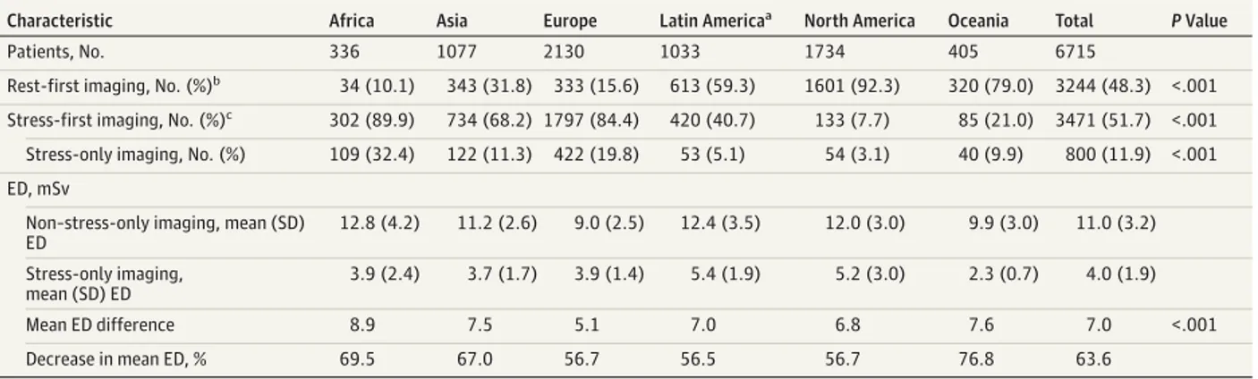
sion tomographic imaging, in which radiation doses are lower and stress-only imaging is generally not warranted. We compared regional rates of stress-only imaging and associated radiation effective doses among the remaining 6715 patients. We modeled the effect on radiation exposure to the US population if stress-only imaging were performed at the same rate as at European INCAPS sites (19.8%), a large US tertiary care center reported by Chang et al2(29.0%), the 90th percentile of all INCAPS laboratories (42.1%), and the 90th percentile of European INCAPS laboratories (60.0%). We also modeled radiation exposure if stress-only imaging were performed in all studies with normal myocardial perfusion,5a theoretical maximum. Modeling assumed that US nuclear cardiology practice is similar to that in the INCAPS population in the United States, that 9.25 million MPI studies are performed annually, and that annual all-source effective dose to the US population averages 6.2 mSv per person (to convert to roentgen equivalent man [rem], multiply by 0.1).1We also modeled the effect on the world nuclear cardiology population. Categorical variables were compared using the Fisher exact test and continuous vari-ables were compared using analysis of variance, with STATA/SE, version 13.1 (StataCorp LP). The Columbia Uni-versity Institutional Review Board approved the study and deemed it exempt from the requirements of US federal regulations for the protection of human subjects, as no indi-vidually identifiable health information was collected. Results|Marked variation existed between regions regarding the use of stress-first and stress-only protocols; the rates of these protocols were lowest in North America (Table 1). Among eligible studies, the mean effective dose decreased 63.6% (11.0 vs 4.0 mSv; P < .001) when a stress-only protocol was used.
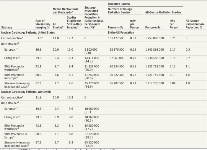
The model estimates a 20.9% reduction in the mean ef-fective dose from MPI if US laboratories were to adopt stress-only imaging at the same rate as the top 10% of INCAPS labo-ratories. This percentage reduction corresponds to a 21.2
million person-mSv reduction in cumulative radiation bur-den to the US population. While risk projection modeling is fraught with uncertainties, as a very rough estimate, the US Food and Drug Administration estimates an increase in the pos-sibility of developing a fatal cancer of 1 in 2000 for a 10-mSv exposure,6suggesting that increased adoption of stress-only MPI might prevent hundreds of cases of cancer annually. Es-timates for each model scenario are presented in Table 2.1,2,4,5 Discussion|Adopting a practice of stress-only imaging among the majority of patients undergoing MPI and who have nor-mal myocardial perfusion and function would dramatically de-crease the average radiation dose to patients, significantly im-proving nuclear cardiology’s radiation safety profile for the US population.
Increasing the rate of stress-only imaging is currently hin-dered by a low rate of performing stress imaging before rest imaging, especially in North America, where stress imaging was performed first in 133 of 1734 eligible studies (7.7%) vs 1797 of 2130 studies (84.4%) in Europe. The ability to perform stress-only imaging requires workflow changes, with real-time phy-sician review of stress images to assess the need for same-day rest imaging, or rest imaging performed, when needed, on a later day than stress imaging, as is common outside the United States.
In addition, current remuneration schemes create disin-centives to performing stress-only MPI. In the United States, there exist 2 Current Procedural Terminology billing codes for single-photon emission computed tomographic scan MPI: 78451 (single study, eg, stress-only imaging performed) and 78452 (multiple studies, eg, both rest and stress imaging per-formed); Medicare global reimbursements for these proce-dures are $355.74 and $492.65, respectively. Given this $137 dif-ference, it is not surprising that the 3.0% rate of stress-only imaging among eligible studies in the United States was far lower than the rates observed worldwide (11.9%) and in Europe (19.8%).
Table 1. Protocol Use and Radiation ED By Geographic Region for Studies Eligible for Stress-Only Protocol
Characteristic Africa Asia Europe Latin Americaa North America Oceania Total P Value
Patients, No. 336 1077 2130 1033 1734 405 6715
Rest-first imaging, No. (%)b
34 (10.1) 343 (31.8) 333 (15.6) 613 (59.3) 1601 (92.3) 320 (79.0) 3244 (48.3) <.001 Stress-first imaging, No. (%)c
302 (89.9) 734 (68.2) 1797 (84.4) 420 (40.7) 133 (7.7) 85 (21.0) 3471 (51.7) <.001 Stress-only imaging, No. (%) 109 (32.4) 122 (11.3) 422 (19.8) 53 (5.1) 54 (3.1) 40 (9.9) 800 (11.9) <.001 ED, mSv
Non–stress-only imaging, mean (SD) ED 12.8 (4.2) 11.2 (2.6) 9.0 (2.5) 12.4 (3.5) 12.0 (3.0) 9.9 (3.0) 11.0 (3.2) Stress-only imaging, mean (SD) ED 3.9 (2.4) 3.7 (1.7) 3.9 (1.4) 5.4 (1.9) 5.2 (3.0) 2.3 (0.7) 4.0 (1.9) Mean ED difference 8.9 7.5 5.1 7.0 6.8 7.6 7.0 <.001
Decrease in mean ED, % 69.5 67.0 56.7 56.5 56.7 76.8 63.6
Abbreviation: ED, effective dose.
SI conversion factor: To convert millisieverts to roentgen equivalent man [rem], multiply by 0.1.
aLatin America includes Mexico.
bRest-first imaging denotes a protocol where technetium Tc 99m rest imaging was performed before technetium Tc 99m stress imaging. c
Stress-first imaging denotes a protocol where technetium Tc 99m stress imaging was performed first, creating the possibility of a stress-only protocol; thus, stress-only imaging constitutes the subset of stress-first imaging in which rest imaging was omitted.
However, equalization of reimbursement for stress-only MPI with multiple-study imaging is also undesirable because it could disincentivize performing rest imaging (which en-tails additional costs) where needed (eg, when attenuation ar-tifacts impede image interpretation). In patients with estab-lished cardiomyopathy, or myocardial infarction and a known scar, stress-only imaging may be inapplicable.
Nevertheless, the present reimbursement system pro-vides strong financial disincentive for many US laboratories to consider stress-only imaging. Increasing reimbursement
for single-study MPI without increasing total payment for MPI (approximately $800 million annually in the Center for Medicare & Medicaid Service’s Outpatient Prospective Pay-ment System) could eliminate this disincentive, thereby decreasing radiation exposure to the US population. In addi-tion, a policy mandating reimbursement for single-study imaging (eg, stress-only imaging), if multiple-study imaging had been preauthorized, would provide physicians perform-ing MPI the flexibility needed to perform stress-only imaging when clinically warranted.
Table 2. Effect of Increase in the Rate of Stress-Only Protocol Use on Radiation Burden to US and World Nuclear Cardiology Populations, and to the Entire US Populationa
Strategy
Rate of Stress-Only Imaging, %
Mean Effective Dose per Study, mSv5 Strategy-Associated Population Reduction in Radiation Dose, Person-mSv, No. (%)d Radiation Burden All-Source Radiation Dose Reduction, % Nuclear Cardiology
Radiation Burden All-Source Radiation Burden
All Studiesb Studies Eligible for Stress-Only Imagingc Person-mSv mSv per Person Person-mSv mSv per Person
Nuclear Cardiology Patients, United States Entire US Population
Current practice4 3.0e 11.0 12.2 0 101 472 500 0.32 1 953 000 000 6.21 0 Rate attainedf European4 19.8 10.0 11.0 9 102 000 (9.0) 92 370 500 0.29 1 943 898 000 6.17 0.5 Chang et alg 29.0 9.4 10.3 14 411 000 (14.2) 87 061 000 0.28 1 938 588 500 6.15 0.7 90th Percentile worldwide4 42.1 8.7 9.4 21 238 000(20.9) 80 234 500 0.25 1 931 762 000 6.13 1.1 90th Percentile in Europe4 60.0 7.6 8.1 31 250 000 (30.8) 70 222 300 0.22 1 921 749 800 6.1 1.6 Stress-only imaging
in all normal casesh 67.6 7.2 7.6 35 270 000(34.9) 66 202 500 0.21 1 917 730 000 6.09 1.8
Nuclear Cardiology Patients, Worldwide Current practice4 11.9 10.0 10.2 0 Rate attainedf European4 19.8 9.4 9.6 10 080 000 (5.1) Chang et alg 29.0 8.9 9.0 20 160 000 (10.1) 90th Percentile worldwide4 42.1 8.2 8.1 35 280 000(17.7) 90th Percentile in Europe4 60.0 7.1 6.8 57 120 000 (28.7) Stress-only imaging
in all normal casesh 67.6 6.7 6.3 65 520 000(32.9)
Abbreviation: INCAPS, International Atomic Energy Agency Nuclear Cardiology Protocols Study. SI conversion factor: To convert millisieverts to roentgen equivalent man [rem], multiply by 0.1.
aThe rates of stress-only imaging and mean effective doses are estimated as per the INCAPS study,4unless otherwise noted. bMean Effective Dose per Study, All Studies includes those not eligible for the stress-only protocol.
c
As detailed in the Methods section, studies eligible for stress-only imaging excludes positron emission tomographic scan studies and studies with rest imaging alone, which have lower radiation effective doses, thus accounting for the generally higher mean radiation dose for studies eligible for stress-only imaging compared with all studies.
d
Our calculations assume 9.25 million nuclear cardiology procedures in the United States (82% eligible for stress-only protocol, as per INCAPS4
), and 20 million worldwide (85% eligible, as per INCAPS4). All calculations based on US and world population estimates at the time of data collection (March 18 through April 22,
2013).
eThis rate is consistent with the Centers for Medicare & Medicaid Services’ observation that 96% of myocardial perfusion imaging is billed with Current Procedural
Terminology code 78452.
f
Stress-only imaging was performed at the same rate as in the following 5 scenarios.
g
Chang et al2
rate is that achieved in a single US center between 1999 and 2007, and is virtually identical to the 75th percentile rate in Europe in INCAPS (30%).
hWe used a conservative estimate of the “normal” rate—67.6%—based on the 2009-2012 period in Duvall et al,5who, in a multicenter study, reported a lower rate of
In conclusion, our findings suggest a clear need for change in the United States to achieve parity with worldwide prac-tice in the use of stress-only imaging, and thereby reduce the radiation burden from MPI.
Mathew Mercuri, PhD Thomas N. B. Pascual, MD John J. Mahmarian, MD Leslee J. Shaw, PhD Maurizio Dondi, MD Diana Paez, MD Andrew J. Einstein, MD, PhD; for the INCAPS Investigators Group
Author Affiliations: Division of Cardiology, Department of Medicine, Columbia University Medical Center, New York–Presbyterian Hospital, New York (Mercuri, Einstein); Section of Nuclear Medicine and Diagnostic Imaging, Division of Human Health, International Atomic Energy Agency, Vienna, Austria (Pascual, Dondi, Paez); Department of Cardiology, Houston Methodist DeBakey Heart and Vascular Center, Houston, Texas (Mahmarian); Division of Cardiology, Department of Medicine, Emory University School of Medicine, Atlanta, Georgia (Shaw); Emory Clinical Cardiovascular Research Institute, Emory University School of Medicine, Atlanta, Georgia (Shaw); Department of Radiology, Columbia University Medical Center, New York–Presbyterian Hospital, New York (Einstein).
Corresponding Author: Andrew J. Einstein, MD, PhD, Division of Cardiology, Department of Medicine, Columbia University Medical Center, New York– Presbyterian Hospital, 622 W 168th St, Office PH 10-203, New York, NY 10032 ([email protected]).
Published Online: December 28, 2015. doi:10.1001/jamainternmed.2015.7106. Author Contributions: Drs Mercuri and Einstein had full access to all the data in the study and take responsibility for the integrity of the data and the accuracy of the data analysis.
Study concept and design: Mercuri, Pascual, Mahmarian, Dondi, Paez, Einstein. Acquisition, analysis, or interpretation of data: Mercuri, Pascual, Mahmarian, Shaw, Einstein.
Drafting of the manuscript: Mercuri, Einstein.
Critical revision of the manuscript for important intellectual content: All authors. Statistical analysis: Mercuri, Einstein.
Obtained funding: Einstein.
Administrative, technical, or material support: Pascual, Mahmarian, Shaw, Paez, Einstein.
Study supervision: Dondi, Einstein.
Conflict of Interest Disclosures: Dr Einstein reported receiving institutional research grants to Columbia University for other research from GE Healthcare, Philips Healthcare, Spectrum Dynamics, and Toshiba America Medical Systems. No other disclosures were reported.
Funding/Support: This study was supported by the International Atomic Energy Agency, the Margaret Q. Landenberger Research Foundation in memory of A. Donny Strosberg, PhD, and the Irving Scholars Program.
Role of the Funder/Sponsor: Drs Pascual, Dondi, and Paez are employed by the International Atomic Energy Agency and contributed as noted above. The other sponsors had no role in the design and conduct of the study; collection, management, analysis, and interpretation of the data; preparation, review, or approval of the manuscript; and decision to submit the manuscript for publication.
Group Information: The INCAPS Investigators Group includes executive committee members A. J. Einstein (chair), T. N. B. Pascual (IAEA project lead), D. Paez (IAEA section head), M. Dondi (IAEA section head); N. Better, S.E. Bouyoucef, G. Karthikeyan, R. Kashyap, V. Lele, F. Mut, V. P. C. Magboo, J. J. Mahmarian, M. Mercuri, M. M. Rehani, and J. V. Vitola, and regional coordinators E. Alexanderson (Latin America), A. Allam (Africa and Middle East), M. H. Al-Mallah (Middle East), N. Better (Oceania), S. E. Bouyoucef (Africa), H. Bom (East Asia), A. Flotats (Europe), S. Jerome (United States), P. A. Kaufmann (Europe), V. Lele (South Asia), O. Luxenburg (Israel), J. Mahmarian (North America), L. J. Shaw (North America), S. R. Underwood (United Kingdom), and J. Vitola (Latin America). Members by region include
W. Amouri, H. Essabbah, S. S. Gassama, K. B. Makhdomi, G. I. E. El Mustapha, N. El Ouchdi, N. Qaïs, N. Soni, and W. Vangu (Africa); R. M. Abazid, B. Adams, V. Agarwal, M. A. Alfeeli, N. Alnafisi, L. Bernabe, G. G. Bural, T. Chaiwatanarat, J. M. Chandraguptha, G. J. Cheon, I. Cho, A. S. Dogan, M. Eftekhari, A. Frenkel, I. Garty, S. George, P. Geramifar, H. Golan, S. Habib, R. Hussain, H. Im, H-J. Jeon, T. Kalawat, W. J. Kang, F. Keng, A. Klaipetch, P. G. Kumar, J. Lee, W. W. Lee, I. Lim, C. M. M. Macaisa, G. Malhotra, B. R. Mittal, M. H. Mohammad, P. Mohan, I. D. Mulyanto, D. Nariman, U. N. Nayak, K. Niaz, G. Nikolov, J. M. Obaldo, E. Ozturk, J. M. Park, S. Park, C. D. Patel, H. K. Phuong, A. P. Quinon, T. R. Rajini, Y. Saengsuda, J. Santiago, H. B. Sayman, A. S. Shinto, V. Sivasubramaniyan, M. H. Son, P. Sudhakar, G. M. S. Syed, N. Tamaki, K. Thamnirat, T. Thientunyakit, S. Thongmak, D. N. Velasco, A. Verma, U. Vutrapongwatana, Y. Wang, K. S. Won, Z. Yao, T. Yingsa-nga, R. Yudistiro, K. T. Yue, and N. Zafrir (Asia); S. C. Adrian, D. Agostini, S. Aguadé, G. Armitage, M. Backlund, M. Backman, M. Baker, M. T. Balducci, C. Bavelaar, M. Berovic, F. Bertagna, R. Beuchel, A. Biggi, G. Bisi, R. Bonini, A. Bradley, L. Brudin, I. Bruno, E. Busnardo, R. Casoni, A. Choudhri, C. Cittanti, R. Clauss, D. C. Costa, M. Costa, K. Dixon, M. Dziuk, N. Egelic, I. Eriksson, G. Fagioli, D. B. de Faria, L. Florimonte, A. Francini, M. French, E. Gallagher, I. Garai, O. Geatti, D. Genovesi, L. Gianolli, A. Gimelli, E. del Giudice, S. Halliwell, M. J. Hansson, C. Harrison, F. Homans, F. Horton, D. Jędrzejuk, J. Jogi, A. Johansen, H. Johansson, M. Kalnina, M. Kaminek, A. Kiss, M. Kobylecka, M. Kostkiewicz, J. Kropp, R. Kullenberg, T. Lahoutte, O. Lang, Y. H. Larsson, M. Lázár, L. Leccisotti, N. Leners, O. Lindner, R. W. Lipp, A. Maenhout, L. Maffioli, C. Marcassa, B. Martins, P. Marzullo, G. Medolago, J. B. Meeks, C. G. Mendiguchía, S. Mirzaei, M. Mori, B. Nardi, S. Nazarenko, K. Nikoletic, R. Oleksa, T. Parviainen, J. Patrina, R. Peace, C. Pirich, H. Piwowarska-Bilska, S. Popa, V. Prakash, V. Pubul, L. Puklavec, S. Rac, M. Ratniece, S. A. Rogan, A. Romeo, M. Rossi, D. Ruiz, N. Sabharwal, B. G. Salobir, A. I. Santos, S. Saranovic, A. Sarkozi, R. P. Schneider, R. Sciagra, S. Scotti, Z. Servini, L. R. Setti, S.-Å. Starck, D. Vajauskas, J. Veselý, A. Vieni, A. Vignati, I. M. Vito, K. Weiss, D. Wild, and M. Zdraveska-Kochovska (Europe); R. N. Agüro, N. Alvarado, C. M. Barral, M. Beretta, I. Berrocal, J. F. Batista Cuellar, T.-M. Cabral Chang, L. O. Cabrera Rodríguez, J. Canessa, G. Castro Mora, A. C. Claudia, G. F. Clavelo, A. F. Cruz Jr, F. F. Faccio, K. M. Fernández, J. R. Gomez Garibo, U. Gonzalez, P. González, M. A. Guzzo, J. Jofre, M. Kapitán, G. Kempfer, J. L. Lopez, T. Massardo, I. Medeiros Colaco, C. T. Mesquita, M. Montecinos, S. Neubauer, L. M. Pabon, A. Puente, L. M. Rochela Vazquez, J. A. Serna Macias, A. G. Silva Pino, F. Z. Tártari Huber, A. P. Tovar, L. Vargas, and C. Wiefels (Latin America); A. Aljizeeri, R. J. Alvarez, D. Barger, W. Beardwood, J. Behrens, L. Brann, D. Brown, H. Carr, K. Churchwell, G. A. Comingore, J. Corbett, M. Costello, F. Cruz, T. Depinet, S. Dorbala, M. Earles, F. P. Esteves, E. Etherton, R. J. Fanning Jr, J. Fornace, L. Franks, H. Gewirtz, K. Gulanchyn, C.-L. Hannah, J. Hays, J. Hendrickson, J. Hester, K. Holmes, S. Jerome, A. Johnson, C. Jopek, H. Lewin, J. Lyons, C. Manley, J. Meden, S. Moore, W. H. Moore, V. Murthy, R. Nace, D. Neely, L. Nelson, O. Niedermaier, D. Rice, R. Rigs, K. Schiffer, E. Schockling, T. Schultz, T. Schumacker, B. Sheesley, A. Sheikh, B. Siegel, A. M. Slim, J. Smith, M. Szulc, N. Tanskersley, P. Tilkemeier, G. D. Valdez, R. Vrooman, D. Wawrowicz, and D. E. Winchester (North America); and A. Alcheikh, B. Allen, E. Atkins, J. Bevan, C. Bonomini, J. Christiansen, L. Clack, E. Craig, H. Dixson, I. Duncan, S. Fredericks, S. Gales, R. Hampson, T. Hanley, K. Hartcher, J. Hassall, B. Kelley, S. Kelly, T. Kidd, T. de Kort, G. Larcos, W. Macdonald, C. McGrath, E. Murdoch, S. O’Malley, M. O’Rourke, M. Pack, R. Pearce, R. Praehofer, S. Ramsay, L. Scarlett, K. Smidt, F. Souvannavong, K. Taubman, G. Taylor, K. Tse, S. Unger, and J. Weale (Oceania).
Additional Contributions: We thank the International Atomic Energy Agency Nuclear Cardiology Protocols Study executive committee, regional
coordinators, and investigators group. Investigators were compensated for time and effort involved in data collection.
1. National Council on Radiation Protection and Measurements. Ionizing Radiation Exposure of the Population of the United States: 2006. Bethesda, MD: National Council on Radiation Protection and Measurements; 2009. NCRP Report No. 160.
2. Chang SM, Nabi F, Xu J, Raza U, Mahmarian JJ. Normal stress-only versus standard stress/rest myocardial perfusion imaging: similar patient mortality with reduced radiation exposure.J Am Coll Cardiol. 2010;55(3):221-230. 3. Einstein AJ, Tilkemeier P, Fazel R, Rakotoarivelo H, Shaw LJ; American Society of Nuclear Cardiology. Radiation safety in nuclear cardiology—current knowledge and practice: results from the 2011 American Society of Nuclear Cardiology member survey.JAMA Intern Med. 2013;173(11):1021-1023. 4. Einstein AJ, Pascual TNB, Mercuri M, et al; INCAPS Investigators Group. Current worldwide nuclear cardiology practices and radiation exposure: results
from the 65 country IAEA Nuclear Cardiology Protocols Cross-Sectional Study (INCAPS).Eur Heart J. 2015;36(26):1689-1696.
5. Duvall WL, Rai M, Ahlberg AW, O’Sullivan DM, Henzlova MJ. A multi-center assessment of the temporal trends in myocardial perfusion imaging.J Nucl Cardiol. 2015;22(3):539-551.
6. US Food and Drug Administration. What are the radiation risks from CT? http://www.fda.gov/Radiation-EmittingProducts
/RadiationEmittingProductsandProcedures/MedicalImaging/MedicalX-Rays /ucm115329.htm.Updated February 10, 2015. Accessed September 1, 2015.
LESS IS MORE
Use of Computed Tomography in Emergency
Departments in the United States:
A Decade of Coughs and Colds
Computed tomography (CT) can be an essential tool in guid-ing the management of acute or life-threatenguid-ing pulmonary disease. Increasing use of CT, however, has raised concerns about the effects of ionizing radiation on organs within the ra-diation field, including the thyroid, lungs, and breast.1 Beyond the risk posed by ion-izing radiation, high resolu-tion CT may have unintended downstream consequences related to incidental findings and overdiagnosis, leading to a costly and potentially harmful diagnostic, therapeutic, or in-terventional cascade.2Increasing use of CT is most concern-ing among patients with the least to gain (eg, patients with ill-nesses of low acuity or at low risk of serious pathological conditions) or the most to lose (eg, young patients in whom CT carries the greatest risk of causing future radiation-related cancers). We sought to examine trends in the use of CT, and in clinical decision-making, for patients presenting to the emergency department (ED) with respiratory symptoms. Methods|Using data from 2001 to 2010 in the National Hospi-tal Ambulatory Medical Care Survey (NHAMCS), an annual na-tional survey that obtains information about patients, pre-senting symptoms, and management for a systematic sample of visits, we identified visits by adults 18 years of age and older
who presented to the ED of a hospital in the United States with a primary respiratory symptom. Data analysis was conducted from November 2013 through February 2015. The complex sampling strategy and analytic design of the NHAMCS allow the derivation of nationally representative information about the use of ambulatory-care services. We stratified ED visits ac-cording to symptom location in the lower respiratory (eg, cough or shortness of breath) vs upper respiratory tract (eg, sore throat or nasal congestion) and acuity of illness based on triage rat-ing and vital signs.
Our primary outcome variable was CT performed during an ED visit. Secondary outcomes included diagnosis and man-agement strategies (antibiotic prescription and hospital ad-mission). We present weighted crude point estimates (in 2-year intervals) and, using multiple logistic regression, time trends, both crude and adjusted for age, race, sex, region, and insur-ance status.
Dartmouth College’s institutional review board waived re-view of this research.
Results|The 23 416 ED visits among adults with respiratory symp-toms recorded in the NHAMCS between 2001 and 2010 repre-sented an estimated 79 million ED sample visits in the United States. Overall use of CT imaging quadrupled during the 10-year period, from 2.2% (2001-2002) to 9.4% (2009-2010) (odds ra-tio, 4.6; 95% CI, 3.4-6.2) of visits. Use of CT increased at least 4-fold within each symptom group, increasing most steeply among patients with the least acute reason for imaging (ie, the lowest absolute CT rates), those with nonacute upper respiratory symptoms, among whom the use of CT increased from 0.5% to 3.6% (odds ratio, 7.4; 95% CI, 1.3-42.0) (Figure). Odds ratios cited represent crude likelihood in 2009-2010 vs 2001-2002. Adjusted odds ratios were comparable (Table).
The use of CT increased comparably across all age strata, in-cluding a 4-fold increase among the youngest patients (aged 18-39 years) (Table). Management (antibiotic prescription and hos-pital admission) did not appear to change, while the proportion of patients discharged without a diagnosis (ie, with a symptom-based diagnosis) increased during the study period.
Figure. Ten Years of CT Use for Adults With Respiratory Symptoms Presenting to EDs of US Hospitals, by Acuity and Symptom Location (2001-2010)
20 18 16 14 12 10 8 6 4 2 2001-2002 2003-2004 2005-2006
National Hospital Ambulatory Medical Care Survey, y National Hospital Ambulatory Medical Care Survey, y
Lower Upper Lower Upper 2007-2008 2009-2010 0 ED Visits at Which CT W as Ordered, % Acute A 20 18 16 14 12 10 8 6 4 2 2001-2002 2003-2004 2005-2006 2007-2008 2009-2010 0 ED Visits at Which CT W as Ordered, % Nonacute B
Crude estimates are shown. CT indicates computed tomography; ED, emergency department; lower, lower respiratory tract; upper, upper respiratory tract. Editorialpage 168
Related articlespages 266 and269